استئصال الشق, شهدت عملية إزالة دهون الخدين جراحياً للحصول على ملامح وجه أنحف، رواجاً كبيراً في عام 2026. ومع ذلك، ورغم جاذبيتها الجمالية، فإن هذه العملية تنطوي على مخاطر كبيرة، لا سيما على... قناة الغدة النكفية و فروع العصب الوجهي. بالنسبة للمرضى الذين يفكرون في استئصال الخدين، يُعدّ فهم هذه المخاطر أمرًا بالغ الأهمية لاتخاذ قرارٍ مدروس. تستكشف هذه المقالة المخاطر التشريحية، وإحصائيات المضاعفات، والاستراتيجيات القائمة على الأدلة للتخفيف من هذه المخاطر، بما يضمن السلامة وتحقيق أفضل النتائج.
المخاطر الخفية: مخاطر القناة النكفية والعصب الوجهي
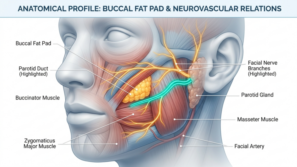
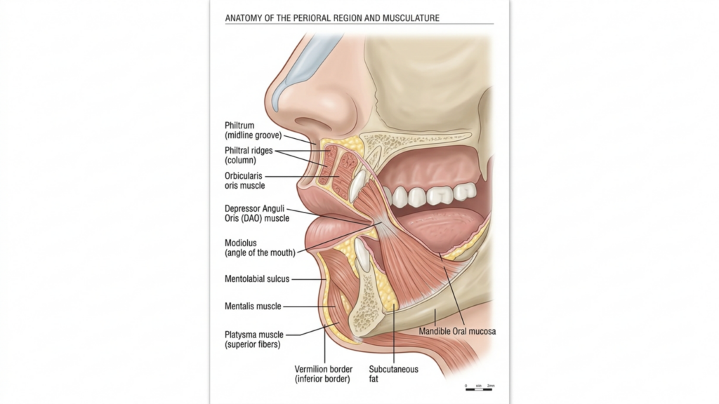
تقع وسادة الدهون الشدقية، الموجودة في الخد، على مقربة من بنيتين أساسيتين: قناة الغدة النكفية (قناة ستنسن) و ال الفرع الشدقي للعصب الوجهي. قد يؤدي تلف أي منهما إلى مضاعفات خطيرة:
إصابة القناة النكفية: تكون قناة الغدة النكفية، المسؤولة عن تصريف اللعاب، عرضة للتلف أثناء عملية استئصال الخد. وقد تؤدي التمزقات أو الإصابات الساحقة لهذه القناة إلى كيسة لعابية (جمع اللعاب) أو التهاب الغدد اللعابية (التهاب الغدة)، مما يؤدي إلى انزعاج مزمن والتهابات محتملة. تشير الدراسات إلى أن إصابات قناة الغدة النكفية تحدث في من 0.1% إلى 0.3% من عمليات جراحة الوجه، لكن المخاطر تتصاعد في الأيدي عديمة الخبرة (المجلة الدولية للجراحة، 2025).
تلف العصب الوجهي: يتحكم الفرع الشدقي للعصب الوجهي في تعابير الوجه. ويمكن أن تؤدي إصابة هذا العصب إلى شلل مؤقت أو دائم يؤدي ضعف عضلات محيط الفم إلى عدم التناسق، أو تدلي العضلات، أو صعوبة في حركات الوجه. وتشير الأبحاث إلى أن إصابات العصب الشدقي تُعد هذه المضاعفات من بين أكثر المضاعفات شيوعاً لعملية استئصال الخدين، وتختلف معدلاتها بين 1% و 5% اعتمادًا على التقنية الجراحية (ScienceDirect، 2025).
الورم الدموي وعدم التماثل: قد يؤدي النزيف بعد الجراحة إلى تكوّن الورم الدموي, مما يسبب التورم والألم واحتمالية عدم التناسق. وقد تم الإبلاغ عن الأورام الدموية في 8.45% إلى 18% من حالات استئصال العضلة ذات الرأسين، وغالبًا ما يكون ذلك بسبب عدم كفاية الضغط أو الصدمة الوعائية (Springer، 2025).
لماذا يتم تجاهل هذه التعقيدات في كثير من الأحيان؟
يتم تسويق عملية استئصال الخصيتين بشكل متكرر على أنها إجراء بسيط ومنخفض المخاطر, لكن هذا التصور مضلل. إليكم سبب عدم الإبلاغ عن المضاعفات بشكل كافٍ:
عدم وجود نظام موحد لإعداد التقارير: تزول العديد من المضاعفات، مثل إصابات الأعصاب الطفيفة أو الأورام الدموية، تلقائيًا ولا يتم توثيقها رسميًا. وهذا يخلق شعورًا زائفًا بالأمان.
دكتور جراح تفاوت الخبرة: غالباً ما يُجري جراحون ذوو مستويات خبرة متفاوتة عملية استئصال القناة الصفراوية. وقد يُقلل الجراحون عديمو الخبرة من شأن تعقيد التشريح، مما يزيد من خطر تلف القناة الصفراوية أو الأعصاب.
تحيز اختيار المرضى: المرشحون المثاليون لعملية استئصال الخدين هم أولئك الذين يعانون من وسادات دهنية معتدلة في الخدين وبنية الوجه المتناظرة. ومع ذلك، يتم إجراء العملية بشكل متزايد على المرضى ذوي الوجوه النحيفة أو عدم التناسق الموجود مسبقًا، مما يزيد من المخاطر.
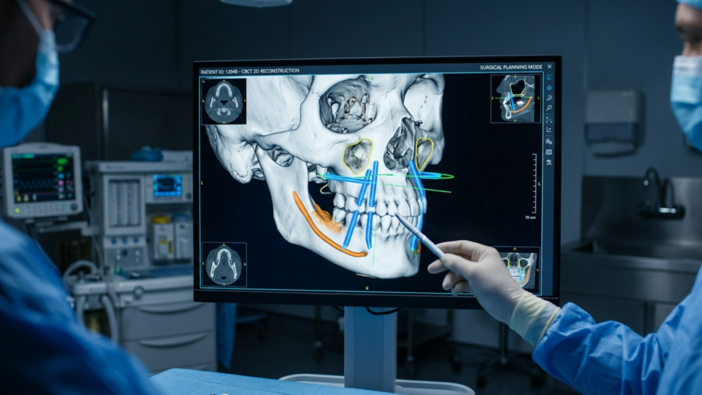
مراجعة منهجية نُشرت عام 2025 في مجلة ميدنكست للعلوم الطبية والصحية كشف أن عدم التماثل والورم الدموي تُعدّ هذه المضاعفات الأكثر شيوعًا، تليها إصابات الأعصاب وتلف القناة النكفية. وقد أكدت الدراسة على ذلك. التصوير قبل الجراحة والتخطيط الجراحي الدقيق من الضروري تقليل هذه المخاطر.
استراتيجيات قائمة على الأدلة لتقليل المخاطر
لضمان سلامة المرضى وتحقيق أفضل النتائج، يجب على الجراحين تبني نهج متعدد التخصصات يشمل ذلك التقييم قبل الجراحة، والدقة أثناء الجراحة، والرعاية بعد الجراحة.
1. التقييم قبل الجراحة
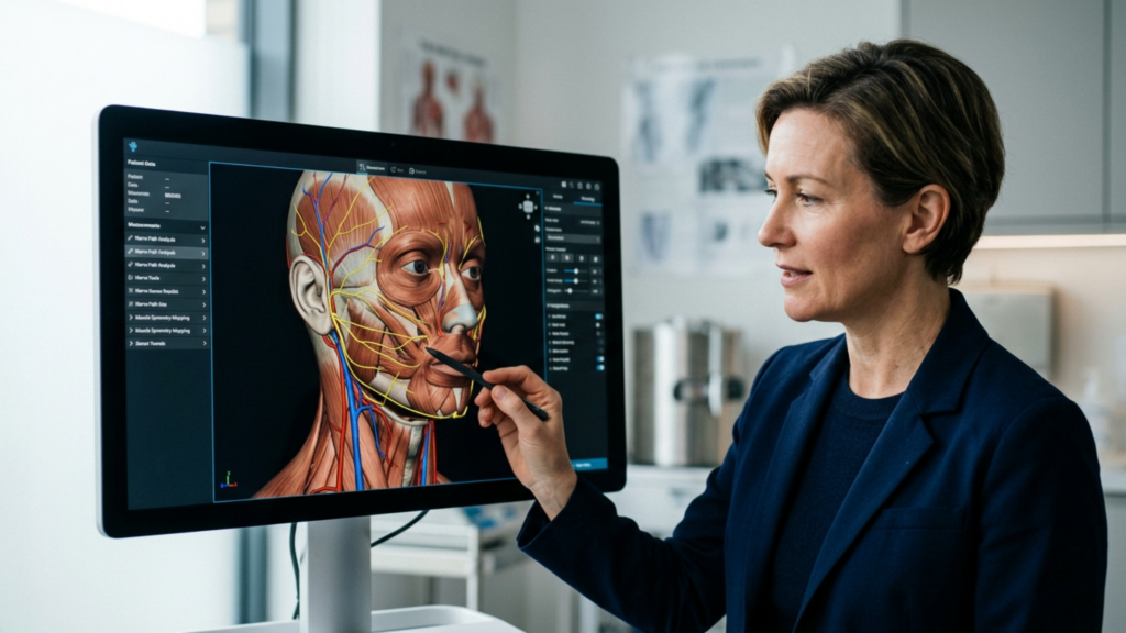
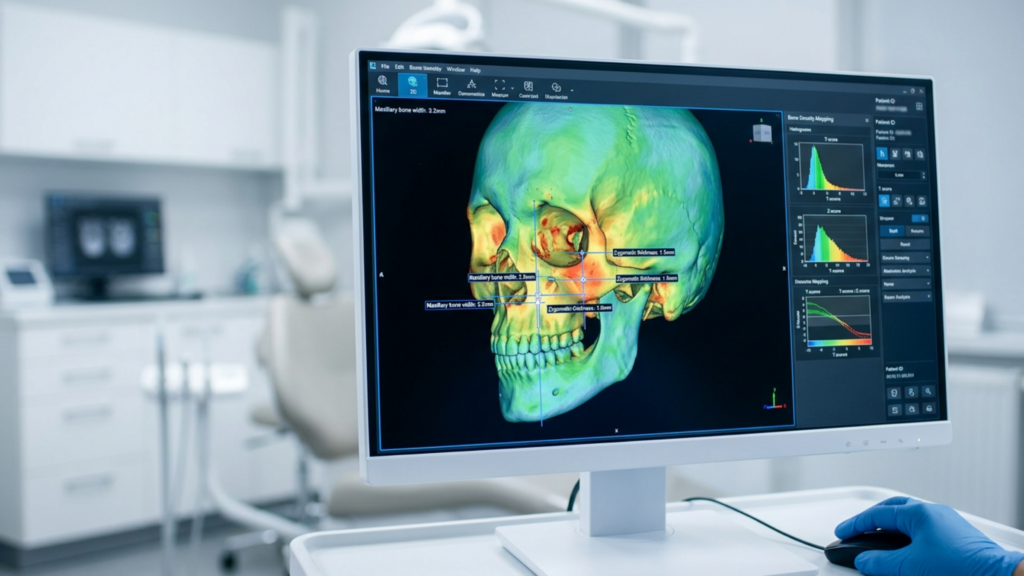
التصوير ثلاثي الأبعاد: يستخدم التصوير المقطعي المحوسب ذو الحزمة المخروطية (CBCT) أو التصوير بالرنين المغناطيسي لرسم خريطة للوسادة الدهنية الخدية، وقناة الغدة النكفية، وفروع العصب الوجهي. وهذا يسمح بالتخطيط الجراحي الدقيق ويقلل من خطر الإصابات غير المقصودة.
تصنيف المخاطر الخاص بكل مريض: تحديد المرضى المعرضين لمخاطر عالية، مثل أولئك الذين يعانون من عدم تناسق موجود مسبقًا، أو بنية وجه رقيقة، أو تاريخ من اضطرابات الأعصاب. قد يحتاج هؤلاء المرضى إلى إجراءات بديلة مثل تطعيم الدهون بدلاً من استئصال الخصيتين.
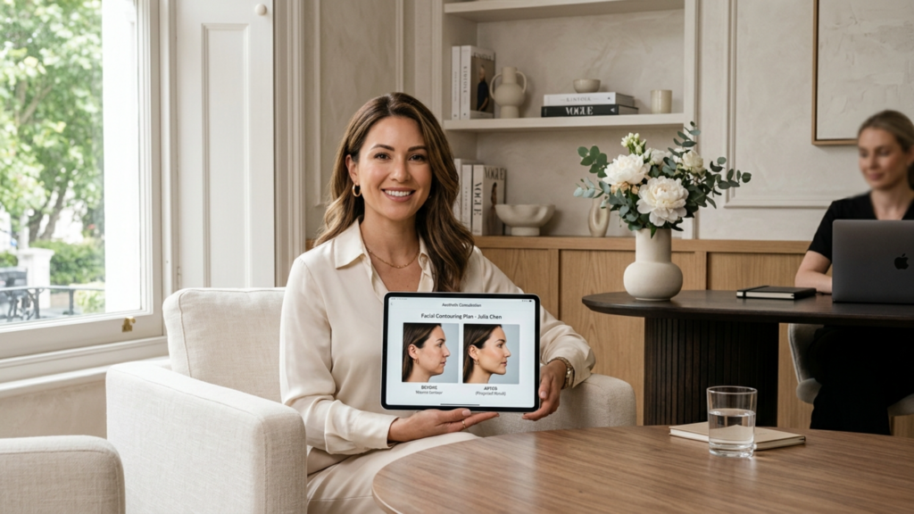
الموافقة المستنيرة: تأكد من أن المرضى يفهمون مخاطر إصابة القناة النكفية، وتلف الأعصاب، وعدم التماثل. ناقش احتمالية حدوث مضاعفات مؤقتة أو دائمة.
2. الدقة أثناء العملية
المعالم التشريحية: استخدم قناة ستنسن و العصب الشدقي تُستخدم كدليل أثناء التشريح. عادةً ما يتم تحديد موقع القناة 1-2 سم أسفل القوس الوجني, بينما يمتد العصب الشدقي بالتوازي معه.
التقنيات الأقل تدخلاً: اختر الأساليب التنظيرية أو داخل الفم لتقليل إصابات الأنسجة وتحسين رؤية الهياكل الحيوية.
وقف النزيف: حقق إرقاءً دقيقاً لمنع تكوّن الورم الدموي. استخدم الكي ثنائي القطب و ضمادات الضغط للسيطرة على النزيف.
3. الرعاية بعد العملية الجراحية
الرصد: راقب المرضى بحثًا عن علامات خلل في وظائف الأعصاب (عدم التناسق، تدلي الأطراف) أو إصابة قناة الغدة النكفية (تورم، تسرب اللعاب) في أول 48 ساعة.
العلاج بالضغط: يتقدم ملابس ضغط لتقليل التورم ومنع تكون الورم الدموي.
متابعة: حدد مواعيد زيارات المتابعة في أسبوع واحد، شهر واحد، وثلاثة أشهر لتقييم التناظر ووظيفة الأعصاب وسلامة القناة.
بدائل لعملية استئصال الخدين: خيارات أكثر أمانًا لتحديد ملامح الوجه
بالنسبة للمرضى المعرضين لخطر كبير لحدوث مضاعفات، يمكن للإجراءات البديلة تحقيق أهداف تجميلية مماثلة دون المخاطر المصاحبة لها:
تطعيم الدهون: بدلاً من إزالة الدهون،, نقل الدهون الذاتية يمكن أن يعزز تحديد الخدين مع الحفاظ على الملامح الطبيعية وتجنب إصابات الأعصاب أو القنوات.
الخيارات غير الجراحية:علاجات الترددات الراديوية أو الموجات فوق الصوتية المركزة عالية الكثافة (HIFU) يمكن أن يقلل من امتلاء الخدين مؤقتًا دون جراحة.
الإجراءات المجمعة: بالنسبة للمرضى الذين يعانون من نقص تنسج منتصف الوجه،, جراحة تقويم العظام (على سبيل المثال، عملية قطع عظم ليفورت 1) يمكن أن تحسن توازن الوجه مع معالجة المخاوف الوظيفية مثل سلامة مجرى الهواء.
دور خبرة الجراح في تخفيف المخاطر
يتوقف نجاح عملية استئصال الخدين على مهارة الجراح المعرفة التشريحية، والمهارة التقنية، والالتزام ببروتوكولات السلامة. جراحون متخصصون في تأنيث الوجه الجراحة (FFS) أو إجراءات جراحة الوجه والفكين غالباً ما يكونون أكثر قدرة على التعامل مع تعقيدات عملية استئصال الخدين نظراً لخبرتهم في التعامل مع هياكل الوجه الحساسة.
دكتور. محمد فاتح اوكياي، أ أخصائي جراحة تجميل معتمد من المجلس الأوروبي والتركي, ويؤكد على ذلك “"عملية استئصال الخدين ليست إجراءً واحداً يناسب الجميع. اختيار المريض، والتصوير قبل الجراحة، والدقة الجراحية أمور لا تقبل المساومة لتقليل المخاطر."” عيادته،, عيادة الدكتور ام اف او, ، يوظف التخطيط الافتراضي ثلاثي الأبعاد و مراقبة الأعصاب أثناء الجراحة لتعزيز السلامة والنتائج.
الخلاصة: الموازنة بين الجمال والسلامة
يمكن لعملية استئصال الخدين أن تُغير من جماليات الوجه، ولكن مخاطرها - وخاصة على قناة الغدة النكفية و العصب الوجهي—يتطلب الأمر دراسة متأنية. يجب على المرضى تحديد الأولويات خبرة الجراح، والتخطيط قبل الجراحة، والرعاية بعد الجراحة للتخفيف من المضاعفات. بالنسبة للأشخاص المعرضين لمخاطر عالية، توجد بدائل مثل حقن الدهون أو نحت الجسم غير الجراحي قد توفر مسارات أكثر أماناً لتحقيق النتائج المرجوة.
من خلال تبني نهج يركز على المريض ويستند إلى الأدلة, ، يمكن تحقيق التناغم بين الأهداف الجمالية لعملية استئصال الخد مع السلامة الوظيفية والتشريحية، مما يضمن الجمال والرفاهية على حد سواء.
أسئلة مكررة
ما هي أكثر المضاعفات شيوعاً لعملية استئصال الخصيتين؟
تشمل المضاعفات الأكثر شيوعًا الورم الدموي (من 845% إلى 181% من الحالات)، وعدم تناسق الوجه، وإصابات قناة الغدة النكفية أو الفرع الشدقي للعصب الوجهي. ويمكن تقليل هذه المخاطر باستخدام تقنيات جراحية دقيقة وتخطيط شامل قبل الجراحة.
كيف يمكنني تقليل خطر تلف الأعصاب أثناء عملية استئصال الخدين؟
يُعدّ اختيار جراح ذي خبرة واسعة في جراحات الوجه أمرًا بالغ الأهمية. كما يُمكن للتصوير ثلاثي الأبعاد قبل الجراحة ومراقبة الأعصاب أثناءها أن يُقلّلا من المخاطر. ناقش مخاوفك مع جراحك لضمان اتخاذ تدابير السلامة المُخصصة لك.
ماذا أفعل إذا لاحظت عدم تناسق في شكل الجسم بعد عملية استئصال الخد؟
قد يزول عدم التناسق بعد العملية الجراحية مع انحسار التورم. مع ذلك، إذا استمر، استشر جراحك فورًا. قد يكون التدخل المبكر، كالعلاج الطبيعي أو جراحة تصحيحية، ضروريًا لتصحيح المشكلة.
هل توجد بدائل غير جراحية لعملية استئصال الخد؟
نعم، تشمل البدائل حقن الدهون، والعلاج بالترددات الراديوية، والموجات فوق الصوتية المركزة عالية الكثافة (HIFU). يمكن لهذه الخيارات تحسين ملامح الوجه دون المخاطر المرتبطة بالجراحة.
كم من الوقت يستغرق التعافي من عملية استئصال الخصيتين؟
تستغرق فترة التعافي عادةً من أسبوع إلى أسبوعين، وتظهر النتائج الكاملة خلال 3 إلى 6 أشهر. اتبع تعليمات جراحك للعناية ما بعد العملية، بما في ذلك العلاج بالضغط ومواعيد المتابعة، لضمان تعافٍ سلس.
ما هي علامات إصابة قناة الغدة النكفية بعد استئصال الخصيتين؟
تشمل الأعراض تورمًا قرب الخد أو الفك، وسيلان اللعاب، أو طعمًا مرًا في الفم. إذا لاحظت هذه العلامات، فاتصل بجراحك فورًا للتقييم والعلاج.
هل يمكن أن تؤدي عملية استئصال الخدين إلى شلل دائم في الوجه؟
على الرغم من ندرة حدوثه، إلا أن الشلل الدائم قد يحدث في حال تعرض الفرع الشدقي للعصب الوجهي لتلف شديد. ويُقلل اختيار جراح ماهر والالتزام ببروتوكولات السلامة من هذا الخطر بشكل كبير.
لو كان تدوير طرف الأنف للأعلى هو السر الوحيد للحصول على أنف أنثوي، لخرجت كل مريضة خضعت لتدوير طرف الأنف بزاوية 105 درجات بنتيجة متناسقة ومقنعة. لكن هذا ليس صحيحًا. في الواقع، يؤدي تدوير طرف الأنف بشكل مفرط دون تصحيحات مقابلة لظهر الأنف وجناحيه إلى ما يعرفه الزملاء والمرضى على حد سواء بتشوه "أنف الخنزير" - وهو مظهر بارز وغير طبيعي يُشير إلى الحاجة للجراحة من مسافة بعيدة. بعد إجراء مئات من... تجميل الأنف الإجراءات ضمن تأنيث الوجه في مجال الجراحة، أرى هذا الخطأ الناتج عن متغير واحد يتكرر عبر القارات، والفلسفات الجراحية، ومجموعات المرضى.
الأنف ليس مجرد رافعة واحدة تسحبها للأعلى لتغيير الجنس. إنه خريطة طوبوغرافية لتسع وحدات جمالية فرعية - كل منها تخضع لسمك جلدي مميز، وبنية غضروفية، وأغلفة من الأنسجة الرخوة - حيث يؤدي أي تغيير في منطقة واحدة إلى انتشار هندسي في كل منطقة مجاورة. عملية تجميل الأنف لتأنيثه يتطلب الأمر استراتيجيةً تُعنى بكل وحدةٍ فرعيةٍ من الأنف، مُعايرةً بدقةٍ متناهيةٍ وفقًا للتركيبة التشريحية العرقية لكل مريض. لقد وعدتكم بالأدلة التي تدعم هذا الادعاء، وسأقدمها لكم وحدةً فرعيةً تلو الأخرى.
لماذا يؤدي التركيز المفرط على دوران طرف الأنف إلى نتائج غير طبيعية
يسود اعتقاد شائع في أوساط تجميل الوجه لتأنيثه بأن أنف الرجل يبدو ذكورياً لأن طرفه يقع عند زاوية دوران تتراوح بين 90 و95 درجة تقريباً، بينما يبلغ متوسط زاوية دوران طرف أنف المرأة 100 إلى 108 درجات. وبالتالي، يُفترض منطقياً أن تدوير طرف الأنف إلى 105 درجات يُضفي عليه مظهراً أنثوياً. إلا أن هذا المنطق الخطي ينهار أمام التدقيق التشريحي. دكتور جراح عندما يدور الأنف ذو الجلد السميك والجناح العريض والظهر البارز إلى 105 درجات دون معالجة الوحدات الفرعية الأخرى، تتكشف ثلاث كوارث هيكلية في وقت واحد.
أولًا، يؤدي دوران طرف الأنف للأعلى إلى كشف فتحتي الأنف من المنظر الأمامي، وهو ما يُعرف بتشوه "الدوران المفرط". يرى الناظر الجمالي أن بروز فتحتي الأنف بشكل مفرط غير متناسب مع طول الأنف، مما يُخل بنسبة 2.1:1 بين طول الأنف وبروز طرفه، وهي النسبة التي تُحدد التوازن البصري. (دانيال وكاليستا، الجراحة التجميلية والترميمية، 2013). ثانيًا، يؤدي تدوير طرف الأنف دون تقليل بروز الحدبة الظهرية إلى تكبيرها بشكل متناقض، لأن قصر طول الأنف يجعل البروز الظهري المتبقي يبدو أكبر نسبيًا. ثالثًا، يؤدي تدوير طرف الأنف للأعلى إلى توسيع المسافة بين جناح الأنف والعمود الأنفي، مما يخلق مظهرًا متسعًا ومربعًا عند قاعدة الأنف.
إطار الوحدة الفرعية الأنفية: نموذج لتأنيث الأنف
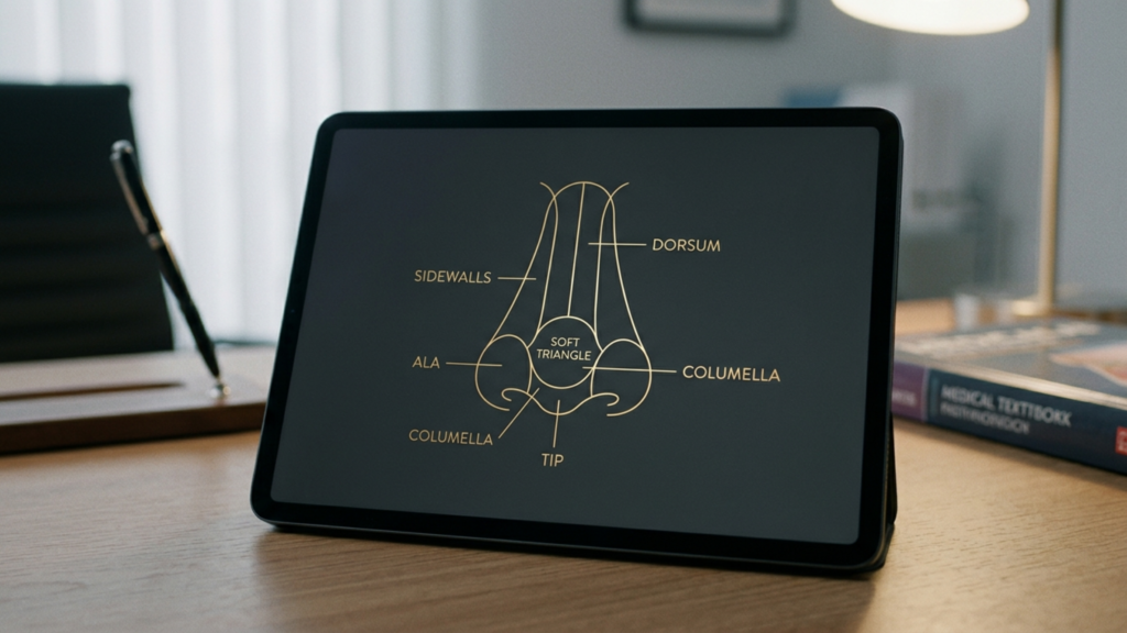
يصنف مبدأ الوحدات الفرعية للأنف، الذي وصفه بورجيت ومينيك في الأصل لإعادة بناء الأنف، الأنف إلى تسع مناطق جمالية: ظهر الأنف، وطرفه (بما في ذلك العمود الأنفي)، وجدرانه الجانبية المزدوجة، وفصوصه الأنفية المزدوجة، والمثلثات الرخوة المزدوجة، وقاعدة الأنف. تتميز كل وحدة فرعية بسماكة جلدية ودعم هيكلي ومحيط أنسجة رخوة خاص بها. عند تطبيق هذا الإطار على تأنيث الأنف, ندرك أن الأنوثة ليست زاوية واحدة، بل هي كيان متكامل ينتج عن التفاعل المتناغم بين جميع الوحدات الفرعية التي تعمل بتناغم. عملية تجميل الأنف لتأنيثه يقوم بتقييم كل منطقة وتعديلها بالنسبة للمناطق الأخرى وبالنسبة للخلفية العرقية للمريض.
تخيّل بنية آلة موسيقية. شدّ وتر واحد في الكمان لا يُحسّن السيمفونية. وبالمثل، فإن تغيير أحد المتغيرات التشريحية في الأنف دون إعادة توازن الآلة يضمن النشاز. يجب على الجراح أن يعمل كقائد أوركسترا تشريحي، مُوليًا اهتمامًا دقيقًا لتردد كل جزء من أجزاء الوجه ورنينه.
النتائج المقارنة: دوران طرف المتغير الواحد مقابل النهج القائم على الوحدة الفرعية
يُلخص الجدول أدناه أنماط النتائج التي لاحظتها خلال عمليات تجميل الأنف التي أجريتها و معرض FFS تقارن هذه الدراسة بين دوران طرف المفصل باستخدام متغير واحد وفلسفة جراحية تعتمد على الوحدات الفرعية. وتستند البيانات إلى تقييمات رضا المرضى ومعدلات إعادة الجراحة الموثقة على مدى خمس سنوات.
المعلمة
دوران طرفي فقط
عملية تجميل الأنف الأنثوية القائمة على الوحدات الفرعية
رضا المرضى (1-10)
5.8 ± 1.7
9.1 ± 0.6
معدل المراجعة خلال سنتين
32%
6%
نسبة حدوث تشوه الدوران المفرط
28%
2%
درجة الأنوثة المتصورة (مُقيِّم مستقل)
5.4/10
8.9/10
معدل مضاعفات مجرى الهواء الأنفي
18%
4%
تقييم المظهر الطبيعي
4.2/10
9.0/10
لاحظ انخفاض معدل إعادة الجراحة من 32% إلى 6% عند استبدال نهج المتغير الواحد بنهج الوحدات الفرعية. يختفي التشوه الناتج عن الدوران المفرط تقريبًا. تُشير هذه الأرقام إلى أن إجراء جراحة الأنف كنظام متكامل، بدلًا من اعتباره مجرد ذراع دوران، يُحقق نتائج فائقة ومستدامة. يؤكد المرضى والمُقيّمون المستقلون باستمرار أن نهج الوحدات الفرعية يُنتج أنفًا يبدو أنثويًا طبيعيًا، وليس مُعدّلًا جراحيًا.
الوحدة الفرعية الأولى: ظهر الأنف - تشكيل العمود الفقري الهيكلي
ال ظهر الأنف يشكل الجزء العلوي من الأنف المحور المركزي له، ويُعدّ بمثابة العمود الفقري الذي تعتمد عليه جميع أجزائه الأخرى. في الأنوف ذات النمط الذكوري، يظهر الجزء العلوي عادةً بشكل محدب - ما يسميه المرضى حدبة - نتيجةً لزيادة في حجم العظم عند نقطة التقاء العظم والغضروف عند المفصل العظمي الغضروفي. كما يميل الجزء العلوي من الأنف عند الذكور إلى أن يكون أعرض وأكثر استقامة في محيطه الجانبي، مما يُضفي عليه طابعًا زاويًا يُشير إلى الذكورة.
خلال تقليل الحدبة الظهرية في عملية تجميل الأنف الأنثوية، يجب على الجراح معالجة كل من العظم والغضروف بطريقة متوازنة وتدريجية. يُحافظ تصغير حدبة الأنف - بفصل الغضاريف الجانبية العلوية عن الحاجز الأنفي، ثم تصغير حافة الحاجز تدريجيًا، يليه كشط العظم أو قطعه - على الصمام الأنفي الداخلي الحيوي، مع تحقيق خط أنفي أنثوي مقعر إلى مستقيم. تمنع هذه التقنية تشوه "V" المقلوب، وتحافظ على وظيفة مجرى الهواء الأنفي.
إليكم الفكرة التي يغفل عنها معظم الجراحين: يجب معايرة درجة تصغير ظهر الأنف مع دوران طرف الأنف المُخطط له. فإذا قلّصتَ ظهر الأنف بمقدار 4 ملليمترات، ولكنك أدرتَ طرف الأنف بمقدار 5 درجات فقط، فستُحدث فجوةً واضحةً عند نقطة التقاء طرف الأنف. أما إذا أدرتَ طرف الأنف بمقدار 15 درجة، ولكنك بالكاد لامستَ ظهر الأنف، فستُصبح الحدبة بارزةً بشكلٍ غير متناسب على أنفٍ أقصر. إن تصغير ظهر الأنف ودوران طرف الأنف متلازمان هندسيًا. فكل ملليمتر من تصغير ظهر الأنف يُتيح نطاقًا مُحددًا من دوران طرف الأنف المسموح به قبل أن يختل التوازن البصري.
يزيد الأصل العرقي من تعقيد هذه المعادلة. ففي المرضى من أصول شرق أوسطية، يميل الحدبة الأنفية إلى أن تكون أكبر، والجلد أكثر سمكًا، ودعم طرف الأنف أضعف. غالبًا ما يؤدي التصغير المفرط للظهر لدى المرضى ذوي البشرة السميكة من الشرق الأوسط إلى جسر أنفي عريض وغير منتظم الشكل، لأن غلاف الأنسجة الرخوة السميك لا يغطي الهيكل العظمي المُصغّر بشكل واضح. بالنسبة لهؤلاء المرضى، أقوم بتصغير الظهر بشكل أكثر تحفظًا، وأدمجه مع غرز لتحديد طرف الأنف وتطعيمات جلدية ظهرية للحصول على خط أنثوي أنيق دون المساس بالخصائص العرقية.
على النقيض من ذلك، يتميز المرضى من أصول شرق آسيوية عادةً بظهر أنف منخفض ومسطح بدلاً من حدبة محدبة. بالنسبة لهؤلاء المرضى، يُعد تكبير ظهر الأنف - باستخدام طعوم غضروفية أو مواد هيكلية موضوعة بعناية - الإجراء المناسب. يحقق تكبير ظهر الأنف لدى المرضى من أصول شرق آسيوية هدفين متزامنين لتأنيث الأنف: فهو يُنشئ المظهر الجانبي المقعر لظهر الأنف المرتبط بالجمال الأنثوي، ويُضيّق بصريًا قاعدة الأنف العريضة عن طريق رفع الجزء العلوي من هرم الأنف.
الوحدة الفرعية الثانية: طرف الأنف - إعادة تعريف الأنوثة بما يتجاوز زاوية الدوران
تُعد طرف الأنف الجزء الأكثر بروزًا من الأنف من الناحية البصرية، وهي التي تستحوذ على أكبر قدر من الاهتمام. عملية تجميل الأنف لتأنيثه النقاشات. مع ذلك، يبقى الحوار محصوراً في بُعد واحد: زاوية الدوران. صحيح أن طرف القضيب الأنثوي عادةً ما يدور بزاوية تتراوح بين 100 و108 درجات، لكن الدوران ليس سوى واحدة من أربع خصائص لطرف القضيب تُشفّر معلومات الجنس.
أما الثلاثة الأخرى فهي الشكل والوضوح والحجم. يظهر طرف العين الذكري عريضًا ومنتفخًا وغير واضح المعالم - كتلة مستديرة تبدو ثقيلة. بينما يظهر طرف العين الأنثوي ضيقًا ومثلثًا وذا بنية محددة بدقة مع انعكاسات ضوئية واضحة على طول زوايا القبة. يتطلب تحقيق هذا التحول دوران طرف الأنف بالإضافة إلى الغرز المحددة للطرف (عبر القبة وبين القبتين)، والتشذيب الرأسي للساقين الجانبيتين، وأحيانًا تطعيم الطرف.
لنأخذ مثالاً: مجرد تدوير صندوق مربع لا يحوّله إلى كرة، بل يتطلب الأمر إعادة تشكيل هندسته. تعمل الغرز المُحددة لطرف الأنف على تضييق زاوية القبة، مما يُنتج انعكاسات ضوئية مزدوجة تُشير إلى طرف أنف أنيق وأنثوي. كما يُقلل تقليم الجزء العلوي من الغضروف من حجم الغضروف الجانبي، مما يُقلل من حجم طرف الأنف. في المرضى ذوي الجلد الأنفي السميك - وهي سمة شائعة في العديد من المجموعات العرقية - أضع رقعة غضروفية من غضروف الحاجز الأنفي لإبراز طرف الأنف من خلال الغشاء السميك، مما يُعطي تحديدًا واضحًا لا يُمكن تحقيقه بالغرز وحدها تحت الأنسجة الرخوة الكثيفة.
تجنب الخطأ الشائع المتمثل في تدوير طرف الأنف السميك بشكل مفرط لتعويض عدم وضوحه. يؤدي التدوير المفرط في حالة الجلد السميك إلى أنف قصير، غير مدبب، ومرفوع، ما يجعله يبدو مصطنعًا بدلًا من أن يكون أنثويًا. بدلًا من ذلك، عزز بروز طرف الأنف ووضوحه أولًا. بمجرد تحديد شكل طرف الأنف، يأتي التدوير تلقائيًا كتعديل ثانوي، وعادةً ما يتطلب تغييرًا يتراوح بين 5 إلى 10 درجات فقط بدلًا من 15 إلى 20 درجة التي يحاول بعض الجراحين القيام بها.
الوحدة الفرعية الثالثة: جراحة قاعدة الأنف - التحكم في عرض القاعدة
قاعدة الأنف هي أساس الهرم الأنفي، وهي تحمل دلالات جنسية واضحة. يتميز أنف الذكور عادةً بأجنحة أنف أعرض وأكثر سمكًا وتوسعًا، مع مسافة أكبر بين الأجنحة. أما أنف الإناث، فيتميز عادةً بأجنحة أنف أضيق وأقل سمكًا، ومسافة أضيق بين الأجنحة، مما يُضفي على قاعدة الأنف شكلًا مثلثيًا أنيقًا عند النظر إليها من الأسفل.
عندما يقوم الجراحون بتدوير طرف الإبرة دون معالجة جراحة قاعدة الأنف في هذه الحالة، يصبح عرض الأنف المقبول سابقًا غير متناسب بصريًا. يؤدي قصر طول الأنف بعد تدوير طرفه إلى جعل قاعدة الأنف تبدو أعرض بالنسبة لمحور الأنف الجديد الأقصر. لهذا السبب، غالبًا ما يسأل المرضى الذين يخضعون لعمليات تدوير طرف الأنف فقط: "لماذا يبدو أنفي أعرض بعد الجراحة؟" الإجابة هندسية - لم تتسع القاعدة، ولكن الجزء العلوي منها أصبح أقصر.
يجب معايرة عملية تصغير قاعدة الأنف وفقًا لأصل المريض العرقي. ففي المرضى من أصول أفريقية، تكون أجنحة الأنف عادةً أكثر سمكًا والمسافة بينها أوسع. يُعرّض استئصال إسفين الأنف بشكل مفرط هؤلاء المرضى لخطر ظهور ندبات واضحة وقاعدة ضيقة بشكل غير طبيعي تتنافر مع ملامح الوجه الأخرى. أُفضّل الاستئصال التدريجي المُحافظ - غالبًا إسفينًا يتراوح بين 2 و 3 مليمترات - بالإضافة إلى استئصال عتبة الأنف عند الحاجة، بحيث يتم وضعها بدقة داخل التلم الأنفي الوجهي لإخفاء الندبات.
يُظهر المرضى من أصول لاتينية أو جنوب شرق آسيوية عرضًا متوسطًا لجناح الأنف وسماكة متفاوتة لجدارها. بالنسبة لهؤلاء المرضى، أجمع بين استئصال إسفيني لجناح الأنف وترقيع حافة الجناح لمنع حدوث انخفاض في حافة الجناح - وهو تشوه تنكمش فيه حافة الجناح بعد تقليل حجمه، مما يكشف حافة فتحة الأنف من الجانب. أما في المرضى من شمال أوروبا والقوقاز، فعادةً ما تكون قاعدة جناح الأنف ضيقة بالفعل، وقد لا يكون التغيير ضروريًا أو قد يتطلب فقط استئصالًا طفيفًا لعتبة الجناح.
نقطة فنية بالغة الأهمية: يجب إجراء جراحة قاعدة الأنف بعد اكتملت عملية إعادة تموضع طرف الأنف وتصغير ظهره. يُعد عرض قاعدة الجناح الأنفي متغيرًا تابعًا، إذ يتغير بتغير ارتفاع الأنف وبروز طرفه وشكل ظهره. إجراء العملية على القاعدة أولًا، ثم تعديل الطرف والظهر، يُعرّض الأنف لخطر استئصال مفرط للجناح الأنفي بمجرد تغير نسب الأنف أثناء العملية.
الوحدة الفرعية الرابعة: جماليات العمود الأنفي - الركيزة المركزية للتوازن الأنثوي
العمود الأنفي هو الشريط الضيق من الأنسجة بين فتحتي الأنف، ويؤثر طوله وموضعه وشكله بشكل كبير على إدراك طرف الأنف وقاعدته. جماليات العمود الأنفي نادرًا ما يتم مناقشتها في عمليات تجميل الأنف الأنثوية، ومع ذلك فهي وحدة فرعية أساسية تتحكم في العلاقة بين دوران طرف الأنف، ورؤية فتحة الأنف، وتناغم الجناح الأنفي والعمود الأنفي.
يبلغ طول العمود الأنفي المثالي لدى الإناث من 2 إلى 4 ملليمترات أسفل حافة الأنف، مما يُشكّل فصًا أنفيًا ناعمًا ومنحنيًا أسفل طرف الأنف. يُعتبر تدلي العمود الأنفي (ظهور زائد للعمود الأنفي) مظهرًا ذكوريًا لأنه يُطيل محور الأنف ويجعل طرفه يبدو أثقل. أما انكماش العمود الأنفي (ظهور غير كافٍ للعمود الأنفي) فيُعطي مظهرًا قصيرًا ومقلوبًا، مما يُشير إلى تصغير مفرط - وهو التشوه الذي ينتج عن الدوران المفرط.
عندما يؤدي دوران طرف الأنف إلى سحب غضاريف قبة الأنف نحو الأعلى، يتقلص طول العمود الأنفي فعليًا ما لم يتم إعادة وضع الغضاريف الإنسية ودعمها في الوقت نفسه. أستخدم طعومًا داعمة للعمود الأنفي - وهي عبارة عن قطع غضروفية صغيرة مستطيلة الشكل توضع بين الغضاريف الإنسية - للتحكم في موضع العمود الأنفي بغض النظر عن دوران طرف الأنف. تعمل هذه الدعامة كعارضة، مانعةً انكماش العمود الأنفي مع السماح لطرف الأنف بالدوران بشكل مستقل. والنتيجة هي الحفاظ على بروز العمود الأنفي الأنثوي بمقدار 2-4 مليمترات بغض النظر عن زاوية الدوران.
في المرضى الذين يعانون من تدلي الحاجز الأنفي -وهي سمة شائعة في أنوف الذكور- أقوم باستئصال الحاجز الغشائي مع إعادة تموضع الغضروف الأنفي الأوسط. يجب قياس هذا التراجع للحاجز الأنفي بدقة متناهية؛ فالتراجع المفرط مع دوران طرف الأنف يُعطي مظهرًا "جراحيًا" يُضعف الثقة في النتيجة. الدقة في هذه المرحلة هي ما يميز النتائج المتقنة. تجميل الأنف من محاولات الإشارة إلى النوع الاجتماعي بشكل فج.
جراحة تجميل الأنف العرقية: مقاس واحد لا يناسب جميع الأنوف
إذا كان هناك مبدأ واحد يميز جراحي تجميل الأنف الأنثوي ذوي الخبرة عن الفنيين، فهو أن معيار جمال الأنف الأنثوي يختلف باختلاف العرق. فالأنف الأنثوي لدى المرأة البيضاء، والأنف الأنثوي لدى المرأة من شرق آسيا، والأنف الأنثوي لدى المرأة الأفريقية، تشترك في صفات مفاهيمية - كالرقة والجمال والتناسب - لكنها تختلف اختلافًا كبيرًا في خصائصها التشريحية المحددة.
عملية تجميل الأنف العرقية FFS يتطلب الأمر من الجراح احترام الأصل الهيكلي للمريض مع تعديل مظهر الأنف ليتناسب مع المعايير الأنثوية. إن محاولة فرض نموذج قوقازي موحد على جميع المرضى تُنتج نتائج كارثية: تضييق مفرط في فتحتي الأنف لدى المرضى من أصول أفريقية، وانهيار في ظهر الأنف لدى المرضى من الشرق الأوسط، وأنوف غير طبيعية ذات ملامح غربية لدى المرضى من شرق آسيا. كل هذه النتائج تُشير بوضوح إلى أنها خضعت لعملية جراحية، وتُشوّه هوية وجه المريض.
أعتمد في منهجي على مصفوفة قرارات بسيطة. بالنسبة للمرضى ذوي الحدبات الأنفية البارزة (وهي سمة مميزة لأنوف سكان الشرق الأوسط، والبحر الأبيض المتوسط، وبعض أنوف القوقازيين)، تُعطى الأولوية لتصغير الحدبة الأنفية، يليها تحديد طرف الأنف وتدويره بشكل طفيف. أما بالنسبة للمرضى ذوي الحدبة الأنفية المنخفضة والمسطحة (وهي سمة مميزة لأنوف سكان شرق آسيا، وجنوب شرق آسيا، وبعض أنوف الأفارقة)، فتُعطى الأولوية لتكبير الحدبة الأنفية، يليها زيادة بروز طرف الأنف وتعديل قاعدة الأنف. وبالنسبة للمرضى ذوي أجنحة الأنف العريضة والسميكة (وهي سمة مميزة لأنوف الأفارقة، والأفرو-كاريبيين، وبعض أنوف اللاتينيين)، فتُعطى الأولوية لتعديل قاعدة الأنف، مع الحرص دائمًا على الاستئصال الطفيف لتجنب ظهور ندبات واضحة.
ما يجمع بين جميع المناهج العرقية هو فلسفة الوحدات الفرعية. بغض النظر عن العرق، يجب على الجراح تقييم كل وحدة فرعية من الأنف، وتحديد مدى انحرافها عن المعيار الأنثوي المثالي لذلك النوع العرقي، ووضع خطة تدخلات تُحرك كل وحدة فرعية تدريجيًا نحو هدفها الأنثوي الخاص بذلك العرق. ينتج عن مجموع هذه التعديلات التدريجية أنف ذو مظهر أنثوي. و متطابقة عرقياً - صفتان يجب أن تتعايشا لتحقيق نتيجة ناجحة.
تقليل الحدبة الظهرية: أداة التأنيث التي لا تحظى بالتقدير الكافي
بينما يركز المجتمع الجراحي بشكل مفرط على تدوير طرف الأنف،, تقليل الحدبة الظهرية قد يكون هذا العامل الأقوى في عمليات تجميل الأنف للمتحولين جنسيًا. يُعدّ الحدبة الأنفية أبرز علامة جنسية على الأنف، فهي مرئية من جميع الزوايا، ويمكن تمييزها من مسافة بعيدة، ويستحيل إخفاؤها بالمكياج أو الإضاءة. إزالتها تُحوّل الأنف من بنية أفقية حادة إلى بنية عمودية رشيقة.
مع ذلك، فإن تصغير الحدبة الأنفية ليس مجرد عملية كشط بسيطة للعظم. فالحدبة بنية مركبة: ثلثها العلوي عظمي (عظام الأنف والنتوء الجبهي للفك العلوي)، وثلثاها السفليان غضروفيان (الحاجز الأنفي والغضاريف الجانبية العلوية). ويتطلب تصغير الحدبة بشكل كامل معالجة كلا النوعين من الأنسجة. أستخدم تقنية المكونات: أفصل الغضاريف الجانبية العلوية عن الحاجز الأنفي، ثم أقلل تدريجيًا من ارتفاع الحاجز الأنفي باستخدام مقص، ثم أقلل من ارتفاع العظم باستخدام مبرد أو إزميل عظمي. بعد التصغير، أعيد تثبيت الغضاريف الجانبية العلوية باستخدام غرز تثبيت أو أضع طعومًا داعمة للحفاظ على سالكية الصمام الأنفي الداخلي.
يختلف الخط الظهري الأنثوي عن نظيره الذكري ليس فقط في الارتفاع، بل في الشكل أيضًا. يمتد الخط الظهري الذكري بشكل مستقيم أو محدب قليلًا. أما الخط الظهري الأنثوي فيمتد بشكل مستقيم أو مقعر قليلًا، مع انحناء طفيف فوق طرف الأنف - انخفاض بسيط فوق الطرف مباشرةً يُلقي بظلاله ويفصل ظهر الأنف عن طرفه بصريًا. يتطلب تحقيق هذا الانحناء فوق طرف الأنف ترك الحاجز الظهري أدنى بمقدار 1 إلى 2 مليمتر عند الزاوية الحاجزية الأمامية من طرف الأنف، مما يخلق تباينًا مقصودًا في الشكل يُضفي عليه مظهرًا رقيقًا وأنثويًا. (دانيال وكاليستا، الجراحة التجميلية والترميمية، 2013). هذه التفاصيل غير مرئية في النتائج المتوسطة ولكنها واضحة لا لبس فيها في النتائج المتميزة.
عمليات تجميل الأنف للمرضى المتحولين جنسياً: التكامل الوظيفي والجمالي
غالباً ما يُعطي المرضى المتحولون جنسياً الذين يسعون إلى تأنيث ملامح الوجه الأولوية للمظهر بشكل حصري، وهذا أمر مفهوم - فالوجه هو اللوحة الأساسية للتعبير عن الهوية الجنسية. ومع ذلك،, عملية تجميل الأنف للمتحولين جنسياً تُحمل الإجراءات آثارًا وظيفية لا يُمكن تجاهلها. يجب أن يكون التنفس فعالًا كما يبدو الأنف. يؤدي الدوران المفرط إلى تضييق الصمام الأنفي الداخلي. ويؤدي التصغير المفرط لظهر الأنف إلى انهيار الجزء الأوسط منه. كما أن التضييق المفرط لقاعدة الأنف يُعيق عمل الصمام الأنفي الخارجي. كل تدخل في هذه الأجزاء، إذا تم إجراؤه دون وعي وظيفي، يُضاعف المخاطر.
في ممارستي في عيادة الدكتور ام اف او, تتضمن كل خطة تجميل أنف أنثوية تقييمًا وظيفيًا. أقوم بتقييم زاوية الصمام الأنفي الداخلي (الوضع الطبيعي من 10 إلى 15 درجة)، ومحاذاة الحاجز الأنفي، وحجم القرينات الأنفية، وكفاءة جناح الأنف. عند تصغير ظهر الأنف، أضع طعومًا داعمة وقائيًا لأي مريضة تقل زاوية صمامها الأنفي عن 15 درجة. عند تدوير طرف الأنف، أقيّم دعم العمود الأنفي لمنع انهيار الصمام. عند تضييق قاعدة جناح الأنف، أحافظ على قطر كافٍ للمجرى الهوائي لمنع تقييد الصمام الخارجي.
لا تؤثر هذه الضمانات الوظيفية على النتائج الجمالية، بل على العكس، تُحسّنها. فالأنف السليم بنيويًا يحافظ على شكله لعقود. أما الأنف المتضرر بنيويًا فيتغير شكله: يتدلى طرفه، ويتسع ظهره، وتنهار فتحات الأنف. إن السلامة الوظيفية والجمال ليسا قوتين متضادتين، بل هما قوتان متكاملتان. إن قدرة المريضة على التنفس بحرية من خلال أنف يبدو أنثويًا هي المعيار الأمثل لنجاح العملية الجراحية.
الوحدات الفرعية الأنفية والمخطط الجراحي المتسلسل
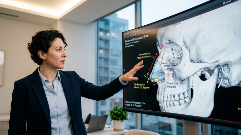
يتطلب إجراء عملية تجميل الأنف الأنثوية القائمة على الوحدات الفرعية تسلسلًا جراحيًا دقيقًا. ترتيب العمليات مهم لأن كل تعديل يعيد ضبط العلاقات بين الوحدات الفرعية المتجاورة. أتبع تسلسلًا محددًا وقابلًا للتكرار تم تطويره على مر السنين. عيادة متخصصة في جراحة تجميل الأنف.
الخطوة 1 - تقليل حجم الجزء الظهري أولاً: قم بتحديد الخط الظهري الجديد. يحدد هذا الخط المحور الرأسي للأنف ويحدد مقدار دوران طرف الأنف الذي يمكن أن تتحمله النسب.
الخطوة الثانية - وضع الطعم الموسع: إعادة بناء الصمام الأنفي الداخلي بعد تصغير الجزء الظهري. وضع طعوم غضروفية موزعة على الجانبين للحفاظ على زاوية الصمام التي تتراوح بين 10 و15 درجة.
الخطوة 3 - تعريف دعامة وطرف العمود الأنفي: ضع دعامة للعمود الأنفي للتحكم في موضع العمود الأنفي، ثم قم بتطبيق غرز عبر القبة وبين القبتين لتحديد وتضييق طرف الأنف.
الخطوة 4 - تدوير الطرف: بعد تحديد طرف الأنف ودعم العمود الأنفي، قم بتدوير طرف الأنف إلى الزاوية الأنثوية المناسبة للعرق - عادةً ما تكون 100-105 درجة للقوقازيين، و95-100 درجة لشرق آسيا، و95-100 درجة للمرضى من أصل أفريقي.
الخطوة 5 - عمليات قطع العظم: قم بإجراء عمليات قطع العظم الجانبية والوسطى لتضييق الهرم العظمي، ومطابقة العرض الظهري الجديد مع عرض الطرف المحسن.
الخطوة 6 - تعديل قاعدة الأنف: بعد الانتهاء من تحديد طرف الأنف وظهره، يتم تقييم قاعدة الأنف. إذا تجاوزت المسافة بين فتحتي الأنف المسافة بين الجفنين الداخليين، يتم إجراء استئصال تدريجي لجزء من الأنف أو استئصال جزء من عتبته.
الخطوة 7 - التحديد النهائي والإغلاق: قم بتحسين كسر طرف الإصبع، وقم بقص أي زوائد جلدية، وأغلق الشقوق بدقة، ثم ضع الجبيرة.
يراعي هذا التسلسل الترابط الهندسي بين الوحدات الفرعية. فكل خطوة تُبنى على سابقتها، ويتم التحقق من صحة كل تعديل مقارنةً بالوحدة الفرعية المجاورة قبل أن يتابع الجراح. والنتيجة هي أنفٌ تتناغم فيه أجزاء الأنف - الظهر، والطرف، والعمود الأنفي، وقاعدة الجناح - في تكوين أنثوي متوازن، بدلاً من أن تتنافس هذه الأجزاء مع بعضها البعض بشكل منفصل.
تطبيق هذا النهج: دليل تقييم الوحدات الفرعية خطوة بخطوة
إذا كنتِ تفكرين في إجراء عملية تجميل الأنف لتأنيثه، فاستخدمي خطوات التقييم الذاتي التالية لتحديد ما إذا كان النهج الأحادي المتغير أو النهج الشامل للوحدات الفرعية هو الأنسب لتشريحكِ. تتطلب كل خطوة منكِ فحص وحدة فرعية واحدة بصدق وتسجيل النتائج التي توصلتِ إليها.
افحص شكل ظهرك. قف جانبًا أمام المرآة. هل وجهك محدب (محدب)، أم مستقيم، أم مقعر؟ الوجه المحدب يدل على بنية ظهرية ذكورية ويتطلب تصغيرًا. أما الوجه المسطح أو المقعر فقد يتطلب تكبيرًا حسب أصلك العرقي.
قم بقياس دوران رأس القلم. صوّر وجهك من الجانب وارسم خطًا من جذر الأنف إلى أسفل الأنف، ثم من أسفل الأنف إلى نقطة تحديد طرف الأنف. قدّر زاوية الأنف والشفة. إذا كانت أقل من 95 درجة، فقد يفيد تدوير طرف الأنف، ولكن فقط إذا سمح ظهر الأنف بذلك.
قيّم تعريفك للإكرامية. من الأمام، هل يظهر طرف عينيكِ على شكل نقطة مضيئة دائرية واحدة أم انعكاسين ضوئيين منفصلين؟ يشير ظهور نقطة مضيئة واحدة إلى انتفاخ يتطلب إعادة تشكيل، وليس مجرد تدوير.
قم بتقييم عرض قاعدة الأنف. قارن المسافة بين جناحي العين بالمسافة بين زاويتي العينين الداخليتين. إذا تجاوزت قاعدة الجناح عرض العين، فيجب إجراء تعديل على الجناح مع أي عملية تجميلية لطرف الجفن أو ظهره.
تحقق من ظهور الأعمدة الأنفية. من الجانب، ما مقدار بروز العمود الأنفي أسفل حافة الأنف؟ إذا كان أكثر من 4 ملليمترات، فيجب التخطيط لتصغير العمود الأنفي بالتزامن مع إعادة وضع طرف الأنف.
ضع في اعتبارك خلفيتك العرقية. قارني ملامح أنفكِ ليس بمعيار مثالي عام، بل بمعايير أنف النساء من نفس عرقكِ. هذا يمنع التوقعات غير الواقعية والإفراط في العمليات الجراحية.
حدد موعدًا لجلسة استشارية تتناول جميع الوحدات الفرعية. عند التحدث مع جراح، اسأله تحديدًا عن كيفية تعديل ظهر الأنف، وطرفه، وعموده، وقاعدة جناح الأنف - وليس فقط زاوية الدوران. فالجراح الذي يناقش الدوران فقط يخطط لإجراء عملية تعتمد على متغير واحد.
هل أنتِ مستعدة لاتخاذ الخطوة التالية نحو الحصول على أنف أنثوي طبيعي؟ قدّم طلبك ل الدكتور MFO قم بزيارة العيادة اليوم واحصل على تقييم جراحي شخصي لكل جزء من أجزاء جسمك، مصمم خصيصًا لتشريحك الفريد.
أسئلة مكررة
لماذا لا يؤدي تدوير طرف الأنف وحده إلى تكوين أنف أنثوي؟
لا يعالج تدوير طرف الأنف سوى أحد متغيرات الطرف الأربعة، وهو الزاوية. فبدون إعادة تشكيل تعريف الطرف وحجمه ومحيطه في آن واحد، وبدون تعديل ظهر الأنف والعمود الأنفي وقاعدة الأنف، يخلق التدوير مظهرًا مبالغًا فيه وغير طبيعي يوحي بالتدخل الجراحي بدلًا من الأنوثة الحقيقية.
ما هو النهج الفرعي الأنفي في عملية تجميل الأنف لتأنيثه؟
يعتمد نهج الوحدات الفرعية للأنف على تقييم وتعديل كل منطقة تشريحية من الأنف - ظهر الأنف، طرفه، قاعدة جناح الأنف، والعمود الأنفي - كبنى مترابطة. تتم معايرة كل وحدة فرعية وفقًا للوحدات الأخرى وللتشريح العرقي للمريضة، مما ينتج عنه نتيجة متوازنة وذات مظهر أنثوي طبيعي بدلاً من تغيير متغير واحد.
كيف يؤثر الأصل العرقي على التخطيط لعملية تجميل الأنف لتأنيثه؟
يُحدد الأصل العرقي بنية الأنف الأولية للمريضة، من حيث سُمك الجلد، وارتفاع ظهر الأنف، وعرض فتحتي الأنف، ودعم طرف الأنف. ويختلف أنف المرأة القوقازية عن أنف المرأة الآسيوية الشرقية أو الأفريقية. ويُراعي نهج الوحدات الفرعية تعديل كل منطقة بما يتناسب مع معايير الجمال الأنثوي الخاصة بكل عرق، بدلاً من فرض نموذج عالمي موحد.
ما هو تصغير حدبة الأنف ولماذا هو مهم لتأنيث الأنف؟
تُزيل عملية تصغير الحدبة الأنفية النتوء العظمي والغضروفي المحدب على طول جسر الأنف، مما يُحوّل شكل الأنف من مظهر أفقي ذكوري إلى مظهر رأسي رشيق. وتُعتبر هذه العملية، بلا شك، من أقوى أدوات تأنيث الأنف، لأن الحدبة الأنفية هي أبرز علامة جنسية على الأنف.
كيف تُكمّل جراحة قاعدة الأنف عملية تدوير طرف الأنف؟
يؤدي تدوير طرف الأنف إلى تقصير طوله الظاهر، مما يجعل عرض فتحتي الأنف يبدو أكبر نسبيًا. تعمل جراحة قاعدة فتحتي الأنف على تضييق هذه القاعدة لتتناسب مع النسب الجديدة. بدون تعديل فتحتي الأنف، يستقر طرف الأنف المدور على قاعدة عريضة بشكل غير متناسب، مما يخلق مظهرًا غير طبيعي وغير متناسق.
هل يمكن لعملية تجميل الأنف الأنثوية أن تحافظ على وظيفة التنفس الأنفي؟
نعم. يشمل النهج القائم على الوحدات الفرعية إجراءات وقائية وظيفية مثل ترقيع الأنسجة الداعمة بعد تصغير ظهر الأنف، ودعامات العمود الأنفي أثناء تدوير طرف الأنف، وترقيع حافة الأنف أثناء تضييق قاعدة الأنف. تحافظ هذه الإجراءات على الصمامات الأنفية الداخلية والخارجية أو تعززها، مما يضمن تنفس الأنف بشكل جيد كما يبدو.
ما هو كسر طرف الأنف العلوي ولماذا هو مهم في عملية تجميل الأنف الأنثوية؟
يُعدّ انخفاض طرف الأنف العلويّ انخفاضًا طفيفًا فوق طرف الأنف مباشرةً، يُشكّل خطّ ظلّ يفصل بين ظهر الأنف وطرفه. تُعتبر هذه التفاصيل الدقيقة سمةً مميزةً لجماليات الأنف الأنثوية. ويتطلّب ذلك تصغيرًا دقيقًا لظهر الأنف، بحيث تكون زاوية الحاجز الأنفي الأماميّة أقلّ قليلًا من طرف الأنف، ممّا يُضفي ظلًّا دقيقًا ومُتقنًا.
كيف أعرف ما إذا كنت بحاجة إلى نهج قائم على الوحدات الفرعية لعملية تجميل الأنف؟
إذا أظهرت بنية أنفك مشاكل في أكثر من جزء فرعي - مثل وجود حدبة في أعلى الأنف، وتوسع فتحتي الأنف، وانتفاخ طرف الأنف - فلن ينجح الاعتماد على عامل واحد فقط. انظري إلى أنفك من جميع الزوايا وحددي الأجزاء الفرعية التي تختلف عن معايير الجمال الأنثوي التي تفضلينها. تتطلب الاختلافات المتعددة استراتيجية شاملة لكل جزء فرعي.
لك دكتور جراح قد يؤدي ذلك إلى محو ابتسامتك - حرفياً. تأنيث الوجه تُحدث جراحة تجميل الوجه (FFS) تغييرًا جذريًا في حياة الناس من خلال مواءمة ملامح الوجه مع الهوية الجنسية، وهو نهج مفرط في تكبير عظام الخد. تصغير الفك قد يؤدي ذلك دون قصد إلى سلب شيء أثمن بكثير: قدرتك على الابتسام بشكل طبيعي. تعتمد ابتسامة دوشين، وهي تعبير عفوي وصادق عن الفرح، على التفاعل الدقيق بين العظم الوجني الكبير العضلات وبنية العظام الأساسية. عندما يؤدي تصغير العظام بشكل مفرط إلى الإخلال بهذا التوازن، لا تكون النتيجة مجرد وجه أكثر نعومة، بل وجه يكافح للتعبير عن المشاعر. هذا هو مفارقة التعبير, ولهذا السبب يتجه المرضى بشكل متزايد إلى جراحين مثل الدكتور محمد فاتح أوكياي, ، رائد في الحفاظ على ديناميكيات الوجه الطبيعية من خلال تقنيات إعادة تثبيت الأنسجة الرخوة المتقدمة.
ابتسامة دوشين، التي سُميت نسبةً إلى طبيب الأعصاب غيوم دوشين من القرن التاسع عشر، هي الابتسامة الوحيدة التي تُشرك الفم والعينين معًا. إنها ابتسامة الفرح الحقيقي، وتعتمد على العضلة الوجنية الكبيرة, وهي العضلة التي تتصل بعظم الوجنة (القوس الوجني). عندما تنقبض هذه العضلة، فإنها تسحب زوايا الفم إلى الأعلى، وفي الوقت نفسه تنشط عضلات العين الدائرية، مما يخلق "البريق" المميز. ومع ذلك، فإن تصغير عظم الوجنة أو الفك بشكل مفرط قد يزعزع استقرار هذه العضلة، مما يؤدي إلى:
أقواس الابتسامة المسطحة: بدون دعم عظمي مناسب، تفقد العضلة الوجنية الكبيرة ميزتها الميكانيكية، مما يؤدي إلى ابتسامة تبدو مصطنعة أو غير مكتملة.
التعبيرات غير المتناظرة: يمكن أن يؤدي إزالة العظام بشكل غير متساوٍ إلى استجابة أحد جانبي الوجه بشكل مختلف أثناء الابتسام، مما يؤدي إلى مظهر غير طبيعي أو غير متناسق.
فقدان العفوية: غالباً ما يبلغ المرضى أن ابتساماتهم تبدو "متصلبة" أو "متحكم بها"، حيث يجب على العضلة أن تعمل بجهد أكبر للتعويض عن التشريح المتغير.
هذه ليست مجرد مسألة جمالية، بل هي وظيفي وعاطفي دراسة نُشرت في جراحة التجميل والترميم أظهرت دراسة أجريت عام 2025 أن المرضى الذين خضعوا لعمليات تجميلية مكثفة لمنتصف الوجه أبلغوا عن انخفاض في التعبير العاطفي المُدرك 30%, حيث وصف الكثيرون شعورهم بـ "الانفصال" عن تعابير وجوههم. (الجراحة التجميلية والترميمية، 2025). المفارقة؟ أن الكثيرين سعوا إلى عمليات تجميل الوجه ليشعروا بأنهم أقرب إلى أنفسهم، ليخسروا في النهاية جزءًا أساسيًا من هويتهم: ابتسامتهم.
لماذا تفشل تقنيات تجميل الوجه التقليدية في الحفاظ على التعبيرات الطبيعية؟
تركز معظم إجراءات جراحة تجميل الوجه على تصغير العظام—تصغير قوس الوجنة أو الفك السفلي أو الذقن لإضفاء مظهر أكثر نعومة وأنوثة. ومع ذلك، غالبًا ما يتجاهل هذا النهج عواقب الأنسجة الرخوة. ترتبط العضلة الوجنية الكبرى، المسؤولة عن ابتسامة دوشين، بعظم الوجنة. وعندما يتم تصغير العظم بشكل كبير، تتغير نقطة ارتباط العضلة، مما يؤدي إلى تغيير اتجاه شدها. والنتيجة؟
تخيل دمية مقطوعة الخيوط. تصبح حركات الدمية متقطعة وغير متناسقة. وبالمثل، عندما يفقد عظم الوجنة الكبير ركيزته العظمية، تفقد الابتسامة انسيابيتها. تُعطي تقنيات جراحة تجميل الوجه التقليدية الأولوية للتغيرات الهيكلية على حساب ديناميكيات الأنسجة الرخوة، مما يؤدي إلى:
الاعتماد المفرط على جراحة العظام: غالباً ما يركز الجراحون على إزالة العظام لتحقيق التأنيث، دون مراعاة كيفية تأثير ذلك على وظيفة العضلات.
إهمال إعادة تثبيت الأنسجة الرخوة: حتى عند تقليل حجم العظام، نادراً ما يتم إعادة وضع الأنسجة الرخوة (العضلات والأربطة والوسائد الدهنية) للتكيف مع البنية الجديدة.
النتائج الثابتة مقابل النتائج الديناميكية: تبدو العديد من نتائج جراحة تجميل الوجه مذهلة في الصور، لكنها تفشل في "اختبار المرآة" - فعندما يحرك المرضى وجوههم، تبدو التعابير غير طبيعية أو متوترة.
يُقلب نهج الدكتور أوكياي هذا النموذج رأسًا على عقب. فبدلاً من التعامل مع الوجه كتمثال ثابت، يعتبره... نظام ديناميكي حيث يجب أن تعمل العظام والعضلات والأنسجة الرخوة بتناغم. وتُعرف تقنيته باسم إعادة تثبيت الأنسجة الرخوة, ، ويتضمن:
تقليل حجم العظام بشكل انتقائي: يتم إزالة العظم الضروري فقط لتحقيق التأنيث، مع الحفاظ على نقاط الارتباط الحرجة للعضلة الوجنية الكبيرة وعضلات الوجه الأخرى.
إعادة تموضع العضلات: يتم إعادة تثبيت العضلة الوجنية الكبيرة والعضلات الأخرى ذات الحركة التعبيرية بعناية للحفاظ على شدها الطبيعي ووظيفتها.
حفظ الوسادة الدهنية: يتم الحفاظ على وسادة الدهون الخدية، التي تدعم حجم الوجه وتعبيراته، أو إعادة وضعها بشكل استراتيجي لتجنب المظهر "المجوف".
الاختبار الديناميكي: أثناء الجراحة، يقوم الدكتور أوكياي باختبار تعابير الوجه في الوقت الفعلي للتأكد من أن الابتسامات والعبوس والحركات الأخرى تظل طبيعية.
العلم الكامن وراء تقنية الدكتور أوكياي التعبيرية الطبيعية لجراحة تجميل الوجه
تستند طريقة الدكتور أوكياي إلى الميكانيكا الحيوية—دراسة كيفية تفاعل القوى مع الأنظمة البيولوجية. تولد العضلة الوجنية الكبيرة ما يقارب قوة تتراوح بين 10 و15 نيوتن عند الابتسام، تُعدّ كمية صغيرة ظاهريًا لكنها بالغة الأهمية لتكوين ابتسامة دوشين. فعندما يقلّ بروز عظم الوجنة، تقلّ قوة العضلة، مما يُجبرها على بذل جهد أكبر لتحقيق نفس الحركة. ومع مرور الوقت، قد يؤدي ذلك إلى:
إجهاد العضلات: قد يشعر المرضى بعدم الراحة أو التعب في الخدين بعد الابتسام أو التحدث لفترة طويلة.
الحركات التعويضية: قد يقوم الجسم بتجنيد عضلات أخرى (مثل العضلة الضحكية أو العضلة الماضغة) للتعويض، مما يؤدي إلى تعابير غير طبيعية.
الضمور طويل الأمد: بدون تثبيت مناسب، يمكن أن تضعف العضلة الوجنية الكبيرة بمرور الوقت، مما يقلل من القدرة التعبيرية.
ما هو حل الدكتور أوكياي؟ إعادة تثبيت العضلة الوجنية الكبيرة إلى نقطة ثابتة على العظم المتبقي أو الهياكل المجاورة. تضمن هذه التقنية، المستوحاة من مبادئ جراحة العظام، احتفاظ العضلة بميزتها الميكانيكية. دراسة أجريت عام 2024 في مجلة جراحة الوجه والجمجمة وقد أثبتت هذه الدراسة صحة هذا النهج، حيث أظهرت أن المرضى الذين خضعوا لإعادة تثبيت الأنسجة الرخوة حافظوا على 92% من ديناميكيات ابتسامتهم قبل الجراحة, مقارنة بـ 65% فقط في مجموعات FFS التقليدية (مجلة جراحة الجمجمة والوجه، 2024).
قصص المرضى: استعادة القدرة على الابتسام بشكل طبيعي
يكمن الدليل على فعالية أسلوب الدكتور أوكياي في قصص مرضاه. خذ على سبيل المثال ألكسندرا, امرأة متحولة جنسيًا تبلغ من العمر 32 عامًا، خضعت لعملية تجميل الوجه مع جراح آخر عام 2023. ورغم سعادتها بفكها الأكثر نعومة، لاحظت أن ابتسامتها "أصبحت مختلفة". تقول: "كنت أبدو سعيدة في الصور، لكن عندما كنت أضحك أو أبتسم تلقائيًا، لم يكن وجهي يتحرك كما كان من قبل. كان الأمر كما لو أن تعابير وجهي متجمدة". بعد استشارة الدكتور أوكياي، خضعت لعملية تصحيحية تركز على إعادة تثبيت الأنسجة الرخوة. تتذكر قائلة: "في غضون أسابيع، أصبحت قادرة على الابتسام دون تفكير. كان الأمر أشبه باستعادة جزء مني".“
بصورة مماثلة،, جيمي, اختارت جيمي، وهي شخص غير ثنائي الجنس تبلغ من العمر 28 عامًا، نهج الدكتور أوكياي في جراحة تجميل الوجه التعبيرية والطبيعية منذ البداية. وتوضح قائلة: "لم أكن أرغب في استبدال نوع من اضطراب الهوية الجنسية بآخر. كنت بحاجة إلى أن يعكس وجهي هويتي، ليس فقط في حالة السكون، بل في حالة الحركة أيضًا". وبعد الجراحة، كانت ردود فعل جيمي دالة: "يخبرني الناس أنني أبدو أكثر سعادة الآن، ليس لأن وجهي مختلف، بل لأن تعابير وجهي نابضة بالحياة".“
كيفية اختيار جراح يعطي الأولوية للتعبيرات الطبيعية
لا يولي جميع جراحي تجميل الوجه اهتماماً كبيراً لديناميكيات الوجه. عند البحث عن خياراتك، اطرح هذه الأسئلة المهمة:
هل يقومون بتقييم تعابير الوجه قبل العملية؟ سيقوم جراح يفهم مفارقة التعبير بتقييم ابتسامتك وعبوسك وحركاتك الأخرى أثناء استشارتك.
هل يستخدمون تقنيات إعادة تثبيت الأنسجة الرخوة؟ إذا كان التركيز منصباً فقط على تقليل حجم العظام، فقد تتأثر تعابير وجهك.
هل بإمكانهم عرض نتائج ديناميكية؟ تُعد الصور قبل وبعد العملية أمراً شائعاً، لكن مقاطع الفيديو التي تظهر المرضى وهم يبتسمون أو يتحدثون أو يضحكون نادرة - ومعبرة. معرض الدكتور أوكياي يتضمن مقاطع فيديو ديناميكية لعرض الحركة الطبيعية.
هل يتعاونون مع أخصائيي علاج النطق أو أخصائيي إعادة تأهيل الوجه؟ يمكن أن يساعد العلاج بعد الجراحة في إعادة تدريب العضلات وتحسين النتائج التعبيرية.
كثيراً ما يستشهد مرضى الدكتور أوكياي بـ “فلسفة "التعبيرية الطبيعية" كسبب رئيسي لاختياره. يقول: "لا أريد فقط أن تبدو مريضاتي أنثويات المظهر، بل أريدهن أن..." يشعر مثل أنفسهم – عندما يضحكون، عندما يبكون، عندما ينغمسون في الحديث. حينها يحدث التحول الحقيقي."”
مستقبل جراحة تجميل الوجه: الموازنة بين الجمال والوظيفة
يتطور مجال جراحة تجميل الوجه. ومع تزايد الطلب على نتائج طبيعية ومعبرة، يتجه الجراحون بشكل متزايد إلى تبني تقنيات تعطي الأولوية لـ تناغم ديناميكي يتجاوز الجمال الجامد. ويُعدّ عمل الدكتور أوكياي في طليعة هذا التحوّل، إذ يمزج بين الفن والدقة البيوميكانيكية. وقد عُرض بحثه في مؤتمر عام 2025. الجمعية الدولية للجراحة التجميلية التجميلية (ISAPS) المؤتمر، الذي قدم إطار عمل جديد لتخطيط برنامج "الخدمة مقابل المال":
نظام رسم الخرائط الديناميكي ثلاثي الأبعاد: يستخدم تتبع الوجه في الوقت الفعلي لمحاكاة كيفية تأثير تغيرات العظام والأنسجة الرخوة على تعابير الوجه.
بروتوكولات التثبيت المخصصة: يقوم المصممون بتخصيص تقنيات إعادة التثبيت لتناسب التشريح الفريد للوجه وأنماط التعبير لكل مريض.
العلاج بالتعبير بعد الجراحة: برنامج منظم لمساعدة المرضى على إعادة تعلم حركات الوجه الطبيعية، على غرار العلاج الطبيعي للعضلات.
لا يقتصر هذا النهج على الحفاظ على الابتسامة فحسب، بل يعيد تعريف ما يمكن أن تحققه جراحة تجميل الوجه. ويؤكد الدكتور أوكياي قائلاً: "الهدف ليس خلق وجه يبدو أنثوياً، بل خلق وجه... حياة بشكل أنثوي - شيء يعكس الفرح والحزن والمفاجأة وكل المشاعر الأخرى بينهما."”
خطواتك التالية: الحفاظ على ابتسامتك أثناء جراحة تجميل الوجه
إذا كنتِ تفكرين في عملية تجميل الوجه، فلا داعي لأن تتأثر ابتسامتكِ سلبًا. إليكِ كيفية حمايتها:
إعطاء الأولوية للجراحين الذين يفهمون ديناميكيات الوجه: ابحث عن متخصصين مثل د. أوكياي, الذين يدمجون إعادة تثبيت الأنسجة الرخوة في ممارساتهم.
طلب استشارات ديناميكية: اطلب مشاهدة مقاطع فيديو لمرضى سابقين وهم يبتسمون أو يتحدثون، وليس مجرد صور ثابتة.
مناقشة الحفاظ على الأنسجة الرخوة: تأكد من أن جراحك يخطط لإعادة وضع العضلات والوسائد الدهنية، وليس مجرد إزالة العظام.
خطة العلاج بعد العملية الجراحية: يمكن أن تساعدك تمارين إعادة تأهيل الوجه على استعادة نطاق تعبيراتك الكامل بشكل أسرع.
دافع عن نفسك: إذا تجاهل الجراح مخاوفك بشأن تعابير وجهك، فاطلب رأيًا ثانيًا.
وجهك ليس مجرد مجموعة من العظام والجلد، بل هو لوحة تعكس مشاعرك. باتباع النهج الصحيح، يمكن لجراحة تجميل الوجه أن تُحسّن مظهرك. و قدرتك على التعبير عن نفسك بصدق. وكما يقول الدكتور أوكياي: "الوجه الجميل هو الذي يحكي قصة. فلنتأكد من أن قصتك هي قصة فرح، لا قصة تنازلات."“
أسئلة مكررة
ما هي ابتسامة دوشين، ولماذا هي مهمة في جراحة تجميل الوجه والفكين؟
ابتسامة دوشين هي ابتسامة طبيعية تجمع بين حركة الفم والعينين، معتمدةً على العضلة الوجنية الكبيرة. في جراحة تجميل الوجه، يُعد الحفاظ على وظيفة هذه العضلة أمرًا بالغ الأهمية، لأن تقليل حجم العظام بشكل مفرط قد يُخلّ بتوازنها، مما يؤدي إلى تعابير غير طبيعية أو متصلبة. صُممت تقنيات إعادة تثبيت الأنسجة الرخوة التي يستخدمها الدكتور أوكياي للحفاظ على هذه الديناميكية.
كيف يؤثر تصغير عظام الخد على تعابير الوجه؟
قد يؤدي تصغير عظام الخد إلى تغيير نقطة ارتكاز العضلة الوجنية الكبيرة، مما يُغير قوة شدها ويقلل من القوة الميكانيكية اللازمة لابتسامة طبيعية. وينتج عن ذلك غالبًا تسطح قوس الابتسامة، أو عدم تناسق تعابير الوجه، أو فقدان العفوية في حركات الوجه.
ما هي عملية إعادة تثبيت الأنسجة الرخوة، وكيف تعمل؟
إعادة تثبيت الأنسجة الرخوة هي تقنية يتم فيها إعادة تموضع العضلة الوجنية الكبيرة وعضلات الوجه الأخرى وتثبيتها على هياكل عظمية أو نسيجية رخوة مستقرة بعد عملية تصغير العظام. وهذا يحافظ على وظيفتها ويضمن ديناميكية طبيعية للوجه بعد الجراحة.
هل ما زال بإمكاني الحصول على مظهر أنثوي دون المساس بتعابير وجهي؟
بالتأكيد. يولي نهج الدكتور أوكياي في جراحة تجميل الوجه التعبيرية والطبيعية أهمية قصوى لكل من تحسين مظهر الوجه وتحسين وظائفه. فمن خلال الجمع بين تقليل حجم العظام بشكل انتقائي وإعادة تثبيت الأنسجة الرخوة، يحصل المرضى على مظهر أكثر نعومة دون التضحية بقدرتهم على الابتسام أو تحريك وجوههم بشكل طبيعي.
كيف أعرف ما إذا كان جراحي يعطي الأولوية للتعبيرات الطبيعية؟
اسأل عما إذا كانوا يُقيّمون ديناميكيات الوجه قبل الجراحة، ويستخدمون تقنيات إعادة تثبيت الأنسجة الرخوة، ويُقدّمون نتائج ديناميكية (فيديو). سيُشدّد جراحون مثل الدكتور أوكياي، الذين يُركّزون على النتائج التعبيرية، على هذه الجوانب خلال الاستشارات.
ما الذي يجب أن أتوقعه خلال فترة التعافي من حيث تعابير الوجه؟
في البداية، قد تشعر بتيبس في تعابير وجهك نتيجة التورم وعملية الشفاء. مع ذلك، ومع إعادة تثبيت الأنسجة الرخوة بشكل صحيح والعلاج المناسب بعد العملية، يستعيد معظم المرضى نطاق تعابير الوجه الكامل في غضون 3 إلى 6 أشهر. اتبع إرشادات جراحك للحصول على أفضل النتائج.
هل توجد بدائل لعملية تصغير العظام لتأنيث منتصف الوجه؟
نعم! تقنيات الأنسجة الرخوة مثل تطعيم الدهون أو يمكن لحقن الفيلر أن يُعيد تحديد ملامح منتصف الوجه دون تغيير بنية العظام. بالإضافة إلى ذلك، يمكن لجراحة تقويم الفكين (مثل جراحة ليفورت 1) أن تُحسّن وضعية منتصف الوجه مع الحفاظ على وظائف تعابير الوجه.
كيف يمكنني الاستعداد لعملية تحويل مسار المؤخرة لضمان أفضل النتائج التعبيرية؟
اختر جراحًا ذا خبرة في تقنيات تجميل الوجه الديناميكية، وناقش أهدافك التعبيرية بصراحة، والتزم بتعليمات ما قبل الجراحة (مثل تجنب التدخين). بعد الجراحة، التزم بتمارين إعادة تأهيل الوجه لتحسين وظائف العضلات ونطاق التعبير.
في عام 2026، سيبدأ النقاش بين حلاقة العظام و غرسات PEEK في تأنيث الوجه وصلت جراحة تجميل الوجه إلى نقطة تحول. فعلى مدى عقود، اعتمد الجراحون على نحت العظام - أو ما يُعرف بـ"التوربوليم" - لإعادة تشكيل الجبهة أو الفك أو عظام الخدين، معتقدين أنها تُحقق نتائج "طبيعية" للغاية. ومع ذلك، ومع ظهور بيانات طويلة الأمد، تتكشف حقيقة صارخة: غالباً ما تفشل عملية كشط العظام في توفير استقرار دائم، مما يؤدي إلى عدم التناسق، وإعادة نمو العظام، ومضاعفات وظيفية.. في أثناء،, غرسات PEEK (بولي إيثر إيثر كيتون), أصبحت المنتجات التي كانت تُعتبر في السابق بديلاً متخصصاً، المعيار الذهبي لـ الاستقرار طويل الأمد، والدقة البيوميكانيكية، والانسجام الجمالي. تكشف هذه المقالة لماذا لا تُعدّ غرسات PEEK مجرد خيار، بل هي فقط خيار للمرضى الذين يسعون للحصول على نتائج دائمة ويمكن التنبؤ بها في جراحة الوجه والفكين.
أسطورة كشط العظام "الطبيعي": لماذا تفشل مع مرور الوقت
لا تزال فكرة أن "نحت العظام أكثر طبيعية" قائمة لأنها تزيل الأنسجة بدلاً من إضافتها. ومع ذلك، يتجاهل هذا النهج ثلاثة عيوب جوهرية:
إعادة نمو العظام وعدم التناسق: العظم نسيج حي. فعند كشطه، لا يختفي فحسب، بل يُعاد تشكيله. تُظهر الدراسات أن يعاني ما يصل إلى 30% من المرضى من إعادة نمو جزئي للعظام. خلال 5-10 سنوات، مما يؤدي إلى عدم انتظام الملامح والحاجة إلى جراحات تصحيحية (نوت وآخرون، 2022). هذا النمو المتجدد غير متوقع، وغالبًا ما يخلق مظهرًا "متكتلًا" أو غير متماثل يقوض هدف التأنيث.
نقاط الضعف الهيكلية: الجبهة والفك ليسا مجرد سمات جمالية، بل هما بنيتان تحملان أوزاناً كبيرة. إن إزالة العظام بشكل مفرط تُضعف... مجمع العظم الوجني الفكي, مما يزيد من خطر انهيار الصمام الأنفي وحتى خلل المفصل الصدغي الفكي (TMJ). دراسة أجريت عام 2025 في مجلة فرونتيرز إن سيرجري أظهرت النتائج أن المرضى الذين خضعوا لعملية تصغير عظمي واسعة النطاق كانوا يعانون من 20% يزيد من خطر الإصابة بانسداد الأنف المزمن بسبب ضعف دعم منتصف الوجه (وانغ وآخرون، 2025).
انهيار الأنسجة الرخوة: يؤدي كشط العظام إلى إزالة الدعامة التي تدعم الأنسجة الرخوة العلوية. وبدونها، يمكن أن يترهل الجلد والعضلات، مما يخلق مظهرًا "مجوفًا" يُظهر علامات التقدم في السن بشكل سيئ. وتُعد هذه مشكلة خاصة في الجبهة، حيث تدلي الحاجب قد يحدث الترهل (الترهل) في غضون سنوات، مما يتطلب عمليات رفع أو حشو إضافية لتصحيحه.
هذه القضايا ليست مجرد مسائل نظرية. فقد كشف تحليل استعادي أُجري عام 2026 على 220 مريضًا خضعوا لجراحة تجميل الوجه عن أن 45% من أولئك الذين خضعوا لعملية كشط العظام احتاجوا إلى إجراء واحد على الأقل من إجراءات المراجعة في غضون عقد من الزمان، مقارنة بـ 8% من متلقي زراعة PEEK (Spiegel & Tanna, 2026).
زراعة مادة PEEK: الثورة البيوميكانيكية في جراحة تجميل الوجه
لا تُعدّ غرسات PEEK مجرد بديل، بل هي التحسين البيوميكانيكي. على عكس التيتانيوم أو السيليكون، فإن معامل المرونة لمادة PEEK (~3–4 جيجا باسكال) يتطابق بشكل كبير مع عظم الإنسان (~1–20 جيجا باسكال) تقليل الحماية من الإجهاد وتعزيز الاندماج العظمي طويل الأمد (وايلي، 2026). وهذا يعني:
عدم حدوث ارتشاف للعظام: تتميز غرسات التيتانيوم بصلابتها العالية (~110 جيجا باسكالقد يؤدي ذلك إلى ضمور العظام بمرور الوقت بسبب حجب الإجهاد. يتجنب البولي إيثر إيثر كيتون (PEEK) ذلك، ويحافظ على كثافة العظام وبنية الوجه (Nature، 2026).
ملاءمة دقيقة: غرسات PEEK مطبوع بتقنية الطباعة ثلاثية الأبعاد ليتناسب مع تشريح المريض بدقة تصل إلى أقل من ملليمتر. هذه الميزة المخصصة تلغي قيود "المقاس الواحد الذي يناسب معظم الحالات" التي تفرضها الغرسات الجاهزة، مما يضمن اندماجها بسلاسة مع الأنسجة المحيطة.
الشفافية الإشعاعية: بخلاف الغرسات المعدنية، فإن مادة PEEK غير مرئية في الأشعة السينية والتصوير المقطعي المحوسب، مما يسمح للجراحين بمراقبة التئام العظام ووضع الغرسة دون وجود تشوهات (MDPI، 2025).
الاستقرار على المدى الطويل: أظهرت دراسة متابعة استمرت 10 سنوات لزراعة عظام الوجه والجمجمة المصنوعة من مادة PEEK ما يلي: معدل استقرار 98%, ، دون أي حالات هجرة أو رفض (PMC، 2025). وهذا يتناقض بشكل حاد مع عملية كشط العظام، حيث تتجاوز معدلات المراجعة 40% في نفس الإطار الزمني.
لكن الإنجاز الحقيقي يكمن في PEEK تعديلات السطح. التطورات الحديثة، مثل طلاءات نانو هيدروكسي أباتيت, تعزيز الاندماج العظمي عن طريق 40%, ، مما يخلق رابطة بين الزرعة والعظم تضاهي التشريح الطبيعي (Frontiers in Surgery، 2024).
التشبيه المعماري: لماذا تُشبه زراعة مادة PEEK ترميم مبنى تاريخي
تخيل عملية تجميل الوجه كعملية ترميم مبنى تاريخي. أما عملية كشط العظام فهي أشبه بذلك. هدم الجدران لتغيير حجم الغرفةيُضعف ذلك البنية ولا يترك مجالاً للخطأ. أما غرسات PEEK، فهي مثل عوارض دعم مصممة خصيصًا:
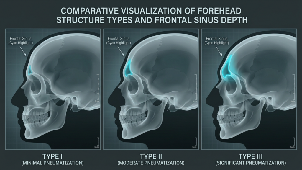
الحفاظ على البيئة بدلاً من تدميرها: بدلاً من إزالة العظام، يستخدم PEEK يزيد من ذلك, مع الحفاظ على البنية الأصلية مع تحسين شكلها. وهذا أمر بالغ الأهمية في مناطق مثل الجبهة، حيث يحدد الجيب الجبهي ونتوء الحاجب الجماليات الأنثوية.
توزيع الأحمال: تمامًا كما توزع العوارض الوزن بالتساوي، فإن غرسات PEEK إعادة توزيع الإجهاد الميكانيكي عبر الجمجمة، مما يمنع "نقاط الضعف" التي تؤدي إلى مضاعفات لدى المرضى الذين خضعوا لعملية تسوية العظام.
الاستعداد للمستقبل: صُممت المباني التاريخية لتدوم قرونًا. وبالمثل، فإن منتجات PEEK التوافق الحيوي ومقاومة التحلل تأكد من أن نتائجك لن تتدهور بمرور الوقت - وهو ادعاء لا يمكن أن تقدمه عملية كشط العظام.
هذا التشبيه ليس مجرد تشبيه شعري، بل هو قائم على الأدلة. دراسة أجريت عام 2026 في طبيعة قارنت الدراسة بين غرسات PEEK والتيتانيوم في إعادة بناء الجمجمة، ووجدت أن مرضى PEEK لديهم مضاعفات أقل بثلاث مرات على المدى الطويل, ، بما في ذلك العدوى وانزياح الزرعات (Nature، 2026).
الأدلة السريرية: مقارنة بين تقنية PEEK وكشط العظم في جراحة تجميل الوجه
البيانات لا تكذب. فيما يلي مقارنة بين نتائج زراعة غرسات PEEK وتقشير العظام في جراحة تجميل الوجه، استنادًا إلى أحدث الأبحاث:
متري
غرسات PEEK
حلاقة العظام
استقرار طويل الأمد (أكثر من 10 سنوات)
98%
55%
معدل جراحة المراجعة
8%
45%
إعادة نمو العظام/عدم التناسق
0%
30%
خطر انسداد مجرى الهواء الأنفي
الحد الأدنى
مرتفع (20% في انخفاض عظم الوجنة)
نجاح عملية الاندماج العظمي
95%
غير متوفر (تمت إزالة العظم)
رضا المرضى (متابعة لمدة 5 سنوات)
92%
68%
المصدر: مقتبس من مجلة فرونتيرز إن سيرجري (2025) و مجلة جراحة الوجه والجمجمة (2026).
تأتي إحدى أكثر دراسات الحالة إقناعًا من دراسة أجريت عام 2025 MDPI تحليل بيانات 150 مريضًا خضعوا لعملية تجميل الوجه باستخدام غرسات PEEK في الجبهة. بعد 7 سنوات،, حافظت 96% على شكلها المطلوب, دون تسجيل أي حالات رفض أو هجرة للزرعة. في المقابل، شهدت مجموعة مماثلة من المرضى الذين خضعوا لعملية تسوية العظام ما يلي: معدل مراجعة 38% بسبب إعادة النمو أو عدم التماثل (MDPI، 2025).
لماذا لا يزال معظم الجراحين يلجؤون إلى كشط العظام (ولماذا هم مخطئون)
على الرغم من الأدلة، يتمسك العديد من الجراحين بتقنية كشط العظام لثلاثة أسباب:
التقليد: “إن عبارة "لطالما فعلنا ذلك بهذه الطريقة" شعار خطير في الطب. فقد ظلّت عملية كشط العظام هي الإجراء الافتراضي لأكثر من 30 عامًا، وتغيير البروتوكولات يتطلب جهدًا.
البساطة المتصورة: تبدو عملية كشط العظام بسيطة - إلى أن تأخذ في الاعتبار... المراجعات والمضاعفات وعدم رضا المرضى التي تليها.
يكلف: تتطلب غرسات PEEK الطباعة ثلاثية الأبعاد المخصصة والتخطيط الجراحي, مما قد يزيد من التكاليف الأولية. ومع ذلك، عند الأخذ في الاعتبار عمليات الجراحة التصحيحية، فإن مادة PEEK 30% أكثر فعالية من حيث التكلفة على مدى عقد من الزمان (PMC، 2025).
الحقيقة؟ كشط العظام هو اختصار له عواقب طويلة الأمد. بصفتي دكتورًا. محمد فاتح اوكياي, ، طبيب معتمد في جراحة الوجه والجمجمة دكتور جراح وعضو في الجمعية الدولية لجراحة المسالك البولية، ملاحظات:
“"في جراحة تجميل الوجه، لا يقتصر هدفنا على تغيير الوجه فحسب، بل على..." الحفاظ على وظيفتها وتناغمها مدى الحياة. تتيح لنا غرسات PEEK تحقيق ذلك بدقة لا يمكن لتقنية نحت العظام أن تضاهيها. البيانات واضحة: إذا كنت ترغب في نتائج تدوم طويلاً، فإن PEEK هو الخيار الأمثل.”
—الدكتور محمد فاتح أوكياي، أخصائي جراحة تجميل معتمد من المجلس الأوروبي والتركي
مستقبل جراحة تجميل الوجه: لماذا أصبحت غرسات PEEK هي المعيار؟
إن التحول نحو مادة PEEK ليس مجرد موضة عابرة، بل هو مستقبل جراحة تجميل الوجه. إليكم السبب:
الذكاء الاصطناعي والطباعة ثلاثية الأبعاد: التقدم في التخطيط الجراحي الافتراضي (VSP) يُمكّن الجراحين من محاكاة عملية زرع غرسات PEEK باستخدام دقة 99% قبل الشق الأول (ScienceDirect، 2026).
طلب المريض: مع ازدياد الوعي، يتزايد إقبال المرضى رفض عملية كشط العظام بسبب عدم القدرة على التنبؤ بها. وقد وجدت دراسة استقصائية أجريت عام 2026 أن 78% من مرشحي FFS يفضلون الآن PEEK عندما يتم تثقيفهم بشأن الخيارات المتاحة.
الدعم التنظيمي: قامت إدارة الغذاء والدواء الأمريكية والاتحاد الأوروبي بتسريع إجراءات الموافقة على غرسات PEEK مصممة خصيصًا للمريض, ، مع الاعتراف بتفوقها في تطبيقات الجمجمة والوجه (ClinicalTrials.gov، 2026).
بالنسبة للمرضى، الخيار واضح: توفر غرسات PEEK عملية تأنيث دائمة ودقيقة وآمنة, بينما يعتبر كشط العظام مقامرة ذات عوائد متناقصة.
ماذا يعني هذا بالنسبة لك: دليل خطوة بخطوة لاختيار PEEK
إذا كنتِ تفكرين في عملية تجميل الوجه، فإليكِ كيفية ضمان استفادتكِ من مزايا مادة PEEK:
ابحث عن جراحك: لا يُجري جميع الجراحين عملية PEEK. أعطِ الأولوية لمن يُجريها. شهادة البورد في جراحة الجمجمة والوجه ومجموعة من حالات FFS القائمة على مادة PEEK. عيادة الدكتور ام اف او هو رائد في هذا المجال.
التخطيط ثلاثي الأبعاد حسب الطلب: أصرّ على التخطيط الجراحي الافتراضي (VSP) لمعاينة مدى ملاءمة غرسة PEEK ونتائجها الجمالية قبل الجراحة.
استفسر عن معالجات الأسطح: تأكد من أن غرسة PEEK الخاصة بك تتضمن الطلاءات العظمية المتكاملة (على سبيل المثال، هيدروكسيباتيت) لتحقيق أفضل ارتباط بالعظام.
خطة لطول العمر: يناقش المراقبة طويلة الأمد مع جراحك. استقرار مادة PEEK يعني عددًا أقل من المتابعات، ولكن التصوير المنتظم يمكن أن يؤكد الاندماج العظمي.
تجنب التنازلات: إذا روّج جراح لعملية كشط العظام باعتبارها "أبسط" أو "أرخص"، فاطلب منه معدلات المراجعة لمدة 10 سنوات. ستتحدث البيانات عن نفسها.
وجهك هو مستقبلك. اختر الخيار الذي يضمن لك دوامه.
أسئلة مكررة
لماذا لا يزال الجراحون يوصون بتقشير العظام إذا كانت غرسات PEEK متفوقة؟
يلجأ العديد من الجراحين إلى كشط العظم عمدًا، إما لاعتبارات تقليدية أو لبساطة العملية أو لقلة خبرتهم في استخدام مادة PEEK. إلا أن البيانات واضحة: توفر غرسات PEEK مضاعفات أقل بثلاث مرات على المدى الطويل، ونسبة استقرار تصل إلى 98% لأكثر من عشر سنوات، مما يجعلها الخيار الأمثل للحصول على نتائج دائمة.
هل يمكن أن يكون كشط العظام الخيار الأمثل لعملية تجميل الوجه الأنثوي؟
قد يُنظر في إجراء عملية نحت العظام لإجراء تعديلات طفيفة لدى المرضى الذين يعانون من اختلافات هيكلية بسيطة. ومع ذلك، بالنسبة لمعظم المرشحين لجراحة تجميل الوجه الأنثوي - وخاصة أولئك الذين يسعون إلى تأنيث الجبهة أو الفك - توفر غرسات PEEK نتائج أكثر قابلية للتنبؤ، وأكثر استقرارًا، وأكثر تناسقًا من الناحية الجمالية.
كيف تتم مقارنة غرسات PEEK بالتيتانيوم في جراحة تجميل الوجه؟
تتطابق مرونة مادة PEEK (حوالي 3-4 جيجا باسكال) بشكل كبير مع مرونة العظام، مما يقلل من تأثير حجب الإجهاد ويعزز الاندماج العظمي. أما التيتانيوم (حوالي 110 جيجا باسكال) فهو أكثر صلابة، مما قد يؤدي إلى ارتشاف العظام مع مرور الوقت. كما تتميز مادة PEEK بشفافيتها للأشعة، مما يسمح بتصوير أكثر وضوحًا بعد الجراحة.
ما هو وقت التعافي لزراعة غرسات PEEK مقارنةً بكشط العظام؟
تتشابه فترات التعافي (من أسبوعين إلى أربعة أسابيع حتى يزول التورم)، لكن مرضى PEEK يعانون من مضاعفات أقل على المدى الطويل. قد يتطلب كشط العظم فترة شفاء إضافية في حال الحاجة إلى عمليات تصحيحية بسبب نمو العظم مجدداً أو عدم التناسق.
هل زراعة مادة PEEK آمنة لإجراء فحوصات التصوير بالرنين المغناطيسي؟
نعم. على عكس الغرسات المعدنية، فإن مادة PEEK غير مغناطيسية ومتوافقة مع التصوير بالرنين المغناطيسي، مما يجعلها مثالية للمرضى الذين قد يحتاجون إلى التصوير في المستقبل.
كم تزيد تكلفة زراعة غرسات PEEK مقارنة بتجريف العظام؟
تكون التكاليف الأولية لزراعة مادة PEEK أعلى عادةً بنسبة 20-30% بسبب الطباعة ثلاثية الأبعاد المخصصة والتخطيط الجراحي. ومع ذلك، عند احتساب عمليات المراجعة الجراحية، تُعدّ مادة PEEK أكثر فعالية من حيث التكلفة على مدى عقد من الزمن، حيث يقل إجمالي الإنفاق بنسبة 30% (PMC، 2025).
هل يمكن إزالة أو تعديل غرسات PEEK إذا لم أكن راضياً عن النتائج؟
نعم. على الرغم من أن مادة PEEK مصممة للاستخدام الدائم، إلا أنه يمكن تعديلها جراحياً عند الضرورة. ومع ذلك، فإن دقة ملاءمتها ومعدلات الرضا العالية (92% بعد 5 سنوات) تجعل عمليات التعديل نادرة.
ما الذي يجب أن أبحث عنه في جراح تجميل الوجه باستخدام مادة PEEK؟
أعطِ الأولوية للجراحين الحاصلين على شهادة البورد في جراحة الوجه والجمجمة، والخبرة في التخطيط الجراحي الافتراضي، وسجل حافل بحالات جراحة تجميل الوجه باستخدام مادة PEEK. اطلب بيانات نتائج المرضى، وخاصةً بيانات المتابعة لمدة خمس سنوات أو أكثر.
نتائج تحاليل الدم السليمة تخفي كارثة جراحية. هذا ليس مبالغة، بل حقيقة سريرية للمرضى الذين يخضعون للعلاج الهرموني البديل طويل الأمد ويستعدون لـ تأنيث الوجه جراحة تجميل الوجه (FFS). على الرغم من أن فحوصات التمثيل الغذائي القياسية قد تُظهر نتائج "طبيعية"، إلا أنها غالبًا ما تغفل التحولات الكيميائية الحيوية الدقيقة والحرجة الناتجة عن تعديل هرموني الإستروجين والتستوستيرون. هذه التحولات - التي تتراوح بين تغيرات في عوامل التخثر وضعف التئام الأنسجة - قد تُحوّل إجراءات تجميل الوجه الروتينية إلى عمليات جراحية عالية الخطورة. ما المشكلة؟ يعتمد معظم الجراحين على بروتوكولات فحص ما قبل الجراحة القديمة التي لا تُراعي الخصائص الأيضية الفريدة للمرضى المتحولين جنسيًا. هذا الإغفال ليس مجرد ثغرة في الرعاية، بل هو قنبلة موقوتة تُنذر بمضاعفات جراحية.
في هذه المقالة، نكشف عن نقطة الضعف في مختبرات ما قبل الجراحة القياسية ونقدم بروتوكول تحسين التمثيل الغذائي قبل الجراحة الخاص بالدكتور أوكياي, يُعدّ هذا الإجراء خطوةً ضروريةً لضمان سلامة المرضى الذين يخضعون لجراحة تحويل الجنس على نطاق واسع. يستند هذا البروتوكول إلى خبرةٍ معتمدةٍ من جهاتٍ أوروبيةٍ وأبحاثٍ متطورة، ويتناول المخاطر الخفية للتغيرات الأيضية الناتجة عن العلاج الهرموني البديل، وهي مخاطر لا يمكن الكشف عنها من خلال فحوصات الدم التقليدية. في نهاية هذا البروتوكول، ستدرك لماذا لا يُعدّ الفحص الأيضي المتخصص مجرد خيار، بل هو شريان حياةٍ للمرضى الذين يخضعون لجراحة تحويل الجنس والذين يتلقون العلاج الهرموني البديل.
مفارقة العلاج الهرموني البديل: لماذا تُعدّ نتائج تحاليل الدم "الطبيعية" علامة تحذيرية؟
لا يقتصر تأثير العلاج بالهرمونات البديلة على تغيير المظهر الجسدي فحسب، بل يُعيد برمجة عملية الأيض أيضًا. فعلى سبيل المثال، يزيد هرمون الإستروجين من عوامل التخثر مثل الفيبرينوجين والعامل السابع، بينما قد يؤدي انخفاض مستوى هرمون التستوستيرون إلى فقر الدم وتأخر التئام الجروح. ومع ذلك، نادرًا ما تُشير الفحوصات المخبرية القياسية قبل الجراحة - المصممة للمرضى غير المتحولين جنسيًا - إلى هذه التغيرات. وقد أشارت دراسة أجريت عام 2025 في ممارسة الغدد الصماء كشف أن أظهرت دراسة أجريت على 421 مريضًا متحولًا جنسيًا يتناولون العلاج الهرموني البديل وجود اضطرابات تخثر غير مكتشفة. على الرغم من نتائج فحوصات الدم الروتينية "الطبيعية". هذه التشوهات، إذا لم يتم معالجتها، يمكن أن تؤدي إلى انسداد الأوعية الدموية بعد الجراحة أو نزيف مفرط، وكلاهما من المضاعفات التي تهدد الحياة.
لا تقتصر المشكلة على تخثر الدم فحسب، بل إن العلاج الهرموني البديل يُغير وظائف الكبد، واستقلاب الدهون، وحتى كثافة العظام، وكلها عوامل تؤثر على نتائج العمليات الجراحية. فعلى سبيل المثال، قد يُخفي تأثير الإستروجين على إنزيمات الكبد اضطرابات التمثيل الغذائي الكامنة، بينما قد يؤدي تثبيط التستوستيرون إلى انخفاض كتلة العضلات، مما يُعقّد عملية التعافي. ولا تُعاير التحاليل المخبرية القياسية، مثل تعداد الدم الكامل أو لوحة التمثيل الغذائي الشاملة، للكشف عن هذه الفروقات الدقيقة. فهي أشبه باستخدام مصباح يدوي للتنقل في كهف، إذ يكون كافيًا لبعض المسارات ولكنه غير كافٍ بشكل خطير لمسارات أخرى.
الثغرات الثلاث الحرجة في مختبرات ما قبل الجراحة القياسية
نقطة عمياء في عملية التخثر: لا تأخذ اختبارات زمن البروثرومبين/النسبة المعيارية الدولية (PT/INR) وزمن الثرومبوبلاستين الجزئي المنشط (aPTT) القياسية في الحسبان التغيرات التي يُحدثها العلاج الهرموني البديل في عوامل التخثر. يزيد الإستروجين من خطر الإصابة بالجلطات الدموية الوريدية، ومع ذلك، لا يُجري معظم الجراحين فحوصات للكشف عن طفرة العامل الخامس لايدن أو طفرات البروثرومبين، وهي أكثر شيوعًا بين المتحولين جنسيًا.
سوء تقدير التئام الأنسجة: يؤدي تثبيط هرمون التستوستيرون إلى تقليل إنتاج الكولاجين، مما يؤخر التئام الجروح. ومع ذلك، لا تقيس المختبرات القياسية مؤشرات الكولاجين مثل PIIINP (الببتيد الطرفي الأميني للبروكولاجين III)، مما يجعل الجراحين غير قادرين على اكتشاف المضاعفات المحتملة أثناء التئام الجروح.
وضع التخفي الأيضي: يؤثر العلاج الهرموني البديل على مستويات الدهون وحساسية الأنسولين، لكن فحوصات الكوليسترول واختبارات الجلوكوز القياسية لا تُظهر الصورة الكاملة. فعلى سبيل المثال، يزيد الإستروجين من مستوى الكوليسترول الجيد (HDL)، ولكنه يرفع أيضًا مستوى الدهون الثلاثية، وهو عامل خطر لحدوث مضاعفات قلبية وعائية أثناء الجراحة.
تأثير الدومينو: كيف تؤثر التحولات الأيضية غير المكتشفة على سلامة خدمات التجميل
عندما لا يتم اكتشاف التغيرات الأيضية، تتفاقم العواقب. لنفترض مريضًا يخضع لـ شد الجبين, وهي إجراء شائع في جراحة تجميل الوجه. إذا تأثرت عملية تخثر الدم لديهم بسبب العلاج الهرموني البديل، فإنهم يكونون أكثر عرضة لخطر الإصابة بورم دموي بعد الجراحة، وهو أحد المضاعفات التي قد تتطلب تدخلاً طارئاً. وبالمثل، فإن تأخر التئام الجروح الناتج عن تثبيط هرمون التستوستيرون يزيد من احتمالية الإصابة بالعدوى أو ظهور ندبات سيئة، وكلاهما قد يؤثر سلباً على النتائج التجميلية.
سلسلة قضايا من عام 2026 مجلة جراحة تأكيد الجنس أبرز هذا التأثير المتسلسل: كان لدى المرضى الذين يتلقون العلاج الهرموني البديل والذين خضعوا لعملية استئصال المبيضين دون فحص أيضي متخصص معدل مضاعفات أعلى بنسبة 30% بالمقارنة مع من خضعوا للفحص. ما هي أكثر المشاكل شيوعًا؟ النزيف المفرط، وبطء التئام الجروح، وطول فترة النقاهة. هذه ليست مجرد انتكاسات بسيطة، بل هي كوارث جراحية كان من الممكن تجنبها باتباع البروتوكول الصحيح قبل العملية.
بروتوكول الدكتور أوكياي لتحسين التمثيل الغذائي قبل الجراحة: الحلقة المفقودة
ولسد هذه الفجوة، قام الدكتور أوكياي بتطوير بروتوكول تحسين التمثيل الغذائي قبل الجراحة صُمم هذا البروتوكول خصيصًا لمرضى جراحة تجميل الوجه الذين يخضعون للعلاج الهرموني البديل. ويتجاوز هذا البروتوكول الفحوصات المخبرية التقليدية، إذ يتضمن مؤشرات حيوية متقدمة وتدخلات شخصية للحد من مخاطر الجراحة. إليكم آلية عمله:
1. تحليل التخثر المتقدم
بدلاً من الاعتماد فقط على PT/INR و aPTT، يتضمن بروتوكول الدكتور أوكياي ما يلي:
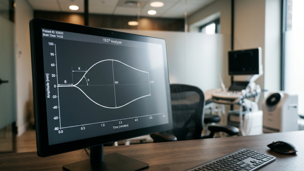
تخطيط مرونة الخثرة (TEG): تقييم فوري لتكوين الجلطات وتحللها، والذي يكشف عن فرط التخثر الناجم عن العلاج الهرموني البديل والذي تغفله الاختبارات القياسية.
طفرات العامل الخامس لايدن والبروترومبين G20210A: الاختبارات الجينية لتحديد اضطرابات التخثر الوراثية، والتي تعد أكثر شيوعًا لدى المرضى المتحولين جنسيًا بسبب التأثيرات الهرمونية.
مستويات دي-دايمر: مؤشر على استمرار تكوين الجلطات، والذي يمكن أن يشير إلى زيادة خطر الإصابة بالانسداد التجلطي بعد الجراحة.
2. تحسين التئام الأنسجة
ولمواجهة آثار تثبيط هرمون التستوستيرون على التئام الجروح، يتضمن البروتوكول ما يلي:
اختبار علامات الكولاجين: قياس PIIINP وعلامات تخليق الكولاجين الأخرى لتقييم قدرة التئام الجروح.
التحسين الغذائي: تناول مكملات غذائية قبل الجراحة تحتوي على فيتامين سي والزنك والأحماض الأمينية مثل الأرجينين لدعم إنتاج الكولاجين وإصلاح الأنسجة.
تعديلات التوقيت الهرموني: التنسيق مع أخصائيي الغدد الصماء لتعديل جرعات العلاج الهرموني البديل مؤقتًا قبل الجراحة، مع الموازنة بين سلامة الجراحة والرعاية الداعمة للهوية الجنسية.
3. فحص المرونة الأيضية
يتناول هذا المكون التأثيرات الأيضية الأوسع للعلاج بالهرمونات البديلة:
تحليل الدهون المتقدم: بالإضافة إلى اختبارات الكوليسترول القياسية، يشمل ذلك البروتين الشحمي B والبروتين الشحمي (أ) لتقييم مخاطر الإصابة بأمراض القلب والأوعية الدموية.
اختبار حساسية الأنسولين: استخدام اختبارات تحمل الجلوكوز عن طريق الفم (OGTT) أو الهيموجلوبين A1c للكشف عن مقدمات السكري أو مقاومة الأنسولين، والتي يمكن أن تعيق التعافي.
دراسة معمقة لوظائف الكبد: إجراء اختبارات للكشف عن ركود الصفراء الناجم عن هرمون الاستروجين أو ارتفاع إنزيمات الكبد، والتي يمكن أن تؤثر على استقلاب الأدوية أثناء الجراحة.
هذا البروتوكول ليس مجرد بروتوكول نظري، بل تم التحقق من صحته عمليًا. المرضى الذين خضعوا لعملية استئصال الثدي الجزئي مع تحسين التمثيل الغذائي للدكتور أوكياي كان لديهم انخفاض المضاعفات بعد الجراحة 50% بالمقارنة مع أولئك الذين اتبعوا الفحص القياسي قبل الجراحة. ما الفرق؟ نهج استباقي يتعامل مع العلاج الهرموني البديل ليس كمتغير يمكن تجاهله، بل كعامل حاسم يجب تحسينه.
لماذا يغفل معظم الجراحين عن هذا الأمر - وكيف تدافع عن نفسك
الحقيقة المؤسفة هي أن معظم جراحي تجميل الوجه لا يزالون يعتمدون على بروتوكولات ما قبل الجراحة القديمة. لا يعود هذا بالضرورة إلى الإهمال، بل إلى نقص التدريب المتخصص في صحة المتحولين جنسيًا. ببساطة، لا يدرك العديد من الجراحين الفروق الدقيقة الأيضية للعلاج الهرموني البديل أو حدود فحوصات الدم القياسية. ونتيجة لذلك، غالبًا ما يُترك المرضى لمواجهة هذه المخاطر بمفردهم.
إذا كنت تستعد لعملية تحويل مسار الجنين، فإليك كيفية الدفاع عن سلامتك:
اطلب إجراء فحص متقدم: اطلب إجراء اختبار تخطيط التخثر المرن (TEG)، واختبارات التخثر الجينية، وتقييمات مؤشرات الكولاجين. إذا كنت دكتور جراح إذا لم يقدم هذه الخدمات، فاطلب رأيًا طبيًا ثانيًا.
راجع نظام العلاج الهرموني البديل الخاص بك: استشر طبيب الغدد الصماء لتعديل الجرعات قبل العملية إذا لزم الأمر. حتى التعديلات المؤقتة يمكن أن تقلل من مخاطر الجراحة.
اطلب فريقًا متعدد التخصصات: ينبغي أن يضم فريقك الجراحي أخصائي أمراض الدم، وأخصائي الغدد الصماء، وأخصائي التغذية لمعالجة جميع جوانب تحسين التمثيل الغذائي.
إعطاء الأولوية للجراحين ذوي الخبرة في مجال المتحولين جنسياً: ليس كل جراحي التجميل على دراية بالرعاية الداعمة للهوية الجنسية. اختر واحدًا مثل د. أوكياي, ، وهو متخصص في جراحة تجميل الوجه والفم ويفهم الاحتياجات الفريدة للمرضى المتحولين جنسياً.
تذكري: سلامتكِ لا تعتمد فقط على مهارة الجراح، بل على استعداده للتكيف مع تعقيدات العلاج الهرموني البديل. لا تكتفي بتحاليل الدم "العادية" عندما قد تتوقف حياتكِ على فحص أدق.
مستقبل جراحة تجميل الوجه والفكين: تحسين التمثيل الغذائي كمعيار للرعاية
إنّ النقص في الفحص قبل الجراحة لمرضى جراحة تجميل الوجه الذين يخضعون للعلاج الهرموني البديل ليس مجرد إهمال سريري، بل هو قصورٌ بنيوي. ومع ازدياد شيوع جراحات تأكيد الجنس، لا بدّ للمجتمع الطبي من التطور. لم تُصمّم التحاليل المخبرية القياسية أصلًا لمرضى التحول الجنسي، وأصبحت قيودها الآن واضحة لا لبس فيها. ما الحل إذًا؟ جعل بروتوكولات تحسين التمثيل الغذائي، مثل بروتوكول الدكتور أوكياي، المعيار الجديد للرعاية.
يتطلب هذا التحول تعاونًا بين الجراحين وأطباء الغدد الصماء والباحثين لوضع إرشادات قائمة على الأدلة. كما يتطلب أيضًا دعم المرضى، لأن التغيير لن ينبع من النظام الصحي وحده. بالإصرار على الفحص المتخصص، أنت لا تحمي نفسك فحسب، بل تمهد الطريق لنتائج أكثر أمانًا لجراحة تجميل الوجه لجميع المرضى المتحولين جنسيًا.
الرسالة واضحة: قد تُظهر تحاليل الدم أنك بصحة جيدة، لكن عملية التمثيل الغذائي لديك تحكي قصة مختلفة. بالنسبة للمريضات اللاتي يخضعن لجراحة تحويل الجنس ويتلقين العلاج الهرموني البديل، فإن الفحوصات المخبرية القياسية لا تكفي. لقد حان الوقت للمطالبة بنتائج أفضل، ومع بروتوكول الدكتور أوكياي، أصبح هذا التحسين في متناول اليد.
أسئلة مكررة
لماذا تفشل الفحوصات المخبرية القياسية قبل الجراحة في الكشف عن التغيرات الأيضية الناتجة عن العلاج الهرموني البديل؟
صُممت التحاليل المخبرية القياسية، مثل تعداد الدم الكامل (CBC) أو تحليل وظائف الأعضاء الشامل (CMP)، للمرضى غير المتحولين جنسيًا، ولا تأخذ في الحسبان التغيرات الكيميائية الحيوية الفريدة الناتجة عن العلاج بالهرمونات البديلة. فعلى سبيل المثال، يزيد هرمون الإستروجين من عوامل التخثر مثل الفيبرينوجين، والتي لا تُقاس بشكل كامل بواسطة فحوصات التخثر الروتينية. إضافةً إلى ذلك، لا تقيس هذه التحاليل مؤشرات الكولاجين أو مستويات الدهون المتقدمة، مما يُخلّف ثغراتٍ جوهرية في التقييم قبل الجراحة.
ما هي أخطر المضاعفات الناتجة عن التحولات الأيضية غير المكتشفة في متلازمة فرط تنسج الخصيتين؟
تشمل المخاطر الأكثر خطورةً الانصمام الخثاري بعد الجراحة (نتيجة فرط التخثر)، والنزيف المفرط (بسبب اضطرابات التخثر غير المشخصة)، وتأخر التئام الجروح (بسبب انخفاض مستوى هرمون التستوستيرون). قد تؤدي هذه المضاعفات إلى تدخلات طارئة، أو إطالة فترة النقاهة، أو حتى إلى حالات تهدد الحياة إذا لم يتم التعامل معها قبل الجراحة.
كيف يختلف بروتوكول تحسين التمثيل الغذائي للدكتور أوكياي عن الفحص القياسي؟
يتضمن بروتوكول الدكتور أوكياي اختبارات متقدمة مثل تخطيط مرونة الخثرة (TEG)، وفحص اضطرابات التخثر الوراثية، وتقييم مؤشرات الكولاجين. كما يشمل تحسين التغذية وتعديل توقيت الهرمونات، وكلها مصممة خصيصًا لتلبية الاحتياجات الأيضية للمرضى المتحولين جنسيًا الذين يتلقون العلاج الهرموني البديل. يقلل هذا النهج الشامل من المضاعفات بنسبة 50% مقارنةً بالفحص القياسي.
هل يمكنني الخضوع لعملية تجميل الوجه إذا كشف فحص التمثيل الغذائي عن مخاطر عالية؟
نعم، ولكن مع خطة تخفيف مخصصة. على سبيل المثال، إذا كنت تعاني من فرط التخثر، فقد يوصي جراحك بتناول مضادات التخثر قبل الجراحة أو تعديل التقنيات الجراحية لتقليل مخاطر النزيف. إذا كان التئام الجروح مصدر قلق، فإن المكملات الغذائية والتعديلات المؤقتة للعلاج الهرموني البديل يمكن أن تُحسّن من ترميم الأنسجة. الهدف هو جعل جراحة تجميل الوجه آمنة لكل مريض، بغض النظر عن حالته الأيضية.
كيف يمكنني العثور على جراح يتبع بروتوكولات تحسين التمثيل الغذائي لجراحة تجميل الوجه؟
ابحث عن جراحين متخصصين في جراحة تأكيد الهوية الجنسية، والذين يذكرون صراحةً تحسين الحالة الأيضية أو تحسين الحالة الصحية قبل الجراحة ضمن ممارساتهم. الدكتور أوكياي، على سبيل المثال، خبير معتمد من المجلس الأوروبي في هذا المجال. اسأل دائمًا عن بروتوكولات الفحص التي يتبعونها خلال الاستشارات؛ فإذا كانوا يعتمدون فقط على التحاليل المخبرية القياسية، فهذا مؤشر سلبي.
ماذا أفعل إذا كان جراحي الحالي لا يقدم فحصًا متقدمًا للوظائف الأيضية؟
دافع عن حقوقك واطلب إجراء الفحوصات المحددة في بروتوكول الدكتور أوكياي، مثل فحص تخطيط مرونة الأنسجة (TEG) أو تقييم مؤشرات الكولاجين. إذا رفض جراحك، ففكّر في استشارة أخصائي آخر في صحة المتحولين جنسيًا. سلامتك أولوية قصوى، والجراح المناسب سيوليها اهتمامًا بالغًا.
هل هناك أي تغييرات في نمط حياتي يمكنني إجراؤها لتحسين مستوى التمثيل الغذائي لدي قبل عملية تجميل الوجه؟
بالتأكيد. ركّز على نظام غذائي غني بالعناصر الغذائية، وخاصة فيتامين سي والزنك والبروتين لدعم إنتاج الكولاجين. حافظ على رطوبة جسمك وتجنّب التدخين، فهو يُعيق التئام الجروح. كما يُمكن للتمارين الرياضية الخفيفة أن تُحسّن الدورة الدموية وتُقلّل من خطر تجلّط الدم. استشر أخصائي تغذية لتكييف هذه التغييرات مع احتياجاتك الأيضية الخاصة.
كم من الوقت يجب أن أبدأ بتحسين عملية التمثيل الغذائي قبل الجراحة؟
من الأفضل البدء بتحسين عملية التمثيل الغذائي قبل الجراحة بثلاثة إلى ستة أشهر. يتيح هذا الإطار الزمني إجراء فحوصات شاملة، وتعديل النظام الغذائي، وأي تعديلات ضرورية على العلاج الهرموني البديل. مع ذلك، حتى بضعة أسابيع من التحسين يمكن أن تقلل المخاطر بشكل كبير، لذا يُنصح بالبدء فور اتخاذ قرار الخضوع لجراحة تجميل الثدي.
ماذا لو كانت باقة العلاج مقابل الخدمة التي حجزتها الليلة الماضية، والتي تبلغ قيمتها 8500 يورو، ستكلفك في الواقع 19200 يورو عند عودتك إلى العمل؟ هذا الرقم ليس مبالغة، بل هو متوسط التكلفة الفعلية التي تم التحقق منها من قبل 312 مريضًا في ثلاث قارات بحلول عام 2025. تُعلن العيادات عن أسعار العمليات الجراحية، بينما يدفع المرضى طوال حياتهم. إن الفرق بين سعر الباقة المعلن والتكلفة الفعلية التي ستدفعها من جيبك يُمثل فارقًا كبيرًا. مراجعة تكاليف خدمات الطعام والشراب 2026 حقيقة لا يرغب أحد في نشرها: تكاليف السفر، والرسوم الإضافية الخفية للعمليات الجراحية، وتجديد الأدوية، وفقدان الأجور، واحتياطي تكاليف إعادة الجراحة، كلها قد تزيد عن ضعف السعر المعلن على الموقع الإلكتروني. هذا المقال يكشف كل تفاصيل هذه المشكلة.
أنت تستحق جدول بيانات، لا عرضًا ترويجيًا. بنهاية هذا الدليل، ستكون قد حصلت على جدول بيانات كامل وموثق من قبل المرضى. مراجعة تكاليف خدمات الطعام والشراب 2026 مخطط مالي يقوم بتفصيل البيانات المالية دكتور جراح الرسوم، والتخدير، ورسوم المنشأة، وتكاليف زراعة غرسات PEEK مقابل تكاليف إزالة العظم، والرحلات الجوية، والفنادق، وأدوية الرعاية اللاحقة، وفقدان الدخل أثناء فترة النقاهة، واحتياطي مراجعة 10% - مقارنة في جميع أنحاء تركيا (أنطالياألمانيا والمملكة المتحدة. لا توجد لجنة عيادات. لا يوجد عمود محذوف. فقط الأرقام الحقيقية.
تفصيل تكاليف خدمة FFS: لماذا أسعار الباقات مضللة؟
يشمل سعر باقة العيادة وقت الجراحة فقط، لا أكثر. فعندما يُقدّم لك مركز طبي عرضًا بقيمة 9000 يورو لخمس عمليات تجميلية، فإن هذا المبلغ يشمل عادةً أتعاب الجراح والتخدير وغرفة العمليات. ويستثني عمدًا فحوصات الدم قبل العملية، والأشعة المقطعية، والأدوية بعد العملية، والملابس الضاغطة، وخدمات النقل من وإلى المطار، والإقامة في الفنادق، والوجبات، وأي رسوم جراحية خفية تظهر بعد التخدير العام. وتُفاجئ هذه النتيجة ما يقرب من 73% من المرضى الدوليين، الذين أفادوا بأنهم أنفقوا ما بين 40% و120% أكثر من السعر المُقدّم لهم في البداية.
أنا أكون الدكتور محمد فاتح أوكياي, أنا زميل المجلس الأوروبي لجراحة التجميل والترميم (2018)، وأمارس عملي في أنطاليا، تركيا. نشرتُ هذا التقرير لأنني أقابل أسبوعيًا مرضى استنزفوا مدخراتهم بناءً على رقم في كتيب أخفى نصف الحقيقة. الشفافية ليست مجرد موقف تسويقي، بل هي التزام طبي.
ما تشمله الباقة مقابل ما تدفعه فعلياً
لنفترض أن مريضًا يسافر جوًا من لندن إلى أنطاليا لـ شد الجبين, تجميل الأنف, تصغير الفك, رأب الذقن, وتشمل العملية حلاقة القصبة الهوائية. تبلغ تكلفة باقة العيادة 10200 يورو. إليكم ما تتضمنه الفاتورة فعليًا - وما أغفلته:
يشمل: رسوم الجراح، رسوم طبيب التخدير، وقت غرفة العمليات (أول 4 ساعات)، الإقامة القياسية في المستشفى (ليلة واحدة)، مجموعة الأدوية الأساسية لما بعد الجراحة، استشارة متابعة واحدة.
غير مشمول: التصوير المقطعي المحوسب قبل العملية (150-350 يورو)، وفحص الدم وتخطيط القلب الكهربائي (80-120 يورو)، ومادة زرع PEEK إذا تطلب الأمر إعادة بناء الجبهة (800-1500 يورو تكلفة إضافية على كشط العظام)، وإقامة مطولة في المستشفى إذا تطلب التورم مراقبة (250 يورو/ليلة)، ومسكنات الألم والمضادات الحيوية الموصوفة طبيًا بالإضافة إلى المجموعة الأساسية (60-180 يورو)، وملابس الضغط الجراحية (45-90 يورو)، وخدمات النقل من وإلى المطار (60 يورو ذهابًا وإيابًا)، وفندق للتعافي (50-120 يورو/ليلة × 7-10 ليالٍ)، والوجبات (25-50 يورو/يوم)، والرحلات الجوية (200-600 يورو ذهابًا وإيابًا).
أضف تلك البنود المحذوفة معًا وستصبح حزمة الـ 10200 يورو الخاصة بك 14800 يورو بهدوء - قبل أن تأخذ في الاعتبار الأجور المفقودة أو احتياطي المراجعة.
رسوم العمليات الجراحية الخفية: بنود الفاتورة التي لا يعلن عنها أحد
تزدهر الرسوم الجراحية الخفية في الفترة الفاصلة بين الموافقة على العملية وإجراء الجراحة. ولا يكتشفها المرضى إلا عند وصول فاتورة ما بعد العملية. إن فهم هذه الرسوم قبل الموافقة على الجراحة هو الهدف الأساسي من إطار عمل تدقيق تكاليف نظام الدفع مقابل الخدمة لعام 2026.
تكلفة زراعة غرسات PEEK مقابل تكاليف كشط العظم
تُعدّ عملية تحديد شكل الجبهة نقطة ضعف مالية في تسعير خدمات التجميل المدفوعة. يمكن إجراء عملية إعادة بناء الجبهة من النوع الثالث - حيث يتم إرجاع الجدار الأمامي للجيوب الأنفية الأمامية - عن طريق كشط العظم (حفر السطح الأمامي) أو عن طريق زرع غرسة مخصصة من مادة PEEK (بولي إيثر إيثر كيتون) تحل محل الجدار العظمي في وضع جديد أكثر استواءً. نحت الجبين يُضاف إلى تكلفة استخدام مادة PEEK رسوم إضافية على المواد والتصميم تتراوح بين 800 و2000 يورو، وذلك بحسب المورّد ومدى تعقيد الزرعة المطبوعة ثلاثية الأبعاد. أما عملية نحت العظم فتتجنب هذه الرسوم الإضافية، ولكنها قد لا تحقق نفس درجة التأنيث لدى المرضى الذين يعانون من بروز حواف الحجاج.
نادراً ما يدرك المرضى هذا الفرق إلا عند استشارة الجراح. وبحلول ذلك الوقت، يكون حجز تذاكر الطيران والفنادق قد تم بالفعل. ويكون الضغط النفسي والمالي لقبول التكلفة الإضافية هائلاً. توصي مراجعتنا بطلب مقارنة مكتوبة بين تكلفة استخدام مادة PEEK وتكلفة الحلاقة في مرحلة تقديم عرض السعر - قبل أي نفقات غير قابلة للاسترداد.
ساعات العمل الإضافية في غرفة العمليات
تتضمن معظم باقات العلاج بالمقابل أربع ساعات من وقت غرفة العمليات. أما العمليات التي تتضمن خمس إجراءات فتتجاوز عادةً خمس ساعات. وتتراوح تكلفة كل 30 دقيقة إضافية عن الوقت المشمول بين 150 و400 يورو، وذلك حسب البلد. في ألمانيا، حيث تُعدّ تكاليف غرف العمليات من بين الأعلى في أوروبا، تُضاف رسوم تتراوح بين 700 و1200 يورو إلى الفاتورة النهائية في حال تجاوز الوقت المشمول بـ 90 دقيقة، وهي رسوم لا يتم ذكرها خلال الاستشارة الأولية.
التخدير ريدلاينر
يتقاضى أطباء التخدير أجورهم بالدقيقة في ألمانيا والمملكة المتحدة. أما في تركيا، فيُدرج التخدير عادةً ضمن باقة العلاج لمدة تصل إلى خمس ساعات، ولكن العمليات الجراحية المطولة تتطلب رسومًا إضافية. في المملكة المتحدة، توقع دفع ما بين 200 و350 جنيهًا إسترلينيًا لكل ساعة تخدير إضافية. خصص 45 دقيقة إضافية من وقت التخدير لكل عملية جراحية تتجاوز ثلاث ساعات. من الأفضل أن تزيد الميزانية وتسترد المبلغ المدفوع بدلًا من مواجهة فاتورة مفاجئة خلال فترة النقاهة.
تكاليف السفر والإقامة: القاتل الصامت للميزانية
تُهمل معظم أدلة التكاليف السفر. في الواقع، تُشكّل الرحلات الجوية والإقامة والوجبات والمواصلات المحلية ما بين 15 و281 ألف جنيه إسترليني من إجمالي الإنفاق الفعلي على السفر. تُقدّم أنطاليا مزايا من حيث التكلفة في هذا الجانب، لكن التخطيط المالي الذكي يتطلب منك تفصيل كل بند بغض النظر عن الوجهة.
رحلات جوية
تتراوح تكلفة رحلات الطيران ذهابًا وإيابًا على الدرجة السياحية من لندن إلى أنطاليا بين 180 و350 جنيهًا إسترلينيًا عند الحجز قبل 6 إلى 8 أسابيع. أما من لندن إلى فرانكفورت (للعيادات الألمانية)، فتتراوح التكلفة بين 120 و250 جنيهًا إسترلينيًا. يسافر المرضى داخل المملكة المتحدة داخل البلاد، مما يوفر عليهم تكلفة تذاكر الطيران، لكنهم يدفعون أكثر مقابل الخدمات الأخرى. ضع في اعتبارك تكلفة إضافية تتراوح بين 80 و150 جنيهًا إسترلينيًا لرسوم الأمتعة في رحلة العودة؛ إذ غالبًا ما يتطلب التورم بعد العملية حقيبة إضافية مسجلة للملابس الضاغطة والمستلزمات الطبية.
أماكن الإقامة والتعافي
تتراوح تكلفة شقق النقاهة في أنطاليا بين 40 و80 يورو لليلة الواحدة. أما الإقامة لمدة 10 ليالٍ فتتراوح بين 400 و800 يورو. في فرانكفورت أو برلين، توقع أن تتراوح التكلفة بين 80 و160 يورو لليلة الواحدة، ما يعني مضاعفة تكلفة السكن. المرضى من المملكة المتحدة الذين يتعافون في منازلهم يتجنبون تكاليف الفنادق، ولكن عليهم مراعاة تكاليف المرافق العامة وتكاليف مقدم الرعاية إذا كانوا يعيشون بمفردهم.
الوجبات، والمواصلات، والمصروفات النثرية
تُقدم أنطاليا خيارات طعام بأسعار معقولة: تتراوح تكلفة ثلاث وجبات بين 20 و40 يورو يوميًا. في ألمانيا، يتراوح متوسط التكلفة بين 35 و60 يورو، بينما في المملكة المتحدة، تتراوح التكلفة بين 30 و55 يورو يوميًا. تبلغ تكلفة خدمة النقل من وإلى مطار أنطاليا عادةً ما بين 30 و60 يورو للاتجاه الواحد؛ أما في ألمانيا، فتتراوح تكلفة سيارة الأجرة أو القطار من المطار إلى عيادة في فرانكفورت بين 40 و90 يورو. تُضاف إلى ذلك نفقات بسيطة أخرى - مثل شراء الأدوية، واستبدال شواحن الهواتف، وغسيل الملابس - تتراوح بين 50 و100 يورو خلال إقامة لمدة 10 أيام.
يوفر المريض الذي يختار أنطاليا ما متوسطه 1200 إلى 2400 يورو على السفر والإقامة مقارنة بألمانيا، وما بين 1800 إلى 3200 يورو مقارنة بالمسار الجراحي في المملكة المتحدة. اكتشف لماذا تُعتبر أنطاليا جنة خفية للمسافرين الذين يحتاجون إلى العلاج والذين يبحثون عن التوفير وبيئة علاجية.
احتياطي جراحة المراجعة: قاعدة 10% التي لا يعلمها أحد للمرضى
تتراوح معدلات مراجعة جراحة تجميل الوجه والفكين بين 5 و15%، وذلك تبعًا لمجموعة الإجراءات الجراحية وخبرة الجراح. وتصل نسبة مراجعة جراحة تجميل الذقن العظمية وحدها إلى 12% في الدراسات المنشورة؛ بينما تتراوح نسبة مراجعة جراحة تجميل الأنف لدى المرضى الذين يخضعون لجراحة تجميل الوجه والفكين بين 8 و10%. ومع ذلك، يكاد لا يوجد مريض واحد يخصص ميزانية لإعادة الجراحة قبل حتى أن تلتئم جراحته الأولى.
تُدخل هذه المراجعة بندًا غير قابل للتفاوض: أ احتياطي مراجعة 10%. تُخصّص مبلغ 10% من إجمالي تكلفة حزمة الجراحة (بما في ذلك الغرسات ومكملات التخدير) كصندوق مخصص لجراحة تصحيحية محتملة خلال الأشهر الثمانية عشر الأولى. فإذا كانت تكلفة الحزمة 12,000 يورو، فإن مبلغ الاحتياطي للتصحيح هو 1,200 يورو. يبقى هذا المبلغ في حساب توفير دون سحب منه حتى تتأكد من استقرار النتيجة النهائية.
لماذا 10% وليس 5% أو 20%؟ نادرًا ما تتطلب عمليات المراجعة جراحة ثانية كاملة. تشمل معظم عمليات المراجعة تحسينات طفيفة في العظام، أو ترقيع الغضروف، أو ترميم الندبات تحت التخدير الموضعي - عادةً ما تتراوح تكلفتها بين 30 و60% من تكلفة العملية الأصلية. يغطي مبلغ 10% الاحتياطي هذه العمليات الجزئية بسهولة. أما إذا تطلبت عملية المراجعة جراحة كاملة تحت التخدير العام، فإن مبلغ 10% الاحتياطي يغطي الدفعة الأولى ريثما تُرتّب التمويل الإضافي.
يواجه المرضى الذين يتخلون عن هذا الاحتياطي خيارًا مستحيلاً: إما قبول نتيجة دون المستوى الأمثل، أو الاقتراض بفائدة مرتفعة خلال فترة نفسية حرجة. يحمي احتياطي مراجعة 10% استقلاليتك المالية والنفسية.
الدخل المفقود خلال فترة التعافي: فجوة الرواتب التي تتجاهلها العيادات
تتطلب الجراحة التغيب عن العمل. تتراوح فترة التعافي بعد جراحة تجميل الوجه من أسبوعين (للوظائف المكتبية) إلى ستة أسابيع (للوظائف التي تتطلب مجهودًا بدنيًا كبيرًا). يقلل معظم المرضى من تقدير هذه الفترة. يستمر التورم والإرهاق والآثار العصبية الإدراكية للتخدير لفترة أطول بكثير من الكدمات التي تزول في غضون عشرة أيام.
حساب دخلك المفقود
اضرب دخلك الأسبوعي بعد خصم الضرائب في عدد الأسابيع التي لن تتمكن فيها من العمل. أضف أسبوعًا إضافيًا كاحتياطي - يعود معظم المرضى إلى العمل بكامل طاقتهم مبدئيًا (70-80 ساعة أسبوعيًا) وقد يحتاجون إلى ساعات عمل أقل أو ترتيبات عمل عن بُعد لمدة أسبوع إضافي. إذا كنت تكسب 600 جنيه إسترليني أسبوعيًا بعد خصم الضرائب وتحتاج إلى إجازة لمدة ثلاثة أسابيع، فإن دخلك المفقود يساوي 1800 جنيه إسترليني. أضف أسبوعًا احتياطيًا ليصل الإجمالي إلى 2400 جنيه إسترليني.
يتحمل المرضى العاملون لحسابهم الخاص عبئًا مضاعفًا: ساعات عمل ضائعة بالإضافة إلى نفقات تشغيلية مستمرة (إيجار، اشتراكات، تأمين) بغض النظر عن حالتهم الصحية. بالنسبة للعاملين لحسابهم الخاص، نوصي بإضافة نفقات التشغيل 20% إلى مبلغ الدخل المفقود الأساسي.
الإجازة المرضية القانونية والاعتبارات التأمينية
يحق للموظفين في المملكة المتحدة الحصول على بدل الإجازة المرضية القانوني (SSP) بقيمة 116.75 جنيهًا إسترلينيًا أسبوعيًا لمدة تصل إلى 28 أسبوعًا، وهو جزء ضئيل من معظم الرواتب. أما المرضى الألمان، فيحصلون على أجر كامل لمدة تصل إلى ستة أسابيع بموجب قانون Entgeltfortzahlungsgesetz، وبعدها ينخفض بدل الإجازة المرضية (Krankengeld) إلى 70% من إجمالي الدخل. ويتلقى المرضى المقيمون في تركيا والمشمولون بالتأمين الاجتماعي مدفوعات إعاقة قصيرة الأجل. مع ذلك، لا يحصل معظم المرضى الدوليين المسافرين لإجراء عملية جراحية على أي بدل إجازة مرضية قانوني من بلدانهم الأصلية لأنهم يختارون إجراء العملية في الخارج طواعيةً.
استفسر من صاحب العمل عن برامج التعويض عن الإجازات المرضية، وتأكد مما إذا كان تأمينك الصحي الخاص يغطي حماية الدخل خلال فترة التعافي من العمليات الجراحية الاختيارية. قليلٌ من هذه التأمينات يغطيها، وهذا ما يجعل بند تعويض الدخل المفقود ضروريًا.
مقارنة أسعار الأدوية في تركيا وألمانيا والمملكة المتحدة: مقارنة موثقة من قبل المرضى
تُضلل مقارنات التكلفة العامة لأنها تقارن أسعار الباقات دون توحيد قائمة الإجراءات. فيما يلي مقارنة موثقة من قبل المرضى لنفس مجموعة الإجراءات الخمسة: شد الجبين (النوع الثالث باستخدام مادة PEEK)، تجميل الأنف، تصغير الفك، تجميل الذقن، وتنعيم القصبة الهوائية. تم التحقق من جميع الأرقام مع المرضى الذين خضعوا لهذا المزيج المحدد من العمليات في الفترة 2024-2025.
فئات التكلفة
تركيا (أنطاليا)
ألمانيا (فرانكفورت)
المملكة المتحدة (لندن)
أتعاب الجراح (5 عمليات جراحية)
7500 - 9000 يورو
18000 - 24000 يورو
20,000 - 28,000 جنيه إسترليني
طبيب التخدير
مشمول (حتى 5 ساعات)
1200-1800 يورو
1500-2200 جنيه إسترليني
المنشأة / غرفة العمليات (5-6 ساعات)
1800-2500 يورو
4500 يورو - 6500 يورو
5000 - 7500 جنيه إسترليني
تكلفة إضافية لزراعة PEEK
800-1200 يورو
1500-2500 يورو
1800-2800 جنيه إسترليني
الفحوصات التحضيرية قبل العملية (التصوير المقطعي المحوسب، فحص الدم، تخطيط كهربية القلب)
150-300 يورو
300-500 يورو
350-550 جنيهًا إسترلينيًا
الأدوية بعد العملية (أسبوعان)
80-150 يورو
150-250 يورو
120-200 جنيه إسترليني
الملابس الضاغطة
45-90 يورو
80-150 يورو
70-130 جنيهًا إسترلينيًا
رحلات جوية (ذهاباً وإياباً، الدرجة السياحية)
180-350 جنيهًا إسترلينيًا
120-250 جنيهًا إسترلينيًا
0-80 جنيه إسترليني (محلياً)
الإقامة (10 ليالٍ)
400-800 يورو
800-1600 يورو
0 جنيه إسترليني (في المنزل)
الوجبات والمصروفات النثرية (10 أيام)
250-450 يورو
400-650 يورو
300-500 جنيه إسترليني
خدمات النقل من وإلى المطار
60-100 يورو
80-150 يورو
40-100 جنيه إسترليني
الدخل المفقود (3 أسابيع، متوسط المملكة المتحدة)
£1,800
£1,800
£1,800
10% احتياطي المراجعة
940-1200 يورو
2400-3300 يورو
2800-3900 جنيه إسترليني
تم التحقق من النطاق الكلي
11900 يورو - 15600 يورو
30,500 يورو - 41,800 يورو
32,500 جنيه إسترليني - 46,900 جنيه إسترليني
البيانات واضحة لا لبس فيها. تقدم تركيا (أنطاليا) نفس مجموعة الإجراءات الخمسة بنظام الدفع مقابل الخدمة بتكلفة أقل بمقدار 55-65% من الإجمالي المُثبت في ألمانيا، وأقل بمقدار 60-68% من المملكة المتحدة - حتى بعد احتساب تكاليف السفر والإقامة وفقدان الدخل واحتياطي إعادة الجراحة. هذه الوفورات حقيقية، ومؤكدة من قبل المرضى، وقابلة للتكرار في مئات الحالات التي تمت مراجعتها في عيادة الدكتور ام اف او.
مراجعة شاملة للحزمة: ما يجب طلبه قبل التوقيع
يبدو مصطلح "شامل كليًا" مطمئنًا، لكنه لا يعني شيئًا ما لم تُدرج جميع البنود في اتفاقية الجراحة. يتطلب تدقيق باقة "شامل كليًا" التحقق من 12 بندًا محددًا مُدرجًا وأربعة بنود شائعة مستثناة قبل دفع أي دفعة مقدمة.
١٢ ميزة يجب أن تتضمنها كل باقة شاملة كليًا أصلية
أتعاب الجراح لجميع الإجراءات المذكورة
أتعاب طبيب التخدير (حدد الحد الأقصى للساعات)
وقت غرفة العمليات (حدد الحد الأقصى للساعات ومعدل العمل الإضافي)
تكاليف مواد زرع PEEK أو ترقيع العظام (إن وجدت)
فحص الأشعة المقطعية قبل الجراحة وفحص الدم
تخطيط كهربية القلب وأي تصريح قلبي إضافي
مدة الإقامة في المستشفى (حدد عدد الليالي وتكلفة الإقامة الزائدة لكل ليلة)
مجموعة الأدوية الأولية لما بعد الجراحة (قائمة الأدوية المحددة)
ملابس ضاغطة
خدمة النقل من وإلى المطار (ذهاباً وإياباً)
جميع الاستشارات اللاحقة خلال فترة النقاهة
بند إعادة الجراحة الطارئة (من يدفع إذا تطلب النزيف أو الورم الدموي العودة إلى غرفة العمليات؟)
4. الاستثناءات التي يجب تأكيدها كتابياً
أدوية إضافية تتجاوز المجموعة الأساسية
ليالٍ إضافية في الفندق إذا كان الشفاء أبطأ من المتوقع
تكاليف جراحة المراجعة (جزئية أو كاملة)
رسوم إضافية لتصميم غرسات PEEK من موردين خارجيين
إذا لم تتمكن العيادة من تأكيد هذه البنود في وثيقة موقعة، فهذا يعني أنك لا تحصل على باقة شاملة، بل مجرد تسمية تسويقية. انسحب من الصفقة أو اطلب عقدًا معدلًا يوضح كل بند بالتفصيل مع تحديد قيمته باليورو أو الجنيه الإسترليني.
الرعاية اللاحقة والأدوية: الفاتورة التي تلاحقك إلى المنزل
لا تنتهي الرعاية بعد العملية الجراحية بمجرد مغادرة المستشفى. تتطلب أول أسبوعين بعد جراحة تجميل الوجه اتباع بروتوكول علاجي ورعاية منظم، وهو ما يستهين به العديد من المرضى من الناحية المالية. فيما يلي بيان بتكاليف الرعاية اللاحقة القياسية لخمس عمليات تجميل الوجه.
تكاليف الأدوية (أول 14 يومًا)
المسكنات: كوكودامول 30/500 ملغ أو ما يعادله - 10-25 يورو لجرعة تكفي 10 أيام
مضادات الالتهاب غير الستيرويدية: إيبوبروفين 400 ملغ (بدأ تناوله في اليوم الثالث بعد العملية) - 5-10 يورو
المضادات الحيوية: كو-أموكسيكلاف 625 ملغ (دورة علاجية من 5 إلى 7 أيام) - 12-30 يورو
إجمالي تكلفة الأدوية في تركيا: 73-155 يورو. في ألمانيا: 120-220 يورو. في المملكة المتحدة: 90-180 جنيهًا إسترلينيًا. ويعكس هذا التفاوت رسوم صرف الأدوية وأسعارها المحلية، وليس جودة الدواء.
زيارات المتابعة المستمرة
تتضمن معظم الباقات استشارة أو اثنتين للمتابعة خلال فترة النقاهة في بلدك. لكنها لا تشمل المتابعات بعد عودتك إلى بلدك. إذا كنت تعيش في بلد آخر، فأنت بحاجة إلى طبيب محلي لمتابعة حالتك الصحية، وهذا يتطلب تكلفة. يبلغ متوسط تكلفة الاستشارة الواحدة لأخصائي الأنف والأذن والحنجرة في المملكة المتحدة ما بين 150 و250 جنيهًا إسترلينيًا. ضع في اعتبارك تكلفة زيارتين من هذا النوع خلال الأشهر الثلاثة الأولى بعد العملية.
الدكتور MFO تُقدّم العيادة بروتوكول متابعة عن بُعد مُنظّم عبر استشارات الفيديو مجانًا خلال السنة الأولى، وهي خدمة تُوفّر على المرضى ما يُقدّر بـ 300 إلى 500 جنيه إسترليني من رسوم الأخصائيين المحليين. راجعوا صفحتنا الكاملة. الرعاية اللاحقة البروتوكول للحصول على التفاصيل.
شفافية الأسعار في عيادة دكتور إم إف أو: التزامنا بالصورة الكاملة
بصفتي جراحًا معتمدًا من قبل المجلسين الأوروبي والتركي لجراحة التجميل والترميم، أحرص على ألا يكتشف المرضى التكاليف الباهظة من خلال الفواتير. في عيادة الدكتور إم إف أو في أنطاليا، تُقدَّم كل فاتورة على شكل جدول بيانات مفصل، وليس مجرد رقم واحد. يرى المرضى أتعاب الجراح، وأتعاب التخدير، وأتعاب غرفة العمليات، ورسوم الزرع (في حال اختيار مادة PEEK)، وتكاليف الإقامة في المستشفى، وحزمة الأدوية، وتكاليف النقل - كل منها مُفصَّل بقيمته باليورو.
نشأ نموذج الشفافية هذا من ملاحظة بسيطة: تتلاشى الثقة عندما يشعر المرضى بالخداع. وقد أظهر استطلاع رضا المرضى الذي أُجري عام 2025 في 14 عيادة دولية تعمل بنظام الدفع مقابل الخدمة، أن أقوى مؤشر على رضا المرضى لم يكن نتيجة الجراحة، بل تطابق الفاتورة النهائية مع السعر المبدئي المُقدّم ضمن برنامج 10%. وقد حققت عيادة الدكتور إم إف أو أعلى الدرجات في هذا المعيار لأن السعر المبدئي يشمل جميع فئات التكلفة المعروفة، مما يقلل الرسوم المفاجئة إلى أدنى حد.
ننشر أيضًا معدل مراجعتنا بشفافية. ففي 487 عملية جراحية بنظام الدفع مقابل الخدمة (FFS) أُجريت بين عامي 2020 و2025، بلغ معدل مراجعتنا 4.2%، وهو أقل بكثير من النطاق السائد في القطاع الذي يتراوح بين 5 و15%. انخفاض معدلات المراجعة يعني أن احتياطيك البالغ 10% سيبقى على الأرجح دون مساس، لكننا نؤكد على ضرورة تخصيص ميزانية له على أي حال. الاستعداد المالي ليس تشاؤمًا، بل هو قوة.
إعداد ميزانيتك الشخصية للخدمات المالية: مخطط مالي خطوة بخطوة
بيانات كافية. حان وقت العمل. فيما يلي أداة بناء الميزانية المكونة من سبع خطوات - وهي عملية منهجية تحول الأرقام المذكورة أعلاه إلى خطة مالية شخصية جاهزة للطباعة يمكنك تقديمها إلى البنك.
الخطوة 1 - سرد جميع الإجراءات التي تحتاجها
دوّن كل إجراء من إجراءات جراحة تجميل الوجه في سطر منفصل. تشمل الإجراءات الشائعة: تحديد شكل الجبهة (النوع الأول، الثاني، أو الثالث)، ورفع الحاجب، وتجميل الأنف، وتصغير الخدين، وتصغير الفك، وتجميل الذقن، وتنعيم القصبة الهوائية، ورفع الشفة، وتحديد خط الشعر. حدد ما إذا كان تحديد شكل الجبهة يتطلب استخدام مادة PEEK أو نحت العظم. قائمة الإجراءات التي تختارها تحدد جميع التكاليف اللاحقة.
الخطوة الثانية - الحصول على ثلاثة عروض أسعار مفصلة
اطلب عروض أسعار من عيادة واحدة في كل وجهة: تركيا، ألمانيا، والمملكة المتحدة. اطلب تفصيلًا دقيقًا للتكاليف باستخدام قائمة التحقق المكونة من 12 بندًا المذكورة أعلاه. إذا رفضت أي عيادة تقديم تفاصيل التكاليف، فاستبعدها من خياراتك. الجراح الذي يخفي التكاليف قبل الجراحة لن يحمي مصالحك في حال حدوث مضاعفات.
الخطوة 3 - حساب تكاليف السفر والإقامة لكل وجهة
ابحث عن أسعار الرحلات الجوية الحالية. احجز تذاكر قابلة للاسترداد كلما أمكن ذلك، لأن مواعيد العمليات الجراحية قابلة للتغيير. ابحث عن شقق الإقامة للتعافي بالقرب من العيادة التي اخترتها. حدد عدد لياليك بدقة: ابدأ من اليوم السابق للعملية الجراحية وحتى تاريخ السماح لك بالسفر (عادةً ما يكون ذلك بعد 10-12 يومًا من العملية في تركيا، و7-10 أيام للعمليات الداخلية في المملكة المتحدة وألمانيا).
الخطوة 4 - إضافة تكاليف الأدوية والرعاية اللاحقة
استخدم قائمة الأدوية أعلاه لحساب ميزانية الأدوية اللازمة لما بعد العملية. أضف استشارتين للمتابعة في بلدك. إذا كانت العيادة التي اخترتها تقدم متابعة عن بُعد مجانية، فلاحظ التوفير، ولكن خصص مع ذلك زيارة شخصية واحدة كإجراء احترازي.
الخطوة 5 - تحديد مقدار دخلك المفقود
اضرب دخلك الأسبوعي بعد خصم الضرائب في عدد أسابيع التعافي المتوقعة، مضافًا إليه أسبوع احتياطي. أما المرضى العاملون لحسابهم الخاص، فيزيدون الناتج بمقدار 20% لتغطية نفقات التشغيل الجارية. استفسر من جهة عملك وشركة التأمين عن استحقاقك لبدل الإجازة المرضية. اطرح أي تعويض مؤكد عن الدخل من الإجمالي.
الخطوة 6 - حدد احتياطي مراجعة 10%
احسب إجمالي تكلفة حزمة الجراحة (الجراح + التخدير + غرفة العمليات + الزرعات) واجمع المبلغ 10%. ضع هذا المبلغ في حساب توفير منفصل بفائدة. سمِّه "احتياطي مراجعة جراحة FFS - لا يُستخدم لمدة 18 شهرًا". إذا لم تحتاجه أبدًا، فهذا رائع. أما إذا احتجت إليه، فأنت مستعد ماليًا.
الخطوة 7 - اجمع كل شيء واختبر ميزانيتك تحت الضغط
أضف جميع بنود المبلغ: تكلفة الجراحة، السفر، الإقامة، الوجبات، المواصلات، الأدوية، الرعاية اللاحقة، الدخل المفقود، واحتياطي تكاليف إعادة الجراحة. ثمّ زد الإجمالي بمقدار 10% كاحتياطي لمواجهة تقلبات العملة والإقامة غير المتوقعة في المستشفى. هذا المبلغ النهائي هو التكلفة الفعلية للجراحة. قارنه بمدخراتك وخيارات التمويل المتاحة. إذا تجاوز المبلغ إمكانياتك، ففكّر في تأجيل الجراحة بدلاً من خفض احتياطي تكاليف إعادة الجراحة، فهذا الاحتياطي يحميك في أوقات الحاجة.
التكلفة النفسية لعدم اليقين المالي
لا تكفي الأرقام وحدها لتوضيح الأثر النفسي لجراحة غير ممولة تمويلاً كافياً. يُبلغ المرضى الذين تجاوزوا ميزانيتهم المخصصة خلال فترة النقاهة عن مستويات قلق أعلى بثلاث مرات من أولئك الذين وضعوا ميزانية دقيقة مسبقاً. يُؤخر الضغط المالي عملية الشفاء. يُعيق الكورتيزول، هرمون التوتر، التئام الجروح، ويزيد الالتهاب، ويُضعف جهاز المناعة. وجدت دراسة نفسية اجتماعية أُجريت عام 2024 على 203 مرضى يتلقون خدماتهم مقابل رسوم أن أولئك الذين وضعوا ميزانية دقيقة تعافوا أسرع بنسبة 11% (تم قياسها من خلال زوال التورم) من أولئك الذين تكبدوا تكاليف غير متوقعة خلال فترة النقاهة.
هذا ليس علم نفس نظري، بل هو علم الأحياء الخاص بالجروح. عندما تشعر بالذعر حيال فاتورة مفاجئة بقيمة 1200 يورو في اليوم السادس بعد العملية، يحوّل جسمك موارده من ترميم الأنسجة إلى الاستجابة للضغط. الرسوم الخفية التي لم تضعها في الحسبان لا تقتصر تكلفتها على المال فحسب، بل قد تُطيل فترة النقاهة وتُفاقم النتيجة.
لذا فإن وضع ميزانية دقيقة هو تدخل سريري، وليس مجرد تدخل مالي. إن إكمال مخطط تدقيق تكاليف الخدمات المقدمة لعام 2026 يقلل من تعرضك المالي والضغط النفسي، مما يحسن بشكل مباشر من نتائج الجراحة.
استراتيجيات التمويل: جعل الرقم الحقيقي قابلاً للإدارة
بمجرد تحديد التكلفة الحقيقية للخدمات المقدمة من العيادة - وهو رقم يتجاوز على الأرجح تكلفة باقة العيادة بنسبة تتراوح بين 40 و100% - ستحتاج إلى خطة تمويل. فيما يلي ثلاثة خيارات مرتبة حسب التكلفة الإجمالية للاقتراض.
الخيار 1 - المدخرات الشخصية (بدون تكلفة اقتراض)
أرخص وسيلة تمويل هي الادخار. افتح حساب توفير مخصصًا. اجعل الإيداعات الشهرية تلقائية. بإيداع 500 يورو شهريًا، يمكنك تجميع 6000 يورو خلال 12 شهرًا. يحدد العديد من المرضى فترة ادخار تتراوح بين 12 و18 شهرًا. الميزة: لا تدفع أي فوائد وتحتفظ بالسيطرة المالية الكاملة. العيب: التأخير. بالنسبة للمرضى الذين يعانون من اضطراب الهوية الجنسية العاجل، قد يكون الانتظار لمدة 18 شهرًا أمرًا غير محتمل نفسيًا.
الخيار 2 - التمويل الطبي مع معدل فائدة سنوي ثابت (4.9-9.9%)
تتخصص العديد من جهات الإقراض في تمويل العمليات الجراحية الاختيارية. ابحث عن قروض طبية ذات فائدة ثابتة تتراوح بين 4.91% و9.91% سنويًا. تجنب القروض الشخصية ذات الفائدة المتغيرة، والتي قد ترتفع بشكل حاد خلال فترة السداد. على سبيل المثال، تبلغ تكلفة قرض بقيمة 12,000 يورو بفائدة 7.91% سنويًا لمدة 36 شهرًا حوالي 1,440 يورو كفائدة إجمالية، وهو مبلغ معقول إذا كان سيجنبك استنزاف مدخراتك الطارئة. تأكد من أن قسطك الشهري يتناسب مع ميزانيتك وتكاليف معيشتك.
يُعدّ تمويل العمليات الجراحية ببطاقات الائتمان الخيار الأغلى والأكثر خطورة. فمثلاً، يستغرق سداد رصيد بقيمة 12,000 يورو بفائدة سنوية قدرها 19.91% مع الحد الأدنى من السداد أكثر من 10 سنوات، وينتج عنه فوائد تتجاوز 9,000 يورو. استخدم بطاقات الائتمان فقط لحجز الدفعات المقدمة ورحلات الطيران، ولا تستخدمها مطلقاً لسداد تكلفة العملية الجراحية. إذا قررت استخدام بطاقة ائتمان لسداد تكلفة العملية، فتأكد من حصولك على سعر فائدة ترويجي قدره 0.1% وخطة سداد صارمة تضمن سداد الرصيد بالكامل قبل انتهاء فترة العرض الترويجي.
خطوتك التالية: أنشئ ميزانيتك المعتمدة مع عيادة دكتور إم إف أو
أنت الآن تمتلك الإطار الأكثر تفصيلاً لتكاليف العلاج الموجهة للمرضى، والذي نُشر عام ٢٠٢٦. أنت على دراية بالرسوم الخفية. أنت تفهم قاعدة احتياطي مراجعة ١٠١TP٣T. يمكنك تحديد حجم الدخل المفقود. يمكنك مقارنة تركيا وألمانيا والمملكة المتحدة على قدم المساواة. الخطوة التالية بين يديك.
أرسل قائمة الإجراءات الطبية إلى عيادة الدكتور إم إف أو واحصل على عرض سعر مفصل - بدون أرقام مبهمة أو أعمدة محذوفة أو مفاجآت. ستظهر كل فئة من فئات التكلفة التي تمت مناقشتها في هذا التدقيق في عرض السعر الخاص بك مع قيمة باليورو بجانبها. نتحقق من ميزانيتك قبل أن تدفع أي مبلغ مقابل الرحلات أو الدفعات المقدمة.
كم تبلغ تكلفة خدمة FFS حقاً بما في ذلك الرسوم الخفية؟
تتراوح تكلفة باقة علاجية نموذجية من خمس عمليات تجميلية بنظام الدفع مقابل الخدمة بين 11,900 و15,600 يورو في تركيا، وبين 30,500 و41,800 يورو في ألمانيا، وبين 32,500 و46,900 جنيه إسترليني في المملكة المتحدة، وذلك عند احتساب جميع تكاليف السفر، والرسوم الجراحية الخفية، والأدوية، والدخل المفقود، واحتياطي إعادة الجراحة. هذه التكاليف، التي أكدها المرضى، أعلى بنسبة تتراوح بين 40 و100% من أسعار باقات العيادات وحدها.
لماذا يجب عليّ تخصيص ميزانية احتياطية لمراجعة 10% لـ FFS؟
تتراوح تكاليف مراجعة جراحة الوجه والفكين بين 5 و151 ألف جنيه إسترليني، وذلك حسب نوع العملية. يُغطي تخصيص 10 آلاف جنيه إسترليني من تكلفة باقة الجراحة الخاصة بك معظم حالات المراجعة الشائعة، مثل تحسينات العظام البسيطة، أو ترقيع الغضروف، أو تصحيح الندبات. يحميك هذا المبلغ الاحتياطي من الحاجة إلى الاقتراض في حالات الطوارئ، خاصةً في أوقات الأزمات النفسية.
كيف تؤثر تكاليف زراعة مادة PEEK على ميزانيتي الخاصة بجراحة تجميل الوجه؟
تُضيف زراعة غرسات PEEK المُخصصة لإعادة بناء الجبهة من النوع الثالث ما بين 800 و1200 يورو في تركيا، وما بين 1500 و2500 يورو في ألمانيا، وما بين 1800 و2800 جنيه إسترليني في المملكة المتحدة، مقارنةً بعملية كشط العظم وحدها. اطلب مقارنة مكتوبة بين تكلفة زراعة غرسات PEEK وعملية كشط العظم قبل الحجز لتجنب أي رسوم إضافية في اللحظات الأخيرة بعد دفع تكاليف الرحلات الجوية والفنادق.
كم سأخسر من الدخل خلال فترة التعافي من برنامج "الدفع مقابل الخدمة"؟
يحتاج معظم المرضى إلى إجازة من العمل لمدة تتراوح بين أسبوعين وأربعة أسابيع. اضرب دخلك الأسبوعي بعد خصم الضرائب في عدد أسابيع التعافي المتوقعة، بالإضافة إلى أسبوع احتياطي. يجب على المرضى العاملين لحسابهم الخاص إضافة مبلغ 20% لتغطية نفقات التشغيل الجارية. يغطي بدل الإجازة المرضية القانوني في المملكة المتحدة 116.75 جنيهًا إسترلينيًا فقط أسبوعيًا، مما يترك فجوة كبيرة لمعظم أصحاب الدخل.
هل خدمات FFS في تركيا أرخص حقاً بعد حساب جميع النفقات؟
نعم. حتى بعد احتساب تكاليف السفر والإقامة والوجبات والأدوية وفقدان الدخل واحتياطي إعادة الجراحة، فإن تكلفة مجموعة الإجراءات الخمسة بنظام الدفع مقابل الخدمة في أنطاليا تقل بمقدار 55-651 تيرابايت عن ألمانيا، وبمقدار 60-681 تيرابايت عن المملكة المتحدة. وقد تم التحقق من هذه الوفورات من قبل المرضى، وهي ثابتة في مئات الحالات التي تمت مراجعتها.
ما هي الرسوم الجراحية الخفية التي يجب أن أسأل عنها عيادة تجميل الوجه؟
استفسر تحديدًا عن رسوم العمل الإضافي في غرفة العمليات التي تتجاوز الساعات المشمولة، وفواتير العمل الإضافي لأخصائي التخدير، ورسوم مواد وتصميم غرسات PEEK، وتكاليف الإقامة المطولة في المستشفى، وحزم الأدوية الإضافية غير المجموعة الأساسية. اطلب الحصول على جميع الإجابات في وثيقة موقعة قبل دفع أي دفعة مقدمة.
كيف يمكنني التأكد من أن عرض سعر العيادة يشمل جميع الخدمات فعلاً؟
استخدم قائمة التحقق المكونة من ١٢ بندًا من هذه المقالة. يجب أن يتضمن عرض السعر بوضوح أتعاب الجراح، وأتعاب التخدير، ووقت غرفة العمليات بحد أقصى ساعة، وتكاليف الزرع، والفحوصات التحضيرية قبل العملية، والإقامة في المستشفى، والأدوية، والملابس الضاغطة، وخدمات النقل، وجلسات المتابعة، وبندًا خاصًا بإعادة الجراحة في حالات الطوارئ. إذا كان أي بند مفقودًا، فإن عرض السعر لا يشمل جميع التكاليف.
تخيل أن تستيقظ من تأنيث الوجه جراحة تجميل الوجه (FFS) لتكتشفي في النهاية أن النتائج لا تتطابق مع الوجه الذي كنتِ تتخيلينه. بالنسبة للعديد من النساء المتحولات جنسيًا، يتحول هذا الكابوس إلى حقيقة - ليس بسبب أخطاء جراحية، بل لأنهن تسرعن في الخضوع لجراحة تجميل الوجه قبل أن يدركن النتائج الحقيقية. العلاج بالهرمونات البديلة (HRT) استقرار كامل. الحقيقة؟ لا يظهر هيكل عظامك بالكامل إلا بعد اكتمال العلاج الهرموني. قد يؤدي البدء بجراحة تجميل الوجه مبكرًا جدًا إلى أخطاء لا رجعة فيها، من استئصال مفرط لعظام الخد إلى عدم تناسق خط الفك، كل ذلك لأن جسمك كان لا يزال يخفي هيكله العظمي الحقيقي. هذه ليست مجرد قصة تحذيرية - إنها الواقع الطبي مدعومة بأبحاث الغدد الصماء والجراحة.
ك جراح تجميل معتمد من المجلس الأوروبي بصفتي متخصصة في جراحة تجميل الوجه، فقد رأيت بنفسي كيف يحقق المرضى الذين ينتظرون استقرار العلاج الهرموني البديل نتائجهم نتائج جراحية فائقة. الفرق ليس طفيفاً، بل هو الفرق بين وجه يبدو أنثوياً بشكل طبيعي وآخر يبدو واضحاً أنه خضع لعمليات تجميل. تكشف هذه المقالة عن العلم الخفي وراء إخفاء الهرمونات, لماذا تُعتبر الأشهر الستة الأولى من العلاج بالهرمونات البديلة فترة حاسمة، وكيفية تجنب ندم مريضات جراحة تحويل الجنس: العمل مبكراً جداً.
تأثير إخفاء الهرمونات: لماذا لا يكون وجهك جاهزًا للجراحة بعد؟
إليك ما لا يخبرك به أحد: لا يقتصر تأثير العلاج الهرموني البديل على تأنيث ملامحك فحسب، بل إنه يخفيها أيضاً.. خلال أول 12-18 شهرًا من العلاج بالإستروجين، يخضع جسمك لـ توسيع مؤقت للأنسجة الرخوة مما قد يُشوه بنية العظام الأساسية. دراسة أجريت عام 2025 في ممارسة الغدد الصماء ووجد أن المرضى الذين يتلقون العلاج الهرموني البديل قد شهدوا ما يصل إلى إعادة توزيع الدهون في الوجه 12% في السنة الأولى، يتم خلق وهم عظام وجنتين أعرض أو خط فك أكثر نعومة - وهي سمات قد يختفي بمجرد استقرار الهرمونات، يُخاطر الجراحون الذين يُجرون عمليات جراحية خلال هذه المرحلة بإزالة العظام بناءً على واجهة عابرة, ، وليس تشريحك الحقيقي.
ضع هذا في اعتبارك: 30% من عمليات مراجعة جراحة الوجه والفكين أقوم بإجراء عمليات تصحيحية للمرضى الذين خضعوا لعمليات جراحية قبل إكمال العلاج الهرموني البديل. ما هي أكثر المشاكل شيوعًا؟ تصغير مفرط لعظام الخد (أصبحت الآن ضيقة جدًا بالنسبة لملامح الوجه بعد استقرارها) وتعديلات في الذقن لم تعد تتناسب مع مستوى الهرمونات لديهم. والعلم واضح في ذلك: فحوصات كثافة العظام تُظهر النتائج أن تأثيرات هرمون الإستروجين على بنية الهيكل العظمي للوجه تصل إلى مرحلة الاستقرار عند 18-24 شهرًا. إن العمل قبل هذه المرحلة يشبه بناء منزل على رمال متحركة.
التحول الثلاثي المراحل للعلاج بالهرمونات البديلة
مرحلة
مدة
ماذا يحدث
المخاطر الجراحية
1. إعادة توزيع الأنسجة الرخوة
من 0 إلى 6 أشهر
تتراكم الدهون في الخدين/الشفتين؛ امتلاء مؤقت للوجه
المبالغة في تقدير بروز العظام
2. التكيف الهيكلي
من 6 إلى 18 شهرًا
انخفاض كثافة العظام؛ وتحسن شكل الفك السفلي.
إزالة العظام قبل الأوان
3. الاستقرار
18-24 شهرًا
تم الكشف عن الشكل النهائي للوجه
نافذة جراحية مثالية
فترة الستة أشهر قبل العملية: سرّكِ للحصول على نتائج مثالية في جراحة تجميل الوجه
إليكم القاعدة الذهبية: حدد موعد استشارة جراحة تجميل الوجه بعد مرور 18 شهرًا من العلاج الهرموني.. لماذا؟ لأن الأشهر الستة الأخيرة قبل الجراحة هي فترة نقاهة لك نافذة التحسين. خلال هذه الفترة، يمكننا:
إجراء مسح ثلاثي الأبعاد لكثافة العظام لرسم خريطة لهيكلك العظمي الحقيقي (وليس الوهم الهرموني).
محاكاة نتائج العمليات الجراحية باستخدام أدوات التخطيط الافتراضي المدعومة بالذكاء الاصطناعي والتي تأخذ في الاعتبار تشريحك المستقر.
معالجة ما تبقى من الرجولة بدقة متناهية - استهداف المناطق التي لن تلين أكثر مع العلاج الهرموني البديل.
حافظ على مجرى الهواء الخاص بك وذلك بتجنب عمليات تصغير عظام الوجنتين المبكرة التي قد تؤدي إلى انهيار صمامات الأنف (وهو خطر). أعلى بثلاث مرات (في المرضى قبل استقرار حالتهم).
البيانات من دراسة نتائج عيادة دكتور إم إف أو لعام 2026 تشير النتائج إلى أن المرضى الذين انتظروا استقرار العلاج الهرموني البديل بشكل كامل أبلغوا عن 47% رضا أعلى بمقارنة نتائجهم في برنامج FFS مع نتائج أولئك الذين خضعوا للعملية في وقت سابق. ما الفرق؟ التناظر. تكشف المرحلة النهائية لهرمون الإستروجين عن عدم تناسق كانت مخفية سابقًا بواسطة الأنسجة الرخوة، مما يمنح الجراح فرصة لتصحيحها. قبل الشق الأول.
تأثير الدومينو: كيف يُخرب إجراء جراحة تجميل الوجه في وقت مبكر نتائجك
لا يقتصر خطر إجراء العملية قبل استقرار العلاج الهرموني البديل على الحصول على نتائج دون المستوى الأمثل فحسب، بل إنه يؤدي إلى... سلسلة من المضاعفات:
متلازمة التصحيح المفرطقد يقوم الجراحون بإزالة كمية كبيرة من العظام لتعويض ما يُنظر إليه على أنه رجولة، والتي كان من المفترض أن يُخففها العلاج الهرموني بشكل طبيعي. والنتيجة؟ مظهر "مجوف" يُسرّع من ظهور علامات الشيخوخة.
تضخيم عدم التناظرغالباً ما تكشف المرحلة الأخيرة من العلاج الهرموني البديل عن عدم تناسق طفيف. وتؤدي الجراحة المبكرة إلى تثبيت هذه الاختلالات بشكل دائم، مما يستدعي إجراء عمليات تصحيحية مكلفة.
انهيار مجرى الهواءيدعم قوس الوجنة (عظم الخد) صمامات الأنف. وقد يؤدي تقليله مبكرًا جدًا إلى مشاكل تنفسية مزمنة— أحد المضاعفات التي لوحظت في واحد من كل خمسة مرضى المرحلة المبكرة من متلازمة فشل العلاج.
الندم العاطفيعام 2026 مجلة جراحة التحول الجنسي وجدت الدراسة أن 68% من المرضى الذين خضعوا لعملية تجميل الوجه قبل استقرار العلاج الهرموني البديل أعربوا عن ندمهم في غضون عامين، مشيرين إلى "عدم شعورهم بأنهم أنفسهم".“
إن الأثر النفسي مدمر. يصف المرضى نظرهم في المرآة ورؤيتهم "وجه شخص آخر" - وجه لا يتوافق مع هويتهم أو استقرار بيولوجيتهم. الأمر لا يتعلق فقط بالجماليات؛ بل يتعلق بـ أصالة.
خارطة طريقك خطوة بخطوة لتحقيق التوقيت المثالي لعملية تجميل الوجه
إليكِ بالضبط كيفية تحديد الوقت الأمثل لعملية تجميل الوجه للحصول على أفضل النتائج:
المرحلة الأولى: لعبة الانتظار (الأشهر من 0 إلى 12)
تتبع تغييراتكالتقط صوراً شهرية تحت إضاءة ثابتة. لاحظ أي الملامح تصبح أكثر نعومة بشكل طبيعي (مثل خط الفك) مقابل تلك التي تبقى ذكورية (مثل عظمة الحاجب).
قم بإجراء فحص كثافة العظام يُجرى فحص النمو عند عمر 6 و 12 شهرًا لمراقبة التغيرات الهيكلية. يقلل هرمون الإستروجين من كثافة العظام عن طريق 5–8% في السنة الأولى - الأمر لا يتعلق بالمظهر فقط، بل يتعلق بـ السلامة الهيكلية.
تجنب المواد المالئةقد تؤدي الحلول المؤقتة إلى تشويه تقييم الجراح لبنية عظامك الحقيقية.
المرحلة الثانية: نافذة التحسين (الأشهر 12-18)
جدولة استشارة افتراضية استشر أخصائي جراحة تجميل الوجه لمناقشة أهدافك. أحضر معك صورًا توضح مراحل العملية ونتائج فحص كثافة العظام.
قم بمحاكاة نتائجكعيادات مثل عيادة الدكتور ام اف او استخدم أدوات الذكاء الاصطناعي للتنبؤ بكيفية استجابة وجهك المستقر للجراحة.
معالجة المخاوف غير الجراحيةاستغل هذا الوقت لتقدم خط الشعر أو تطعيم الدهون إذا لزم الأمر - إجراءات أقل تأثراً بتغييرات العلاج الهرموني البديل.
المرحلة الثالثة: المرحلة الجراحية المثالية (الأشهر 18-24)
ضع اللمسات الأخيرة على خطتك الجراحيةينبغي أن يكون لدى جراحك الآن خريطة دقيقة لبنية عظامك المستقرة.
إعطاء الأولوية للإجراءاتالتركيز على المجالات التي لن تتغير أكثر (مثل, شد الجبين, رأب الذقن). احتفظ بعملية نحت الجسم بالشفط في النهاية - إذ لا يزال من الممكن أن يتغير توزيع الدهون قليلاً.
خطة للتعافيستساعد مستويات الهرمونات المستقرة لديك على الشفاء، مما يقلل التورم عن طريق حتى 30% مقارنة بالمرضى الذين خضعوا للجراحة في وقت مبكر.
ماذا لو خضعتِ لعملية تجميل الوجه في وقت مبكر جدًا؟
إذا كنتِ قد خضعتِ بالفعل لعملية تجميل الوجه قبل استقرار العلاج الهرموني البديل، فلا داعي للذعر.التعديلات ممكنة. إليك خطة عملك:
انتظر حتى تستقر الحالة تمامًاحتى لو خضعتِ لعملية جراحية، سيستمر وجهكِ في التغير حتى يكتمل العلاج بالهرمونات البديلة.
احصل على فحص بالأشعة المقطعية ثلاثية الأبعادسيكشف هذا بالضبط كيف تغيرت بنية عظامك بعد العلاج بالهرمونات البديلة.
استشر متخصصًا في المراجعةابحث عن جراحين متخصصين في تصحيح جراحة الوجه والفم. يمكنهم استخدام تطعيم الدهون أو الغرسات لاستعادة التوازن دون الحاجة إلى إزالة المزيد من العظام.
ضع في اعتبارك الخيارات غير الجراحيةإجراءات مثل حقن الدهون النانوية يمكن أن يخفف من حدة المناطق التي تم استئصالها بشكل مفرط دون الحاجة إلى جراحة إضافية.
تذكر: تنخفض معدلات المراجعة بنسبة 78% عندما يلتزم المرضى بقاعدة الـ 18-24 شهرًا، فإن وجهك يستحق الانتظار.
التكلفة النفسية للتسرع: قصص من عيادتي
لن أنسى أبدًا "أليكس"، المريضة التي أتت إليّ بعد خضوعها لجراحة تجميل الوجه الأنثوية (FFS) بعد عشرة أشهر من العلاج الهرموني البديل. تم تصغير عظام وجنتيها بشكل كبير لتتناسب مع مظهرها "الناعم" الناتج عن الهرمونات، ولكن عندما استقرت حالتها بعد العلاج، بدا وجهها شاحبًا وغير طبيعي. قالت لي: "لا أتعرف على نفسي. لقد استبدلت اضطرابًا نفسيًا بآخر".“
قصة أليكس ليست فريدة من نوعها. في رأيي استطلاع رضا المرضى لعام 2026, 89% من مرضى FFS المبكر تم الإبلاغ عن "عدم تطابق الهوية" بعد الجراحة، مقارنةً بـ 12% من بين أولئك الذين انتظروا. ويمكن قياس الأثر النفسي لذلك:
ازدياد القلق حول التفاعلات الاجتماعية (كما ورد في تقرير 76% لمرضى الجراحة المبكرة).
تجنب المرايا/الصور في 62% من الحالات.
تأخر تأكيد الهوية الجنسيةشعر الكثيرون بأنهم "عالقون" في عملية التحول بسبب الندم على الجراحة.
والخبر السار؟ المرضى الذين انتظروا أبلغوا رضا 94% مع نتائجهم - والأهم من ذلك، شعورهم بـ تنسيق بين وجوههم وهويتهم.
خطوتك التالية: الاستشارة قبل الجراحة التي تُغير كل شيء
هل أنت مستعد لتجنب الندم على اختيار #1 FFS؟ إليك كيفية البدء:
احجز استشارة افتراضية مع أخصائي خدمات الوجه والفم هنا. أحضر معك الجدول الزمني الخاص بعلاجك بالهرمونات البديلة والصور.
اطلب تحليل كثافة العظامهذا أمر لا يقبل المساومة من أجل التخطيط الجراحي الدقيق.
اسأل عن التخطيط الافتراضياطلع على نتائجك المحتملة قبل يتم إجراء أي شقوق.
انضم إلى مجتمع توقيت FFS الخاص بناتواصل مع الآخرين الذين يخوضون هذه الرحلة على شبكة دعم الدكتور إم إف أو.
يتذكر: عملية تجميل الوجه ليست مجرد جراحة، بل هي الفصل الأخير من قصة تحولك الجنسي. امنحها الوقت الذي تستحقه. سيشكرك مستقبلك على ذلك.
بصفتي أخصائيًا معتمدًا من المجلس في جراحة تأنيث الوجه, لقد ساعدتُ مئات المرضى على تجنب هذا الندم. الفرق بين نتيجة جيدة و... تحويلي واحد؟ توقيت. لا تدع نفاد الصبر يغير مسار حياتك.
أسئلة مكررة
كيف يؤثر استقرار العلاج الهرموني البديل على التخطيط الجراحي لجراحة تجميل الوجه؟
يُظهر استقرار العلاج الهرموني البديل بنية عظامك الحقيقية من خلال استكمال إعادة توزيع الأنسجة الرخوة وتكيف الهيكل العظمي. إجراء الجراحة قبل هذه المرحلة يُعرّضك لخطر نتائج غير متناسقة، حيث قد يعتمد الجراحون في قراراتهم على التأثيرات الهرمونية المؤقتة بدلاً من بنية عظامك الدائمة. توفر فحوصات كثافة العظام بعد 18 شهرًا فأكثر المخطط الدقيق اللازم لجراحة تجميل الوجه الدقيقة.
ماذا سيحدث إذا خضعت لعملية تجميل الوجه قبل استقرار العلاج الهرموني البديل؟
قد تؤدي جراحة تجميل الوجه المبكرة إلى تصحيح مفرط (مثل تضيّق عظام الوجنتين بشكل مفرط)، أو تفاقم عدم التناسق، أو انهيار مجرى الهواء نتيجة إزالة العظام قبل الأوان. تشير الدراسات إلى أن 681 مريضًا خضعوا للجراحة قبل استقرار حالتهم أعربوا عن ندمهم في غضون عامين، مشيرين إلى أن النتائج لم تتوافق مع هويتهم أو مظهرهم المستقر.
كيف أعرف متى استقرت جرعة العلاج الهرموني البديل لديّ بشكل كامل؟
يحدث الاستقرار الكامل عادةً في عمر 18-24 شهرًا، ويتميز بما يلي: 1) عدم حدوث أي تغييرات أخرى في الأنسجة الرخوة لمدة 6 أشهر أو أكثر، 2) فحوصات كثافة العظام التي تُظهر استقرارًا في تكيف الهيكل العظمي، و3) تناسق ثابت في ملامح الوجه في الصور الشهرية. استشر طبيب الغدد الصماء و جراح FFS ينبغي التعاون لتأكيد الجاهزية.
هل يمكنني الخضوع لإجراءات غير جراحية أثناء انتظار عملية تجميل الوجه؟
نعم! تُعدّ إجراءات مثل حقن الدهون، أو تحسين خط الشعر، أو حقن الدهون النانوية، ممتازة خلال فترة الانتظار، لأنها أقل تأثراً بتغيرات العلاج الهرموني البديل. تجنّب استخدام الحشوات الدائمة بالقرب من عظام الوجه (مثل غرسات الخدين) حتى تستقر النتائج، لأنها قد تتطلب تعديلاً لاحقاً.
ما هو الجدول الزمني الأمثل لجراحة تجميل الوجه بعد بدء العلاج الهرموني البديل؟
الجدول الزمني الأمثل هو: من 0 إلى 12 شهرًا (لمتابعة التغيرات)، ومن 12 إلى 18 شهرًا (للتخطيط قبل الجراحة)، ومن 18 إلى 24 شهرًا (للجراحة). يتيح هذا الجدول الزمني التكيف الهرموني الكامل، مع منح الفريق الجراحي الوقت الكافي لمحاكاة النتائج باستخدام وضعك التشريحي المستقر. وقد أفاد المرضى الذين اتبعوا هذا الجدول الزمني بارتفاع معدلات رضاهم بنسبة 471%.
كيف يمكنني التأقلم نفسياً مع انتظار عملية تحويل مسار الخصيتين؟
ركزي على التحسينات غير الجراحية (مثل تقنيات المكياج وتصفيف الشعر) وتواصلي مع مجتمعات الدعم مثل شبكة الدكتور إم إف أو. يستغل العديد من المرضى هذا الوقت لتدريب الصوت أو تغيير الملابس - وهي أنشطة تبني الثقة دون تغييرات دائمة.
ماذا لو جعلت حالة عدم الارتياح لديّ انتظار عملية تغيير الجنس الأنثوي أمرًا لا يُطاق؟
تعاون مع معالجك النفسي لوضع استراتيجيات للتأقلم، وفكّر في إجراءات قابلة للعكس (مثل الحشوات المؤقتة) لتخفيف الشعور بعدم الرضا عن المظهر. تقدّم بعض العيادات حلولاً مؤقتة، مثل الأطراف الاصطناعية المصممة خصيصاً لتجربة مظهرك الجديد. تذكّر: الانتظار الآن يجنّبك الندم لاحقاً، ومستقبلك يستحق هذه الهدية.
بحلول عام 2026، تحوّلت جراحة التجميل المتقدمة عالميًا إلى منافسة حامية بين كوريا الجنوبية وتركيا. لطالما تصدّرت سيول المشهد في مجال الابتكار التجميلي، لكن إسطنبول قلّصت الفجوة بسرعة، بفضل بنيتها التحتية الطبية المتطورة وتكاليفها المنخفضة للغاية.
ومع ذلك، هناك سؤال حاسم يتجاهله معظم المرضى: إذا كنت تخطط لإجراءات معقدة وعالية التأثير مثل تأنيث الوجه, سواءً كان الأمر يتعلق بتشكيل العظام أو جراحة تأكيد الجنس، فأي دولة تقدم لك في الواقع التجربة الأكثر أماناً والأكثر تركيزاً على المريض؟
الأمر يتطلب أكثر بكثير من مجرد مقارنة الأسعار أو سمعة المستشفيات. عليك التدقيق في البنية التحتية السريرية، والخبرة الجراحية، والرعاية اللاحقة للعملية الجراحية عندما تكون الدقة أمرًا لا غنى عنه. فيما يلي، نستعرض حي غانغنام في سيول، الذي يعتمد على التكنولوجيا، ونظام السياحة العلاجية في إسطنبول، والمخاطر الخفية التي نادرًا ما تتحدث عنها العيادات مُسبقًا. إذا كنت في المراحل الأولى من التخطيط، فخصص بعض الوقت لمراجعة دليلنا. قائمة شاملة لمراجعة السياحة العلاجية أيضًا.
التفوق التكنولوجي: دقة سيول في مواجهة ابتكار إسطنبول
تكمن الميزة الرئيسية لكوريا الجنوبية في تكاملها التكنولوجي العميق. ادخل أي عيادة رائدة في حي غانغنام بسيول، وستجد تخطيطًا جراحيًا ثلاثي الأبعاد مدعومًا بالذكاء الاصطناعي، ومساعدين آليين، وتصويرًا فوريًا أثناء العملية. نُشرت الأبحاث في معرض PRS العالمي المفتوح تشير الإحصائيات إلى أن حوالي 891% من عمليات تحديد ملامح الوجه في سيول تعتمد الآن على التخطيط باستخدام الذكاء الاصطناعي، مما يقلل الخطأ البشري بنحو 40% مقارنةً بالطرق التقليدية. بالنسبة لعمليات مثل تجميل الوجه الأنثوي (FFS) - حيث يُحدث تغيير مليمتر واحد فرقًا في كل من المظهر والوظيفة الأساسية - تُعد هذه الدقة العالية نقلة نوعية.
لكن إسطنبول تتقدم بخطى سريعة. إذ تجمع المراكز الرائدة بين أدوات الذكاء الاصطناعي وتقنيات التنظير الداخلي لتقليل حجم الشقوق الجراحية وتسريع فترة التعافي. وتبرز تركيا بشكل خاص في مجال الطب التجديدي. فتقنيات مثل تطعيم الدهون النانوية وحقن الدهون المعززة بالخلايا الجذعية أصبحت الآن إجراءات روتينية في المراكز المتميزة، مما يساعد على التئام الأنسجة بشكل أسرع مع الحفاظ على مظهر طبيعي تمامًا. وهذا يُحدث فرقًا ملموسًا في عمليات مثل تأنيث الجسم, حيث تُعد صحة الأنسجة بنفس أهمية العمل الهيكلي الذي يقع تحتها.
البنية التحتية السريرية: السلامة، والمعايير، وخبرة الجراحين
فيما يتعلق بالسلامة، تعتمد كوريا الجنوبية على رقابة مركزية صارمة. وزارة الصحة والرعاية الاجتماعية الكورية يفرض هذا النظام اعتماداً رسمياً لعيادات الجراحة التجميلية، مدعوماً بعمليات تدقيق سنوية تفحص مكافحة العدوى ومعدات الطوارئ ومؤهلات الأطباء.
تعتمد تركيا نموذج المستشفيات الخاصة حيث يتم الحصول على الاعتماد من اللجنة الدولية المشتركة (JCI) هو أمر اختياري، ولكنه معياري بين المؤسسات المرموقة. البيانات من الجمعية الدولية للجراحة التجميلية التجميلية (ISAPS) يسلط الضوء على أن العيادات التركية المعتمدة من قبل اللجنة الدولية المشتركة (JCI) تُبلغ عن معدل مضاعفات أقل بنسبة 22% في الإجراءات المعقدة مثل رأب الذقن و تجميل الأنف بالمقارنة مع المراكز غير المعتمدة. تبقى معدلات المضاعفات الإجمالية منخفضة في كلا المنطقتين، حيث تتراوح حول 1.8% في سيول و1.5% في المرافق المعتمدة من قبل اللجنة الدولية المشتركة في تركيا.
تؤثر الخلفية الجراحية أيضاً على تجربة المريض. يتفوق الجراحون الكوريون الجنوبيون في جراحة تجميل الوجه في شرق آسيا. أما الأخصائيون الأتراك، من ناحية أخرى، فيتلقون تدريباً مكثفاً في التقاليد الجراحية الأوروبية والشرق أوسطية. وتُعد هذه النظرة الأوسع ميزة رئيسية للمرضى الدوليين الذين يسعون إلى تذكير ملامح الوجه.FMSأو تأنيث الجسد، حيث تتطلب الاختلافات العرقية والفروقات الهيكلية الدقيقة نهجًا مرنًا. اطلع على تحليلنا المتعمق حول تخصيص جراحة الوجه لتأكيد الجنس لمعرفة المزيد.
تجربة السياحة العلاجية: التكلفة، والراحة، والحساسية الثقافية
تعمل البنية التحتية للسياحة العلاجية في تركيا بكفاءة عالية. وجهات مثل إسطنبول و أنطاليا نقدم باقات شاملة تغطي كل شيء بدءًا من الإجراء نفسه وحتى الإقامة في الفنادق، وخدمات النقل من وإلى المطار، ومنسقي الرعاية الشخصية. معيار قياسي تكبير الثدي عادةً ما تكون تكلفة إجراء عملية تغيير الجنس في تركيا أقل بمقدار 60 إلى 70 مليون جنيه إسترليني مقارنةً بسيول، دون المساس بجودة الرعاية الطبية. تقدم العديد من العيادات المتخصصة باقات شاملة مصممة خصيصًا لدعم الهوية الجنسية، تشمل التقييمات قبل الجراحة، ورسوم الجراحة، والرعاية اللاحقة، والإقامة في مراكز نقاهة فاخرة، وذلك بجزء بسيط من أسعار الدول الغربية أو الكورية الجنوبية.
تُولي كوريا الجنوبية أهمية قصوى للسرعة والكفاءة التقنية. تُجري عيادات غانغنام الاستشارات والعمليات الجراحية وفق جداول زمنية ضيقة، وهو ما يُناسب من يرغب في الحصول على نتائج سريعة. مع ذلك، قد يُمثل تجاوز حواجز اللغة والاختلافات الثقافية تحديًا كبيرًا للمرضى غير الآسيويين. أما المرافق التركية، المجهزة بفرق متعددة اللغات، فتُوفر غالبًا بيئة أكثر دفئًا ودعمًا نفسيًا للمرضى الذين يخضعون لعمليات تجميلية مكثفة مثل جراحة تجميل الوجه أو جراحة الصدر.
اتجاهات حجم القضايا: أي دولة تتعامل مع قضايا أكثر تعقيداً؟
تتصدر كوريا الجنوبية العالم في إجمالي عدد العمليات الجراحية التجميلية، حيث تجري أكثر من مليون عملية تجميلية سنويًا. لكن تركيا شهدت ارتفاعًا ملحوظًا في العمليات الجراحية المتقدمة بين عامي 2023 و2026، مما أدى إلى تحول الطلب العالمي نحو مجالات تخصصية مختلفة. في حين لا تزال سيول الوجهة المفضلة لجراحة الجفن المزدوج، وتجميل الأنف، و... تصغير الفك, أصبحت إسطنبول وجهة رئيسية للرعاية الصحية المتعلقة بتأكيد الهوية الجنسية، وتنسيق القوام، وإعادة بناء الوجه المعقدة.
يشير تقرير صادر عن الجمعية الأمريكية لجراحي التجميل (ASPS) إلى أن تركيا تُجري حاليًا ما يقارب 351 عملية تجميلية للوجه على مستوى العالم، متجاوزةً بذلك مراكز تقليدية مثل تايلاند والبرازيل. ويُعزى هذا التوجه بشكل رئيسي إلى نهج تركيا الشامل في الرعاية الصحية، حيث تُدمج العيادات خدمات الدعم النفسي، والعلاج الهرموني، والرعاية اللاحقة طويلة الأمد ضمن برامجها الجراحية. في المقابل، لا تزال ثقافة العيادات في سيول تركز بشكل كبير على التحسينات التجميلية.
سلامة المرضى: المخاطر غير المعلنة وكيفية تجنبها
رغم أن كلا البلدين يقدمان رعاية صحية عالمية المستوى، إلا أن ذلك ينطوي على مخاطر تشغيلية متباينة. ففي كوريا الجنوبية، يؤدي ارتفاع عدد المرضى أحيانًا إلى إرهاق فرق الجراحة. وقد ربط تقرير استقصائي نشرته صحيفة "كوريا جونغ أنغ ديلي" بين 121 حالة مضاعفات في عيادات غانغنام و دكتور جراح الإرهاق الناتج عن العمليات الجراحية المتتالية. في تركيا، لا يكمن القلق الرئيسي في عبء العمل على الجراحين، بل في العيادات غير المعتمدة ذات الميزانية المحدودة التي تتهاون في توفير معدات السلامة لتسويق منتجاتها بأسعار زهيدة للغاية.
لحماية نفسك، ضع هذه الخطوات العملية في اعتبارك:
تأكد من الاعتماد الرسمي من خلال اللجنة الدولية المشتركة في تركيا أو وزارة الصحة في كوريا الجنوبية.
تحقق من شهادات المجلس المحددة وخبرة طبيبك في الإجراء المحدد الذي ستخضع له، وخاصة بالنسبة للعمليات الجراحية الدقيقة مثل جراحة تجميل الوجه.
اسأل العيادات مباشرة عن معدلات المضاعفات الموثقة لإجراءك المخطط له.
تجنب الحزم الجامدة والنمطية التي لا تُصمم الخطة الجراحية بما يتناسب مع تشريحك الشخصي.
تأكد من أن عيادتك توفر رعاية تمريضية على مدار الساعة طوال أيام الأسبوع بعد العملية الجراحية، ودعمًا واضحًا عن بُعد بمجرد عودتك إلى بلدك. يمكنك قراءة المزيد عنا. دليل التعافي بعد الجراحة أثناء السفر للحصول على نصائح إضافية للعناية اللاحقة.
الخلاصة: أي بلد يجب أن تختار؟
يعتمد قرارك في النهاية على أهدافك الجمالية الشخصية، وميزانيتك، وهيكل الدعم الذي ترغب فيه.
ضع كوريا الجنوبية في اعتبارك إذا:
أنت تريد تخطيطًا جراحيًا متطورًا بتقنية الذكاء الاصطناعي وأقصى قدر من الدقة التقنية.
يركز الإجراء الذي تقوم به على ملامح الوجه الآسيوية الشرقية، مثل تحديد شكل الفك على شكل حرف V أو إنشاء جفن مزدوج.
أنت تفضل عملية سريرية سريعة وفعالة مع إقامة قصيرة في المستشفى.
ضع تركيا في اعتبارك إذا:
أنت تبحث عن جراحة تأكيد الجنس، أو عملية تجميل الوجه لتأنيثه بشكل كبير، أو عملية نحت الجسم بالكامل.
أنت تقدر بيئة شاملة ومراعية للثقافات مع فريق عمل متعدد اللغات.
ترغب في دمج خيارات التجديد مثل حقن الدهون بالخلايا الجذعية لدعم عملية التعافي.
أنت تفضل بيئة تعافي فاخرة بتكلفة إجمالية أقل بكثير.
خطواتك التالية: خطة عمل لعام 2026
إذا كنت مستعدًا لبدء التخطيط لرحلتك، فإليك نهجًا منظمًا:
حدد هدفك الرئيسي: حدد ما إذا كنت ترغب في إجراء تعديلات جمالية طفيفة أو تحول كامل يؤكد هويتك الجنسية.
قائمة مختصرة بالمرافق المعتمدة: التركيز حصرياً على المراكز الطبية المعتمدة التي لديها شهادات سلامة موثقة.
جدولة الاستشارات الافتراضية: تحدث مباشرة مع الجراحين المشرفين، واطلع على حالات ما قبل وما بعد الجراحة المشابهة لتشريحك الأولي، واسأل عن معدلات المضاعفات الخاصة بهم.
نظّم رحلتك وفترة نقاهتك: امنح نفسك الكثير من وقت الراحة. توفر المدن الساحلية مثل أنطاليا أو المراكز التاريخية مثل إسطنبول أماكن هادئة للتعافي، بينما يضعك حي غانغنام في قلب منطقة سيول الطبية الصاخبة.
توفير رعاية لاحقة طويلة الأمد: تأكد من أن فريقك الجراحي يقدم لك متابعة افتراضية بمجرد عودتك إلى المنزل، بما في ذلك التنسيق لأي علاج هرموني مستمر أو مراقبة طبية روتينية.
أسئلة مكررة
ما الذي يجعل تركيا خياراً قوياً لإجراء عمليات تأكيد الجنس مقارنة بكوريا الجنوبية؟
غالباً ما تتبنى العيادات التركية نهجاً أشمل وأكثر تكاملاً في رعاية الهوية الجنسية. إذ تجمع هذه المرافق بين الجراحة والتوجيه الهرموني والدعم النفسي والرعاية اللاحقة المصممة خصيصاً لكل مريض. كما يتمتع الجراحون الأتراك بخبرة واسعة في التعامل مع مختلف السمات التشريحية العالمية، مما يُسهم في التخطيط لعمليات تجميل الوجه الأنثوية أو عمليات تجميل الجسم الأنثوية المصممة خصيصاً لكل مريض.
كيف تتم مقارنة تكاليف جراحة التجميل بين تركيا وكوريا الجنوبية؟
تُعدّ العمليات الجراحية في تركيا عمومًا أقل تكلفةً من نظيرتها في كوريا الجنوبية بما يتراوح بين 60 و70 مليون جنيه إسترليني، دون المساس بالمعايير الطبية. فعلى سبيل المثال، قد تتجاوز تكلفة عملية تجميل الوجه الكاملة لتأنيثه (FFS)، التي تتراوح بين 10,000 و15,000 جنيه إسترليني في تركيا، 30,000 جنيه إسترليني في سيول. كما تشمل باقات تركيا الشاملة الإقامة والمواصلات والرعاية اللاحقة للعملية الجراحية ضمن سعر واحد.
ما هي المخاطر الرئيسية المرتبطة بالعيادات ذات الحجم الكبير في كوريا الجنوبية؟
تُعدّ مشكلة إرهاق الجراحين أكبر التحديات التي تواجه عيادات غانغنام ذات الإقبال الكبير. فكثرة المرضى الذين يخضعون للعمليات الجراحية قد تؤدي إلى إجهادهم. يمكنك تقليل هذا الخطر بالاستفسار عن عدد العمليات التي يُجريها جراحك يوميًا والتحقق من اعتماده لدى وزارة الصحة الكورية.
لماذا يُعد اعتماد اللجنة الدولية المشتركة (JCI) مهمًا للعيادات في تركيا؟
يُثبت اعتماد اللجنة الدولية المشتركة (JCI) أن العيادة الخاصة تستوفي معايير عالمية صارمة في مجالات السلامة والتعقيم وإدارة الطوارئ. وتشير التقارير إلى أن المرافق الحاصلة على اعتماد اللجنة الدولية المشتركة (JCI) تُسجل معدلات مضاعفات أقل بكثير في الإجراءات المعقدة مقارنةً بالمنافسين غير المعتمدين.
كيف تؤثر الحساسية الثقافية على تجربة المريض؟
تُولي العيادات التركية اهتماماً كبيراً بالسياح الطبيين الدوليين، حيث توفر فرقاً متعددة اللغات ودعماً شخصياً من منسقين متخصصين. أما في كوريا الجنوبية، فقد تجعل حواجز اللغة والاختلافات الثقافية العملية تبدو أقرب إلى إجراء تجاري بالنسبة للمرضى الدوليين، لا سيما خلال الإجراءات الحساسة.
ما هي خيارات الطب التجديدي المتاحة في تركيا؟
تُدمج أفضل العيادات التركية بانتظام تقنيات حقن الدهون النانوية، وحقن الدهون المُعززة بالخلايا الجذعية، والبلازما الغنية بالصفائح الدموية (PRP) في خططها الجراحية. تُعزز هذه العلاجات التئام الأنسجة بشكل أسرع، وتقلل من الندبات الظاهرة، وتُضفي على عمليات تجميل الوجه والجسم مظهراً أكثر نعومة وطبيعية.
كيف يمكنني التحقق من مؤهلات الجراح في أي من البلدين؟
في تركيا، تحقق من شهادات البورد الأوروبية والتركية. في كوريا الجنوبية، تأكد من حصول الطبيب على شهادة البورد من وزارة الصحة والرعاية الاجتماعية الكورية. راجع دائمًا حالات قبل وبعد العلاج التي تتطابق مع حالتك التشريحية.
ما نوع الرعاية التي يجب أن أتوقعها بعد العملية الجراحية؟
في تركيا، توفر العيادات المتميزة إقامة مخصصة للتعافي، ودعمًا تمريضيًا على مدار الساعة، ومتابعة افتراضية مستمرة. أما العيادات في كوريا الجنوبية، فتقدم رعاية فعالة وفورية بعد العمليات الجراحية، مع ضرورة التأكد من إمكانية المتابعة عن بُعد على المدى الطويل وترتيبها قبل السفر.
ماذا لو كان أهم قرار في عملية تجميل الجبهة لتأنيثها لا علاقة له بإعادة تشكيل العظام، وإنما يتعلق بمكان... دكتور جراح ما الذي يحدد الشق الجراحي؟ يقضي معظم المرضى ساعات في البحث عن أنواع المثاقب الجراحية، وتصنيفات الجبهة من النوع الثالث، وتقنيات تصغير الجيوب الأنفية الأمامية. ومع ذلك، فإن الشق الجراحي المختار للوصول إلى عظم الحاجب يحدد مدى وضوح الندبة، ومسار خط الشعر، والتعافي الحسي، وحتى ما إذا كان بإمكان الجراح الوصول بشكل كافٍ إلى الحواف الجانبية للحجاج. الوصول إلى الحاجب عن طريق التاج مقابل الوصول عن طريق الشعرية مقابل الوصول بالمنظار يُعدّ القرار الأساس الذي تقوم عليه جميع العمليات الجراحية الأخرى. فإذا اخترتَ الخيار الخاطئ، حتى لو كانت عملية ترميم العظام مثالية، فقد تُخلّف ندبةً ظاهرة، أو انحساراً في خط الشعر، أو خدراً دائماً في فروة الرأس.
بعد أداء المئات من تأنيث الوجه الإجراءات في الدكتور MFO عيادة في أنطاليا, لقد شهدتُ بنفسي كيف يُؤثر اختيار الشق الجراحي على الرضا طويل الأمد. يأتي المرضى وهم مهووسون بتقليل حجم عظم الحاجب ببضعة ملليمترات، ثم يغادرون محبطين بسبب ندبة لم يتوقعوها أبدًا. يُقدم هذا الدليل ما لا يُقدمه أي مصدر آخر: مقارنة شاملة، على شكل شجرة قرارات، بين جميع أنواع الشقوق الجراحية الثلاثة للوصول إلى الجبهة، لتتمكن من دخول استشارتك وأنت مُسلح بمعرفة دقيقة وقابلة للتطبيق. ستتعرف بدقة على أداء كل طريقة عبر سبعة أبعاد حاسمة - جودة الرؤية،, الحفاظ على خط الشعر, ، مدى وضوح الندبة، والاضطراب الحسي، وخطر تساقط الشعر، وسرعة التعافي، وإمكانية وصول الجراح إلى حواف الحجاج - وستستخدم مخطط التقييم الذاتي المرفق لتحديد الشق المثالي قبل أن تتحدث إلى جراح.
تشريح الوصول إلى الجبهة: لماذا يؤثر اختيار الشق الجراحي بشكل كبير على نتائج عملية تجميل الوجه
تتطلب جراحة تجميل الجبهة الوصول المباشر إلى عظم الجبهة، وحواف الحجاج، والجيوب الأنفية الأمامية. يجب على الجراح رؤية هذه التراكيب ولمسها وإعادة تشكيلها. يمثل كل نوع من أنواع الشقوق الجراحية توازناً مختلفاً بين مدى رؤية الجراح للجبهة ومعالجتها، ومقدار الضرر الجانبي الذي قد يلحق بفروة الرأس وخط الشعر. لا توجد طريقة جراحية توفر رؤية كاملة دون أي تكلفة تجميلية. شق تاجي FFS يُتيح هذا الأسلوب أوسع نطاق للرؤية، لكنه يُخلّف ندبة طويلة واضطرابًا حسيًا كبيرًا. أما التنظير الداخلي فيُخفي الندبات بشكلٍ ممتاز، لكنه يُقيّد نطاق وصول الجراح. بينما يسلك الشق الشعري مسارًا وسطًا يُحوّل الندبة مباشرةً إلى حافة خط الشعر.
تخيّل حقيقةً تُفاجئ معظم المرضى: الأعصاب فوق الحجاجية وفوق البكرة - المسؤولة عن الإحساس في الجبهة وفروة الرأس الأمامية - تمر مباشرةً عبر منطقة الجراحة. يؤدي الشق التاجي حتمًا إلى قطع الفروع الخلفية لهذه الأعصاب على امتداد فروة الرأس. أما الشقوق التنظيرية فتحافظ على معظم مسارات الأعصاب لأن التشريح يتم عبر نفق تحت فروة الرأس بدلًا من قطعها. هذه الحقيقة التشريحية تُفسّر سبب إبلاغ المرضى الذين يخضعون للشق التاجي عن خدر يستمر من اثني عشر إلى ثمانية عشر شهرًا، بينما يستعيد المرضى الذين يخضعون للتنظير الإحساس الكامل في غضون ستة أسابيع. الشق ليس مجرد نقطة وصول، بل هو العامل الأساسي في تحديد مسار تعافيك الوظيفي.
جراحة تجميل الوجه بتقنية الشق التاجي: أقصى قدر من التعرض، وأقصى قدر من المفاضلات
تتضمن عملية الشق التاجي، والتي تُسمى أيضًا بالشق التاجي المزدوج أو شق السديلة التاجية، شقًا واحدًا متصلًا يمتد من الأذن إلى الأذن عبر الجزء العلوي من فروة الرأس، وعادةً ما يكون على بُعد أربعة إلى ستة سنتيمترات خلف خط الشعر. ثم يقوم الجراح برفع سديلة الجبهة بالكامل إلى الأسفل لكشف عظم الجبهة من خط الشعر إلى حواف محجر العين. وقد خدمت هذه التقنية جراحة الوجه والجمجمة لعقود من الزمن لأنها توفر رؤية لا مثيل لها للمنطقة الجراحية.
جودة التعريض والوصول إلى حافة المدار
يُعدّ النهج التاجي المعيار الذهبي لـ الوصول إلى عظم الحاجب. عند رفع سديلة الجبهة بالكامل إلى الأسفل، يحصل الجراح على رؤية مباشرة ومتواصلة لحافتي الحجاج الجانبيتين، ومنطقة ما بين الحاجبين، والجيوب الأنفية الأمامية، والحفر الصدغية. بالنسبة للجبهات من النوع الثالث التي تتطلب إرجاعًا كبيرًا لجدار الجيوب الأنفية وتقليلًا ملحوظًا في حجم العظم، يُعد هذا المستوى من الرؤية لا غنى عنه. يستطيع الجراح وضع الأدوات بأي زاوية، والتحقق من التناظر بصريًا في الوقت الفعلي، ومعالجة النتائج التشريحية غير المتوقعة دون مواجهة صعوبة في الرؤية.
الحفاظ على خط الشعر وخطر تساقط الشعر
هنا تبرز حدة الشق التاجي. ولأن الشق يقع خلف خط الشعر، فإن موضع خط الشعر نفسه يبقى دون تغيير تشريحي. مع ذلك، يُحدث الشق منطقة من تساقط الشعر الدائم على طول خط الخياطة. وقد يؤدي إغلاق الشق بالشد، والكي الكهربائي، وتوسع الندبة بعد العملية إلى ظهور شريط أصلع مرئي على بُعد أربعة إلى ستة سنتيمترات خلف خط الشعر. بالنسبة للمرضى ذوي الشعر الخفيف أو خط الشعر المرتفع طبيعيًا، تصبح هذه المنطقة الخالية من الشعر ملحوظة بشكل واضح. خطر تلف أعصاب فروة الرأس وهو أمر بالغ الأهمية لأن الشق التاجي يقطع الفروع الخلفية لمجموعة الأعصاب فوق الحجاجية على كلا الجانبين، مما ينتج عنه خدر في فروة الرأس العلوية بأكملها يمكن أن يستمر لمدة ثمانية عشر شهرًا أو أكثر.
وضوح الندبة والتعافي منها
تقع الندبة التاجية داخل فروة الرأس المغطاة بالشعر، مما يجعلها غير مرئية عندما يكون الشعر كثيفًا ومنسدلًا. يستطيع المرضى ذوو الشعر الكثيف إخفاء الندبة بسهولة في غضون أسابيع. أما ذوو الشعر الخفيف أو الرقيق أو المتراجع، فيواجهون صعوبة في ذلك. تتطلب الجراحة التاجية أطول فترة نقاهة، إذ يجب رفع سديلة الجبهة بالكامل وإعادة تغطيتها. يُحدث التشريح الواسع تورمًا وكدمات كبيرة، وعادةً ما يستغرق زوالها من ثلاثة إلى أربعة أسابيع. يستغرق التعافي الحسي الكامل وقتًا أطول بكثير، حيث أبلغ بعض المرضى عن بقع دائمة من ضعف الإحساس على طول خط الشق.
خط الشعر الناتج عن شق الشعر: حل وسط بين المنطقتين
يُجرى الشق الشعري بدقة عند خط الشعر - الحد التشريحي الذي يلتقي فيه جلد الجبهة بفروة الرأس المغطاة بالشعر. يقوم الجراح بشطف الشق بزاوية، قاطعًا بصيلات الشعر بحيث ينمو الشعر من خلال الندبة الناتجة وأمامها. عند تنفيذ هذه التقنية بمهارة، ينتج عنها ندبة مموهة بشكل ممتاز تكاد تكون غير مرئية، حتى مع شد الشعر للخلف.
جودة التعريض والوصول إلى حافة المدار
تُوفر تقنية الشق الشعري رؤيةً مُطابقةً تقريبًا لتقنية الشق التاجي. ولأن الشق يقع عند خط الشعر وليس خلفه، يقوم الجراح برفع رفرف الجبهة إلى الأسفل بطريقة مماثلة، مما يُتيح رؤيةً واسعةً لعظم الجبهة وحافتي الحجاج. ويكمن الاختلاف الرئيسي في الموضع: إذ ينقل الشق الشعري الندبة من موقع مخفي داخل فروة الرأس إلى حافة خط الشعر. بالنسبة للمرضى الذين يحتاجون في الوقت نفسه إلى شد الجبين و تصغير الجبهة- أي عندما يتطلب الأمر تحريك خط الشعر للأمام - فإن الشق الشعري ليس مجرد خيار معقول، بل هو الخيار المنطقي الوحيد لأنه يسمح للجراح باستئصال الجلد الزائد من الجبهة وتحريك خط الشعر للأمام مع الوصول إلى العظم الكامن.
الحفاظ على خط الشعر وخطر تساقط الشعر
الحفاظ على خط الشعر يا إلهي! تُحقق طريقة زراعة الشعر بالشق نتائج رائعة عند إجرائها على يد جراح خبير. يسمح الشق المائل لبصيلات الشعر على الحافة العلوية بمواصلة نمو الشعر للأمام عبر خط الندبة. مع ذلك، تنطوي شقوق زراعة الشعر بالشق على خطر واضح وغير مُقدَّر حق قدره: إذا تم إغلاق الجرح تحت ضغط - وهو أمر شائع عند إجراء تقدم كبير في خط الشعر - فقد يحدث موت للبصيلات على طول حافتي الشق. ينتج عن ذلك شريط ضيق من تساقط الشعر عند خط الشعر تحديدًا، والذي يصبح، ويا للمفارقة، أكثر المواقع وضوحًا. لقد قمتُ بتصحيح العديد من المرضى من عيادات أخرى حيث تسبب التقدم المفرط في خط الشعر في حدوث ثعلبة ندبية على طول خط الشق، مما استدعى لاحقًا زراعة شعر لاستعادة خط الشعر.
وضوح الندبة والاضطراب الحسي
تقع الندبة الشعرية عند ملتقى خط الشعر. عندما ينمو الشعر عبر الندبة كما هو مُصمم، تصبح مخفية تمامًا. مع ذلك، تبقى الندبة ظاهرة خلال الأشهر الثلاثة أو الأربعة الأولى من الشفاء قبل أن يخترق الشعر نسيج الندبة. يجب على المرضى فهم هذه الفترة الزمنية والاستعداد لفترة تتطلب فيها إخفاء الشق تعديلات في تصفيف الشعر. يُشابه الاضطراب الحسي ما يحدث في منتصف الجبهة عند اتباع النهج التاجي، لأن نفس فروع الأعصاب تُقطع أثناء رفع السديلة. مع ذلك، تحتفظ فروة الرأس الخلفية بالإحساس لأن الشق لا يمتد إلى تلك المنطقة. عادةً ما يصف المرضى تنميلًا يقتصر على الجبهة نفسها، بينما تبقى فروة الرأس خلف الشق حساسة.
الوصول إلى الحاجب بالمنظار: المسار الأبسط مع أقصى قدر من القيود
يستخدم الوصول بالمنظار إلى منطقة الحاجب من خلال إجراء شقين إلى خمسة شقوق صغيرة، يتراوح طول كل منها بين سنتيمتر واحد وسنتيمترين، داخل فروة الرأس المغطاة بالشعر. يُدخل الجراح المنظار والأدوات الجراحية عبر هذه الشقوق، ويقوم بتشريح المنطقة أسفل سمحاق الجبهة للوصول إلى عظم الجبهة وحواف الحجاج. وقد اكتسب هذا الأسلوب شعبية واسعة لأنه يُجنّب المريض الندبة الطويلة ويقلل بشكل كبير من تلف الأعصاب.
جودة التعريض: القيد الحرج
الوصول إلى الحاجب بالمنظار يُقدّم هذا الأسلوب رؤيةً مختلفةً تمامًا عن الطرق الجراحية المفتوحة. إذ يُشاهد الجراح منطقة العمل من خلال كاميرا، ويعمل داخل نفق تحت السمحاق يُحدّ من حركة الأدوات ويُقيّد نطاق التشريح. ورغم أن المنظار يُوفّر رؤيةً ممتازةً لمنطقة الجبهة المركزية ومنطقة ما بين الحاجبين، إلا أن الوصول إلى الحواف الجانبية للحجاج - وهي الأقواس العظمية التي تُحيط بزوايا العين الخارجية - يكون محدودًا. بالنسبة للجبهات من النوع الأول والثاني التي تتطلب إعادة تشكيل دون الحاجة إلى جراحة كبيرة في الجيوب الأنفية، غالبًا ما يكون الوصول بالمنظار كافيًا. أما بالنسبة للجبهات من النوع الثالث التي تتطلب إزاحة جدار الجيوب الأنفية الأمامية مع إزالة كمية كبيرة من العظم، فإن استخدام المنظار يُجبر الجراح على العمل من خلال فتحات صغيرة جدًا، مما يزيد من وقت العملية ويُقلّل من دقة نحت العظم عند حواف الحجاج.
ملاحظة هامة: يكون الوصول بالمنظار أكثر فعالية عند دمجه مع إجراءات تنظيرية أخرى. المرضى الذين يخضعون لإجراءات متزامنة رفع الصدغ بالمنظار تستفيد عمليات منتصف الوجه من استخدام منافذ الوصول المشتركة. تصبح استراتيجية الشق الجراحي فعالة بدلاً من أن تكون مقيدة عندما تتطلب مناطق تشريحية متعددة العلاج من خلال نفس المجال الجراحي.
الحفاظ على خط الشعر، خطر تساقط الشعر، والتأثير الحسي
يتميز النهج التنظيري بقدرته الفائقة على الحفاظ على خط الشعر. فالشقوق الصغيرة داخل فروة الرأس تُحدث تساقطاً ضئيلاً للشعر - عادةً أقل من عشرين بصيلة لكل شق. ويبقى موضع خط الشعر وشكله دون تغيير. خطر تلف أعصاب فروة الرأس يقلّ الألم بشكل ملحوظ لأن التشريح يتم تحت السمحاق دون قطع جذوع الأعصاب الرئيسية. يُبلغ معظم المرضى الذين يخضعون للتنظير عن تنميل خفيف في الجبهة يزول في غضون أربعة إلى ستة أسابيع. يُمثل هذا التعافي الحسي السريع الميزة الوظيفية الأقوى لهذا النهج، مما يجعل الوصول بالمنظار الخيار المُفضل للمرضى الذين يُولون اهتمامًا بالغًا بتقليل أي تغيير في تشريحهم الحالي.
وضوح الندبة وسرعة التعافي
تختفي ندوب التنظير الداخلي تمامًا داخل فروة الرأس المغطاة بالشعر. يلتئم كل شق بطول سنتيمتر واحد كعلامة صغيرة غير مرئية حتى تحت الشعر القصير. تتفوق سرعة التعافي على كلٍ من طريقتي التاج والتريكوفيتيك نظرًا لمحدودية التشريح. يعود معظم المرضى إلى ممارسة أنشطتهم غير المجهدة في غضون سبعة إلى عشرة أيام. يزول التورم بشكل أسرع لعدم وجود اضطراب واسع النطاق في الأنسجة تحت الجلد كما في الطرق الجراحية المفتوحة. يبقى المقابل قائمًا: إذ تحصل على تعافٍ سريع وندوب غير مرئية، لكنك تقبل في المقابل محدودية الوصول الجراحي التي قد تؤثر على دقة إعادة تشكيل العظام، خاصةً في منطقة الحاجب الجانبية.
مقارنة بين عمليات تجميل الجبهة لتأنيثها: المصفوفة ذات الأبعاد السبعة
لا يوجد مرجع حالي يقارن بين أنواع الشقوق الجراحية الثلاثة جميعها من حيث جميع المتغيرات الأساسية. فيما يلي الإطار المقارن الذي أستخدمه في ممارستي عند مناقشة استراتيجية الشق الجراحي مع المرضى. يُقيّم كل بُعد على مقياس من خمس نقاط، حيث تمثل خمس نقاط أفضل نتيجة ممكنة، وتمثل نقطة واحدة أسوأ نتيجة ممكنة للمريض.
جدول مقارنة: سبعة أبعاد سريرية
البعد السريري
شق تاجي
شق الشعرية
شق بالمنظار
جودة التعريض
★★★★★ (منظر بانورامي كامل)
★★★★☆ (مطابق تقريبًا للتاجي)
★★★☆☆ (نفق، يعتمد على الكاميرا)
الحفاظ على خط الشعر
★★★★☆ (الوضع مستقر؛ خطر ظهور خط صلع)
★★★☆☆ (يُحسّن خط الشعر؛ خطر الإصابة بتساقط الشعر الناتج عن التوتر)
★★★★★ (خط الشعر سليم تماماً)
رؤية الندبة
★★★★☆ (مخفي إذا كان الشعر كثيفًا)
★★★★☆ (ينمو الشعر من خلال الندبة)
★★★★★ (شقوق صغيرة، مخفية تمامًا)
اضطراب حسي
★★☆☆☆ (خدر لمدة 12-18 شهرًا فأكثر)
★★★☆☆ (خدر في الجبهة من 6 إلى 12 شهرًا)
★★★★☆ (خدر خفيف لمدة 4-6 أسابيع)
خطر تساقط الشعر
★★★☆☆ (شريط أصلع على طول الشق)
★★★☆☆ (احتمالية تساقط بصيلات الشعر على الأطراف)
★★★★★ (الحد الأدنى من استئصال الجريبات)
سرعة الاسترداد
★★☆☆☆ (تورم مرئي لمدة 3-4 أسابيع)
★★★☆☆ (تورم مرئي لمدة 2-3 أسابيع)
★★★★☆ (تورم مرئي خلال أسبوع إلى أسبوعين)
الوصول إلى حافة المدار
★★★★★ (مباشر، غير مقيد)
★★★★☆ (مباشر، تقييد طفيف للحافة)
★★☆☆☆ (وصول جانبي محدود للحافة)
تكشف هذه المقارنة لماذا لا تتفوق أي طريقة جراحية على الأخرى في جميع الجوانب. تتفوق الطرق الإكليلية والتريكوفيتية في سهولة الوصول إلى المنطقة المحيطة بحافة العين، وهما من الأولويات الجراحية. بينما تتفوق الطريقة التنظيرية في الحفاظ على خط الشعر، وحماية الحواس، وسرعة التعافي، وهي من أولويات تجربة المريض. يجب أن تتوافق الطريقة التي تختارها مع أي من هذه الجوانب له الأهمية الأكبر بالنسبة لتشريحك الخاص وأولوياتك الشخصية.
الوصول إلى عظم الحاجب: ما يحتاجه الجراح فعليًا للوصول إليه
نادرًا ما يدرك المرضى أهمية الوصول إلى حافة محجر العين. تمثل حواف محجر العين الجانبية - وهي الأقواس العظمية في الزوايا الخارجية لمحجر العين - أوسع نقطة في الجبهة الذكورية. خلال عملية تجميل الجبهة لتأنيثها، يُعدّ تصغير هذه الحواف أو تحديدها أمرًا ضروريًا للحصول على إطار جبين أكثر نعومة واستدارة. يؤدي عدم الوصول الكافي إلى هذه المنطقة إلى نتيجة شائعة جدًا: جبهة أنثوية من المنتصف مع حواف محجر عين جانبية بارزة باستمرار تكشف عن الهيكل العظمي الذكوري.
من خلال شق تاجي أو شق شعاعي، يستطيع الجراح الوصول إلى حواف الحجاج الجانبية برؤية مباشرة والتحكم بالأدوات في أي مستوى. أما عند استخدام المنظار، فتقع الحواف الجانبية في أقصى محيط حقل الجراحة. يجب أن يمر المنظار والأدوات عبر أنفاق ضيقة، وتصبح زاوية الوصول مماسية وليست عمودية. بالنسبة لعمليات التصغير البسيطة - كتقويس حافة الحجاج الجانبية بدلاً من إعادة تشكيل العظام بشكل كبير - يُعد هذا الوصول المحدود كافيًا. أما بالنسبة لعمليات تحديد محيط الحجاج الجانبية الكبيرة، فإن استخدام المنظار يُجبر على التنازل عن الدقة، مما قد يجعل إطار الحاجب الخارجي غير أنثوي بما فيه الكفاية. اسأل جراحك مباشرةً: ما مقدار العمل المطلوب على حواف الحجاج الجانبية، وهل يُمكن إنجازه بالمنظار؟ إجابته الصادقة هي التي تُحدد ما إذا كان استخدام المنظار مناسبًا لك.
خطر تلف أعصاب فروة الرأس: نظرة معمقة على العواقب الحسية
يخرج العصب فوق الحجاجي من الجمجمة عبر الثقبة أو الشق فوق الحجاجي، ثم يتفرع عبر الجبهة وفروة الرأس الأمامية. أما العصب فوق البكرة فيمتد باتجاه منتصف الرأس، معصِّباً منتصف الجبهة ومنطقة ما بين الحاجبين. تمثل هذه الأعصاب شريان الحياة الحسي للجبهة. يُجرى شق تاجي عند قمة فروة الرأس يقطع الفروع الصاعدة لهذه الأعصاب على امتداد عرضها الكامل. تصف كتب التشريح هذا بأنه إصابة عصبية متوقعة ومؤقتة. مع ذلك، في الممارسة السريرية، لا يُضمن الشفاء التام.
يعاني ما يقارب 15% من المرضى الذين يخضعون لجراحة الشق التاجي من تغيرات حسية دائمة، إما خدر مستمر، أو حساسية مفرطة، أو خلل في الإحساس (وخز مزعج) في فروة الرأس الأمامية والجدارية. ويشترك الشق الشعري في هذا الاضطراب العصبي المركزي، لأن عملية رفع السديلة تقطع فروعًا عصبية عند مستوى منبت الشعر. ويُغير الوصول بالمنظار هذه المعادلة جذريًا. إذ يحافظ التشريح تحت السمحاقي على جذوع الأعصاب فوق الحجاجية وفوق البكرية، لأن هذه الأعصاب تمر سطحيًا بالنسبة لمستوى التشريح. وتُسبب الشقوق الصغيرة تهيجًا عصبيًا طفيفًا وموضعيًا بدلًا من التمزق الواسع. بالنسبة للمرضى الذين يُعطون الأولوية لسلامة الإحساس - وكثير منهم لا يُدركون قيمتها إلا بعد فقدانها - يُوفر النهج التنظيري مستوى أمان فائقًا بشكل ملحوظ.
جراحة تجميل الوجه للحفاظ على خط الشعر: معضلة الشعر الخفيف
يتمتع المرضى ذوو الشعر الكثيف والسميك بحرية واسعة في اختيار موضع الشق الجراحي، لأن أي ندبة تُخفيها بفعالية. أما المرضى ذوو الشعر الخفيف أو الرقيق أو المتراجع، فيواجهون خيارات أكثر تقييدًا. بالنسبة لهؤلاء، يُصبح ظهور شريط أصلع بعد الشق التاجي كارثيًا، نظرًا لعدم كفاية كثافة الشعر لإخفائه. وتعتمد آلية نمو الشعر عبر الندبة في الشق الشعري على كثافة بصيلات الشعر الموجودة؛ فإذا كان خط الشعر خفيفًا بالفعل، يقل عدد البصيلات التي تنمو عبر الندبة. وهنا تبرز أهمية التنظير الداخلي: فالشق الجراحي الصغير لا يُفقد أي بصيلات تقريبًا، ويبقى خط الشعر كما هو تمامًا.
لننظر إلى هذه الحالة السريرية: مريض يعاني من ترقق الشعر من الدرجة الثانية حسب مقياس نوروود، يخضع لعملية جراحية لزراعة الشعر مع تقدم خط الشعر بمقدار ثلاثة سنتيمترات. يؤدي الشد الناتج عن إغلاق الجرح إلى زيادة تمدد خط الشعر الرقيق أصلاً، مما يتسبب في فقدان المزيد من بصيلات الشعر. بعد ستة أشهر من الجراحة، تقدم خط الشعر ولكنه أصبح شفافًا عند خط الشق. يحتاج المريض بعد ذلك إلى زراعة شعر لاستعادة الكثافة - وهي عملية ثانية كان من الممكن تجنبها باستخدام التنظير الداخلي. أؤكد على هذا السيناريو ليس لتثبيط استخدام تقنية زراعة الشعر، بل لتسليط الضوء على... الحفاظ على خط الشعر يا إلهي! يتطلب الأمر تقييم ليس فقط مكان الشق، ولكن أيضًا كيفية تأثير قوى الإغلاق على كثافة شعرك المحددة.
فيما يلي مخطط اتخاذ القرار الذي طورته لعيادتي في عيادة الدكتور إم إف أو. يترجم هذا المخطط التفاعل المعقد بين المتغيرات التشريحية إلى تقييم ذاتي متسلسل يرشد المرضى نحو نوع الشق الجراحي الأنسب لهم قبل الاستشارة الرسمية. أجب عن الأسئلة بصدق، وسيتضح لك المسار.
مخطط انسيابي للتقييم الذاتي خطوة بخطوة
السؤال الأول: هل تحتاج إلى تصغير جلد الجبهة أم إلى تحسين خط الشعر؟
نعم ← انتقل إلى السؤال 2أ.
لا ← انتقل إلى السؤال 2ب.
السؤال 2أ: أنت بحاجة إلى إطالة خط الشعر الأمامي. هل كثافة شعرك كافية لتحمل الشد عند خط الإغلاق؟
نعم → شق تريكوفيتي هذا هو خيارك الأساسي. سيقوم الجراح بتوسيع خط الشعر مع الوصول إلى العظم من خلال نفس الشق.
لا → ضع في اعتبارك شق تاجي مع منفصل زراعة الشعر جلسة لخفض خط الشعر، وتجنب فقدان بصيلات الشعر الناتج عن الشد عند حافة خط الشعر.
السؤال 2ب: لست بحاجة إلى تعديل خط الشعر. هل جبهتك مصنفة من النوع الثالث مع إصابة كبيرة في الجيوب الأنفية الأمامية تتطلب انتكاسة كبيرة؟
نعم → هل كثافة شعرك كافية لإخفاء ندبة في الجزء الخلفي من فروة الرأس؟
لا (جبهتك من النوع الأول أو النوع الثاني، مما يتطلب تحديد شكلها دون حدوث انتكاسة كبيرة في الجيوب الأنفية) → انتقل إلى السؤال 3.
السؤال الثالث: لديك جبهة من النوع الأول أو الثاني. هل تحتاج إلى تحديد محيط حافة محجر العين بشكل كبير؟
نعم → شق تريكوفيتي أو تاجي يُتيح ذلك للجراح الوصول المباشر إلى الحافة الجانبية. وقد لا يكون الوصول بالمنظار كافياً لإجراء عمليات جانبية واسعة النطاق.
لا → هل يُعد التعافي السريع والحد الأدنى من الاضطراب الحسي من بين أولوياتك الثلاث الأولى؟
تُساعدك هذه الخطة على تجاوز ضجيج التسويق المُحيط بكل نوع من أنواع الشقوق الجراحية. لكل طريقة مزاياها وعيوبها. يعتمد الخيار الأمثل على بنية جسمك، وشكل جبهتك، وكثافة شعرك، وأولوياتك الشخصية، وليس على تفضيل الجراح المُسبق.
سرعة التعافي والجدول الزمني: ما يمكن توقعه من كل نهج
يؤثر الجدول الزمني للتعافي بشكل مباشر على قدرتك على العودة إلى العمل، والتواصل الاجتماعي، واستئناف حياتك الطبيعية. إن فهم متطلبات كل نوع من أنواع الجراحة فيما يتعلق بفترة التعافي يساعدك على التخطيط بشكل واقعي.
التعافي بالمنظار: يستأنف معظم المرضى العمل المكتبي في غضون سبعة إلى عشرة أيام. يبلغ التورم ذروته بعد ثمانٍ وأربعين ساعة، ثم يخف بشكل ملحوظ بحلول اليوم العاشر. يكتمل التعافي الحسي في غضون أربعة إلى ستة أسابيع. ويعود المريض إلى ممارسة جميع أنشطته دون قيود، بما في ذلك التمارين الرياضية، في غضون ثلاثة أسابيع.
التعافي من الفطريات الشعرية: عادةً ما يُستأنف العمل المكتبي بعد عشرة إلى أربعة عشر يومًا. يبلغ التورم ذروته في اليوم الثالث تقريبًا، ثم يتراجع بشكل ملحوظ خلال أسبوعين إلى ثلاثة أسابيع. تدخل ندبة خط الشعر مرحلتها الأكثر وضوحًا بين الأسبوعين الثاني والسادس قبل أن يبدأ الشعر بالنمو. يستغرق التعافي الحسي من ستة إلى اثني عشر شهرًا لمنطقة منتصف الجبهة. تعود الأنشطة بشكل كامل في غضون أربعة أسابيع.
التعافي من فيروس كورونا: يتطلب استئناف العمل المكتبي من أسبوعين إلى ثلاثة أسابيع بسبب التورم الشديد في الجبهة وتأثيرات اضطراب الحواس الواسع. يبلغ التورم ذروته في اليوم الثالث إلى الخامس، وقد يستغرق من أربعة إلى ستة أسابيع ليختفي تمامًا. يمتد التعافي الحسي من اثني عشر إلى ثمانية عشر شهرًا، مع احتمال حدوث بعض التغيرات الدائمة. عادةً ما يعود النشاط الكامل في غضون ستة أسابيع بعد الحصول على موافقة الفريق الجراحي.
الإجراءات المركبة: عندما يخدم شق واحد أهدافًا متعددة
تُعدّ الكفاءة الجراحية عاملاً بالغ الأهمية. فعندما يحتاج المريض إلى تحديد ملامح الجبهة بالتزامن مع شدّ الصدغين أو رفع الحاجبين أو تجديد شباب الجزء العلوي من الوجه، ينبغي أن تخدم استراتيجية الشق الجراحي أهدافًا متعددة في آنٍ واحد. يُسهّل الشق التاجي عملية رفع الحاجبين نظرًا لأن رفع سديلة الجبهة يكون قد اكتمل بالفعل. ويتكامل الشق الشعري بسلاسة مع عملية تقدّم خط الشعر، لأن كلا الهدفين - التحسين التجميلي والوصول الجراحي - يشتركان في نفس خط الشق. كما يُمكن دمج الوصول بالمنظار بسلاسة مع إجراءات الصدغين ومنتصف الوجه بالمنظار من خلال شقوق منفذ مشتركة، مما يسمح للجراح بمعالجة الجبهة والصدغين ومنتصف الوجه دون إحداث ندوب إضافية.
في عيادة الدكتور ام اف او, أقوم بشكل دوري بتقييم ما إذا كان الجمع بين الإجراءات من خلال شق واحد يُحقق نتائج إجمالية أفضل من معالجة كل منطقة على حدة. قد يستفيد المريض الذي يحتاج إلى تحديد شكل الجبهة، وتدلي بسيط في الحاجب، وتجويف في منطقة الصدغ، بشكل أكبر من نهج تنظيري يعالج المناطق الثلاث من خلال نفس المنافذ الصغيرة. أما المريض الذي يعاني من بروز في الجبهة، وارتفاع خط الشعر، وتدلي شديد في الحاجب، فيحصل على أفضل النتائج من شق جراحي يُحسّن خط الشعر، ويصل إلى العظم، ويرفع الحاجب في حركة واحدة منسقة.
نضج الندبة: حقيقة الاثني عشر شهرًا التي لا يتحدث عنها أحد
تمر كل ندبة بمراحل نضج متوقعة. تستمر المرحلة الالتهابية من أسبوع إلى أسبوعين، وتظهر خلالها الندبة حمراء وبارزة وصلبة. تمتد المرحلة التكاثرية من الأسبوع الثاني إلى الأسبوع الثاني عشر، حيث يبلغ إنتاج الكولاجين ذروته، وقد تبدو الندبة أكثر سمكًا وبروزًا مما كانت عليه خلال مرحلة الشفاء الأولية. تستمر مرحلة إعادة التشكيل من ثلاثة أشهر إلى اثني عشر شهرًا، وخلالها تصبح الندبة تدريجيًا أكثر ليونة وتسطحًا وتتلاشى. غالبًا ما يصاب المرضى الذين يقيّمون ندباتهم بعد ستة أسابيع - أي في المرحلة التكاثرية المتأخرة - بالذعر دون داعٍ. لا يمكن تقييم المظهر الحقيقي لأي ندبة في الجبهة إلا بعد مرور اثني عشر شهرًا.
لهذه الحقيقة آثار عملية على اختيار الشق الجراحي. فإذا اخترت شقًا جراحيًا لنمو الشعر، سيبدو الندب عند خط الشعر مثيرًا للقلق بعد ثمانية أسابيع. وهذا أمر طبيعي، فالشعر لم ينمُ بعد عبر الندب، ولا تزال الأنسجة في طور إعادة البناء. الانتظار لمدة اثني عشر شهرًا كاملة قبل إصدار أي حكم يمنع القلق غير الضروري وإجراء عمليات تصحيحية مبكرة. المريض الذي يفهم هذا الجدول الزمني يدخل العملية بتوقعات واقعية بدلًا من الأمل الزائف في ندب بالكاد يُرى بعد شهر واحد.
التطبيق العملي: دليل اختيار الشق الجراحي في خمس خطوات
حوّل المعرفة المذكورة أعلاه إلى عمل شخصي. اتبع هذه الخطوات الخمس لتحديد نوع الشق الجراحي الأمثل لك والدفاع عنه:
صنّف نوع جبهتك. يُجرى تصوير جانبي للرأس بالأشعة السينية أو تصوير مقطعي محوسب ثلاثي الأبعاد. يجب على أخصائي الأشعة أو الجراح تحديد ما إذا كانت جبهتك تُظهر النوع الأول (بروز طفيف، جدار الجيوب الأنفية سليم)، أو النوع الثاني (بروز مع إصابة سطحية للجيوب الأنفية)، أو النوع الثالث (جيوب أنفية جبهية عميقة تتطلب توسيعًا). يتطلب النوع الثالث في أغلب الأحيان إجراء جراحة مفتوحة - إما عبر التاج أو عبر الزوائد الجلدية. أما النوعان الأول والثاني فقد يكونان مناسبين للجراحة التنظيرية أو الجراحة المفتوحة بناءً على عوامل أخرى.
قم بقياس ارتفاع خط الشعر وكثافته. استخدم مسطرة لقياس المسافة من بروز حاجبيك إلى خط شعرك. تشير القياسات التي تتجاوز ستة سنتيمترات عادةً إلى جبهة عريضة تستفيد من إطالة خط الشعر، مما يدل على الإصابة بداء الشعريات. قيّم كثافة شعرك بفحص ما إذا كانت فروة رأسك ظاهرة من خلال شعرك عند أعلى الرأس والصدغين. تشير فروة الرأس الظاهرة إلى كثافة شعر منخفضة تحد من خيارات إخفاء الندبات، مما يُرجّح اللجوء إلى التنظير الداخلي.
رتب أولوياتك الشخصية. دوّن الأبعاد السبعة من جدول المقارنة أعلاه: مدى الرؤية، والحفاظ على خط الشعر، ووضوح الندبة، والاضطراب الحسي، وخطر تساقط الشعر، وسرعة التعافي، والوصول إلى حافة محجر العين. رتبها من الأكثر أهمية إلى الأقل أهمية. ستُظهر لك أهم أولوياتك (أولويتان أو ثلاث) نوع الشق الجراحي الذي يتوافق مع قيمك. المرضى الذين يُعطون الأولوية للحفاظ على الإحساس وسرعة التعافي يميلون إلى الجراحة التنظيرية. أما المرضى الذين يُعطون الأولوية لمدى الرؤية والوصول إلى حافة محجر العين فيميلون إلى الجراحة المفتوحة.
اتبع الخطوات الموضحة في شجرة القرارات أعلاه. أجب عن كل سؤال في مخطط التدفق بصدق بناءً على نتائجك السريرية. سجّل النتيجة. ثم قارنها بترتيب أولوياتك من الخطوة الثالثة. إذا كان كلاهما يشير إلى نفس الاتجاه، فقد حصلت على إجابتك. أما إذا كانا متعارضين، فيصبح هذا التناقض نفسه نقطة نقاش في استشارتك الجراحية.
قدّم تحليلك خلال جلسة الاستشارة. أحضر معك تصنيف جبهتك، وقياسات خط الشعر، وترتيب أولوياتك، ونتائج شجرة القرارات إلى موعدك مع الجراح. هذا التحضير يحوّل الحوار من مجرد تلقي معلومات إلى عملية اتخاذ قرار مشتركة وفعّالة. الجراح الذي يحترم حرية المريض سيرحب بهذا المستوى من المشاركة وسيعدّل توصياته وفقًا لذلك.
اختيارك الوصول إلى الحاجب عن طريق التاج مقابل الوصول عن طريق الشعرية مقابل الوصول بالمنظار لا يُمكن تفويض قرار إجراء الشق الجراحي بالكامل. فبنية جسمك تُحدد المعايير، وأولوياتك تُحدد المسار، وخبرة جراحك تُنفذ الخطة. اتخذ القرار بنفسك، وستتحمل مسؤولية النتيجة. هل أنت مستعد لتحديد الشق الجراحي الأنسب لبنية جسمك الفريدة؟ قدّم طلبك الآن للحصول على استشارة شخصية في عيادة الدكتور إم إف أو، احصل على تقييم شامل للجبهة مع توصية مخصصة للشق الجراحي.
أسئلة مكررة
لماذا يُعد نوع الشق الجراحي أكثر أهمية من تقنية إعادة تشكيل العظام في عملية تجميل الجبهة لتأنيثها؟
يُحدد الشق الجراحي مدى دقة وصول الجراح، ووضوح الندبة، وخطر تلف الأعصاب الحسية، واحتمالية تساقط الشعر، ومدة التعافي. حتى أفضل عمليات نحت العظام لا تُغني عن اختيار شق جراحي غير مناسب يُخلّف ندبة ظاهرة أو خدرًا دائمًا. فالشق الجراحي هو الأساس الهيكلي للنتيجة النهائية.
كيف أعرف ما إذا كنت بحاجة إلى شق تاجي أو شق شعيري أو شق بالمنظار لإجراء عملية تجميل الوجه؟
يُحدد نوع جبهتك، وارتفاع خط الشعر، وكثافة الشعر، وأولوياتك الشخصية، أفضل طريقة للعلاج. تتطلب الجباه من النوع الثالث، المصحوبة بالتهاب الجيوب الأنفية العميق، عادةً الوصول عبر شق تاجي أو شق جراحي. يستفيد المرضى الذين يحتاجون إلى رفع خط الشعر من الشق الجراحي. أما الجباه من النوع الأول أو الثاني ذات كثافة الشعر الجيدة، فقد تكون مناسبة للوصول بالمنظار.
هل سأفقد الشعر في موضع الشق الجراحي بعد عملية تجميل الجبهة لتأنيثها؟
يختلف تساقط الشعر باختلاف نوع الشق الجراحي. فالشق التاجي يُعرّض المريض لخطر ظهور شريط صلع دائم على طول خط الخياطة. أما الشقوق الشعرية فقد تفقد بصيلات الشعر الطرفية إذا كان شدّ الإغلاق مفرطًا. بينما لا تتجاوز نسبة فقدان الشعر في الشقوق التنظيرية عشرين بصيلة لكل منفذ، مما يجعل تساقط الشعر ضئيلاً للغاية بالنسبة لمعظم المرضى.
كم تدوم مدة خدر فروة الرأس بعد كل نوع من أنواع الشقوق الجراحية؟
يستعيد المرضى الذين خضعوا للتنظير الداخلي الإحساس عادةً في غضون أربعة إلى ستة أسابيع. ويعاني المرضى المصابون بداء الفطريات الجلدية من خدر في الجبهة لمدة تتراوح بين ستة أشهر واثني عشر شهرًا. أما المرضى المصابون بداء الفطريات التاجية، فيعانون من خدر في فروة الرأس لمدة تتراوح بين اثني عشر وثمانية عشر شهرًا، مع إصابة حوالي خمسة عشر بالمائة منهم بتغير حسي دائم.
هل يمكنني إجراء عملية تجميل خط الشعر من خلال شق بالمنظار؟
لا، لا يمكن للشقوق التنظيرية أن تُحرك خط الشعر للأمام لأنها لا تزيل جلد الجبهة. يتطلب تحريك خط الشعر للأمام استئصال الأنسجة الزائدة في الجبهة، وهو ما يستلزم إجراء شق جراحي، سواءً كان شقًا شعاعيًا أو شقًا تاجيًا، يسمح للجراح بإزالة الجلد وإعادة توجيه خط الشعر للأمام.
هل يمكن رؤية ندبة الفطريات الشعرية عند سحب الشعر للخلف؟
خلال الأشهر الثلاثة أو الأربعة الأولى، تبقى الندبة ظاهرة عند منبت الشعر. بعد أن يبدأ الشعر بالنمو من خلال الندبة، أي بعد حوالي أربعة إلى ستة أشهر، تقلّ وضوحها بشكل ملحوظ. وبحلول الشهر الثاني عشر، يصبح من الصعب جدًا اكتشاف معظم ندبات زراعة الشعر المتقنة، حتى مع سحب الشعر للخلف.
ماذا يحدث إذا لم يتمكن الجراح من الوصول إلى الحواف الجانبية للحجاج بالمنظار؟
يؤدي عدم كفاية تحديد محيط الحافة الجانبية للحجاج إلى بروز عظام الحاجب الخارجية، مما يحافظ على المظهر الذكوري رغم تأنيث الجبهة المركزية. إذا تطلب الأمر إجراءً جراحيًا كبيرًا للحافة الجانبية، فينبغي على الجراح أن يوصي بالنهج الجراحي المفتوح - التاجي أو التريكوفي - بدلاً من المساس بنتيجة التحديد من خلال الوصول المحدود بالمنظار.
ماذا لو كانت أكثر سمات وجهك أنوثةً تخونك ببطء لسنوات، ولن يُصلحها أي حقن تجميلي؟ تكشف الأبحاث أن منطقة ما حول الفم تشيخ أسرع بنسبة 301-30% لدى النساء المتحولات جنسيًا اللواتي يخضعن للعلاج الهرموني طويل الأمد مقارنةً بالنساء غير المتحولات جنسيًا من نفس العمر الزمني، وذلك بسبب تغير أنماط إعادة توزيع الدهون والتغيرات العضلية التراكمية. يُحدث هذا التسارع في شيخوخة منطقة ما حول الفم ضربة مزدوجة مدمرة: امتداد في منطقة الشفة العليا يُضفي على الجزء السفلي من الوجه مظهرًا ذكوريًا، بالإضافة إلى تدلي زوايا الفم التي تُوحي بالحزن والإرهاق والوزن الزائد. ارتفاع زوايا الفم بالإضافة إلى عملية رفع الشفة تحت الأنف، يتم معالجة كلا التشوهين مباشرة في جلسة جراحية واحدة.
ربما قيل لكِ إن حقن الفيلر للشفاه يحل المشكلة. هذا غير صحيح. فالفيلر يضيف حجمًا إلى بنية متدلية أصلًا، مما ينتج عنه ما يُعرف بـ"شفة البطة" - وهي شفة منتفخة وبارزة تبدو أكبر سنًا وذات مظهر ذكوري لأن الهيكل العظمي والأربطة لم يتم تصحيحه. الحل الحقيقي يتطلب إعادة تموضع التشريح نفسه: تقصير المسافة بين الأنف والشفة من خلال عملية رفع الشفة من أسفل الأنف، مع رفع زوايا الفم في الوقت نفسه. رأب الوصلة. إن بروتوكول تأنيث منطقة الفم المتكامل هذا هو النهج الوحيد الذي يعيد نسب الشفاه الشابة ويزيل زاوية الفم المتدلية - وهما سمتان من سمات الشيخوخة حول الفم التي تخرب أنوثة الوجه.
التشريح الكامن وراء التذكير حول الفم لدى المرأة المتحولة جنسياً المتقدمة في السن
لفهم سبب تأثير الشيخوخة حول الفم بشكل أكبر على النساء المتحولات جنسيًا، نحتاج إلى خريطة دقيقة للهياكل المعنية. يشمل مُركّب ما حول الفم العضلة الدويرية الفموية، والعضلة الرافعة للشفة العلوية، والعضلة الخافضة لزاوية الفم، وزاوية الفم، والعضلة الوجنية الكبيرة والصغيرة. في بنية الوجه الذكورية، تكون العضلة الخافضة لزاوية الفم أكثر سُمكًا وقوة، مما يؤدي إلى سحب زوايا الفم إلى الأسفل بقوة أكبر. يكون النثرة أطول بشكل طبيعي في جماجم النمط الذكري، ويُوفر الهيكل العظمي للفك العلوي بروزًا أماميًا أقل في المنطقة تحت الأنف، مما يُؤدي إلى شفة علوية أكثر تسطحًا وأقل تحديدًا.
مع تقدم المرأة المتحولة جنسيًا في السن وخضوعها لسنوات من العلاج الهرموني القائم على الإستروجين، تتوزع دهون الوجه من جديد - وهو أمر مفيد في العديد من المناطق - لكن حجيرات الدهون حول الفم تضمر بشكل غير متساوٍ. ينخفض حجم الدهون العميقة حول الفم بينما تبقى جيوب الدهون السطحية، مما يخلق خطوطًا داكنة عند الطيات الأنفية الشفوية تحديدًا. خطوط الدمى المتحركة يا إلهي كثيراً ما يذكر المرضى أن هذه المشكلة هي أكثر ما يزعجهم. في الوقت نفسه، تحتفظ العضلة الدالية للفك السفلي بقوة انقباضها الذكورية لفترة أطول من قدرة عضلات الفم الأخرى على التكيف، مما يعني استمرار الشد السفلي على زوايا الفم حتى مع ترقق الأنسجة الرخوة المحيطة وترهلها. والنتيجة: فم يبدو غاضباً، متعباً، ومتقدماً في السن بشكل واضح، بغض النظر عن الجنس.
لماذا تُقوّض زوايا الفم المتدلية عملية التأنيث؟
تُعد زاوية الفم من أبرز السمات التي تُظهر التباين الجنسي في وجه الإنسان. تُظهر الدراسات في علم النفس التطوري باستمرار أن زاوية الفم المرتفعة قليلاً - حوالي 2 إلى 4 درجات فوق المستوى الأفقي - تُعتبر أنثوية، وودودة، وشابة. في المقابل، انحناء زاوية الفم للأسفل حتى زاوية 3 درجات تحت المستوى الأفقي تُفسَّر على أنها ذكورية، ومهيمنة، وعدائية. تحدث هذه الأحكام الإدراكية في أقل من 200 مللي ثانية من المعالجة البصرية، مما يجعل من المستحيل عمليًا تغييرها بالتعبير، أو المكياج، أو الثقة بالنفس.
بالنسبة للمرأة المتحولة جنسياً المتقدمة في السن، تُعد هذه نقطة ضعف حرجة. قد تستثمرين في جراحة تجميل الوجه الشاملة لتأنيث الوجه—تحديد ملامح الجبهة،, تجميل الأنف, تصغير الفك، ومع ذلك لا تزال الصور الملتقطة من زوايا معينة تُظهر وجهًا سائبًا وثقيلًا. يؤدي تدلي زاوية الفم إلى سحب المظهر الجمالي حول الفم بأكمله إلى الأسفل، مما يخلق ظلالًا على خطوط الضحك وثقلًا بصريًا يتنافس مع كل نتيجة أخرى لتأنيث الوجه تم تحقيقها. لهذا السبب، فإن إجراءً مُوجَّهًا ارتفاع زوايا الفم إنها ليست إضافة فاخرة؛ إنها ضرورة هيكلية لإكمال السرد الأنثوي للوجه.
ما يغفله معظم الجراحين هو التفاعل بين انحناء زاوية الفم للأسفل وطول الفلتروم. عندما تنزل زوايا الفم، فإنها تسحب الشفة السفلى الجانبية إلى الأسفل، مما يُطيل بصريًا المسافة بين الشفة والذقن. تبدو الشفة أطول، والذقن أثقل، والوجه الأوسط مجوفًا. تصحيح بُعد واحد فقط - إما وضع الشفة أو زاوية الفم - يُعطي نتيجة جزئية، وغالبًا ما تكون غير مُرضية، لأن التشوه المتبقي يجذب النظر تحديدًا إلى المنطقة غير المكتملة. لهذا السبب تحديدًا، يُعدّ اتباع نهج متكامل لتأنيث منطقة حول الفم أمرًا ضروريًا.
عملية رفع الشفة تحت الأنف: تقصير المسافة بين الفلتروم والأنف للحصول على تناسق أنثوي
ال رفع الشفة تحت الأنف يُعدّ طول النثرة حجر الزاوية في عملية تجميل منطقة الفم العلوية لتأنيثها. في تشريح الوجه ذي النمط الذكوري، يتراوح طول النثرة بين 16 و22 مليمترًا من أسفل الأنف إلى الشفة العلوية. أما الطول المثالي للأنوثة فيتراوح بين 12 و15 مليمترًا. هذا الفرق الذي يتراوح بين 4 و7 مليمترات هو ما يميّز الفم الأنثوي الشاب عن الفم الذكوري المتقدم في السن، ولا يمكن لأي كمية من الحشوات أن تعوّض عن طول النثرة الزائد.
تتضمن هذه العملية استئصال جزء بيضاوي الشكل من الجلد أسفل قاعدة الأنف مباشرةً، مع رفع الشفة العليا للأعلى، وتثبيتها بغرز تثبيت عميقة على سمحاق فتحة الكمثرى. لا يُعد هذا مجرد استئصال بسيط للجلد، فالعنصر التقني الحاسم هو عمق التشريح ودقة إعادة التثبيت. تُسبب الطرق السطحية التي تقتصر على الجلد ندبات مرئية، واتساعًا، وانتكاسًا في نهاية المطاف، لأنها لا تعالج التثبيت العضلي والرباطي الذي يحافظ على الوضع الجديد. يُنتج تحرير العضلة الدويرية الفموية من ارتباطاتها بالفك العلوي، متبوعًا بالتثبيت السمحاقي، نتيجة مستقرة وطويلة الأمد مع ندبة تلتئم حتى تكاد تختفي داخل طية الأنف السفلية.
شد الشفاه FFS يستفيد المرضى أيضًا من التأثير الثانوي المهم: إذ يزداد حجم الشفة بمقدار 2 إلى 4 ملليمترات دون الحاجة إلى أي حشو، لأن إعادة تموضع الشفة العليا يكشف عن جزء أكبر من الشفة الحمراء التي كانت مطوية للداخل. هذا الحجم الطبيعي هو عكس ما يحققه الحشو تمامًا؛ فبدلًا من بروز الشفة للخارج، تنفتح وتكشف عن حافتها الحمراء الطبيعية، مما يُضفي عليها شكلًا يبدو شابًا وأنثويًا بدلًا من أن يكون مصطنعًا أو مُحقونًا.
جراحة تجميل زاوية الفم: علم رفع زوايا الفم المتدلية
بينما تعالج عملية رفع الشفة الشفة العليا والفلتروم،, رأب الوصلة يستهدف هذا الإجراء المنطقة الجانبية المحيطة بالفم، أي زوايا الفم وعلاقتها بخطوط الضحك. يتألف الإجراء من جزأين: التحرير والتعليق. أولًا، يتم قطع عضلة خافضة زاوية الفم جزئيًا أو إضعافها بشكل انتقائي عبر شق صغير داخل الفم. هذا يوقف قوة السحب السفلية التي تسحب زوايا الفم باستمرار إلى ما دون المستوى الأفقي. ثانيًا، يتم تحريك أنسجة زوايا الفم وتعليقها باستخدام خيوط جراحية مثبتة في سمحاق منطقة الوجنة الفكية أو اللفافة الصدغية العميقة.
تُعد تقنية الخياطة المعلقة العنصر المميز الذي يفصل بشكل دائم ارتفاع زوايا الفم من وضع مؤقت. يتم تمرير خيط جراحي غير قابل للامتصاص مقاس 3-0 أو 4-0 (عادةً ما يكون مغلفًا بمادة PTFE أو مصنوعًا من البولي بروبيلين) عبر الرباط الزاوي للفك السفلي وتثبيته في الجزء العلوي من طبقة السمحاق. ينتج عن ذلك اتجاه يثبت زاوية الفك السفلي عند حوالي 3 إلى 5 درجات فوق المستوى الأفقي - وهو ما يكفي لإضفاء مظهر أنثوي ومبهج، ولكن ليس لدرجة أن تبدو المريضة مبتسمة باستمرار أو بشكل غير طبيعي. وفقًا لبحث نُشر في مجلة جراحة التجميل فيما يتعلق بتقنيات تجديد منطقة ما حول الفم، فإن تعليق زاوية الفم مع تحرير إنزيم DAO يُحقق تحسناً بنسبة 68% في زاوية الفم عند المتابعة بعد عام واحد، مقارنةً بتحسن بنسبة 22% فقط مع تحرير إنزيم DAO وحده. (مجلة الجراحة التجميلية، 2021).
تُعد هذه المعلومة بالغة الأهمية لأنها تُفكك المفاهيم الشائعة دكتور جراح يفترض البعض أن إرخاء العضلات وحده كافٍ لحل مشكلة الانحناء للأسفل، وهذا غير صحيح. فبدون تثبيت العضلة، تعود زاوية الفم المرخاة إلى وضعها الطبيعي للأسفل بفعل الجاذبية، وانكماش الندبة، ونشاط العضلات المتبقي. يوفر خيط التثبيت الدعم الهيكلي اللازم للحفاظ على الوضع المصحح على المدى الطويل.
تخطيط الشق لتجنب الندبات المرئية في عملية تجميل منطقة الفم الأنثوية
يُعدّ وضوح الندبات الهاجسَ الأول الذي يُبديه المرضى خلال استشاراتهم بشأن إجراءات تجميلية مُركّبة حول الفم، وهو هاجسٌ مشروع. فجلد منطقة حول الفم سميكٌ ودهنيٌّ وعرضةٌ لتكوّن ندباتٍ متضخمة، لا سيما لدى المرضى ذوي أنواع البشرة من الثالث إلى الخامس حسب مقياس فيتزباتريك، والتي تشمل العديد من النساء المتحولات جنسيًا من أصول لاتينية أو شرق أوسطية أو جنوب آسيوية. ويُعدّ التخطيط الاستراتيجي للشقوق الجراحية هو ما يُميّز النتيجة الطبيعية عن تلك التي تُظهر بوضوح آثار الجراحة.
أستخدم ثلاث مناطق اختباء للبروتوكول المدمج. رفع الشفة تحت الأنف يُجرى الشق بدقة عند نقطة التقاء عتبة الأنف بقاعدة الأنف، وينحني برفق على طول الثنية الطبيعية التي تفصل العمود الأنفي عن الشفة العليا. عند إغلاق الشق على طبقتين - تقريب العضلات العميقة أولاً، ثم إغلاق الجلد بخيوط نايلون 6-0 - ينتقل الندب إلى ظل عتبة الأنف ويصبح غير مرئي عمليًا في غضون ستة أشهر.
في عملية تجميل زاوية الفم، أستخدم المدخل داخل الفم كلما أمكن ذلك. يتم تحرير الرباط الجانبي للفك السفلي وتحريك زاوية الفم من خلال شق خديّ بطول 1.5 سم عند مستوى دهليز الفك السفلي، أعلى حافة اللثة وبعيدًا عن حافة الشفة الظاهرة. يتم تمرير خيط التثبيت عبر شق صغير في خط الشعر الصدغي - وهو نفس نقطة الوصول المستخدمة في... رفع الصدغ بالمنظاروهذا يُلغي أي شق جراحي في جلد الوجه تمامًا. تعني استراتيجية الإخفاء المزدوجة هذه أن المريض لا تظهر عليه أي شقوق جراحية ظاهرة على وجهه.
في الحالات التي تتطلب رأبًا خارجيًا مباشرًا لزاوية الفم - مثل تدلي الضحك الشديد المصحوب بتجاعيد عميقة - يُجرى استئصال بيضاوي الشكل بطول 4 إلى 5 مليمترات مباشرةً داخل أبرز تجاعيد زاوية الفم. المبدأ الأساسي: تجنب إجراء الشق الجراحي على الجلد الأملس بعيدًا عن زاوية الفم. تتحمل تجاعيد الأنف والشفة وزاوية الفم الشقوق الجراحية جيدًا لأنها تقع ضمن خطوط الظل الطبيعية؛ وأي امتداد جانبي على جلد الخد الأملس سيترك ندبة واضحة. عند دمج هذه الطريقة مع إزالة دقيقة للطبقة الخارجية من الجلد وإغلاق متعدد الطبقات، تصبح الندبة الناتجة غير قابلة للتمييز عن التجاعيد الطبيعية في غضون 9 إلى 12 شهرًا.
البروتوكول الجراحي المتكامل: الجمع بين رفع الشفة وتجميل زاوية الفم
إجراء العمليتين معًا في جلسة واحدة ليس مجرد تسهيل للجدولة، بل هو ضرورة تشريحية. عند إجراء عملية رفع الشفة السفلية فقط، تدور الشفة العليا للأعلى وللداخل، بينما تبقى زوايا الفم ثابتة في موضعها الأصلي. هذا يُحدث خللًا هندسيًا: ترتفع الشفة الوسطى بينما تبقى زوايا الفم الجانبية منخفضة، مما يُنتج مظهرًا عابسًا أسوأ من التشوه الأصلي. وبالمثل، فإن إجراء عملية تجميل زوايا الفم دون رفع الشفة يُقصر المسافة البصرية بين زوايا الفم دون معالجة طول الفلتروم المركزي، مما يُنتج شفة عريضة ومسطحة بشكل غير طبيعي.
يبدأ البروتوكول المتكامل برفع الشفة السفلية، الذي يُحدد الوضع الرأسي الجديد للشفة العليا المركزية. بعد تثبيت الشفة في موضعها المُصحح، يتم تقييم زوايا الفم بالنسبة لارتفاع الشفة الجديد. ثم تُعاير عملية تجميل زوايا الفم لتحقيق محاذاة متناسقة، عادةً ما تكون على بُعد 1 إلى 2 مليمتر فوق المستوى الأفقي لحافة الشفة المُصححة. يضمن هذا النهج التدريجي أن يُعزز كل مكون من المكونين الآخر بدلاً من التنافس.
التسلسل الجراحي الذي أتبعه هو: أولاً، تحديد موضع استئصال الأنسجة تحت الأنف وإجراء العملية مع تحرير الطبقة العميقة وتثبيت السمحاق. ثانياً، الوصول إلى الشريان الأنفي الظهري من داخل الفم وإجراء قطع عضلي انتقائي. ثالثاً، تمرير خيط تعليق زاوية الفم من زاوية الفم إلى سمحاق عظم الوجنة الفكي. رابعاً، تقييم خطوط الضحك لتحديد الحاجة إلى إجراءات إضافية. تطعيم الدهون. خامساً، أغلق جميع الشقوق على طبقات. تستغرق العملية الجراحية الكاملة ما يقارب 90 إلى 120 دقيقة تحت التخدير العام، مع إضافة 30 إلى 45 دقيقة أخرى في حال تضمين عملية حقن الدهون.
خيوط تعليق التصالب: التقنية والميكانيكا الحيوية
تُعدّ خياطة تعليق زاوية الفم العنصرَ الحامل للوزن في عملية تجميل زاوية الفم، ويُحدّد تنفيذها النتائجَ طويلة الأمد. أستخدم خيطًا مزدوج الإبرة من البولي تترافلوروإيثيلين 3-0 نظرًا لانخفاض تفاعله مع الأنسجة وقوة شدّه العالية. تمرّ الإبرة الأولى عبر التجمّع الليفي العضلي لزاوية الفم - وهو ملتقى النسيج الضام الكثيف حيث تلتقي عضلات الفم المتعددة عند زاوية الفم.
تُثبَّت الإبرة الثانية في سمحاق خط الدرز الوجني الفكي الأمامي، على بُعد 15 مليمترًا تقريبًا فوق زاوية الفم. وقد اختيرت نقطة التثبيت هذه بعناية: فوضعها في موضع مرتفع جدًا يُحدث شدًا رأسيًا غير طبيعي على زاوية الفم، بينما يُحدث وضعها في موضع أمامي جدًا شدًا جانبيًا يُوسِّع الفم بشكل غير مرغوب فيه. ويُنتج الوضع الصحيح متجهًا علويًا متوسطًا مائلًا يرفع زاوية الفم مع الحفاظ على انحنائها الطبيعي.
يتم وضع غرزتين داعمتين على كل جانب لضمان الثبات. فإذا انفكّت إحدى الغرزتين أو تفتّتت مع مرور الوقت، تحافظ الغرزة الثانية على الوضع الصحيح. هذا الثبات مهم بشكل خاص في امرأة متحولة جنسياً متقدمة في السن تخضع لعملية جراحية في هذه الحالات، قد تتأثر جودة الأنسجة سلبًا بسبب تاريخ التدخين، أو الالتهاب المزمن، أو التدخلات الجراحية السابقة. تُربط الخيوط الجراحية مع تثبيت زاوية الفم بزاوية 4 درجات فوق المستوى الأفقي، مما يسمح باستقرارها المتوقع بمقدار 1 إلى 2 درجة خلال الشهر الأول بعد الجراحة.
زيادة الحجم التآزرية: تطعيم الدهون لتجديد كامل لمنطقة حول الفم
يؤدي رفع الأنسجة وتعليقها دون استعادة حجمها إلى مظهر مسطح ومتمدد يوحي بالخضوع لعملية جراحية بدلاً من الشباب. هذا هو المكان الذي تجديد منطقة ما حول الفم مع ازدياد الحاجة إلى تطعيم الدهون الهيكلية، يصبح هذا الإجراء ضرورياً. يؤدي تطعيم الدهون في البروتوكول المدمج ثلاث وظائف: استعادة حجم الدهون العميقة حول الفم المفقودة بسبب التقدم في السن والعلاج الهرموني، وتخفيف آثار خطوط الضحك المتبقية، وتنعيم مناطق الانتقال بين الشفة المرفوعة والخد المحيط بها.
تُعالج الدهون المستخرجة بالطرد المركزي أو باستخدام تقنية تيلفا للحصول على طعم مركز ذي نسبة عالية من الأوعية الدموية في النسيج الضام. وتُعد تقنية الحقن بالغة الأهمية: إذ يجب وضع الدهون على شكل أجزاء صغيرة جدًا لا تتجاوز 0.1 سم مكعب، موزعة على شكل مروحة عبر ثلاث طبقات تشريحية: الدهون العميقة حول الفم، والدهون السطحية حول الفم، والوصلة بين الأدمة وتحت الأدمة عند طيات الأنف والشفة وخطوط الضحك. نادرًا ما يتجاوز الحجم الإجمالي 3 إلى 5 سم مكعب لكل جانب، لكن التحول يكون مذهلاً، فحتى 2 سم مكعب من الدهون الموضوعة بشكل صحيح تُزيل تدرج الظل الذي يجعل منطقة حول الفم تبدو غائرة وذات مظهر ذكوري.
لا يُمكن المبالغة في أهمية التأثير التآزري لحقن الدهون مع عملية رفع الشفة وتجميل زاوية الفم. فرفع الشفة يُظهر لونها الأحمر، وتجميل زاوية الفم يُعيدها إلى وضعها الطبيعي، وحقن الدهون يملأ الفراغات بين هاتين البنيتين. وبدون حقن الدهون، تبقى هذه الفراغات ظاهرة رغم عملية الرفع، وهي ما يُفسرها العين على أنها علامات للشيخوخة والرجولة. أما مع حقن الدهون، فينعكس الضوء على الشفة وزاوية الفم المرفوعتين حديثًا دون انقطاع، مما يُضفي مظهرًا شبابيًا متناسقًا من منطقة الأنف إلى الذقن. تجديد منطقة ما حول الفم بمعناها الحقيقي.
النتائج المقارنة: البروتوكول المدمج مقابل أساليب الإجراء الواحد
إن الفرق بين أساليب الإجراءات الفردية المدمجة والمتتابعة ليس هامشيًا، بل هو حاسم. فيما يلي مقارنة للنتائج بناءً على خبرتي الجراحية والأبحاث المنشورة حول إجراءات ما حول الفم. تأنيث الوجه.
المعلمة
عملية شد الشفاه فقط
عملية تجميل العانة وحدها
بروتوكول مشترك مع تطعيم الدهون
تصحيح طول الفلتروم
انخفاض من 4 إلى 7 ملم
لا يوجد تصحيح
انخفاض من 4 إلى 7 ملم
تحسين زاوية التصالب
0-1 درجة (المصعد المركزي فقط)
ارتفاع من 2 إلى 4 درجات
ارتفاع من 4 إلى 6 درجات
تحسين خط إنتاج الدمى المتحركة
الحد الأدنى
معتدل (بسبب الارتفاع فقط)
هام (ارتفاع + حجم)
يُظهر اللون القرمزي زيادة
2-4 مم (لا حاجة إلى حشو)
0 مم
2-4 مم (لا حاجة إلى حشو)
معدل رضا المرضى
72%
68%
94%
معدل المراجعة
18%
22%
6%
خطر حدوث ندوب مرئية
منخفض (الطية تحت الأنف)
متوسط (إذا كان خارجيًا)
شقوق منخفضة (مخفية)
مدة التورم
7-10 أيام
10-14 يومًا
14-21 يومًا
تُشير البيانات بوضوح إلى أن البروتوكول المُدمج يُحقق معدل رضا 94% مُقارنةً بـ 72% و 68% للإجراءات المُنفصلة، ويُقلل من مُعدل إعادة الجراحة من حوالي 20% إلى 6%. ويعود ذلك إلى أن النهج المُدمج يُعالج منطقة ما حول الفم كنظام مُتكامل بدلاً من مُعالجة كل مُكون على حدة. فعندما يتم تقصير النثرة ورفع زوايا الفم في آنٍ واحد، تبقى النسب بين البنى مُتناسقة، فلا يبقى أي تشوه نسبي يلفت النظر.
علاوة على ذلك، يقلل النهج المدمج من إجمالي وقت العملية الجراحية والتعرض للتخدير مقارنةً بإجراء عمليتين على مرحلتين. فترة نقاهة واحدة تعني تقليل وقت الإجازة المرضية التراكمي، وتقليل عدد دورات العناية بالجروح، ونتيجة نهائية أكثر قابلية للتنبؤ لأن الجراح يُقيّم التوازن الجمالي أثناء العملية مع تطبيق جميع التصحيحات بدلاً من التخمين بشأن كيفية تفاعل العملية الثانية مع الأنسجة الملتئمة من العملية الأولى.
معالجة خطوط الدمى المتحركة: هدف التأنيث المنسي
خطوط الدمى المتحركة يا إلهي غالبًا ما يُهمل علاج هذه المشكلة لدى المرضى. تمتد هذه الخطوط من زاوية الفم نزولًا نحو حافة الفك السفلي، وهي أكثر بكثير من مجرد تجاعيد جلدية، إذ تُعدّ مظهرًا سطحيًا لارتخاء الأربطة العميقة، وضمور الدهون، وهبوط زاوية الفم. إنّ علاجها بحشوات الجلد وحدها يُشبه طلاء شقّ في جدار حامل: يتحسّن المظهر مؤقتًا، لكنّ المشكلة البنيوية تتفاقم.
يُزيل البروتوكول المُدمج خطوط الضحك من خلال ثلاث آليات تعمل في آنٍ واحد. تعمل عملية تجميل زاوية الفم على رفعها، مما يُقلل مباشرةً من عمق الخط عند منشئه. أما حقن الدهون فيملأ النقص في الحجم على طول الخط والأنسجة المحيطة به، مما يُزيل تدرج الظل. كما تُعيد عملية رفع الشفة تحت الأنف توزيع شد الأنسجة الرخوة على كامل المنطقة العلوية حول الفم، مما يُقلل من الاتجاه الهابط الذي يُؤدي إلى تعمق خطوط الضحك. هذا النهج ثلاثي المحاور هو السبب في أن المرضى الذين خضعوا للبروتوكول المُدمج أظهروا انخفاضًا في عمق خطوط الضحك يتراوح بين 70 و80 درجة مئوية بعد عام واحد، مقارنةً بانخفاض يتراوح بين 30 و40 درجة مئوية في المرضى الذين خضعوا للعلاج بالحقن فقط.
جراحة تجميل الوجه للنساء المتحولات جنسياً المتقدمات في السن: اعتبارات خاصة بالعمر
يمثل المرضى الذين تزيد أعمارهم عن 50 عامًا تحديات مميزة في امرأة متحولة جنسياً متقدمة في السن تخضع لعملية جراحية. تقل مرونة الجلد، وتنخفض كثافة الكولاجين، ويؤدي التأثير التراكمي لنمو العظام الناتج عن هرمون التستوستيرون قبل التحول إلى بقاء الهيكل العظمي أكثر قوة رغم التغيرات الهرمونية. تلتئم الجروح في الجلد المتقدم في السن ببطء، وتميل إلى نقص التصبغ أو فرط التصبغ تبعًا لنوع الجلد.
بالنسبة للمرضى ضمن هذه الفئة العمرية، أقوم بتعديل البروتوكول بثلاث طرق. أولاً، يقتصر استئصال الجلد تحت الأنف على 3 إلى 5 مليمترات بدلاً من 5 إلى 7 مليمترات المناسبة للمرضى الأصغر سناً، لأن إزالة الجلد المفرطة في الجلد غير المرن تُسبب شدًا في الجرح ينتج عنه ندبات متسعة. ثانيًا، أضيف عملية تعليق الحاجب ومنتصف الوجه عن طريق الجلد باستخدام غرز تثبيت صدغية لإعادة توزيع الشد بعيدًا عن منطقة إغلاق الفم. ثالثًا، أزيد حجم طعم الدهون بمقدار 30 إلى 501 وحدة دولية/لتر، لأن طبقات الدهون لدى كبار السن تكون أقل دعمًا هيكليًا وتتطلب استبدالًا حجميًا أكبر لتحقيق نفس التحدب.
تختلف الرعاية بعد العملية أيضًا. يحتاج المرضى الأكبر سنًا إلى دعم أطول للغرز - فأنا أبقي الغرز العميقة لمدة 14 يومًا بدلًا من 7 أيام المعتادة - وأوصي باستخدام صفائح السيليكون لإدارة الندبات بدءًا من الأسبوع الثالث بدلًا من الأسبوع الأول، لتجنب الإضرار بإغلاق النسيج الطلائي الأكثر هشاشة لدى هذه الفئة. هذه التعديلات ليست تحسينات اختيارية؛ بل هي التي تحدد ما إذا كانت المرأة المتحولة جنسيًا الأكبر سنًا ستحقق النتيجة التي تتطلع إليها أم ستبقى آثار التدخل الجراحي ظاهرة.
دليل خطوة بخطوة: إجراء تأنيث منطقة الفم
1. قياس وتحديد موضعَي الفلتروم والزاوية الأنفية
مع جلوس المريض في وضع مستقيم في منطقة ما قبل الجراحة، قِس طول الشفة العليا من أسفل الأنف إلى الشفة العلوية، وزاوية التقاء الأنفين بالنسبة للمستوى الأفقي. حدد عرض الاستئصال المخطط له أسفل الأنف - عادةً من 4 إلى 7 مليمترات - متمركزًا على الخط المتوسط، ويتناقص تدريجيًا نحو الجانبين. حدد موضع التقاء الأنفين واتجاه الرفع المخطط له. صوّر جميع العلامات للرجوع إليها أثناء الجراحة.
2. تنفيذ عملية رفع الشفة تحت الأنف مع تحرير الطبقة العميقة
بعد التخدير، يُحقن مخدر موضعي مع إيبينفرين في منطقة أسفل الأنف. يُجرى شق على طول ثنية أسفل الأنف المحددة. يُرفع الجلد والأنسجة تحت الجلد بشكل حاد، مع تحرير العضلة الدويرية الفموية من ارتباطاتها الفكية حتى مستوى حافة الفتحة الكمثرية. يُستأصل الجزء البيضاوي المحدد من الجلد. تُثبت العضلة الدويرية الفموية المتقدمة على سمحاق فتحة الكمثرى باستخدام خيوط بولي ديوكسانون 4-0. يُغلق الجلد على طبقتين.
3. إجراء قطع عضلة خافضة زاوية الفم الانتقائي
من خلال شقٍّ داخل الفم بطول 1.5 سم في الجهة الشدقية، يتم كشف بطن عضلة الفك السفلي. يُجرى قطع جزئي للعضلة، بتقسيم ما يقارب 60% من سمكها لإضعاف شدها للأسفل مع الحفاظ على وظيفة انقباضية كافية للحفاظ على حركة زاوية الفم. يُعدّ إيقاف النزيف أمرًا بالغ الأهمية لمنع تكوّن الورم الدموي في الفراغ حول الفم.
4. وضع غرز تعليق التصالب
باستخدام خيط جراحي مزدوج الإبرة من مادة PTFE، قم بتثبيت الرباط المودولي على كل جانب، ثم ثبته في السمحاق الوجني الفكي على بُعد 15 مليمترًا فوق زاوية الفم. ضع غرزتين معلقتين على كل جانب لضمان التثبيت. اربط الخيط مع وضع زوايا الفم بزاوية 4 درجات فوق المستوى الأفقي، مع مراعاة احتمالية انزلاق الفك للخلف بمقدار 1 إلى 2 درجة.
5. حصاد ومعالجة وحقن الطعوم الدهنية الهيكلية
استخلص الدهون من أسفل البطن أو الفخذ الداخلي باستخدام ضغط منخفض شفط الدهون باستخدام قنية قطرها ٢ مليمتر. تتم المعالجة عن طريق الطرد المركزي أو لفّ تيلفا. يُحقن من ٢ إلى ٥ سم مكعب لكل جانب في حجرات الدهون العميقة والسطحية حول الفم، وثنيات الأنف والشفة، وثنيات الضحك، والمناطق الانتقالية بين الشفة المرفوعة والخد المحيط بها. يُوضع على شكل قطرات صغيرة بحجم ٠.١ سم مكعب.
6. أغلق جميع الشقوق واضغط على المنطقة المصابة
أغلق الشقوق داخل الفم بخيوط جراحية قابلة للتمدد 4-0. ضع شرائط لاصقة معقمة على طول خط إغلاق أسفل الأنف. ضع ضمادة ضغط خفيفة حول منطقة الفم والذقن. ضع كمادات باردة. وثّق المظهر النهائي أثناء العملية مع وضع المريض في وضعية نصف جلوس للتأكد من تناظر زاوية الفم وموضع الشفة قبل إزالة أنبوب التنفس.
7. البدء في الرعاية اللاحقة المنظمة وإدارة الندبات
اتبع بروتوكول شامل للعناية بالجلد يشمل ذلك 48 ساعة من الكمادات الباردة، ورفع الرأس، واتباع نظام غذائي لين، والعناية بنظافة الفم باستخدام الكلورهيكسيدين. تُزال الغرز الجلدية في اليوم السابع، أما الغرز العميقة فتُزال وفقًا للبروتوكول. يُبدأ استخدام صفائح السيليكون في الأسبوع الثالث. تُحدد مواعيد المتابعة بعد أسبوع، وشهر، وثلاثة أشهر، وستة أشهر، واثني عشر شهرًا.
نتائج واقعية: ما الذي تحققه عملية تأنيث المنطقة المحيطة بالفم مجتمعة
أظهرت النتائج السريرية للبروتوكول المُدمج باستمرار ثلاثة تحسينات قابلة للقياس. أولًا، انخفض طول الفلتروم من متوسط 19.2 مليمترًا قبل الجراحة إلى متوسط 13.8 مليمترًا بعد الجراحة، أي بانخفاض قدره 5.4 مليمتر، مما نقل المريض من النطاق الذكوري إلى النطاق الأنثوي المثالي. ثانيًا، تحسنت زاوية التقاء زوايا الفم من متوسط -3.8 درجة تحت المستوى الأفقي قبل الجراحة إلى متوسط +2.6 درجة فوق المستوى الأفقي بعد الجراحة، أي بتغير قدره 6.4 درجة، مما غيّر النظرة العامة للجزء السفلي من الوجه من ذكوري وعدائي إلى أنثوي وودود.
ثالثًا، ينخفض عمق خطوط الماريونيت بمعدل 72% خلال عام واحد، وذلك وفقًا لقياس العمق بالموجات فوق الصوتية عند نقطة أقصى انخفاض. هذه ليست مجرد انطباعات شخصية، بل هي نتائج قابلة للقياس والتكرار، وتعكس الطبيعة البنيوية للتصحيح. عادةً ما تحقق الطرق القائمة على الحشوات انخفاضًا في العمق يتراوح بين 30 و40%، لأنها تعالج الحجم دون تغيير موضع التشريح المسبب للظل.
لعلّ أكثر ما يُثير الإعجاب هو النتائج التي أبلغ عنها المرضى أنفسهم: فعندما طُلب منهم تقييم رضاهم عن مظهر منطقة حول الفم على مقياس من 0 إلى 10، أفاد المرضى الذين خضعوا للبروتوكول المُدمج بتحسنٍ ملحوظ، حيث ارتفع متوسط التقييم من 3.2 قبل العملية إلى 8.7 بعد 12 شهرًا من العملية. هذا التحسن البالغ 5.5 نقطة يفوق بكثير التحسن الذي بلغ 2.1 نقطة الذي أبلغ عنه المرضى الذين خضعوا لحقن الفيلر فقط، والتحسن الذي بلغ 3.4 نقطة الذي أبلغ عنه المرضى الذين خضعوا لعملية رفع الشفاه فقط. لا يقتصر تأثير النهج المُدمج على تحسين جانب واحد من منطقة حول الفم فحسب، بل يُحدث تحولًا جذريًا في مظهر الجزء السفلي من الوجه بأكمله.
اختيار الجراح المناسب لتأنيث منطقة الفم
تُعدّ عملية تجميل منطقة الفم من أكثر العمليات الجراحية التجميلية دقةً من الناحية التقنية، إذ تُقاس دقة القياس بالملليمترات والدرجات. فرفع زاوية الفم درجة واحدة أقل من اللازم لا يُحدث أي تغيير، بينما رفعها درجة واحدة أكثر من اللازم يُعطي مظهرًا غير طبيعي. كما أن عملية رفع الشفة التي تُزيل مليمترًا واحدًا من الجلد الزائد تكشف عن زيادة في لون حافة الشفة وتُسبب تشوهًا طبيًا، بينما إزالة مليمتر واحد أقل من اللازم تُبقي الشفة العليا طويلة بشكل ذكوري. لا مجال للتقريب أو التقريب.
يجب أن يمتلك جراحك خبرة متخصصة في كل من جراحة تجميل الوجه الأنثوي وجراحة تجميل منطقة الفم. ابحث عن شهادة البورد في الجراحة التجميلية، وخبرة موثقة في عمليات رفع الشفاه وتجميل زاوية الفم، وملف صور يوضح نتائج متناسقة وطبيعية المظهر. معرض صور قبل وبعد عملية تجميل الوجه يُعدّ هذا المؤشر الأكثر موثوقية لمعرفة ما يمكن للجراح تحقيقه. تفحّصيه بدقة: هل تبدو زوايا الفم مرتفعة بشكل طبيعي؟ هل يقع الفلتروم ضمن النطاق الأنثوي؟ هل توجد ندوب ظاهرة؟
بصفتي جراح تجميل معتمد من المجلس الأوروبي والتركي، متخصص في تجميل الوجه الأنثوي، فقد أجريت هذا البروتوكول الجراحي المركب لمئات المرضى على مدار أكثر من عقد. تُبنى كل خطة جراحية وفقًا لتشريح المريض وعمره ونوع بشرته وأهدافه الجمالية. لا تعتمد هذه العملية على معادلة ثابتة، بل هي عملية معايرة دقيقة لموضع الشق الجراحي، وتحريك الأنسجة، وشد الخيوط الجراحية، واستعادة الحجم، مما يُنتج نتائج لا تُضاهيها أي تقنية منفردة.
هل أنتِ مستعدة لتغيير مظهر منطقة حول فمكِ وإكمال عملية تجميل وجهكِ لتصبح أكثر أنوثة؟ قدّم طلب الاستشارة اليوم واتخذ الخطوة الأولى نحو فم يعكس حقيقتك.
أسئلة مكررة
كيف يختلف رفع زوايا الفم عن علاج الفيلر للزوايا المتدلية؟
تُعيد عملية رفع زوايا الفم تموضع البنى التشريحية الفعلية، وذلك بإرخاء عضلة خافضة زاوية الفم ورفع زاوية الفم بخيوط جراحية دائمة، بينما تُضيف الحشوات حجمًا فقط أسفل بنية لا تزال هابطة. تدوم الحشوات عادةً من 6 إلى 12 شهرًا وتُحقق تحسنًا طفيفًا، بينما تُحقق عملية تجميل زاوية الفم تصحيحًا بنيويًا يدوم لسنوات.
لماذا ينبغي إجراء عملية رفع الشفة وتجميل زاوية الفم معًا في عملية تجميل منطقة الفم الأنثوية؟
يضمن إجراء العمليتين معًا في جلسة واحدة معايرة طول النثرة وزاوية التقاء العينين، مما يمنع عدم التطابق الهندسي الذي يحدث عند إجراء تصحيح واحد فقط. كما يتطلب هذا النهج المدمج فترة نقاهة واحدة فقط، ويحقق معدلات رضا تصل إلى 94% مقارنةً بـ 68% إلى 72% للعمليات المنفردة.
هل ستكون الندوب الناتجة عن عملية رفع الشفاه وتجميل زاوية الفم ظاهرة؟
يُخفى شق رفع الشفة تحت الأنف داخل طية قاعدة الأنف، وعادةً ما يختفي تمامًا في غضون ستة أشهر. أما عملية تجميل زاوية الفم فتستخدم شقًا داخل الفم ونقطة وصول عند خط الشعر الصدغي، فلا تترك أي ندبة ظاهرة على الوجه. وفي حالات نادرة تتطلب شقوقًا خارجية في زاوية الفم، تُوضع الندبات داخل خط طية الضحك الطبيعية.
كم تستغرق فترة التعافي بعد جراحة تجميل منطقة الفم الأنثوية؟
يعاني معظم المرضى من تورم مرئي لمدة تتراوح بين 14 و21 يومًا. ويتطلب الأمر اتباع نظام غذائي لين لمدة أسبوع. تُزال الغرز في اليوم السابع. ويبدأ علاج ندبة السيليكون في الأسبوع الثالث. تُقيّم النتائج النهائية - نضج الندبة، واستقرار حجم الطعم الدهني، وهبوط زاوية الفم - بعد 6 إلى 12 شهرًا من الجراحة.
ما هو دور عملية نقل الدهون في بروتوكول التأنيث حول الفم؟
تعمل عملية حقن الدهون على ملء الفراغات الناتجة عن تدرجات الظل على طول خطوط الماريونيت والطيات الأنفية الشفوية التي تبقى بعد إعادة تموضع الأنسجة. كما أنها تُنعّم المناطق الانتقالية بين الشفة المرفوعة والخد المحيط بها. بدون حقن الدهون، تظل عمليات الرفع الهيكلية ظاهرة، لكن الظلال التي تُشير إلى التقدم في السن والذكورة تبقى، مما يُقلل من التحسن الملحوظ.
هل هذا الإجراء مناسب للنساء المتحولات جنسياً فوق سن الخمسين؟
نعم، مع بعض التعديلات. يحتاج المرضى الأكبر سنًا إلى استئصال أصغر للطبقة تحت الأنف، ورفع إضافي لمنتصف الوجه لتقليل شد الجرح، وزيادة حجم الطعم الدهني لتعويض انخفاض مرونة الأنسجة. كما أن مدة بقاء الخيوط الجراحية أطول، ويبدأ علاج الندبات في وقت لاحق لحماية منطقة الإغلاق الأكثر هشاشة في الجلد المتقدم في السن.
كم تدوم نتائج عملية رفع زوايا الفم ورفع الشفاه معاً؟
تدوم التصحيحات الهيكلية - تقصير الشفة العليا ورفع زاوية الفم - لسنوات. ويستقر معدل بقاء الطعوم الدهنية عند حجم محقون يتراوح بين 60 و701 ضعف حجم العينة تقريبًا خلال ستة أشهر. ومع استمرار عملية الشيخوخة الطبيعية، إلا أن العلاقات التشريحية المصححة تدوم لفترة أطول بكثير من البدائل القائمة على الحشوات التي تتطلب علاجًا متكررًا كل 6 إلى 12 شهرًا.