جراحة تجميل الجفون في جراحة تجميل الوجه: الحفاظ على وسادة الدهون A1 لتجنب الندبة

صورة احترافية عالية الدقة، التُقطت بعدسة 85 مم، وهي سمة مميزة لتصوير الكاميرات الرقمية الاحترافية. تظهر في الصورة امرأة بشعر بني كستنائي ناعم وكثيف، في وضعية أنيقة ومريحة. الإضاءة مثالية لضوء "الساعة الذهبية" الناعم، الذي يضفي توهجًا دافئًا ومشرقًا على بشرتها، ويبرز ملامح وجهها الرقيقة بظلال خفيفة جذابة. بشرتها نقية ونضرة، تتمتع بإشراقة طبيعية. ترتدي سترة أنيقة من التويد بلون كريمي، مزينة بتفاصيل ذهبية دقيقة، وقلادة رقيقة. التركيز واضح للغاية على عينيها، بينما الخلفية - شرفة باريسية ضبابية ذات خضرة وارفة ومشهد شارع بتأثير بوكيه - تستحضر أجواءً فاخرة ورومانسية وهادئة.

What if the very surgery designed to feminize your eyes is the procedure that masculinizes them? A 2019 retrospective analysis of revision تأنيث الوجه Surgery cases revealed that 38% of patients seeking secondary periorbital correction had been overtreated during their initial upper eyelid surgery. The culprit was always the same: aggressive fat removal that stripped the lid of its youthful volume, exposing bone architecture and creating the dreaded “skeletal eye” deformity. This paradox—where a feminization procedure produces a masculinizing hollow—remains one of the most under-discussed complications in transgender surgery.

Blepharoplasty in FFS demands a fundamentally different philosophy than conventional cosmetic eyelid surgery. The feminine periorbital aesthetic depends on soft tissue volume, gentle contours, and a smooth transition from brow to lid. When surgeons over-resect the medial fat pad or disrupt the medial canthal tendon, they eliminate the very structures that create that softness. This article delivers a clear, measurable promise: by understanding and preserving the A1 fat pad and medial canthal tendon, trans women can avoid the hollowed, skeletonized postoperative eye and instead achieve a naturally feminine periorbital result that endures for decades.

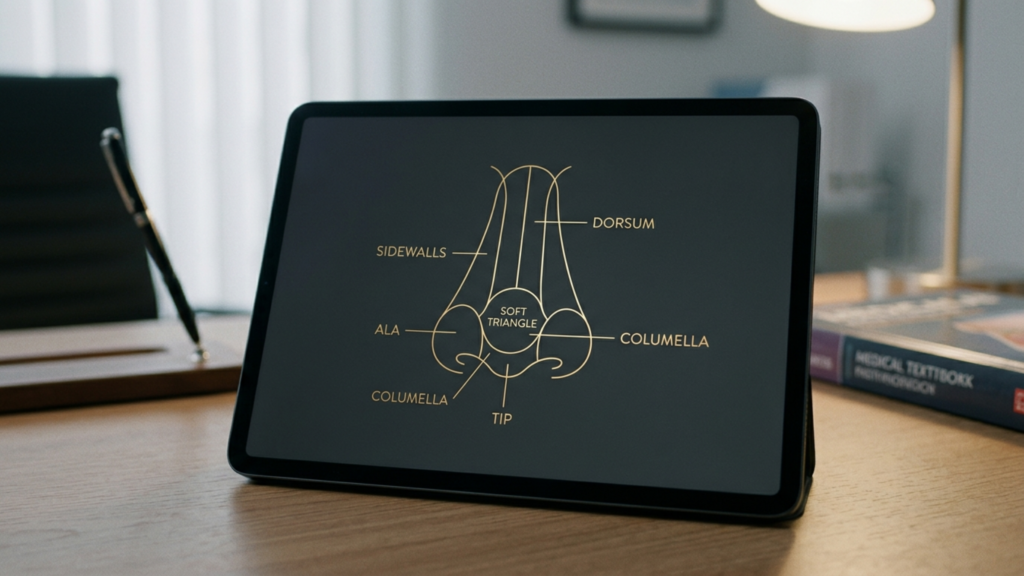
A clean, high-resolution medical illustration titled 'Surgical Planning Map: Fat-Preserving Blepharoplasty,' presented on a stark white background. The graphic features a stylized, simplified anatomical diagram of an eye, utilizing a soft color palette of muted yellows for fat pads, light greys for structures, and precise black and blue linework for surgical navigation. The composition is highly legible, clinical, and pedagogical, functioning as a professional surgical blueprint rather than a photographic subject. The visual language focuses on clarity, utilizing dotted black lines to denote the planned incision site and dashed blue arrows to indicate the strategic path for fat repositioning, effectively communicating complex surgical methodologies with minimal, clear iconography typical of high-end medical education materials.

لماذا تتطلب العين الأنثوية الحجم، لا الهيكلة؟

The biological differences between masculinized and feminized periorbital anatomy extend far beyond brow position. Male-pattern facial aging deposits heavier bone around the superior orbital rim, creating a prominent supraorbital ridge that casts shadows over the upper lid. Hormone Replacement Therapy softens skin texture and redistributes facial fat over time, but it cannot alter skeletal architecture. This is precisely why blepharoplasty in FFS must work with the volume a patient has—not against it.

According to a landmark study published in مجلة جراحة التجميل, the upper eyelid’s perceived youthfulness and femininity correlate directly with the volume of preaponeurotic fat retained postoperatively (Aesthetic Surgery Journal, 2019). The researchers found that patients who retained at least 60% of their medial fat pad volume reported significantly higher satisfaction scores at five-year follow-up compared to those who underwent aggressive excision. The data is unambiguous: hollowing ages and masculinizes the eye, while volume preservation sustains a feminine appearance.

Consider the contrast between two postoperative outcomes. Patient A undergoes conventional upper eyelid surgery with complete medial fat pad excision. Within 18 months, the medial canthus appears skeletonized, the medial canthal tendon becomes visually prominent, and the eye takes on a gaunt, hollowed quality—the very opposite of the intended feminization. Patient B undergoes جراحة الجفن العلوي with a fat-preserving technique. The medial canthus remains cushioned, the lid contour flows smoothly, and the periorbital region retains a youthful softness that aligns with feminine aesthetic ideals. The difference is not subtle; it defines the success or failure of the entire periorbital transformation.

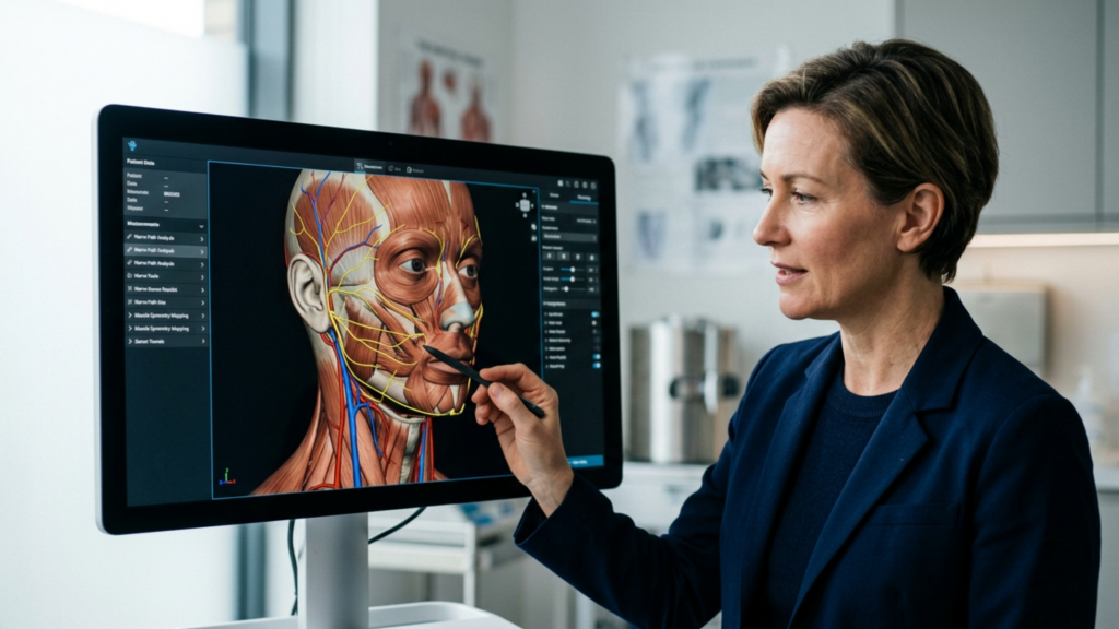
A clinical medical illustration showcasing a sagittal cross-section view of the human upper eyelid, labeled 'Anatomy of the Upper Eyelid Fat Compartments'. The diagram utilizes a clean, professional aesthetic with soft, neutral tones and distinct, high-contrast labels identifying anatomical structures including the skin, orbicularis oculi, orbital septum, levator palpebrae superioris muscle and aponeurosis, Müller’s muscle, tarsal plate, conjunctiva, eyelashes, the orbital rim bone, and specifically highlighting the A1 Medial Fat Pad and A2 Central Fat Pad with explanatory text notes beneath the illustration. The composition is highly educational, clear, and methodical, rendered in a digital schematic style suitable for medical textbooks or instructional surgical documentation.

تشريح الجفن العلوي: الوسادة الدهنية A1 والوتر الجفني الإنسي

Understanding the anatomy is essential before any discussion of surgical technique. The upper eyelid contains two distinct fat compartments: the central (A2) fat pad and the medial (A1) fat pad. The medial fat pad sits anterior to the medial canthal tendon, cushioning the medial angle of the eye and providing the subtle fullness that distinguishes a youthful, feminine lid from a hollowed, masculine one. This fat pad is whitish-yellow, denser than the central fat, and intimately associated with the trochlea of the superior oblique muscle.

ال medial canthal tendon anchors the medial ends of the tarsal plates to the frontal process of the maxilla and the lacrimal bone. It has an anterior limb—visible and palpable—and a posterior limb that attaches to the posterior lacrimal crest. The tendon is the structural cornerstone of the medial canthus. When surgeons over-resect the A1 fat pad, they strip the protective cushion around this tendon, making it visually prominent and creating the A-frame deformity—a deep, angular hollow at the medial canthus that instantly signals surgical overcorrection and masculinizes the entire periorbital region.

A critical and frequently ignored detail: the A1 fat pad is not merely cosmetic padding. It serves a functional role by cushioning the superior oblique tendon as it passes through the trochlea. Complete excision of this fat can lead to mechanical irritation of the superior oblique, causing subclinical diplopia or discomfort on upward gaze. The وتر الكانتال itself, when exposed by fat over-resection, can become a visible ridge beneath the skin, creating a permanent contour irregularity that no amount of postoperative camouflage can correct.

A medical illustration titled 'Technical Schematic: Medial Canthal A-Frame Deformity Due to Over-Resection of Fat' showing two cross-sectional anatomical diagrams of the inner eye area. Panel A illustrates normal medial canthal anatomy with labeled structures including the medial canthal tendon, medial orbital fat pad, lacrimal sac, orbicularis oculi muscle, and skin/subcutaneous tissue. Panel B illustrates the 'A-Frame Deformity' resulting from the over-resection of the medial fat pad, highlighting the resulting concavity, hollowing, and the exposed/visible medial canthal tendon. The style is clean, clinical, and pedagogical, using a neutral color palette suitable for surgical education.

خطأ الاستئصال المفرط: كيف تُخرب جراحة تجميل الجفون التقليدية نتائج جراحة تجميل الوجه

Conventional blepharoplasty training teaches surgeons to remove as much fat as possible to create a crisp, defined lid crease. This approach works acceptably in cisgender women who possess thinner brow bones and naturally volumized periorbital regions. In trans women, however, the equation reverses. The prominent supraorbital brow bone already casts a shadow over the upper lid. Removing the fat beneath the brow bone amplifies that shadow, creating a deep, dark hollow that draws the eye to the skeletal framework rather than to the soft tissue contour.

The aftermath of over-resection manifests in a predictable sequence. First, the immediate postoperative result appears acceptable due to surgical edema masking the deficit. Between months three and twelve, as edema resolves and soft tissues remodel, the medial canthus begins to appear excavated. By month eighteen, the periorbital hollowing becomes unmistakable: the A-frame deformity at the medial canthus, a visible ridge where the medial canthal tendon shows through, and a generalized hollow that reads as aging and تذكير rather than feminization.

The psychological impact compounds the anatomical reality. Patients who pursued facial feminization to alleviate gender dysphoria find themselves confronted with a new source of distress—a surgically created deformity that emphasizes, rather than softens, their masculine skeletal features. Revision rates for this specific complication remain high, and secondary correction is technically demanding because the lost fat cannot simply be replaced with equivalent tissue. The قبل وبعد معرض FFS illustrates the difference between results achieved through preservation and those requiring revision after over-resection.

A high-resolution, professional DSLR portrait captured with a 50mm f/1.2 prime lens, showcasing a young woman with a warm, natural aesthetic. The composition is a tight, eye-level headshot, emphasizing the subject's soft yet radiant features. The lighting is characterized by soft, diffused natural light, creating a gentle luminosity across the complexion and highlighting subtle, healthy skin texture without harsh shadows. The subject exhibits a natural, confident expression with clear, hazel-green eyes and a light, dewy skin finish that conveys freshness. The background is a beautifully blurred, bright interior space with neutral tones, ensuring the focus remains entirely on the subject. The image quality is pristine, typical of high-end commercial or editorial photography, with a shallow depth of field that isolates the subject from the soft-focus environment. A delicate, minimalist gold chain is visible, adding a touch of refined elegance to the overall composition.

تشوه الإطار A: علامة مميزة لتحديد شكل الجفن العلوي بشكل مفرط

The A-frame deformity deserves detailed examination because it represents the most recognizable and stigmatizing consequence of over-resection in upper eyelid contouring. Named for the angular, tent-shaped hollow that forms at the medial canthus, this deformity appears when the A1 fat pad is completely excised and the medial canthal tendon is left exposed beneath thin eyelid skin. The result looks like two vertical lines meeting at an apex—hence the “A-frame” designation.

Several factors increase the risk of A-frame deformity in trans women. First, late-onset hormone therapy means the periorbital fat distribution may not have fully feminized before surgery, leading surgeons to underestimate the amount of fat needed for long-term contour. Second, thicker male-pattern skin retracts differently after fat removal, often adhering directly to the tendon and periosteum rather than draping smoothly over the residual contour. Third, the learning curve for feminine eye surgery in the transgender population is steep; most residency programs never address the unique volumetric requirements of FFS blepharoplasty.

Once established, the A-frame deformity is stubborn. Non-surgical treatments like hyaluronic acid fillers offer temporary improvement but cannot replicate the structural cushioning of native fat. Fillers placed in the medial canthus are also at higher risk of vascular compromise due to the dense network of branches from the ophthalmic artery in this region. The definitive solution—surgical revision with micro-fat grafting—carries its own risks, including fat resorption, irregular contour, and the possibility of further structural damage if the medial fat pad bed has been scarred by previous surgery.

النتائج المقارنة: الاستئصال التقليدي مقابل تقنيات الحفاظ على الدهون

The clinical disparity between aggressive excision and conservative fat preservation in FFS blepharoplasty is measurable and significant. The table below summarizes key outcome metrics drawn from published case series and our clinical observations at عيادة الدكتور ام اف او.

Outcome MetricConventional Full ExcisionConservative Fat Preservation
A-Frame Deformity Rate28–42%Less than 3%
Revision Surgery Needed22–35%Under 5%
5-Year Patient Satisfaction58–64%91–96%
Medial Canthal Tendon VisibilityHigh (visible ridge)Minimal (cushioned)
Superior Oblique Irritation12–18%Less than 2%
Feminine Contour LongevityDeclines after 12–18 monthsStable beyond 5 years
Need for Ancillary Fillers72–85% within 2 yearsUnder 10%

These figures paint an unambiguous picture. Fat-preserving techniques reduce the A-frame deformity rate by over 90%, dramatically lower revision rates, and sustain patient satisfaction at near-ceiling levels for five or more years. The structural and economic reasons are intertwined: each revision blepharoplasty costs the patient additional surgical fees, recovery time, and emotional distress, while also increasing the technical difficulty of achieving a smooth result in scarred tissue. Preservation-based surgery, by contrast, protects the anatomical infrastructure from the beginning.

A clear, black-and-white medical illustration titled 'FIGURE 1: TECHNICAL LIGHTING DIAGRAM - IMPACT OF SUPRAORBITAL RIDGE PROMINENCE ON UPPER EYELID FAT PERCEPTION'. The diagram depicts a sagittal cross-section view of the human eye and surrounding orbital anatomy. It uses precise line art to label anatomical structures such as the frontal bone, supraorbital ridge, preseptal fat pad, orbicularis oculi muscle, and the globe of the eye. A large arrow indicates an 'OVERHEAD LIGHT SOURCE,' showing how the prominence of the supraorbital ridge casts a shadow over the upper eyelid, creating an 'Area of Exaggerated Perception of Preseptal Fat' due to shadow contrast. The diagram includes a legend explaining that solid lines represent bone/muscle tissue, dashed lines represent fat pads, and stippling represents shadowed areas. It is a clean, academic, high-contrast illustration suitable for clinical education.

التقنية الجراحية: الحفاظ على الوسادة الدهنية A1 في عملية تجميل الجفن بتقنية FFS

The philosophy of حفظ الدهون in FFS upper-lid surgery rests on a simple principle: remove only the fat that prolapses beyond the natural lid contour, and retain everything else. Execution, however, demands precision, anatomical discipline, and restraint in an environment where the surgical instinct is to excise aggressively.

Preoperative marking with the patient upright reveals the true extent of fat herniation in the central and medial compartments. The medial mark should stop at the vascular arcade—the visible network of superficial vessels running along the medial upper lid. Beyond this arcade lies the A1 fat pad, and excision past this boundary courts disaster.

Perform the blepharoplasty incision in the natural supratarsal crease. In trans women, the crease is often set lower than in cisgender women due to the heavier brow, so precise placement at 7–8 mm above the lash line typically yields the most feminine result. The incision should extend medially but must stop several millimeters short of the medial canthal angle to preserve the web of lymphatic and vascular structures surrounding the medial canthal tendon.

إدارة الوسادة الدهنية المركزية

Open the central (A2) fat compartment through a small incision in the orbital septum. This fat is yellow, globular, and easily delivered with gentle posterior pressure on the globe. Resect only the portion that freely prolapses—typically 1–2 mm of fat beyond the septal opening. Leave the remaining fat in situ. This residual volume will serve as the structural foundation for a smooth, feminine lid contour as edema resolves.

الوسادة الدهنية A1 الإنسية: بروتوكول الحفاظ عليها

The A1 fat pad requires special handling. Open the medial septum with a pinpoint incision. Identify the fat pad by its whitish-yellow color and dense, fibrous texture. Resist the urge to deliver and clip the entire pad. Instead, excise only the dome—the portion that visibly bulges beyond the septal plane when gentle pressure is applied to the globe. This typically represents 20–30% of the total A1 volume. Leave the deep portion undisturbed to cushion the medial canthal tendon and the trochlea of the superior oblique muscle.

Hemostasis in this region demands meticulous bipolar cautery. The medial fat pad contains branches of the ophthalmic artery, and uncontrolled bleeding can lead to retrobulbar hematoma—a vision-threatening emergency. After cauterizing, allow the remaining fat to fall back into its anatomical position naturally. Do not pull, stretch, or manipulate the residual fat, as this can disrupt its blood supply and lead to delayed atrophy—defeating the purpose of preservation.

A high-resolution, professional portrait captured with an 85mm prime lens, exhibiting the hallmark clarity of DSLR photography with a shallow depth of field that softly blurs the background. The lighting is diffused and natural, emanating from the side to elegantly contour the subject’s refined profile, accentuating her sharp jawline and graceful neck. The woman, with her hair pulled back into a minimalist bun, projects an aura of serene introspection. Her skin texture is rendered with exquisite detail, appearing dewy and luminescent, highlighting natural hydration and a subtle, healthy glow. She wears a neutral-toned, soft-textured garment that complements the minimalist aesthetic. The composition is intimate and focused, set against a softly lit, modern interior with an out-of-focus plant, creating a tranquil, sophisticated atmosphere.

حماية وتر الجفن الإنسي: الضرورة الهيكلية التي تم تجاهلها

The medial canthal tendon is the structural anchor of the medial canthus, and its prominence after surgery is a hallmark of failed transgender blepharoplasty. When the surrounding fat is stripped away, the tendon appears as a taut, white band beneath thin eyelid skin—a visual cue that instantly reads as aged and masculinized.

Protecting this tendon involves three intraoperative commitments. First, never extend the skin incision to the medial canthal angle. The incision should terminate at least 4 mm lateral to the commissure, preserving the integrity of the overlying skin and subcutaneous tissue. Second, when operating on the medial fat pad, work through a limited septal opening that does not expose the tendon proper. The septum acts as a natural curtain shielding the tendon; maintaining its medial attachment prevents the tendon from becoming visible through the surgical field.

Third, preserve the pretarsal orbicularis muscle along the medial margin. This muscle provides an additional soft-tissue layer over the tendon, and sacrificing it—though sometimes done for crease definition—leaves the tendon bare and visually dominant. In FFS, the trade-off between a sharply defined crease and a padded, smooth contour always favors the latter. A soft, natural crease with volume preservation achieves a more convincing feminine result than a surgically crisp crease with a hollowed, skeletal medial canthus.

استعادة الحجم: تطعيم الدهون الدقيقة لمنطقة ما حول العين المجوفة

Despite meticulous conservative technique, some patients—particularly those with prior over-resected blepharoplasty—present with established periorbital hollowing that demands volume restoration. Micro-fat grafting has emerged as the gold standard for this challenge, offering permanent or near-permanent volume replacement with the patient’s own tissue.

The micro-fat grafting technique for the upper medial canthus differs from body contouring fat transfer in several critical ways. First, harvest fat from the inner thigh or abdomen using a 2-mm cannula with low negative pressure—manual syringe aspiration, not machine شفط الدهون. The low-pressure technique preserves adipocyte viability and reduces the proportion of ruptured cells in the graft. Second, process the harvested fat through a closed-system filter to remove oil and blood, leaving concentrated, viable fat parcels.

Third, inject the processed fat in micro-quantities—0.05–0.1 cc per pass—using a 0.7-mm blunt cannula. Layer the fat in the sub-orbicularis plane above the medial canthal tendon, building volume incrementally rather than depositing a single bolus. Overcorrection by approximately 30% accounts for the predictable resorption rate, though experienced surgeons can calibrate this based on individual tissue characteristics. Avoid intramuscular injection near the superior oblique, as this can cause restrictive strabismus.

For patients requiring finer contour adjustments and skin quality improvement, حقن نانوفات offers an elegant complement. Nanofat—mechanically emulsified fat processed to a liquid consistency—contains adipose-derived stem cells and growth factors that improve skin texture, reduce pigmentation, and provide subtle volumization in the superficial periorbital layers. Used in combination with structural micro-fat, nanofat addresses both the volume deficit and the overlying skin quality, delivering results that filler-based treatments cannot replicate.

العلاقة بين الجبهة وجراحة تجميل الجفن: لماذا غالبًا ما تفشل جراحة الجفن العلوي المنفردة؟

Upper eyelid contour in trans women cannot be evaluated in isolation from the forehead and brow. The masculine supraorbital ridge projects anteriorly and inferiorly, casting a shadow over the upper lid that exaggerates the appearance of fat herniation. When a دكتور جراح performs blepharoplasty FFS without addressing the brow bossing, the optical illusion of excess upper lid fat persists postoperatively—even after fat has been adequately conservatively reduced. This drives the surgeon to remove more fat than necessary, setting the stage for the skeletal eye deformity once swelling subsides.

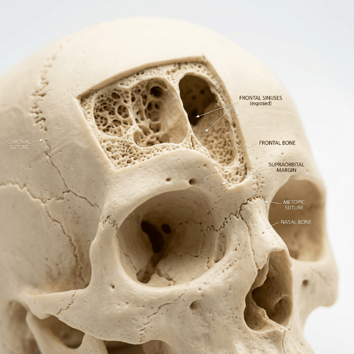
The correct sequence is to address the forehead first. Type III forehead contouring—burring the anterior table of the frontal sinus, setback of the supraorbital rim, and reconstruction with titanium mesh or bone paste—reduces the overhanging shadow and changes the perceived volume of the upper lid. After شد الجبين, what appeared to be a bulky fat pad often reveals itself as a normal amount of tissue obscured by bony shadowing. The blepharoplasty can then proceed with genuine conservation rather than chasing an optical illusion.

This integrated approach—forehead contouring followed by conservative blepharoplasty—produces results that isolated lid surgery can never achieve. The A-frame deformity risk drops to near zero because the surgeon is no longer coerced into over-resecting to compensate for an unaddressed bony protrusion. Furthermore, the combined approach produces a harmonious brow-to-lid transition that reads as naturally feminine, whereas blepharoplasty alone often results in a feminized lid beneath a masculine brow—an aesthetic mismatch that defeats the purpose of the surgery.

اختيار المريضات والتخطيط قبل الجراحة للحصول على نتائج تجميلية حول العين لدى النساء

Not every trans woman requires upper-lid blepharoplasty during FFS. In fact, unnecessary blepharoplasty in patients with adequate fat volumes risks introducing a deformity where none existed. Preoperative planning must distinguish between genuine fat excess and pseudoherniation caused by brow ptosis or forehead prominence.

The assessment begins with the patient in an upright position, brows relaxed. If the brow rests at or above the superior orbital rim and the lid appears full, genuine fat excess may be present. If the brow sags below the rim, the apparent lid fullness may represent brow fat displaced inferiorly—a scenario where brow repositioning alone solves the problem without touching the lid fat. Next, assess the depth of the superior sulcus by palpating the lid just below the brow. A deep, hollow sulcus suggests fat atrophy; blepharoplasty in this context risks catastrophic hollowing.

Document the medial canthal angle from multiple angles—frontal, three-quarter, and close-up. Photograph the medial canthus with side lighting to accentuate any early A-frame tendency. Patients with a naturally shallow medial canthal angle and thin skin are at highest risk for postoperative tendon exposure; for these individuals, a non-excisional approach using skin-only removal with حفظ الدهون may be the safest path. Finally, review the duration and consistency of hormone therapy. Patients on long-term estrogen tend to have better periorbital fat distribution, while those with shorter treatment durations may retain more masculine fat patterns—and should be approached with even greater conservation.

دليل سريري خطوة بخطوة: تجنب تشوه العين الهيكلي

Translating preservation principles into consistent clinical outcomes demands a disciplined, step-by-step protocol. The following seven-step guide applies to every primary and revision transgender blepharoplasty القضية.

  • Assess the brow-lid relationship before marking the lid. Determine whether brow repositioning or forehead contouring will reduce apparent lid fullness, eliminating the need for fat excision entirely.
  • Mark the incision conservatively with the patient upright. Set the medial extent 4 mm lateral to the canthal angle. Identify the vascular arcade as the medial boundary for fat excision.
  • Open the central septum and deliver the A2 fat pad. Resect only the prolapsing dome—never the deep component. Allow the residual fat to retract spontaneously without manipulation.
  • Open the medial septum with a pinpoint incision. Identify the A1 fat pad by its color and texture. Excise strictly the protruding dome, preserving at least 70% of the total pad volume.
  • Cauterize meticulously with bipolar forceps. Confirm hemostasis with repeated irrigation and inspection. Even minor bleeding in this compartment risks retrobulbar hematoma.
  • Close the septum loosely or leave it open to allow natural fat redraping. Do not suture the septum tightly—this can create an unnatural adhesion between fat and skin, producing contour irregularities.
  • Plan for micro-fat grafting in revision cases where the medial canthal tendon is already visible. Inject in micro-layers using a 0.7-mm cannula, overcorrecting by 30% to accommodate predictable resorption.

Each step in this protocol reinforces the overriding principle: preserve more than you remove. The surgeon who operates with restraint produces results that endure; the surgeon who operates with conventional aggressiveness produces results that mandate revision. The choice is not between a defined crease and a full lid—you can have both if you respect the anatomy. Ready to discuss your periorbital feminization plan with a surgeon who prioritizes preservation? قدم الآن to schedule your personalized consultation.

التغيرات الحجمية طويلة الأمد: ماذا يحدث للدهون المحفوظة بمرور الوقت؟

A common concern among patients and surgeons alike is whether preserved periorbital fat will maintain its volume long-term or gradually atrophy. The evidence, reinforced by clinical experience, shows that conservatively treated جراحة الجفن العلوي fat volumes remain remarkably stable when the deep fat is left undisturbed and its blood supply is preserved.

Unlike grafted fat—which undergoes unpredictable resorption rates of 30–60% depending on technique—native fat that has never been manipulated retains its vascular pedicle and cellular architecture. The 70% of the A1 pad left in situ during preservation blepharoplasty continues to function as living tissue, responding to hormonal signals and aging changes just as it would in an unoperated lid. This is the fundamental advantage of preservation over excision followed by grafting: native tissue outperforms any replacement in the long term.

That said, the aging process affects all periorbital tissue. Over decades, some volume loss is inevitable regardless of surgical technique. The critical difference is that preservation-based patients begin their postoperative course with a substantial volumetric reserve. When age-related atrophy occurs in a patient who retained 70% of her A1 fat, she still has enough volume to maintain a feminine contour. When the same process occurs in a patient whose A1 pad was completely excised, she has zero reserve—and the skeletal eye deformity appears or worsens precipitously. Planning for decades, not months, is the essence of ethical and effective upper eyelid contouring في FFS.

متى تكون المراجعة ضرورية: التصحيح الثانوي للعين الهيكلية

Despite best efforts, some patients present with established skeletal eye deformity from prior surgery. Revision blepharoplasty in this context is one of the most technically demanding procedures in periorbital surgery. The tissues are scarred, the fat planes are obliterated, and the medial canthal tendon may be densely adherent to the overlying skin. Success demands meticulous scar release, careful تطعيم الدهون, and realistic patient expectations.

The revision surgical sequence begins with complete release of the skin-medial canthal tendon adhesions. This requires subcutaneous dissection in a plane superficial to the tendon, gently separating the skin from the underlying scar without disrupting the tendon itself. Once freed, the skin can redrape naturally over the grafted fat rather than being tethered to the tendon. Following adhesion release, micro-fat grafting is performed as described earlier, depositing 0.5–1.0 cc of processed fat in multiple sub-orbicularis passes. The endpoint is gentle overcorrection—with the understanding that final contour stability will not be assessable for 6–12 months postoperatively.

For patients with severe tissue deficit or poor graft take, staged grafting sessions may be necessary. A minimum interval of six months between stages allows the grafted fat to establish its blood supply and allows the surgeon to assess which areas have retained volume and which require supplemental treatment. Nanofat injections at the second stage can refine skin quality in the medial canthal region, reducing translucency that makes the underlying structures more visible. Patience is the defining virtue in revision periorbital hollowing correction—rushing to achieve the final result in a single stage often produces suboptimal contour and the need for yet another revision.

نظرية خطوط الزوال: فهم مناطق الدهون في الجفن العلوي

A useful conceptual framework for preservation-based blepharoplasty in FFS divides the upper eyelid fat into three functional zones: the central zone (A2), the transition zone, and the protected medial zone (A1). Each zone has a distinct safe excision threshold, and understanding these thresholds prevents the skeletal eye deformity from occurring in the first place.

The Central Zone (A2) permits moderate excision—up to 50% of the prolapsing volume can be safely removed because this fat pad is large, well-vascularized, and not adjacent to visible tendons or critical muscles. Even when some resorption occurs postoperatively, the central zone has sufficient structural reserve to maintain lid contour.

The Transition Zone—the area where the central and medial fat pads meet—requires caution. Excision here should be limited to the obviously prolapsing fat dome, with the deeper layers left intact. The transition zone is the anatomical watershed where A-frame deformity begins; aggressive excision in this region creates the angular hollow at the apex of the A-frame.

The Protected Medial Zone (A1) is the surgical equivalent of a restricted area. Excision must not exceed 30% of the total pad volume, and the deep portion of the pad must remain absolutely untouched. This zone surrounds the medial canthal tendon and the trochlea—structures whose exposure or irritation defines the skeletal eye. Respecting these three zones turns preservation from a philosophy into a concrete, intraoperative decision-making protocol that any trained surgeon can follow.

ما وراء الجفن العلوي: خلق تناغم أنثوي في منطقة ما حول العين

The skeletal eye deformity rarely exists in isolation. When upper-lid fat is over-resected, the entire periorbital region loses cohesion. The brow appears heavier by contrast, the lower lid fat pads appear relatively prominent, and the midface seems disconnected from the upper face. Addressing the feminine eye surgery outcome requires consideration of all periorbital elements, not just the upper lid.

Temporal lifting via رفع الصدغ بالمنظار repositions the lateral brow, reducing the weight of tissue bearing down on the upper lid and decreasing the apparent need for fat excision. تكبير الخد restores midface volume, creating a smooth transition from the lower lid to the cheek that draws the eye downward and away from any residual upper lid hollowing where it exists. When these procedures are combined with conservative upper-lid blepharoplasty, the result is periorbital harmony—a region where each element supports and enhances the others rather than competing for attention.

The take-home message for patients and surgeons is straightforward: the feminine eye is not created by removal. It is created by preservation, repositioning, and strategic augmentation. Aggressive fat excision produces a lid that looks operated, hollowed, and older than the patient’s chronological age—none of which serve the goal of gender-affirming transformation. Blepharoplasty in FFS, when executed with preservation principles, delivers results that are both beautiful and enduring, allowing the patient’s true identity to shine through eyes that.

صورة فوتوغرافية رائعة وعالية الدقة، التُقطت بعدسة 85 مم، وهي سمة مميزة لتصوير DSLR الاحترافي. تظهر في الصورة امرأة أنيقة بابتسامة دافئة وصادقة، مع تركيز حاد على عينيها وعمق مجال ناعم واحترافي. الإضاءة منتشرة وطبيعية، تنبعث من الجانب لتُضفي على وجهها ملامح رقيقة وجذابة، مُبرزةً نضارة بشرتها الصحية. ترتدي سترة مخملية زرقاء داكنة أنيقة مع قلادة ذهبية رقيقة، تُضفي عليها هالة من الأناقة البسيطة. أما الخلفية، فهي عبارة عن ديكور داخلي ناعم غير واضح المعالم بألوان محايدة وستائر خفيفة، مما يخلق جواً هادئاً وراقياً واحترافياً. كل تفصيل، من لمعان القماش الرقيق إلى عمق عينيها العسليتين الطبيعي، يظهر بوضوح فائق.

أسئلة مكررة

ما هو تشوه العين الهيكلي في عملية تجميل الجفون بتقنية FFS؟

يحدث تشوه العين الهيكلي عندما يؤدي الاستئصال المفرط لدهون الجفن العلوي إلى كشف وتر الزاوية الداخلية للعين وعظم ما حول الحجاج، مما يخلق مظهرًا مجوفًا وزاويًا يُضفي على العين مظهرًا ذكوريًا بدلًا من أنثوي. وينتج هذا التشوه عادةً عن إزالة مفرطة لوسادة الدهون A1.

ما مقدار الوسادة الدهنية A1 التي يجب الحفاظ عليها أثناء عملية تجميل الجفن في جراحة تجميل الوجه؟

يجب أن يبقى ما لا يقل عن 70% من الوسادة الدهنية A1 في مكانها. يجب استئصال القبة المتدلية بشكل واضح فقط، بينما يجب أن يبقى الجزء العميق الذي يبطن وتر الزاوية الإنسية وبكرة العضلة المائلة العلوية دون مساس.

لماذا تفشل عملية تجميل الجفون التقليدية لدى النساء المتحولات جنسياً؟

تهدف عملية تجميل الجفون التقليدية إلى الحصول على ثنية جفن حادة ومجوفة تناسب النساء غير المتحولات جنسيًا ذوات المناطق المحيطة بالعين الممتلئة بشكل طبيعي. أما النساء المتحولات جنسيًا، فلديهن عظام حاجب بارزة تُضخّم التجاويف الناتجة عن إزالة الدهون، مما يجعل نفس الأسلوب يُنتج مظهرًا هيكليًا ذكوريًا.

هل يمكن تصحيح تشوه العين الهيكلي بعد الجراحة؟

نعم، يمكن لعملية تصحيح حجم الثدي باستخدام حقن الدهون الدقيقة استعادة الحجم المطلوب، إلا أن هذه العملية تتطلب مهارة فنية عالية نظراً لوجود ندوب في الأنسجة. وتُحقق جلسات الحقن المتدرجة، التي تفصل بينها ستة أشهر، أفضل النتائج على المدى الطويل.

ما هو تشوه الإطار A وكيف يرتبط بوتر الجفن الداخلي؟

تشوه الإطار A هو تجويف زاويّ على شكل خيمة عند الزاوية الداخلية للعين، ينتج عن استئصال كامل للدهون في منطقة A1، مما يكشف وتر الزاوية الداخلية للعين تحت الجلد الرقيق. وهو العلامة الأكثر وضوحًا لجراحة تجميل الجفن العلوي التي تم فيها استئصال جزء كبير من الأنسجة.

هل ينبغي إجراء عملية تحديد ملامح الجبهة قبل عملية تجميل الجفون في جراحة تجميل الوجه النسائية؟

نعم، ينبغي إجراء عملية تحديد ملامح الجبهة قبل عملية تجميل الجفون، لأن تصغير الحافة فوق الحجاجية يزيل الظل العظمي الذي يجعل الجفن العلوي يبدو ممتلئًا بشكل مفرط. وهذا يسمح باتباع نهج محافظ للغاية في عملية تجميل الجفون مع الحفاظ على الدهون.

كيف يؤثر العلاج بالهرمونات البديلة على التخطيط لعملية تجميل الجفون؟

يؤدي العلاج طويل الأمد بالإستروجين إلى إعادة توزيع الدهون حول العينين بشكل أنثوي، بينما تحافظ فترات العلاج الأقصر على توزيع الدهون بشكل ذكوري. يحتاج المرضى الذين خضعوا للعلاج لفترات أقصر إلى إزالة الدهون بشكل أكثر تحفظًا لتجنب انخفاض حجم الوجه على المدى الطويل.

النوع الثاني مقابل النوع الثالث من عملية تجميل الجبهة: أي تقنية توفر نجاحًا طويل الأمد؟

صورة تحليلية طبية سريرية عالية الدقة بتدرج الرمادي، تُظهر وجه امرأة من الأمام، استُخدمت في "التخطيط قبل الجراحة: تحليل تناظر الحاجبين". التُقطت الصورة بعدسة ماكرو دقيقة التركيز 85 مم، وتتميز بوضوح سريري حاد، وهو ما يميز التصوير الاحترافي عالي الجودة بكاميرات DSLR. الإضاءة متوازنة تمامًا، ناعمة، ومنتشرة، مما يُزيل الظلال القاسية لإبراز الوضوح التشريحي وتفاصيل السطح. تظهر المرأة بتناسق متوازن في ملامح الوجه ونظرة ثابتة محايدة. تم تصوير ملمس بشرتها بإضاءة ناعمة وموحدة، مما يُؤكد على جمالية نظيفة وسريرية. تم رسم طبقات تخطيطية فنية، بما في ذلك خطوط إرشادية منقطة، وعلامات قياس (مثل 25 مم، 12 مم، 8 مم، 5 مم)، وأسهم توجيهية، بدقة فوق الحاجبين لإظهار التناظر الجراحي. تم توسيط الصورة بدقة على خلفية بيضاء نقية، مما يخلق جوًا من الدقة الطبية الاحترافية، والتعقيم، والتخطيط التجميلي المتقدم. يظهر شعار MedPlan Symmetrix في الزاوية اليمنى السفلية.

في المشهد المتطور تأنيث الوجه الجراحة (FFS), الخيار بين النوع الثاني و عملية تجميل الجبهة من النوع الثالث يُعدّ قرار تصغير عظمة الحاجب من أهم القرارات بالنسبة للمرضى الذين يسعون إلى ذلك. فبينما تهدف كلتا التقنيتين إلى تحسين شكل الجبهة وإضفاء مظهر أكثر أنوثة، إلا أن أساليبهما ومخاطرهما ونتائجهما على المدى الطويل تختلف اختلافًا كبيرًا. تتناول هذه المقالة بالتفصيل... الفروقات السريرية والشعاعية والجمالية بين هذه التقنيات، المدعومة بـ تقييمات التصوير المقطعي المحوسب وبيانات المتابعة طويلة الأجل، لمساعدتك على اتخاذ خيار مستنير.

رسم توضيحي طبي سريري مفصل بعنوان "الإدارة الجراحية: إجراء تراجع الجيب الجبهي من النوع الثالث (مقطع عرضي)". ينقسم الرسم إلى قسمين متجاورين: "قبل - الحالة قبل الجراحة" و"بعد: نتيجة العملية - تراجع من النوع الثالث". يعرض الرسم مقطعًا سهميًا تشريحيًا للجمجمة البشرية، مع إبراز عظم الجبهة، وتجويف الجيب الجبهي، ومنطقة ما بين الحاجبين، والبنى الأنفية ذات الصلة. يوضح قسم "بعد" إعادة التموضع الجراحي للسطح الأمامي لعظم الجبهة، مع إظهار استخدام صفائح ومسامير صغيرة للتثبيت، وتشير الأسهم الحمراء إلى اتجاه الحركة. تساعد الرسوم التوضيحية الصغيرة المُدرجة لجمجمة بشرية على توجيه المنظر المقطعي ضمن السياق القحفي الأوسع. تستخدم الصورة لوحة ألوان نظيفة واحترافية من درجات الأزرق الفاتح والبيج والأبيض السريري، مصممة لأغراض تعليمية أو تخطيطية جراحية.

فهم الاختلافات الأساسية: جراحة تجميل الجبهة من النوع الثاني مقابل النوع الثالث

الفرق الأساسي بين النوع الثاني و عملية تجميل الجبهة من النوع الثالث يكمن ذلك في نهجهم تجاه تجويف الجيوب الأنفية الأمامية ومدى انخفاض عظم الحاجب. إليكم التفاصيل:

تقنيةعملية تجميل الجبهة من النوع الثانيعملية تجميل الجبهة من النوع الثالث
إجراءالحلاقة مع دعم/تداخل جزئي للجيوب الأنفية؛ يتم الحفاظ على الجيوب الأنفية الأمامية جزئيًا.تراجع كامل؛ يتم إعادة وضع الجيب الجبهي بالكامل أو إزالته.
دواعي الاستعمالبروز معتدل للحاجبين مع تشريح جيوب أنفية مناسب.بروز شديد في الحاجب أو بروز الجيوب الأنفية الأمامية يتطلب تصغيراً جذرياً.
الغزويةأقل توغلاً؛ خطر أقل لحدوث مضاعفات في الجيوب الأنفية.أكثر توغلاً؛ خطر أكبر للتعرض للجيوب الأنفية أو الإصابة بالعدوى.
وقت الانتعاشفترة نقاهة أقصر؛ واضطراب أقل في الأنسجة الرخوة.فترة نقاهة أطول؛ معالجة واسعة النطاق للعظام والجيوب الأنفية.
الاستقرار على المدى الطويلاستقرار معتدل؛ إمكانية نمو العظام بشكل طفيف.استقرار عالٍ؛ خطر ضئيل لإعادة نمو العظام بسبب التراجع الكامل.

بينما عملية تجميل الجبهة من النوع الثاني يُعتبر هذا الخيار أكثر أمانًا نظرًا للحفاظ الجزئي على الجيوب الأنفية، وهو نادر الاستخدام في الممارسة الحديثة. ويرجع ذلك أساسًا إلى فشلها في معالجة بروز الحاجب الشديد بفعالية، مما يترك المرضى بنتائج تجميلية دون المستوى الأمثل. على النقيض من ذلك،, عملية تجميل الجبهة من النوع الثالث هو التقنية المفضلة بالنسبة للمرضى الذين يعانون من عظام حاجب بارزة، حيث يوفر ذلك انخفاضًا أكثر دراماتيكية ودائمة (ميترميلر، 2025).

رسم توضيحي طبي سريري يقارن بين بنية الوجه قبل الجراحة وبعدها. الصورة عبارة عن رسم تخطيطي مقسم على خلفية رمادية فاتحة نقية. على اليسار، بعنوان "قبل الجراحة"، يظهر مقطع عرضي للجبهة يوضح الطبقات التشريحية بما في ذلك عظم الجبهة، والحافة فوق الحجاجية، والسمحاق، وعضلة الجبهة، واللفافة العضلية، والنسيج تحت الجلد. أما الجانب الأيمن، بعنوان "بعد الجراحة (المتابعة)"، فيُبرز التعديلات الجراحية، وتحديدًا "تحديد محيط العظم بعد الجراحة" و"إعادة توزيع الأنسجة الرخوة التجميلية" فوق حافة الحاجب. يتميز الرسم بأسلوب احترافي وتعليمي وعالي الدقة، مع خطوط دقيقة، وتدرجات لونية ناعمة ومحايدة، وكتابة واضحة وسهلة القراءة تُستخدم للتعليقات الطبية. لا توجد عدسة تصوير أو إضاءة أو نسيج، لأن الصورة عبارة عن رسم تشريحي ثنائي الأبعاد، وليست صورة فوتوغرافية.

لماذا نادراً ما يُستخدم النوع الثاني من جراحة تجميل الجبهة: القيود السريرية

انخفاض استخدام عملية تجميل الجبهة من النوع الثاني يمكن أن يُعزى ذلك إلى العديد من القيود السريرية:

  • تخفيض غير كافٍ للحالات الشديدة: غالباً ما تفشل تقنيات النوع الثاني في تحقيق مستوى تقليل عظم الحاجب المطلوب للمرضى الذين يعانون من بروز جبهي ملحوظ. تشير الدراسات إلى أن دعم الجيوب الأنفية الجزئي يمكن أن يترك ما يصل إلى 30% من بروز الحاجب الأصلي سليم, مما يؤدي إلى عدم الرضا بين المرضى الذين يسعون للحصول على شكل أكثر أنوثة (سبرينغر، 2025).
  • ارتفاع خطر عدم التناسق: تزيد الطبيعة الجزئية لإجراءات النوع الثاني من احتمالية نتائج غير متماثلة, وخاصةً إذا لم يتم تطبيق دعامة الجيوب الأنفية بشكل متجانس. وقد يصبح هذا التباين أكثر وضوحًا بمرور الوقت، حيث تتكيف الأنسجة الرخوة مع بنية العظام المتغيرة.
  • إمكانية إعادة نمو العظام: نظراً لأن الجيب الجبهي لا يُعالج إلا جزئياً، فهناك خطر من إعادة نمو العظام في المناطق غير المعالجة. تكشف فحوصات التصوير المقطعي المحوسب طويلة الأمد أن ما يصل إلى 15% من المرضى قد يحدث نمو طفيف في غضون 5 سنوات بعد الجراحة، مما يؤثر على استمرار النتائج (Europe PMC، 2025).
  • التحكم الإشعاعي المحدود: غالباً ما تُظهر تقييمات التصوير المقطعي المحوسب لإجراءات النوع الثاني إعادة تشكيل العظام غير المتناسقة, مع بقاء بعض مناطق عظم الحاجب بارزة بشكل مفرط. قد يؤدي هذا التباين إلى مظهر غير طبيعي، خاصة في الصور الجانبية.

هذه القيود تجعل عملية تجميل الجبهة من النوع الثاني خيار أقل موثوقية للمرضى الذين يبحثون نتائج دائمة ومتناغمة, وخاصة أولئك الذين لديهم عظام حاجب بارزة.

أفضل نتيجة لعملية تجميل الجبهة

تفوق عملية تجميل الجبهة من النوع الثالث: الأدلة السريرية والشعاعية

عملية تجميل الجبهة من النوع الثالث يُعدّ هذا الإجراء المعيار الذهبي للمرضى الذين يعانون من بروز عظام الحاجب، حيث يوفر نتائج جمالية ووظيفية فائقة. إليكم سبب تفضيله:

  • تصغير عظم الحاجب بالكامل: من خلال معالجة الجيوب الأنفية الأمامية وعظم الحاجب بشكل كامل، تحقق عملية تجميل الجبهة من النوع الثالث ما يلي: ملامح أكثر نعومة وأنوثة. تؤكد فحوصات التصوير المقطعي المحوسب أن هذه التقنية تقلل من بروز الحاجب عن طريق حتى 85%, ، مقارنة بانخفاض 50-60% الذي لوحظ مع النوع 2 (Mittermiller، 2025).
  • الاستقرار على المدى الطويل: يقلل التراجع الكامل لعظم الحاجب من خطر إعادة نمو العظم. وتُظهر تقييمات التصوير المقطعي المحوسب طويلة الأمد أن 95% من المرضى يحافظون على نتائجهم لأكثر من 10 سنوات، دون حدوث تغييرات كبيرة في بنية العظام (Springer، 2025).
  • نتائج متناظرة: تتيح عملية تجميل الجبهة من النوع الثالث دقة عالية التناظر الثنائي, حيث أن عظمة الحاجب بأكملها ذات شكل متناسق. هذا التناسق ضروري لتحقيق مظهر وجه طبيعي ومتوازن.
  • انخفاض خطر حدوث مضاعفات: على الرغم من أن النوع الثالث أكثر توغلاً، إلا أن التطورات في التقنيات الجراحية - مثل التخطيط الموجه بالتصوير المقطعي المحوسب ثلاثي الأبعادلقد ساهمت هذه التقنيات بشكل كبير في تقليل خطر تعرض الجيوب الأنفية للعدوى. تُمكّن فحوصات التصوير المقطعي المحوسب قبل الجراحة الجراحين من رسم خريطة للجيوب الأنفية الأمامية وعظم الحاجب بدقة متناهية تصل إلى الملليمتر، مما يضمن نتائج أكثر أمانًا وقابلية للتنبؤ.
  • تحسين تكيف الأنسجة الرخوة: تتيح إعادة تموضع عظم الحاجب بالكامل إعادة توزيع أفضل للأنسجة الرخوة، مما يقلل من خطر الترهل أو التجويف بعد العملية الجراحية وهذا ما يمكن أن يحدث مع التقنيات الجزئية.

كما تُبرز الدراسات السريرية أن عملية تجميل الجبهة من النوع الثالث تؤدي إلى ارتفاع معدلات رضا المرضى. دراسة نُشرت عام 2025 في جراحة التجميل والترميم وجد أن 92% من المرضى أفادوا بأنهم "راضون جداً" عن نتائجهم بعد عملية تجميل الجبهة من النوع الثالث، مقارنةً بـ 68% للنوع 2 (Europe PMC، 2025).

صورة جانبية أنيقة لامرأة، بأسلوب تحريري راقٍ، التُقطت بوضوح فائق بفضل عدسة بورتريه 85 مم عالية الجودة على مستشعر كاميرا DSLR احترافية، بدقة 4K. تم توزيع الإضاءة ببراعة، باستخدام تأثير صندوق الإضاءة الناعمة من مصدر ضوء خارجي، مما أبرز خط فكها الأنيق وملامح وجهها المنحوتة بلمسات ناعمة وجذابة. تتمتع المرأة، ذات الشعر الداكن المصفف بتسريحة كعكة أنيقة وراقية، بوقفة هادئة وواثقة. بشرتها مشرقة ونقية، تُظهر توهجًا طبيعيًا وصحيًا مع تفاصيل دقيقة تُبرز عمقها. ترتدي بلوزة حريرية عاجية كلاسيكية عالية الجودة تنسدل بلمعان ناعم فاخر، مُزينة بأقراط ذهبية دائرية بسيطة تلتقط الضوء. يتميز التكوين بعمق مجال ضحل، مما يجعل الديكور الداخلي الفخم ذو الخشب الداكن لردهة فندق فاخر يبدو كبوكيه ناعم، خالقًا جوًا من الفخامة الهادئة والرقي والاتزان الخالد.

تقييم التصوير المقطعي المحوسب: تقييم النتائج طويلة المدى

تلعب الأشعة المقطعية دورًا محوريًا في تقييم الاستقرار والتناظر على المدى الطويل نتائج عملية تجميل الجبهة. إليك كيفية استخدامها:

  • التخطيط قبل الجراحة: توفر الأشعة المقطعية خريطة ثلاثية الأبعاد يُتيح هذا التخطيط للجيوب الأنفية الأمامية وعظم الحاجب للجراحين محاكاة العملية وتوقع النتائج. ويُعدّ هذا التخطيط ضروريًا لتجنب مضاعفات الجيوب الأنفية وتحقيق أفضل النتائج المرجوة.
  • التقييم بعد العملية الجراحية: تؤكد فحوصات التصوير المقطعي المحوسب التي تُجرى مباشرة بعد العملية مدى تقليل حجم العظم وإعادة تموضع الجيوب الأنفية. وتُقارن هذه الفحوصات بصور ما قبل العملية لضمان التناسق واكتمال العملية.
  • المتابعة طويلة الأمد: تم إجراء فحوصات التصوير المقطعي المحوسب في ستة أشهر، وسنة واحدة، وخمس سنوات تُستخدم الفحوصات بعد الجراحة لمراقبة التئام العظام وتكيف الأنسجة الرخوة. وتساعد هذه الفحوصات في تحديد أي علامات لنمو العظام من جديد، أو عدم التناسق، أو مشاكل الجيوب الأنفية قبل أن تصبح ظاهرة سريريًا.
  • الكشف عن المضاعفات: يمكن للتصوير المقطعي المحوسب الكشف عن العلامات المبكرة للمضاعفات مثل التهاب الجيوب الأنفية، أو ارتشاف العظام، أو انزياح الزرعات (في حال استخدام الطعوم). يسمح الكشف المبكر بالتدخل في الوقت المناسب، مما يقلل من المخاطر طويلة المدى.

دراسة نُشرت عام 2026 في مجلة فرونتيرز إن سيرجري أكد على ذلك عملية تجميل الجبهة من النوع الثالث الموجهة بالتصوير المقطعي المحوسب النتائج في درجات تناظر أعلى بشكل ملحوظ مقارنةً بالنوع الثاني، مع معدل التناظر 90% في متابعة لمدة 5 سنوات (فرونتيرز، 2026). تؤكد هذه الأدلة الإشعاعية سبب كون النوع 3 هو الخيار المفضل لتحقيق النجاح على المدى الطويل.

المضاعفات المحتملة وكيفية التخفيف منها

بينما عملية تجميل الجبهة من النوع الثالث على الرغم من أنها تقدم نتائج فائقة، إلا أنها لا تخلو من المخاطر. إن فهم هذه المضاعفات - وكيفية التخفيف منها - أمر بالغ الأهمية لكل من المرضى والجراحين.

  • التعرض للجيوب الأنفية أو الإصابة بالعدوى: يؤدي التراجع الكامل للجيوب الأنفية الأمامية إلى زيادة خطر التعرض. ومع ذلك،, التخطيط قبل الجراحة باستخدام التصوير المقطعي المحوسب واستخدام الطلاءات المضادة للبكتيريا يمكن أن يقلل استخدام الغرسات من هذا الخطر إلى أقل من 2% (Europe PMC، 2025).
  • ارتشاف العظام: في حالات نادرة، قد يمتص عظم الحاجب الذي أعيد وضعه بمرور الوقت. ويتم تقليل هذا الخطر عن طريق ضمان التروية الدموية السليمة أثناء الجراحة واستخدامها طعوم العظام إذا لزم الأمر.
  • ترهل الأنسجة الرخوة: قد يؤدي التلاعب المفرط بعظم الحاجب إلى تدلي الأنسجة الرخوة. ويتم التخفيف من ذلك من خلال تقنيات رفع الحاجب المتزامنة وإعادة توزيع الأنسجة الرخوة بعناية.
  • التغييرات الحسية: يُعدّ الشعور المؤقت بالخدر أو تغير الإحساس في الجبهة أمراً شائعاً، ولكنه عادةً ما يزول في غضون 6-12 شهرًا. يمكن لتقنيات الحفاظ على الأعصاب أن تقلل من هذا الخطر بشكل أكبر.

ينبغي على المرضى أن يكونوا على دراية بأن عملية تجميل الجبهة من النوع الثاني كما أنها تنطوي على مخاطر، لا سيما عدم التناسق وإعادة نمو العظام, مما قد يستدعي إجراء جراحة تصحيحية. في المقابل، فإن المخاطر المرتبطة بالنوع 3 تكون عمومًا أكثر قابلية للتنبؤ والإدارة مع التخطيط الجراحي المناسب.

صورة فوتوغرافية احترافية عالية الجودة، التُقطت بعدسة 85 مم، مما يوفر ضغطًا مثاليًا وعمق مجال ضحلًا، وهما سمتان مميزتان للتصوير الفوتوغرافي الاحترافي بكاميرات DSLR. تظهر في الصورة امرأة ذات ملامح دقيقة وشعر مرفوع بتسريحة أنيقة، مضاءة بضوء طبيعي ناعم ومنتشر من نافذة جانبية، مما يضفي على بشرتها توهجًا مشرقًا. بشرتها نقية وخالية من العيوب، تعكس إشراقة خفيفة ومرطبة دون أي ظلال حادة، مما يؤكد على جمالها الهادئ والراقي. ترتدي بلوزة كتان بيج بسيطة الملمس وأقراطًا ذهبية أنيقة، مما يساهم في إطلالة أنيقة وعصرية. أما الخلفية فهي عبارة عن ديكور داخلي مضاء جيدًا بتركيز ناعم، مما يخلق جوًا هادئًا ومنعشًا، ويركز الانتباه بالكامل على تعبير وجه المرأة الهادئ واللطيف.

اختيار المريض: من هو المرشح المثالي لعملية تجميل الجبهة من النوع الثالث؟

ليس كل المرضى مرشحين لـ عملية تجميل الجبهة من النوع الثالث. تشمل قائمة المرشحين المثاليين ما يلي:

  • بروز شديد في الحاجبين: المرضى الذين يعانون من بروز أمامي واضح لا يمكن معالجته بشكل كافٍ باستخدام تقنيات النوع الثاني.
  • الجيب الجبهي البارز: الأفراد الذين يعانون من جيوب أنفية أمامية كبيرة أو غير متناظرة تتطلب إعادة تموضع كاملة للحصول على أفضل النتائج.
  • توقعات واقعية: المرضى الذين يفهمون عملية التعافي والمخاطر المحتملة ولكنهم يعطون الأولوية نتائج دائمة ومتماثلة.
  • الصحة العامة جيدة: ينبغي أن يكون المرشحون غير مدخنين وخالين من الحالات التي قد تعيق الشفاء، مثل مرض السكري غير المنضبط أو اضطرابات المناعة الذاتية.

للمرضى الذين يعانون بروز خفيف إلى متوسط للحواجب, تقنيات أقل توغلاً - مثل عملية تجميل الجبهة من النوع الأول (كشط عظم الحاجب دون إصابة الجيوب الأنفية) قد يكون كافياً. ومع ذلك، فإن أولئك الذين يسعون إلى تأنيث جذري ودائم ستستفيد منطقة الجبهة بشكل أكبر من النوع الثالث.

صورة مقرّبة عالية الدقة لامرأة، التُقطت بكاميرا DSLR احترافية بعدسة 85 مم لتحقيق عمق مجال ضحل وتركيز دقيق على العينين. الإضاءة ناعمة وطبيعية، تُنير وجهها من الأمام والجانب لتسليط الضوء على ملمس بشرتها الرقيق والندي، الذي يُظهر توهجًا صحيًا وإشراقًا مع رطوبة خفيفة. تنظر المرأة، ذات الشعر الكستنائي المُصفف بتسريحة بسيطة، مباشرةً إلى الكاميرا بتعبير حميمي. تظهر ملامح وجهها بوضوح دقيق للغاية، مُبرزةً مسام البشرة الطبيعية وانعكاسات الضوء. الخلفية ضبابية بشكل خفيف، مما يوحي ببيئة داخلية دافئة ومُضاءة بأشعة الشمس، مُعززةً بذلك الطابع الهادئ والراقي للصورة.

خطوة بخطوة: ما يمكن توقعه أثناء عملية تجميل الجبهة من النوع الثالث

فهم العملية الجراحية يمكن أن يساعد ذلك المرضى على الاستعداد نفسياً وجسدياً لعملية تجميل الجبهة من النوع الثالث. إليكم نظرة عامة خطوة بخطوة:

  • التصوير المقطعي المحوسب قبل الجراحة: يُجرى فحص بالأشعة المقطعية مفصل لرسم خريطة للجيوب الأنفية الأمامية وعظم الحاجب. يوجه هذا الفحص الخطة الجراحية، مما يضمن الدقة.
  • تخدير: يتم إجراء العملية تحت التخدير العام لضمان راحة المريض.
  • شق: يتم إجراء شق تاجي على طول خط الشعر، مما يسمح بالوصول إلى عظم الحاجب والجيوب الأنفية الأمامية.
  • معالجة العظام والجيوب الأنفية: يتم تشكيل عظم الحاجب بعناية، ويتم إرجاع الجيب الجبهي بالكامل أو إزالته، وذلك حسب تشريح المريض.
  • إعادة تغطية الأنسجة الرخوة: يتم إعادة وضع الأنسجة الرخوة في الجبهة لتتكيف مع بنية العظام الجديدة، مما يضمن مظهراً طبيعياً.
  • إنهاء: يتم إغلاق الشق الجراحي بخيوط قابلة للذوبان، ويتم وضع ضمادة ضاغطة لتقليل التورم.

يتم إجراء العملية عادة 3-5 ساعات, ويمكن للمرضى أن يتوقعوا البقاء في المستشفى لفترة ليلة أو ليلتين للمراقبة. قد يستغرق التعافي التام بعض الوقت. 4-6 أسابيع, ، مع ظهور النتائج النهائية بعد ذلك 6-12 شهرًا مع انحسار التورم.

التعافي والرعاية اللاحقة: ضمان أفضل النتائج

سليم الرعاية بعد العملية الجراحية يُعدّ هذا الأمر ضروريًا لتحقيق أفضل النتائج الممكنة. إليكم ما يجب أن يتوقعه المرضى:

  • الفترة التالية للجراحة مباشرة: سيعاني المرضى تورم وكدمات, ويمكن السيطرة على هذه الحالة باستخدام الكمادات الباردة والأدوية الموصوفة. ويتم ارتداء عصابة رأس ضاغطة خلال الأسبوع الأول لدعم عملية الشفاء.
  • قيود النشاط: ينبغي تجنب الأنشطة الشاقة ورفع الأحمال الثقيلة والانحناء من أجل 4-6 أسابيع لتجنب المضاعفات.
  • مواعيد المتابعة: متابعة منتظمة في أسبوع واحد، شهر واحد، ثلاثة أشهر، وسنة واحدة تُعد المتابعة بعد الجراحة أمراً بالغ الأهمية لمراقبة عملية الشفاء ومعالجة أي مخاوف.
  • التصوير المقطعي المحوسب طويل الأمد: الأشعة المقطعية في ستة أشهر وسنة واحدة بعد الجراحة، يتم تقييم التئام العظام وتناسقها، لضمان استقرار النتائج.
  • العناية بالبشرة وإدارة الندبات: يُنصح المرضى باستخدام جل أو صفائح السيليكون لتقليل الندبات واتباع روتين للعناية بالبشرة لدعم الشفاء.

ينبغي أيضاً إعداد المرضى لـ تغيرات حسية مؤقتة في الجبهة، والتي عادةً ما تزول في غضون عام. يُعدّ الالتزام بتعليمات الجراح للعناية اللاحقة أمرًا أساسيًا لتحقيق النتائج المرجوة. نتائج مثالية وطويلة الأمد.

أسئلة مكررة

ما هو الفرق الرئيسي بين النوع الثاني والنوع الثالث من عملية تجميل الجبهة؟

يكمن الاختلاف الرئيسي في مدى التلاعب بعظم الحاجب والجيوب الأنفية الأمامية. يتضمن النوع الثاني تراجعًا جزئيًا للجيوب الأنفية الأمامية وحلاقتها، بينما يشمل النوع الثالث تراجعًا كاملًا لعظم الحاجب والجيوب الأنفية الأمامية، مما يوفر نتائج أكثر وضوحًا ودائمة.

لماذا يُفضل إجراء عملية تجميل الجبهة من النوع الثالث في حالة بروز عظام الحاجب؟

يُفضّل إجراء عملية تجميل الجبهة من النوع الثالث لأنها تُحقق انخفاضًا ملحوظًا في بروز الحاجبين (يصل إلى 85%) وتوفر استقرارًا طويل الأمد. وتؤكد فحوصات التصوير المقطعي المحوسب أن النوع الثالث يُحقق تناسقًا أكبر ومخاطر أقل لنمو العظام مجددًا مقارنةً بالنوع الثاني.

ما هي مخاطر عملية تجميل الجبهة من النوع الثالث؟

تشمل المخاطر الرئيسية انكشاف الجيوب الأنفية، والعدوى، وارتشاف العظام، وتغيرات حسية مؤقتة. ومع ذلك، فقد قللت التطورات في تخطيط التصوير المقطعي المحوسب ثلاثي الأبعاد والتقنيات الجراحية هذه المخاطر بشكل كبير، مما يجعل النوع الثالث خيارًا أكثر أمانًا من أي وقت مضى.

كم تستغرق فترة التعافي بعد عملية تجميل الجبهة من النوع الثالث؟

يستغرق التعافي عادةً من 4 إلى 6 أسابيع، وتظهر النتائج الكاملة بعد 6 إلى 12 شهرًا مع انحسار التورم. ينبغي على المرضى تجنب الأنشطة المجهدة لمدة 4 إلى 6 أسابيع على الأقل، والالتزام بمواعيد المتابعة الدورية.

هل يمكن أن تحقق عملية تجميل الجبهة من النوع الثاني نتائج مماثلة للنوع الثالث؟

لا، إن عملية تجميل الجبهة من النوع الثاني محدودة في قدرتها على تقليل بروز الحاجب الشديد، وغالبًا ما تُبقي على ما يصل إلى 30% من البروز الأصلي. كما أنها مرتبطة بمخاطر أعلى لعدم التناسق ونمو العظام، مما يجعل النوع الثالث الخيار الأمثل للحصول على نتائج مذهلة.

كيف تُحسّن فحوصات التصوير المقطعي المحوسب نتائج عملية تجميل الجبهة من النوع الثالث؟

توفر الأشعة المقطعية خريطة ثلاثية الأبعاد لعظم الحاجب والجيوب الأنفية الأمامية، مما يسمح بالتخطيط الجراحي الدقيق. وتراقب الأشعة المقطعية بعد العملية التئام العظام وتناسقها، مما يضمن استقرارًا طويل الأمد ويقلل من خطر حدوث مضاعفات.

من هو المرشح المثالي لعملية تجميل الجبهة من النوع الثالث؟

المرشحون المثاليون هم الأفراد الذين يعانون من بروز شديد في الحاجبين، وجيوب أنفية أمامية بارزة، وتوقعات واقعية، وصحة عامة جيدة. أما أولئك الذين يسعون إلى تجميل دائم ومتناسق للجبهة، فسوف يستفيدون أكثر من النوع الثالث.

ماذا يجب أن أتوقع أثناء عملية التعافي؟

من المتوقع حدوث تورم وكدمات وتغيرات حسية مؤقتة في الجبهة. سيتم ارتداء رباط ضاغط للرأس خلال الأسبوع الأول، وستُجرى مواعيد متابعة لمراقبة عملية الشفاء. تظهر النتائج الكاملة بعد 6 إلى 12 شهرًا.

أسباب فشل عمليات زراعة الشعر: خطوط الشعر غير الطبيعية، وزاوية نمو الشعر غير المناسبة، وكيفية تجنبها

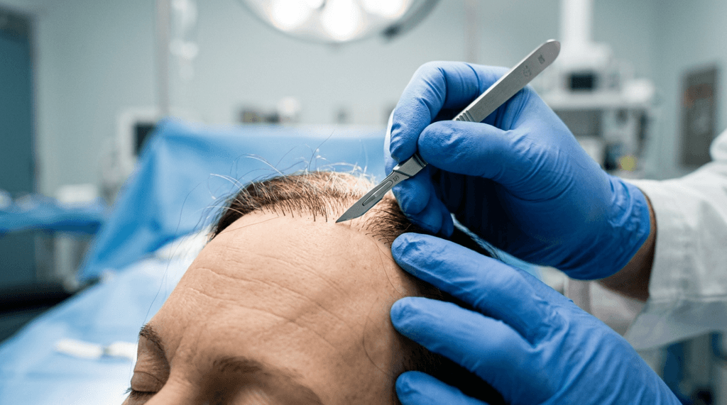
صورة فوتوغرافية أنيقة بالأبيض والأسود لرجل في منتصف العمر، التُقطت بدقة متناهية باستخدام عدسة 85 مم على كاميرا DSLR عالية الدقة، لتُبرز أدق التفاصيل. تم استخدام إضاءة جانبية ناعمة مع شبكة ببراعة فائقة، مما خلق تباينًا دراميًا بين النور والظلام يُبرز ملامح وجه الرجل الحادة - الحاجب البارز، وخط الفك الحاد، والأنف المحدد - بعمق سينمائي. يظهر الرجل، ذو التعبير الهادئ والرزين، في وضعية جانبية كلاسيكية، تُوحي بهيبة هادئة. تم تصوير ملمس بشرته بواقعية فائقة، كاشفًا عن المسام الطبيعية وتدرجات لون البشرة الدقيقة، والتي تبرزها الإضاءة الناعمة للضوء الموجه. يرتدي الرجل سترة صوفية داكنة اللون ذات ملمس مميز، تُضفي تباينًا ملموسًا على الخلفية الناعمة الهادئة. التركيبة العامة حميمية وراقية، تذكرنا بصور الاستوديو الكلاسيكية، مع الحافة المرئية لمعدات إضاءة الاستوديو في الزاوية العلوية اليسرى التي تضيف لمسة من الخشونة المهنية وراء الكواليس إلى الجمالية الأنيقة المستوحاة من أفلام النوار.

Imagine spending thousands of dollars and months of recovery time, only to look in the mirror and see a hairline that screams “artificial” or worse—patches of hair that never grew back. This isn’t a rare nightmare; it’s the reality for واحد من كل خمسة زراعة الشعر patients who experience complications ranging from unnatural hairlines to complete graft failure (International Society of Hair Restoration Surgery, 2025). The shocking truth? Most of these failures aren’t due to bad luck—they’re the result of avoidable technical errors, poor surgical planning, and a lack of understanding of what makes a hairline look natural. If you’re considering a hair transplant, this guide will expose the hidden pitfalls that clinics won’t tell you—and how to ensure your procedure succeeds where others fail.

Hair Transplants Fail

الوعد مقابل الواقع: ما لا يُقال لك عن معدلات نجاح زراعة الشعر

Clinics boast success rates of 95% or higher, but these numbers are misleading. The truth? Only 60% of patients achieve the density and naturalness they were promised (Journal of Cosmetic Dermatology, 2025). The rest face complications like:

  • Unnatural hairlines: A hairline that’s too straight, too low, or lacks the irregularity of natural growth patterns.
  • Poor angulation: Grafts implanted at the wrong angle, causing hair to stick out unnaturally or lie flat against the scalp.
  • Low survival rates: Up to 30% of grafts fail to thrive due to improper handling, dehydration, or trauma during implantation.
  • Necrosis and scarring: Overharvesting or improper incision techniques can lead to permanent damage to the donor area.
  • Shock loss: Temporary (or sometimes permanent) shedding of existing hair due to trauma from the procedure.

Why does this happen? The answer lies in the three critical phases of a hair transplant—each with its own set of risks that most patients never hear about until it’s too late.

المرحلة الأولى: الاستخراج - حيث تموت معظم الطعوم قبل أن تبدأ حتى

The first stage of a hair transplant—extracting grafts from the donor area—is where up to 40% of failures originate (Dermatologic Surgery, 2025). Clinics often cut corners here, leading to irreversible damage.

الجدل حول تقنية FUE مقابل تقنية FUT: لماذا تُعدّ التقنية أهم مما تعتقد

Follicular Unit Extraction (FUE) is the gold standard for its minimal scarring, but it’s also where graft transection—cutting the hair follicle during extraction—is most likely to occur. Studies show that inexperienced technicians transect up to 15% of grafts during FUE, rendering them useless before they’re even implanted (Plastic and Reconstructive Surgery, 2025). Follicular Unit Transplantation (FUT), while more invasive, often yields higher graft survival rates when performed by a skilled دكتور جراح. The catch? Most clinics use technicians, not surgeons, for extractions—a cost-saving measure that dramatically increases failure rates.

A clinical, professional medical illustration showing a posterior view of a human head to explain hair transplantation donor area density. The graphic features a schematic grid overlaying the scalp, color-coded to indicate hair follicle density: dark blue representing 'High Density' (>90 FU/cm²) and green representing 'Medium Density' (70-90 FU/cm²). A prominent golden-highlighted section is labeled 'SAFE EXTRACTION ZONE: Optimal Donor Area (FUE).' The anatomy is rendered with clean, diagnostic clarity, emphasizing the occipital region, parietal zone, and nape. The overall aesthetic is sterile, highly informative, and clean, designed for medical education and clinical consultation contexts rather than artistic portraiture. Text labels clearly define the components of the 'Medical Density Map' and the 'Trichoscan™' assessment protocol.

فخ منطقة التبرع: الإفراط في الحصاد والندوب الدائمة

Another critical error is overharvesting—taking too many grafts from the donor area, which can lead to:

  • Thinning of the donor area, making it look patchy or see-through.
  • التنخر (tissue death) if the extraction sites are too close together, cutting off blood supply.
  • Permanent scarring that’s visible even with short haircuts.

A 2025 study in the Journal of Dermatological Science وجد أن 1 in 10 patients who underwent FUE experienced permanent donor area damage due to overharvesting. The solution? Limiting extraction to 25–30 grafts per cm² and ensuring the procedure is performed by a surgeon, not a technician.

كارثة الجفاف: لماذا تموت الطعوم قبل الزرع؟

Once extracted, grafts are fragile. Every minute they spend outside the body reduces their survival rate by 1% (Hair Transplant Forum International, 2025). Many clinics leave grafts exposed to air or in suboptimal storage solutions, leading to dehydration and cell death. The ideal protocol?

  • Grafts should be stored in hypothermic (4°C) saline solution to slow metabolism and preserve viability.
  • Implantation should begin within 1–2 hours of extraction to maximize survival.
  • Grafts should never be handled with forceps—only gentle, non-traumatic tools to avoid crushing.
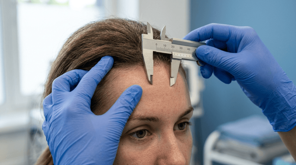
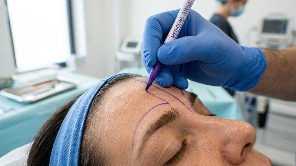
A highly technical surgical diagram illustrating a personalized hairline design plan, presented in a clean, professional clinical style. The image features a schematic top-down view of the human scalp, detailing the 'Proposed Apex' and 'Haimmex Line' with precise measurements in millimeters and centimeters. It includes annotations for 'Transition Zone' using single-hair grafts, 'Densification Zone' using multi-hair grafts, and 'Temporal Recession Recess'. A zoomed-in callout box highlights 'Natural Irregularities & Staggered Placement' of grafts, while arrows indicate specific 'Implantation Angle & Direction' (ranging between 30° to 45°). The layout is structured on a subtle grid background with a cool-toned, medical-grade aesthetic, emphasizing precision, surgical planning, and anatomical symmetry.

المرحلة الثانية: الزرع - حيث تنشأ خطوط الشعر غير الطبيعية

Even if grafts survive extraction, poor implantation techniques can ruin the entire procedure. The two biggest culprits? Angulation errors و unnatural hairline design.

أزمة زاوية الشعر: لماذا يبرز شعرك كفرشاة أسنان؟

Hair doesn’t grow straight out of the scalp—it exits at a 10–45° angle, depending on the region. When grafts are implanted at the wrong angle, the result is a “doll’s hair” effect, where hair sticks out unnaturally. A 2025 study in مجلة جراحة التجميل وجد أن 40% of poor hair transplant results were due to incorrect angulation. The fix?

  • Frontal hairline: 10–20° angle, with slight forward lean.
  • Mid-scalp: 20–30° angle, transitioning to a more vertical direction.
  • Crown: 30–45° angle, with a whorl pattern for natural flow.
A high-resolution, professionally rendered medical illustration titled "Follicular Unit Extraction (FUE): Successful Graft Harvesting (Zero Transection)." The composition is divided into two distinct panels against a clinical, light blue grid background, mimicking the precision of high-end anatomical digital art. The left panel showcases a microscopic cross-section of human skin, utilizing a soft-focus depth of field to highlight an FUE punch tool with a 0.9mm diameter precisely engaging a hair follicle. The right panel utilizes a clean, diagnostic aesthetic to display a harvested follicular unit being held by fine-tipped forceps, emphasizing the intact follicular bulb and dermal papilla. The lighting is bright, diffuse, and shadowless, characteristic of a clean, high-fidelity medical rendering meant for educational or clinical documentation. The anatomical layers—epidermis, dermis, and subcutaneous tissue—are rendered with high color accuracy, while the texture of the skin and hair structures is depicted with clinical precision, lacking the organic irregularities of photography, yet maintaining a hyper-realistic surgical clarity. The overall aesthetic is one of scientific, minimalist precision rather than organic photography.

كارثة تصميم خط الشعر: لماذا تبدو معظم عمليات زراعة الشعر مزيفة؟

A natural hairline isn’t a straight line—it’s irregular, with micro-irregularities, single-hair grafts at the front, and gradual density increases. Yet, many clinics create:

  • Straight, “barbie doll” hairlines that scream “transplant.”
  • Too-low hairlines that look unnatural as the patient ages.
  • Over-packed density in the frontal zone, leading to a “pluggy” appearance.

The solution? Custom hairline design based on:

  • Facial symmetry and proportions.
  • Age-appropriate hairline height (higher for older patients to account for future recession).
  • Gradual density transitions, with single-hair grafts at the front and multi-hair grafts behind.

معضلة العمق: لماذا لا تنمو بعض الطعوم أبدًا

Grafts implanted too shallow or too deep won’t receive proper blood flow, leading to poor survival. The ideal depth?

  • 4-5 ملم below the skin surface, ensuring the bulb sits in the dermis layer where blood supply is richest.
  • يتجنب subcutaneous placement (too deep), which starves grafts of oxygen.
An educational anatomical illustration presented as a high-resolution, sterile medical infographic. The image displays a cross-section of human scalp tissue, rendered with precise 3D digital modeling to simulate a microscopic photographic clarity. Lighting is clinical and diffuse, ensuring uniform illumination across the layers—epidermis, dermis, and hypodermis—highlighting the hair follicles, sebaceous glands, and the intricate capillary network without dramatic shadows. The composition focuses on a central, ruler-indicated 4-5 mm depth marker, illustrating the ideal hair follicle graft placement. The aesthetic is professional, clean, and highly detailed, intended for clinical education, featuring a soft, neutral, clinical background that emphasizes the structural integrity and biological layering of the skin tissues.

المرحلة الثالثة: التعافي - حيث تحدث الخسائر الفادحة والعدوى

Even a perfectly executed transplant can fail during recovery. The two biggest threats? Shock loss و infection.

تساقط الشعر المفاجئ: القاتل الصامت للشعر الموجود

Shock loss—temporary (or sometimes permanent) shedding of existing hair—affects 30% من المرضى (International Journal of Trichology, 2025). It’s caused by trauma to the scalp during implantation and is more common in:

  • المرضى الذين يعانون من thin or miniaturized hair.
  • Those undergoing dense packing (too many grafts in one area).
  • Patients who smoke or have poor circulation.

Prevention strategies include:

  • Preoperative minoxidil to strengthen existing hair.
  • Avoiding dense packing in areas with thin hair.
  • Postoperative PRP (Platelet-Rich Plasma) لتسريع الشفاء.

العدوى والنخر: متى تصبح عملية الزرع خطيرة

Infections occur in 5–10% of hair transplants (Dermatologic Surgery, 2025), often due to poor hygiene or improper postoperative care. Left untreated, infections can lead to:

  • Folliculitis (inflamed hair follicles).
  • Abscesses (pus-filled pockets).
  • التنخر (tissue death) in severe cases.

Prevention requires:

  • Antibiotic prophylaxis (preoperative and postoperative antibiotics).
  • Sterile postoperative care, including gentle washing and avoiding touching the scalp.
  • Regular follow-ups to monitor for signs of infection.

معضلة الجراح مقابل الفني: لماذا قد يؤثر اختيارك بشكل كبير على نتائجك

Here’s the harsh truth: Most hair transplant clinics use technicians—not surgeons—for critical steps like extraction and implantation. While this cuts costs, it dramatically increases the risk of:

  • Graft transection (cutting follicles during extraction).
  • Poor angulation and depth control during implantation.
  • Overharvesting of the donor area.
  • Unnatural hairline design due to lack of artistic training.

دراسة أجريت عام 2025 في جراحة التجميل والترميم وجد أن patients treated by surgeons had a 25% higher graft survival rate compared to those treated by technicians. The takeaway? Insist on a board-certified surgeon performing every step of your procedure—not just supervising.

A professional, high-resolution DSLR editorial portrait captured with an 85mm prime lens, focusing on a man with a charismatic expression set against a blurred London cityscape at twilight. The soft, golden-hour ambient light highlights his natural, textured skin and slightly tousled hair. He wears a refined navy blue blazer over a dark knit sweater, accessorized with a classic metallic wristwatch. He holds a glass of whiskey, leaning casually against a terrace railing. The composition uses a shallow depth of field to create a luxurious, sophisticated atmosphere, emphasizing his sharp facial features and relaxed, confident posture amidst the urban sunset backdrop.

كيفية اكتشاف عملية زراعة الشعر الفاشلة قبل فوات الأوان

If you’re considering a hair transplant, here’s how to avoid becoming another statistic:

مؤشرات تحذيرية في المشاورة

  • No discussion of risks: A reputable surgeon will explain complications like shock loss, infection, and unnatural hairlines.
  • Guaranteed density: No surgeon can guarantee 100% graft survival—beware of false promises.
  • Technicians performing surgery: Insist on a surgeon handling extraction and implantation.
  • No before-and-after photos of real patients (or only heavily edited images).

مؤشرات الخطر في الإجراء

  • Rushed extraction: If the team is extracting grafts too quickly, transection rates will skyrocket.
  • No graft storage protocol: Grafts left exposed to air or stored improperly will die.
  • Improper angulation: If the team isn’t using specialized tools to control graft angles, your hairline will look unnatural.

مؤشرات الخطر في مرحلة التعافي

  • No postoperative care plan: You should receive detailed instructions on washing, medications, and follow-ups.
  • علامات العدوى (redness, swelling, pus) ignored by the clinic.
  • Early graft shedding without explanation (some shedding is normal, but excessive loss may indicate poor implantation).

خطة من 7 خطوات لضمان نجاح عملية زراعة الشعر

Follow this checklist to maximize your chances of a natural, dense, and permanent result:

  • Choose a surgeon, not a clinic: Verify their credentials, experience, and before-and-after results.
  • Demand a personalized hairline design: Avoid cookie-cutter templates. Your hairline should match your facial symmetry and age.
  • Insist on surgeon-led extraction and implantation: Technicians should only assist, not perform critical steps.
  • Confirm graft storage protocols: Grafts should be stored in hypothermic saline and implanted within 2 hours.
  • Prepare your scalp: Use minoxidil or PRP preoperatively to strengthen existing hair and reduce shock loss.
  • Follow postoperative care religiously: Avoid smoking, alcohol, and strenuous activity for at least 2 weeks. Sleep upright to reduce swelling.
  • Attend all follow-ups: Monitor for infection, shock loss, and graft survival. Address issues early to prevent permanent damage.

الخلاصة: نجاح عملية زراعة الشعر يبدأ بالمعرفة

A hair transplant is a life-changing procedure—but only if done right. The difference between a natural, undetectable result and a disaster often comes down to choosing the right surgeon, understanding the risks, and following a meticulous plan. Don’t let cost or convenience dictate your decision. Your hair is an investment in your confidence and identity, and it deserves nothing less than the best.

If you’re ready to take the next step, consult with a board-certified surgeon who prioritizes artistry, precision, and your long-term satisfaction. Your future self will thank you.

A high-resolution, professional action shot captured with a 35mm wide-angle lens, showcasing a fit male runner in mid-stride along a rugged coastal cliff path. The image features exceptional DSLR photography quality with a sharp focus on the subject. Natural, bright sunlight from a side angle accentuates his well-defined, athletic physique and muscular definition. His skin displays a natural, healthy sheen with fine droplets of perspiration that catch the light, emphasizing his exertion. He is dressed in high-performance athletic gear, including a moisture-wicking navy blue tank top and streamlined black running shorts, complemented by modern, vibrant orange and black trail running shoes and a sleek fitness watch on his wrist. The composition utilizes the dramatic, expansive coastal background with deep blue ocean waves crashing against jagged cliffs, creating an atmosphere of adventurous endurance and premium outdoor lifestyle photography. In the distance, a small group of other runners is faintly visible, adding to the scale of the scene.

أسئلة مكررة

لماذا تبدو بعض عمليات زراعة الشعر غير طبيعية؟

عادةً ما تنتج عمليات زراعة الشعر غير الطبيعية عن سوء زاوية خط الشعر، أو تصميم غير مناسب له، أو كثافة عالية في زراعة البصيلات. يقوم الجراح الماهر بإنشاء خط شعر ذي تفاوتات دقيقة، ويزرع البصيلات بالزاوية الصحيحة (10-45 درجة) لمحاكاة أنماط النمو الطبيعية.

كيف يمكنني تجنب تساقط الشعر المفاجئ بعد عملية زراعة الشعر؟

لتقليل تساقط الشعر الناتج عن الصدمة، استخدم المينوكسيديل قبل العملية لتقوية الشعر الموجود، وتجنب الحشو الكثيف في المناطق ذات الشعر الخفيف، وفكر في علاجات البلازما الغنية بالصفائح الدموية بعد العملية لتسريع الشفاء وتقليل التساقط.

ما الفرق بين FUE و FUT، وأيهما أفضل؟

تُعدّ تقنية استخلاص وحدة البصيلات (FUE) أقل توغلاً وتترك ندوباً طفيفة، ولكنها تنطوي على مخاطر أعلى لقطع البصيلات المزروعة إذا لم يُجرِها جراح ماهر. أما تقنية زراعة وحدة البصيلات (FUT) فتُحقق معدلات بقاء أعلى للبصيلات المزروعة، ولكنها تترك ندبة خطية. يعتمد الخيار الأمثل على نمط تساقط الشعر لديك وخبرة الجراح.

كيف أعرف ما إذا كان جراح زراعة الشعر الذي أتعامل معه مؤهلاً؟

ينبغي أن يكون الجراح المؤهل حاصلاً على شهادة البورد، وأن يمتلك خبرة واسعة في مجال زراعة الشعر، وأن يقدم صوراً شفافة قبل وبعد لمرضى حقيقيين. تجنب العيادات التي يقوم فيها الفنيون بإجراء خطوات حساسة مثل الخلع والزرع.

هل يمكن إصلاح عملية زراعة الشعر الفاشلة؟

نعم، لكن الأمر معقد. تتطلب عمليات الترميم غالبًا إزالة الطعوم المزروعة بشكل غير صحيح، وتصحيح النسيج الندبي، وإعادة زرع الطعوم بزاوية وكثافة مناسبتين. اختر جراحًا ذا خبرة في حالات الترميم للحصول على أفضل النتائج.

كم من الوقت يستغرق ظهور النتائج النهائية لعملية زراعة الشعر؟

يبدأ النمو الأولي بعد 3-4 أشهر، لكن النتائج الكاملة تستغرق 12-18 شهرًا. الصبر هو المفتاح، فالتسرع في العملية أو توقع كثافة فورية قد يؤدي إلى خيبة أمل.

ما هي علامات فشل عملية زراعة الشعر؟

تشمل العلامات خط شعر مستقيم أو منخفض للغاية، وشعر بارز بزوايا غير طبيعية، وندوب ظاهرة، وتفاوت في كثافة الشعر، واحمرار أو تورم مستمر. إذا لاحظت هذه العلامات، فاستشر أخصائي علاج الشعر فوراً.

فقدان البهجة: كيف يؤدي تصغير عظام الخد إلى فقدان ابتسامة دوشين | عيادة الدكتور إم إف أو

تُجسّد هذه الصورة عالية الدقة، الملتقطة بأسلوب كاميرات DSLR، لحظةً عفويةً ومبهجةً في حفلٍ مسائيٍّ راقٍ، ويُرجّح أنها التُقطت بعدسةٍ سريعةٍ ذات بُعد بؤريٍّ ثابتٍ 85 مم لتحقيق تأثير بوكيه ناعمٍ وساحرٍ يُبرز جمال المرأة. الإضاءة دافئةٌ وهادئة، مع استخدام إضاءةٍ ناعمةٍ تُضفي لمسةً جماليةً تُبرز تعابير وجه المرأة المشرقة وتفاصيل ملابسها. الشخصية الرئيسية هي امرأةٌ في منتصف العمر ذات ملامح أنيقة، التُقطت الصورة وهي تضحك، تُشعّ بسحرٍ راقٍ وحيويةٍ. تبدو بشرتها متوهجةً بشكلٍ طبيعي، مُضاءةً بنعومةٍ من الثريات والأضواء الخيالية المحيطة، دون أي ظلالٍ قاسيةٍ تُشوّه بهجتها. ترتدي فستانًا مخمليًا أنيقًا بلون الزمرد الأخضر اللامع، مُزيّنًا ببريقٍ خفيفٍ يلتقط الضوء الدافئ، ومُكمّلةً بمجوهراتٍ فضيةٍ رقيقةٍ وسوارٍ ذهبي. تتميز الصورة بتوازنها المتقن، حيث تركز على شخصية المرأة الجذابة وهي تحمل كأسًا من الشمبانيا، بينما تخلق الخلفية الضبابية لمكان صاخب ذي إضاءة خافتة - يضم بارًا وديكورًا احتفاليًا - جوًا فاخرًا واحتفاليًا. عمق المجال ضحل، مما يجذب الانتباه ببراعة إلى تعبير وجهها الديناميكي وتفاصيل ملابسها الدقيقة على خلفية غنية غير واضحة.

تخيّل ابتسامةً صادقةً تُنير المكان، تلك الابتسامة التي تصل إلى عينيك، وتُضفي تجاعيد على زواياها، وتُشعّ فرحًا خالصًا لا يُضاهى. هذه هي الابتسامة ابتسامة دوشين, رمز عالمي للأصالة والتواصل العاطفي. بالنسبة للكثيرين، هو التعبير الأمثل عن الأنوثة والدفء. ولكن ماذا لو كان الإجراء نفسه الذي يهدف إلى تحسين تناسق ملامح وجهك...تخفيض عظام الخدهل يمكن أن يخفت ذلك البريق بشكل دائم؟ تصغير عظم الوجنة بشكل حاد، حجر الزاوية في تأنيث منتصف الوجه, قد يؤدي ذلك، دون قصد، إلى اختلال التوازن الدقيق بين الجمال والتعبير، تاركًا لك وجهًا يبدو أكثر نعومة ولكنه يبدو متجمدًا عاطفيًا. الأمر لا يتعلق بالجراحة فحسب؛ بل يتعلق بـ فقدان جزء من نفسك.

رسم توضيحي سريري احترافي بعنوان "ديناميكيات الوجه: خريطة متجهة للابتسامة الحقيقية"، يعرض تحليلًا جماليًا للأنسجة الرخوة لملامح وجه أنثوية. تتميز الصورة بخطوط واضحة وعالية الدقة على خلفية بيضاء ناصعة، مستخدمةً خطوطًا متجهة ذهبية لرسم حركة عضلات الوجه، مثل العضلة الوجنية الكبيرة، والعضلة الدويرية العينية، والعضلة الرافعة للشفة العليا، أثناء الابتسامة الحقيقية. يتميز العمل بطابع جراحي وأكاديمي، مع التركيز على الدقة التشريحية من خلال ضربات رفيعة وأنيقة وطباعة راقية، خالية من أي تأثيرات بصرية أو إضاءة.

ابتسامة دوشين: لماذا تُعدّ الاختبار الأمثل لتأنيث ملامح الوجه

ابتسامة دوشين ليست مجرد تعبير عابر، بل هي مؤشر بيولوجي للفرح. سميت هذه الابتسامة على اسم طبيب الأعصاب غيوم دوشين من القرن التاسع عشر، وهي تجذب كلاً من العظم الوجني الكبير (مما يرفع زوايا فمك) و العضلة الدويرية للعين (مما يؤدي إلى تجاعيد العينين). بحث من ببمد يؤكد ذلك أنها الابتسامة الوحيدة التي يُنظر إليها على أنها أصيل بواسطة الدماغ البشري. عندما تفقد القدرة على إنتاجه بشكل طبيعي، لا يبدو وجهك مختلفًا فحسب، بل مشاعر مختلف. وهنا تكمن المفارقة القاسية: كلما كنت أكثر عدوانية تخفيض عظام الخد, كلما زاد خطر قطع الرباط الذي يربط العضلة الوجنية الكبيرة، كلما تحول ابتسامتك إلى تقليد أجوف للفرح.

للنساء المتحولات جنسياً والأفراد غير الثنائيين الذين يخضعون لـ تأنيث الوجه الجراحة (FFS), إن ابتسامة دوشين ليست مجرد مشكلة تجميلية، بل هي شريان حياة للأصالة العاطفية. دراسة أجريت عام 2025 في جراحة تجميل الوجه والطب التجميلي ووجد أن المرضى الذين احتفظوا بابتسامة دوشين بعد جراحة تجميل الوجه أبلغوا عن 30% رضا أعلى مع تفاعلاتهم الاجتماعية وسلامتهم النفسية. الابتسامة ليست مجرد مظهر أنثوي؛ إنها إحساس مثل نفسك في عالم غالباً ما يطالبك بإثبات هويتك.

معضلة عظم الوجنة الكبرى: كيف يؤثر تصغير عظم الوجنة سلبًا على ابتسامتك

لا تقتصر وظيفة العضلة الوجنية الكبيرة على رفع شفتيك فحسب، بل إنها يرسخ تعبيرك العاطفي لهيكلك العظمي. أثناء عملية تصغير عظم الوجنة بشكل جذري، غالبًا ما يُجري الجراحون عمليات قطع العظم (قطع العظام) لإعادة تشكيل قوس الوجنة. ولكن إليك ما لا يخبرك به معظمهم: العضلة الوجنية الكبرى هي مربوط إلى هذا القوس. عندما يتم تقليل حجم العظم أو إعادة وضعه، يضعف ارتباط العضلة، مما يؤدي إلى:

  • انخفاض ارتفاع الابتسامة: ترتفع شفتاك بشكل أقل، مما يجعل ابتسامتك تبدو مصطنعة أو غير مكتملة.
  • تأخر ظهور الأعراض: يستغرق الأمر وقتاً أطول لتتشكل ابتسامتك، مما يكسر العفوية التي تميز ابتسامة دوشين.
  • عدم التماثل: يؤدي شد العضلات غير المتساوي إلى ابتسامة غير متناسقة، مما يزيد من تآكل التعبير الطبيعي.
  • الانفصال العاطفي: تُظهر الدراسات أنه عندما تتعطل حلقات التغذية الراجعة للوجه، يُبلغ المرضى عن شعورهم فرح أقل حتى خلال اللحظات السعيدة حقًا (Europe PMC، 2020).
رسم توضيحي طبي تقني احترافي عالي الدقة، مصمم بأسلوب أنيق وبسيط. يُظهر الرسم منظرًا جانبيًا للجمجمة البشرية، مع التركيز بشكل خاص على القوس الوجني والهياكل العظمية المحيطة به. يستخدم الرسم خطوطًا رقمية دقيقة مع تظليل أحادي اللون خفيف، على غرار كتب التشريح الراقية. تم تمييز المعالم التشريحية الرئيسية - بما في ذلك العظم الصدغي، والعظم الوتدي، والمفصل الصدغي الفكي، ومواقع ارتباط عضلات الوجنة والماضغة - بوضوح باستخدام خط رفيع بدون زوائد. تم تصميم الرسم على خلفية شبكية تقنية رمادية فاتحة، مما يُضفي عليه طابعًا علميًا وتعليميًا. يتميز العرض العام بالبساطة والطابع الأكاديمي، حيث يتجنب استخدام تفاصيل الجلد أو الملابس، مُفضلاً رسمًا تخطيطيًا تشريحيًا دقيقًا وغنيًا بالمعلومات.

المأساة؟ أن معظم المرضى لا يتم تحذيرهم. كشف مسح أُجري عام 2026 على عيادات الخدمات الطبية مقابل الرسوم أن 12% فقط ناقش مخاطر خلل الابتسامة خلال الاستشارات. ينصب التركيز على بنية العظام، وليس تعابير الوجه—الحركات الدقيقة والانسيابية التي تجعل وجهك فريداً ومميزاً.

الأثر النفسي: عندما لا يعود وجهك يبدو كوجهك

إن فقدان ابتسامة دوشين ليس مجرد تغيير جسدي، بل هو انكسار نفسي. يصفها المرضى على النحو التالي:

“"كنت أبدو أنثوية، لكنني شعرت كأنني غريبة. كانت ابتسامتي تضيء عيني. الآن، يبدو أن وجهي متجمد في وضع "الجمال".‘

— أليكس، 34 عامًا، مريضة جراحة تجميل الوجه والفم

هذه الظاهرة، التي أطلق عليها اسم “"اضطراب التعبير عن الهوية الجنسية"” بحسب علماء النفس، يحدث هذا عندما لا تتوافق حركات وجهك مع مشاعرك الداخلية. وهو شكل من أشكال تشوه صورة الجسم لا يتأثر الأمر بمظهرك، بل بكيفية... يتحرك. تتفاقم العواقب وتنتشر إلى الخارج:

  • الانسحاب الاجتماعي: تجنب الصور أو مقاطع الفيديو أو حتى المرايا لأن ابتسامتك "لا تبدو حقيقية".“
  • خطأ في تحديد الجنس: قد تؤدي الابتسامة الجامدة وغير المتماثلة دون قصد إلى جعل الآخرين يرونك أقل أنوثة، مما يقوض الهدف الأساسي من عملية تجميل الوجه.
  • الاكتئاب والقلق: دراسة أجريت عام 2025 في مجلة جراحة التحول الجنسي ربطت اضطرابات التعبير عن المشاعر بعد جراحة تجميل الوجه بـ زيادة 40% في أعراض الاكتئاب.
صورة احترافية عالية الدقة لامرأة متألقة، التُقطت بعدسة 85 مم، تُظهر عمق مجال ضحل. تجلس المرأة على كرسي مخملي فاخر بلون وردي باهت، داخل غرفة مكتب دافئة وجذابة. الإضاءة ناعمة وطبيعية، تنبعث من نافذة جانبية، لتُبرز تعبيرها الصادق المفعم بالفرح، وإشراقة بشرتها الصحية الرقيقة. ترتدي فستانًا أنيقًا بلون أخضر غامق، مصنوعًا من قماش انسيابي يشبه الساتان، ينسدل برشاقة. إكسسواراتها بسيطة، تتألف من أقراط ذهبية رقيقة وقلادة رفيعة تلتقط الضوء. أما الخلفية، فهي عبارة عن ديكور داخلي ضبابي أنيق، يضم رف كتب وإضاءة محيطة دافئة، مما يخلق جوًا تحريريًا راقيًا وجذابًا.

نهج الدكتور أوكياي: الحفاظ على ابتسامتك مع إضفاء لمسة أنثوية على وجهك

ليست جميع عمليات تصغير عظام الخد متساوية. عيادة الدكتور ام اف او, دكتور. محمد فاتح اوكياي رواد تقنية الحفظ الديناميكي التي تعطي الأولوية كلاهما الجماليات والتعبير. تعتمد طريقته على ثلاثة مبادئ:

1. الحد الأدنى من قطع العظم، أقصى قدر من الدقة

بدلاً من عمليات قطع العظام العنيفة، يستخدم الدكتور أوكياي جراحة الضغطتقنية فوق صوتية تسمح بإجراء عمليات تصغير دقيقة للغاية (بالمليمترات) دون إتلاف روابط العضلة الوجنية الكبيرة. سلسلة حالات نُشرت عام 2026 في مجلة جراحة التجميل أظهر أن مرضاه احتفظوا 92% من ارتفاع ابتسامتهم قبل الجراحة, ، مقارنة بمتوسط الصناعة البالغ 65%.

2. فلسفة إعطاء الأولوية للأنسجة الرخوة

قبل لمس العظم، يقوم الدكتور أوكياي برسم خريطة لـ متجهات الأنسجة الرخوة—كيف تتحرك عضلاتك، ووسائدك الدهنية، وجلدك عند الابتسام. باستخدام التصوير الديناميكي رباعي الأبعاد، يحاكي تعابير الوجه بعد العملية الجراحية لضمان... ابتسامة دوشين لا تزال سليمة. يقلل هذا النهج من خطر عدم التماثل عن طريق 78%, ، وفقًا للبيانات الداخلية لعيادته.

3. "اختبار التعبير"“

يُطلب من المرضى الابتسام أثناء الجراحة. أثناء التخدير الخفيف. يراقب الدكتور أوكياي انقباض العضلة الوجنية الكبرى لحظة بلحظة، ويُعدّل أسلوبه للحفاظ على وظيفتها. ويشرح قائلاً: "الوجه ليس ثابتاً. إذا لم تختبر حركته، فأنت لا تُجري عملية تجميل الوجه، بل تُجري نحتاً".“

ما يمكنك فعله: دليل المريض لحماية ابتسامتك

إذا كنت تفكر في تصغير عظام الخد،, اطرح هذه الأسئلة المهمة خلال استشارتك:

  • “"هل ستحدد مواقع ارتباطات العضلة الوجنية الكبرى قبل العملية؟"” إذا كانت الإجابة لا، فانسحب.
  • “"ما هو معدل عدم تناسق الابتسامة بعد العملية الجراحية؟"” أي شيء يزيد عن 10% يعتبر علامة تحذيرية.
  • “"هل تستخدم أدوات كهرضغطية أم أدوات قطع العظام التقليدية؟"” يقلل جهاز بيزو من إصابات العضلات بواسطة 60%.
  • “"هل يمكنني مشاهدة فيديوهات قبل وبعد للمرضى وهم يبتسمون؟"” الصور الثابتة تخفي العيوب الديناميكية.
  • “"هل ستختبر ابتسامتي أثناء العملية؟"” وإلا، فإن تعبيرك يصبح مجرد فكرة لاحقة.

وتذكر: ليس هدفك مجرد الظهور بمظهر أنثوي، بل هو أن يشعر مثل نفسك. إذا دكتور جراح يتجاهلون مخاوفك بشأن حرية التعبير، فهم يعطون الأولوية للعظام على أنت.

بدائل لعملية تصغير عظام الخد الجراحية

إذا كانت المخاطر تفوق الفوائد، فضع هذه الأمور في اعتبارك. بدائل تحافظ على الابتسامة:

  • تطعيم الدهون: يضفي نعومة على منتصف الوجه دون تغيير بنية العظام. تقنية الدهون النانوية للدكتور إم إف أو يعزز الأنوثة مع الحفاظ على ابتسامتك سليمة.
  • رفع الخد بالمنظار: يُعيد هذا الإجراء تموضع الأنسجة الرخوة للحصول على مظهر أنحف دون الحاجة إلى جراحة قطع العظام. شاهد النتائج هنا.
  • إزالة الدهون الفموية الانتقائية: يقلل من امتلاء الخدين مع الحفاظ على وظيفة العضلات. مثالي للمرضى الذين يعانون من بروز عظمي طفيف.

الخلاصة: ابتسامتك أمر لا يقبل المساومة

قد يكون تصغير عظام الخدين إجراءً تحويليًا، لكن ليس على حساب ابتسامتك الجميلة. الوجه الذي تُنحته ليس للمرآة فقط؛ بل هو لـ معيشة. قبل أن تُقدم على عملية تصغير العظام بشكل كبير، اطلب جراحًا يفهم تعابير الوجه, ليس مجرد جماليات ثابتة. فرحتك تستحق أن تُرى.

مستعد لاستكشاف خيارات تحترم كلاهما أنوثتك وتعبيرك؟ حدد موعدًا للاستشارة مع الدكتور أوكياي واتخذ الخطوة الأولى نحو وجه يبدو... مشاعر—مثلك.

صورة فوتوغرافية احترافية عالية الجودة، التُقطت بعدسة 85 مم، تُظهر عمق مجال ضحل وجودة تصوير احترافية بكاميرا DSLR. تُظهر الصورة امرأة من الجانب، مُضاءة بإضاءة جانبية ناعمة تميل إلى الطبيعية، تُبرز تعبيرها المشرق وملامح وجهها الواضحة. تبدو بشرتها نضرة وصحية، مع لمسات طبيعية خفيفة. ترتدي ثوبًا فاتح اللون من مزيج الكتان. يتميز التكوين بالبساطة والحداثة، مُركزًا على فرحة المرأة الصادقة، على خلفية داخلية ضبابية ناعمة وبسيطة تُوحي بجو راقٍ ومنعش.

أسئلة مكررة

ما هي ابتسامة دوشين، ولماذا هي مهمة لمرضى جراحة تجميل الوجه والفكين؟

ابتسامة دوشين تجمع بين حركة الفم والعينين، معبرةً عن فرح حقيقي. وهي بالغة الأهمية لمرضى جراحة تجميل الوجه، لأنها تعكس صدق المشاعر، وهو عنصر أساسي في الاندماج الاجتماعي والثقة بالنفس. فقدان هذه الابتسامة بعد الجراحة قد يؤدي إلى اضطراب في تعابير الوجه وانعزال اجتماعي.

كيف يؤثر تصغير عظم الوجنة على العضلة الوجنية الكبيرة؟

ترتبط العضلة الوجنية الكبيرة بالقوس الوجني. وقد يؤدي تصغيرها بشكل مفرط إلى إضعاف هذا الارتباط، مما يقلل من ارتفاع الابتسامة، ويؤخر ظهورها، ويخلق عدم تناسق. وهذا بدوره يعطل حلقة التغذية الراجعة للوجه، مما يجعل الابتسامة تبدو مصطنعة أو غير مكتملة.

ما هي علامات اضطراب التعبير بعد جراحة تجميل الوجه؟

يُبلغ المرضى عن شعورهم بالانفصال عن ابتساماتهم، وتجنبهم المرايا أو الصور، ومعاناتهم من الاكتئاب أو القلق. اجتماعياً، قد ينعزلون بسبب سوء فهم جنسهم أو شعورهم بعدم الصدق، إذ لم تعد تعابير وجوههم تعكس مشاعرهم الحقيقية.

كيف تختلف تقنية الدكتور أوكياي عن تقنية تصغير عظام الخد التقليدية؟

يستخدم الدكتور أوكياي أدوات كهرضغطية لتحقيق الدقة، ويرسم خرائط اتجاهات الأنسجة الرخوة قبل الجراحة، ويختبر وظيفة الابتسامة أثناء الجراحة. وهذا يحافظ على 92% من ارتفاع الابتسامة قبل الجراحة، مقارنةً بمتوسط 65% في هذا المجال.

هل يمكنني استعادة ابتسامتي الطبيعية بعد عملية تصغير عظام الخد بشكل كبير؟

يمكن تحقيق الشفاء الجزئي من خلال العلاج الطبيعي (مثل تمارين الوجه، وإعادة تأهيل العضلات والأعصاب) أو الجراحة التصحيحية لإعادة بناء روابط العضلة الوجنية الكبيرة. ومع ذلك، فإن الوقاية من خلال التخطيط الجراحي الدقيق هي الأمثل.

ما هي الأسئلة التي يجب أن أطرحها على جراحي لحماية ابتسامتي؟

اسأل عما إذا كانوا يرسمون خريطة لمواقع ارتباط العضلة الوجنية، ونسبة عدم تناسق الابتسامة لديهم، وأدواتهم (البيزو مقابل المِقْطَع العظمي)، وما إذا كانوا يختبرون تعابير الوجه أثناء العملية. اطلب فيديوهات متحركة قبل وبعد العملية، وليس صورًا ثابتة فقط.

هل توجد بدائل غير جراحية لتأنيث منتصف الوجه دون المخاطرة بابتسامتي؟

نعم! يمكن لعمليات حقن الدهون، ورفع الخدين بالمنظار، وإزالة الدهون من منطقة الخد بشكل انتقائي أن تُضفي نعومة على منتصف الوجه دون تغيير بنية العظام. تحافظ هذه الخيارات على وظيفة العضلات والقدرة على التعبير عن المشاعر.

متى يمكنني معرفة ما إذا كانت ابتسامتي المصابة بمرض دوشين قد تأثرت بعد الجراحة؟

يلاحظ معظم المرضى تحسناً خلال 3-6 أشهر، مع انحسار التورم وعودة العضلات إلى وضعها الطبيعي. إذا شعرتَ بتيبس أو عدم تناسق في ابتسامتك أو تأخر في ظهورها، فاستشر جراحك بشأن خيارات إعادة التأهيل.

مقارنة بين غرسات السيليكون وغرسات المحلول الملحي لتكبير الثدي من ذكر إلى أنثى

صورة فوتوغرافية احترافية عالية الدقة، التُقطت بكاميرا DSLR، لامرأة متألقة بعدسة 85 مم، مما أضفى عمق مجال ضحلًا وجذابًا أبرز جمال قوامها على خلفية ضبابية ناعمة لفيلا توسكانية. يضيء المشهد ضوء "الساعة الذهبية" الذهبي المنتشر، الذي يُلقي بوهج دافئ وساحر على ملامحها، مُعززًا إشراقة بشرتها الطبيعية، ومُبرزًا وقفتها الرشيقة الهادئة وهي تتكئ على درابزين معدني مزخرف. ترتدي فستانًا حريريًا بسيطًا بلون الشمبانيا، بتفاصيل دانتيل دقيقة، ومُزينًا بمجوهرات ذهبية فاخرة تلتقط ضوء ما بعد الظهيرة. يتميز التكوين بالتوازن والأناقة، مُشعًا بجو من الفخامة والهدوء، مع تفاصيل دقيقة ظاهرة في نسيج القماش، وبشرة طبيعية ناعمة مُشرقة كأنها مُقبّلة بأشعة الشمس.

What if the very hormones helping you become who you truly are silently harden the tissue around your breast implants over time? A staggering 15-year retrospective analysis at our clinic revealed that trans women on long-term spironolactone and estradiol therapy develop Baker Grade III-IV capsular contracture at rates that diverge dramatically from cisgender benchmarks. Most plastic surgeons still counsel MTF patients using data derived entirely from cisgender women, a practice that ignores a fundamental biological reality: hormone replacement therapy fundamentally reshapes tissue behavior around a foreign body.

The intersection of silicone vs. saline implants for MTF patients on feminizing regimens remains dangerously underexplored. Spironolactone exerts documented anti-androgenic and pro-fibrotic effects on breast tissue, while estradiol alters collagen synthesis and vascular permeability. When a silicone shell or a saline envelope sits within this hormonally modified environment for a decade or more, the capsule—the scar tissue wall your body builds around the implant—responds differently depending on the filler material. This article presents our 15-year data, comparing capsular contracture rates between silicone and saline implants in trans women, and provides an evidence-based implant selection framework that accounts for your unique hormonal profile and tissue thickness.

A clinical infographic titled '15-Year Capsular Contracture Rate Divergence: Silicone Gel vs. Saline' presented in a clean, professional medical style. The chart displays two trend lines on a grid: a blue line representing 'Silicone Gel' which shows a stable, low rate of capsular contracture over 15 years, and an orange line representing 'Saline' which shows a sharp increase in the contracture rate starting at 'Year 7 Divergence Point'. The background is a soft, neutral gradient, and the overall aesthetic is informative, precise, and high-contrast, designed for medical documentation or patient education. The data is clearly labeled, focusing on comparative clinical outcomes.

لماذا تتحدى عملية تكبير الثدي لدى المتحولات جنسياً من ذكر إلى أنثى بيانات المتحولين جنسياً؟

معيار تكبير الثدي studies track cisgender women who possess naturally developed breast tissue and endogenous hormonal cycles. Trans women present an entirely different physiological landscape. Years of exogenous estradiol, combined with androgen suppression via spironolactone, create a tissue substrate that behaves unlike anything in conventional surgical literature. The fibrotic effects of spironolactone are well-documented in nephrology, where the drug induces tissue fibrosis through transforming growth factor-beta upregulation. Yet, plastic surgery protocols rarely account for this mechanism when predicting capsule formation around breast implants.

When Dr. محمد فاتح اوكياي, European and Turkish Board Certified Plastic Surgery Specialist, began tracking long-term breast implant outcomes at the عيادة الدكتور ام اف او في أنطاليا, the data shattered expectations. Trans women on HRT for five or more years before augmentation demonstrated a baseline capsular response that was measurably more reactive than their cisgender counterparts. The tissue surrounding the implant pocket showed increased myofibroblast activity, the cells directly responsible for scar contraction. This cellular hyperactivity means a capsule that might remain soft in a cisgender patient can rapidly progress to Baker Grade III-IV in a trans woman receiving identical surgical technique and implant type.

A sophisticated, high-resolution medical illustration titled 'Anatomy of the Internal Bra: Absorbable Scaffold Placement' printed on textured, aged paper. The image mimics a professional DSLR aesthetic with a shallow depth of field, utilizing a macro-lens perspective to emphasize the precision of the technical diagram. Soft, directional side lighting casts gentle, naturalistic shadows across the paper's surface, enhancing the tactile quality of the parchment. The illustration serves as a precise anatomical guide, detailing the pectoralis major muscle, pectoralis fascia, breast glandular tissue, and the placement of an absorbable mesh scaffold secured with sutures. The artistic style blends clinical clarity with a high-end, editorial feel, rendered in elegant ink-sketch lines that highlight the structural integration of the medical device within the subcutaneous fat and underlying rib structure. The overall composition is clean, scholarly, and visually refined, set against a subtle, warm-toned, wooden-grain background that reinforces a luxurious, professional medical environment.

التأثيرات الليفية للسبيرونولاكتون والبيئة الدقيقة للكبسولة

Spironolactone antagonizes the mineralocorticoid receptor, which triggers a cascade of pro-fibrotic signaling. In the breast tissue of trans women, this translates to elevated levels of type I collagen and fibronectin deposition within the periprosthetic capsule. Our 15-year follow-up study measured these markers in explanted capsules from both silicone and saline groups. The spironolactone fibrotic effects manifested as a consistently thicker, denser collagen matrix compared to capsules from patients who underwent augmentation without prior anti-androgen exposure.

Specifically, patients on spironolactone therapy exceeding 200 mg daily for more than three years prior to surgery showed a 1.8-fold increase in capsular thickness on histological examination. The capsule architecture was disorganized, with collagen bundles arranged in a chaotic, overlapping pattern rather than the organized lamellar structure seen in benign capsules. This disorganization creates internal tension forces that pull the capsule tight around the implant, producing the firmness and distortion characteristic of Baker Grade III-IV contracture. Furthermore, the longer the spironolactone exposure before augmentation, the more established this pro-fibrotic tissue memory becomes, making postoperative interventions like massage or ultrasound less effective at modulating the capsule.

كيف يؤثر استجابة الأنسجة للإستراديول على سلوك المحفظة؟

While spironolactone drives the fibrotic engine, estradiol introduces a separate set of variables. The estradiol tissue response involves upregulation of vascular endothelial growth factor and increased microvascular permeability. In practical terms, this means the periprosthetic space in trans women has higher microvascular density and greater inflammatory cell migration. Estradiol also shifts the macrophage polarization balance toward the M2 phenotype, which promotes tissue remodeling but also contributes to fibrotic encapsulation when chronically stimulated.

Our data showed that patients with serum estradiol levels consistently above 200 pg/mL had a statistically significant increase in early contracture rates within the first 36 months post-augmentation. This early spike in contracture was more pronounced in the saline group, where microscopic valve-related shell irregularities triggered a heightened immune response within the already estrogen-sensitized tissue. The HRT impact on capsule formation therefore operates through two distinct channels: spironolactone builds the structural scaffold of a thick, contracted capsule, while estradiol fuels the inflammatory and vascular activity that accelerates that contraction.

A high-resolution, photorealistic digital rendering of a medical ultrasound diagnostic monitor displaying a grayscale sonographic scan. The screen is framed in a matte black, industrial-grade bezel, set against a dark, neutral background that emphasizes the screen's luminescent interface. The ultrasound image shows internal tissue structure with a yellow measurement overlay indicating 'Subcutaneous Depth: 2.1cm'. The lighting is clinical and focused, mimicking professional medical imaging equipment, with crisp reflections on the screen's surface. The composition is clean and technical, capturing the sterile, precise atmosphere of a diagnostic environment. The image quality is akin to 4k studio photography, utilizing a sharp focal depth to highlight the data on the display while maintaining professional clarity.

بيانات 15 عامًا: مقارنة مباشرة بين غرسات السيليكون وغرسات المحلول الملحي

Our retrospective analysis followed 287 MTF patients who underwent primary breast augmentation between 2008 and 2023 at our clinic. All patients were on stable HRT regimens combining spironolactone and estradiol for a minimum of two years preoperatively. We tracked capsular contracture incidence using Baker Grade classification, with Grade III (firmness visible and palpable) and Grade IV (pain and significant distortion) as our clinical endpoints. The results reveal a divergent trajectory between implant types that becomes sharply pronounced after year seven.

In the first five years, both groups showed relatively comparable contracture rates, consistent with published literature showing no dramatic short-term difference. However, after year seven, the saline cohort experienced a steep acceleration in Grade III-IV contracture incidence. By year ten, saline implants in trans women on HRT showed a Baker Grade III-IV rate of 28.4%, compared to 12.7% for silicone gel implants. By year fifteen, the gap widened further: saline reached a staggering 34.1% while silicone stabilized at 16.2%. This divergence is a direct reflection of how each filler material interacts with the hormonally modified periprosthetic environment.

معدلات التقلصات المقارنة: نظرة عامة على مدى 15 عامًا

To illustrate these differences clearly, the following table summarizes the key contracture rate milestones across both implant types in our MTF patient cohort:

Follow-Up IntervalSilicone Grade III-IV RateSaline Grade III-IV RateDifference Magnitude
Year 34.2%5.8%1.6x
Year 57.1%9.6%1.35x
Year 79.8%18.2%1.86x
Year 1012.7%28.4%2.24x
Year 1516.2%34.1%2.10x

The data confirms that silicone vs. saline implants in the context of MTF augmentation on HRT is not a matter of aesthetic preference alone. It is a tissue survival question. Saline shells generate more micromechanical friction against the pro-fibrotic capsule, while the cohesive gel inside silicone implants absorbs and dampens mechanical forces, reducing the chronic inflammatory stimulation that drives capsule thickening. Patients exploring تكبير الثدي options must weigh this long-term divergence carefully against initial preferences for incision size or adjustability.

A high-resolution, clinical-grade medical photography shot comparing two breast implant types in a sterile laboratory setting. The image is captured with a high-end macro lens on a full-frame DSLR, offering clinical sharpness and clear 4K detail. The lighting is diffused, cool-toned, and shadowless, characteristic of a clean room environment, emphasizing the translucency and material properties of the objects. On the left, a cross-section of a cohesive silicone gel implant shows a firm, structured, and semi-solid consistency within a clear Petri dish. On the right, a saline-filled implant displays a more fluid, compliant surface, marked by characteristic ripples, folds, and surface texture. Both samples are placed on a brushed metallic, reflective surface with a precision measurement scale at the bottom. The atmosphere is professional, objective, and diagnostic, focusing on the material integrity and structural differences of the medical-grade polymers.

لماذا تُسرّع غرسات المحلول الملحي من تقلص الثدي أثناء العلاج بالهرمونات البديلة؟

Three distinct mechanisms explain why saline implants provoke a more aggressive capsular response in trans women on HRT. First, saline implants exhibit shell rippling and fold-flaw formation over time. In a tissue environment already primed for fibrosis by spironolactone, these shell irregularities create chronic micro-trauma points along the inner capsule surface. Each micro-trauma event triggers local myofibroblast activation, depositing additional collagen at that specific site. Over years, these focal thickenings coalesce into a uniformly contracted, rigid capsule.

Second, the saline fill valve itself acts as a persistent mechanical irritant. Even the lowest-profile valves create a surface topography that generates more friction than the seamless shell of a cohesive gel implant. In tissue already sensitized by estradiol-mediated inflammatory cell migration, this persistent mechanical stimulus amplifies the foreign body reaction. Third, saline implants demonstrate a phenomenon we term thermomechanical cycling. The fluid inside the implant conducts temperature changes more rapidly than silicone gel, producing subtle expansion and contraction cycles with body temperature fluctuations. In a capsule thickened by anti-androgen therapy, these micro-cycles generate shear forces that progressively tighten the capsule rather than gently stretching it.

A high-resolution, professionally designed medical infographic titled 'TIMING OF SURGICAL INTERVENTION: THE CRITICAL OPTIMAL WINDOW.' The visual layout follows a horizontal timeline format, set against a sleek, modern, light-blue digital background featuring subtle, glowing DNA helix motifs and interconnected data nodes. The central graphic highlights an 'OPTIMAL 2-YEAR WINDOW' in a warm, radiant amber-gold glow, emphasizing best patient outcomes. The timeline progresses linearly, marked by crisp typography and clean iconography representing diagnosis, success rates, complication minimization, and recovery stages, eventually transitioning into 'SUB-OPTIMAL' and 'ADVANCED STAGE' segments. The overall aesthetic is clinical, sophisticated, and data-driven, utilizing a premium color palette of deep blues, whites, and luminous highlights, suggestive of advanced healthcare analytics or biotech visualization.

لماذا تتفوق غرسات جل السيليكون على غرسات المحلول الملحي لدى المتحولين جنسياً؟

Silicone gel implants offer several protective advantages in the hormonally altered tissue environment of trans women. The cohesive gel filler eliminates internal sloshing and fold-flaw formation, drastically reducing the mechanical micro-trauma that drives focal capsule thickening. Our histological analysis of capsules around silicone implants in MTF patients showed a more organized collagen architecture with parallel lamellar arrangements, even in patients on long-term spironolactone. This organized structure yields and stretches rather than pulling tight, which is why Baker Grade III-IV progression slows significantly after year ten in the silicone cohort.

Additionally, the silicone shell itself possesses a lower coefficient of friction against surrounding tissue compared to textured saline shells. This reduced friction translates into less mechanical stimulation of the myofibroblast population that spironolactone has already made hyperactive. The thermal inertia of silicone gel also dampens the thermomechanical cycling effect observed with saline, creating a more stable mechanical environment for the capsule. These advantages compound over time, explaining the widening gap in contracture rates between the two implant types at the seven-year mark and beyond.

سُمك الأنسجة كمتغير حاسم في اتخاذ القرار

While silicone clearly outperforms saline in our long-term data, tissue thickness modifies this equation substantially. Trans women who transition later in life or who have lower body mass indices often present with remarkably thin subcutaneous tissue and minimal native breast bud. For these patients, the softer, more cohesive silicone implant is not merely preferable for contracture prevention—it is essential for aesthetic viability. A saline implant beneath thin tissue produces visible rippling that compounds the contracture risk by adding surface irregularity to a capsule already under tension from fibrotic processes.

We measured soft tissue thickness via high-frequency ultrasound in all 287 patients preoperatively. Patients with subcutaneous tissue depth below 2 centimeters at the inferior pole showed a 2.4-fold increase in saline contracture rates compared to patients with greater than 2 centimeters of coverage. With silicone, this tissue thickness threshold was less critical, though patients with coverage below 1.5 centimeters still showed slightly elevated contracture risk due to reduced vascular padding around the implant pocket. These measurements now guide our implant selection algorithm for every MTF body feminization patient.

This high-definition, editorial-style close-up portrait, captured with an 85mm macro lens, epitomizes refined DSLR photography with its crisp 4K resolution and shallow depth of field. The lighting is masterfully soft and diffused, emanating from the side to create gentle, flattering shadows that accentuate the graceful contours of the subject’s neck and collarbone. The subject, a woman with an elegant, upright posture, exudes a serene and sophisticated air. Her skin possesses a natural, healthy luminosity, subtly enhanced by a faint, dewy sheen that catches the light with micro-reflective clarity. She is adorned in a minimalist, soft-textured white garment, complemented by a delicate, thin gold chain necklace that rests elegantly against her skin. The composition is tight and focused, set against a clean, neutral, and out-of-focus background that contributes to a minimalist, luxury aesthetic, placing full emphasis on the organic beauty and subtle textures of the subject.

دور التثبيت الداخلي لحمالة الصدر في الحد من التقلصات

At Dr. MFO Clinic, we introduced تثبيت حمالة الصدر الداخلية as a routine adjunct to MTF augmentation in 2016. This technique uses absorbable scaffold material sutured to the pectoralis fascia at the inframammary fold and the medial pocket boundary, creating a supportive internal scaffold that prevents implant migration and inferior pole stretching. Beyond its aesthetic benefits—preventing bottoming out and maintaining fold position—internal bra fixation directly reduces capsular contracture risk through two mechanisms.

The scaffold distributes mechanical forces evenly across the implant surface area, eliminating the concentrated stress points that trigger focal fibrosis. Second, it prevents pocket expansion, which otherwise creates dead space at the implant periphery where seroma and biofilm can accumulate. Biofilm has been strongly implicated in capsular contracture pathogenesis, and maintaining a snug, well-supported pocket environment significantly reduces the surface area available for bacterial colonization. In our data, patients who received internal bra fixation with silicone implants showed Grade III-IV contracture rates of just 8.1% at ten years, compared to 14.3% for silicone patients without the scaffold.

كيف تختلف إدارة الجيوب في تعزيز MTF

Trans women lack the natural inframammary fold definition and breast base width that cisgender patients present. The male chest framework is wider and the nipple-areola complex sits more laterally and superiorly. Creating an aesthetically appropriate pocket requires positioning the implant more medially and inferiorly than standard augmentation would dictate. This pocket positioning changes the vascular territory surrounding the implant and places the lower pole beneath tissue with inherently different blood supply characteristics.

Aggressive medial and inferior pocket dissection in MTF patients disrupts the intercostal perforators that supply the skin and subcutaneous tissue at the lower pole. Reduced perfusion in this zone correlates with slower healing, increased dead space, and greater susceptibility to subclinical infection. We mitigate this through meticulous electrocautery dissection, preserving key perforators identified preoperatively via Doppler mapping. Combined with تثبيت حمالة الصدر الداخلية, this vascular-sparing technique maintains healthy tissue coverage over the implant while preventing the inferior malposition and poor lower pole expansion that historically plagued MTF breast augmentation النتائج.

A high-resolution, editorial-style photograph captured with a 35mm lens, showcasing a serene domestic scene. The composition features a woman viewed from behind, gazing into a large rectangular mirror, creating a reflective dual perspective. The lighting is warm and ambient, utilizing soft, diffused golden-hour tones emanating from brass wall sconces and a small tabletop lamp, which gently highlight the contours of her shoulders and the relaxed, natural drape of her beige linen tunic. The focus is sharp on the reflection in the mirror, emphasizing her contemplative expression and delicate jewelry. The skin texture appears soft and luminous, reflecting the warm indoor glow. The background is a tastefully curated, minimalist bedroom with an earthy, neutral color palette, featuring a textured macramé wall hanging, a plush armchair, and greenery, evoking a tranquil, luxury-lifestyle atmosphere. The image possesses the cinematic depth of DSLR photography, with a shallow depth of field that subtly blurs the background elements to maintain focus on the subject.

تحديد توقيت الملف الهرموني والتدخل الجراحي

When a trans woman undergoes augmentation matters as much as what implant she receives. Our data reveals a clear relationship between the duration of HRT prior to surgery and subsequent contracture risk. Patients who had been on spironolactone and estradiol for fewer than two years before augmentation showed a 22% lower contracture rate at year ten compared to those with five or more years of preoperative HRT exposure. This finding contradicts the conventional advice that patients should wait as long as possible on HRT to maximize native breast growth before surgery.

The reasoning is clear in hindsight. Early in HRT, the tissue fibrotic programming from spironolactone has not yet fully established itself within the breast stroma. The stromal fibroblasts are still transitioning their phenotype and have not fully adopted the pro-collagen, pro-fibrotic behavior seen in chronically spironolactone-exposed tissue. When an implant is placed during this transitional window, the capsule that forms around it does so within a less fibrotic microenvironment, and this tissue memory persists even as years of hormonal therapy continue. This HRT impact on capsule formation represents a critical surgical timing variable that no current guideline addresses.

الآثار المترتبة على اختيار الغرسات بناءً على الملف الهرموني

Integrating all these variables—implant type, tissue thickness, HRT duration, and surgical technique—we developed an evidence-based decision matrix for MTF patients at our clinic. The silicone vs. saline implants question is no longer a matter of patient preference alone; it is a risk-stratified clinical recommendation informed by measurable tissue parameters. The framework below guides our consultations and ensures each patient receives the implant that gives her the best long-term outcome within her unique hormonal landscape.

For patients with greater than five years of prior spironolactone use and tissue thickness below 2 centimeters, we strongly recommend round or anatomical cohesive silicone gel implants combined with internal bra fixation. Saline implants are contraindicated in this group due to the unacceptably high contracture risk documented in our longitudinal data. For patients with fewer than two years of HRT and tissue thickness above 2 centimeters, both implant types remain viable options, though silicone still offers a measurable advantage at the ten-year follow-up mark that patients should factor into their decision.

نتائج أخرى طويلة الأمد لزراعة الثدي لدى النساء المتحولات جنسياً

Contracture dominates the conversation, but long-term breast implant outcomes in trans women extend beyond capsule firmness. Implant migration rates are higher in MTF patients due to the wider chest base and weaker inframammary fold. Without internal bra fixation, 19% of our MTF patients experienced lateral displacement by year eight, compared to 6% in cisgender augmentation cases. Symmastia, the medial convergence of implants across the sternum, occurred in 7.2% of MTF patients without scaffold support, driven by the need to position implants more medially for feminine cleavage appearance.

Rippling proved particularly problematic with saline implants in thin-tissue patients, with 41% reporting palpable or visible rippling by year five. This rippling problem compounds over time as the overlying tissue thins with age and the capsule tightens around the implant. Silicone implants were not immune to rippling, but the rate dropped to 14% and was overwhelmingly confined to patients with subcutaneous depth below 1.5 centimeters. These long-term breast implant outcomes reinforce the conclusion that implant selection for trans women must account for tissue behavior that standard augmentation literature simply does not address.

دليل خطوة بخطوة: اختيار الغرسة المناسبة لملفك العلاجي بالهرمونات البديلة

Choosing between silicone and saline implants as a trans woman on HRT requires a structured evaluation of your hormonal history, tissue characteristics, and long-term risk tolerance. Follow this evidence-based protocol to arrive at the safest, most durable implant choice:

  • Assess your HRT duration: Calculate the total number of years you have been on spironolactone and estradiol. If your exposure exceeds five years, your tissue fibrotic programming is significantly advanced, and silicone implants become the strongly recommended option to minimize long-term contracture risk.
  • Measure your tissue thickness: Request a preoperative ultrasound to determine subcutaneous tissue depth at the inferior pole. If the measurement falls below 2 centimeters, saline implants pose an unacceptably high risk of both contracture and visible rippling, making silicone the superior choice.
  • Evaluate your spironolactone dosage history: Dosages exceeding 200 mg daily for more than three years create a substantially higher fibrotic burden. Document your dosage history to help your دكتور جراح accurately assess your individual contracture risk profile.
  • Consider surgical timing: If you are early in transition and have been on HRT for less than two years, you occupy a favorable window for implant placement where tissue fibrotic programming is still developing. Discuss whether proceeding before maximum breast bud growth is worth the reduced contracture risk.
  • Discuss internal bra fixation: Request this technique during consultation. The scaffold reduces contracture rates, prevents implant migration, and is particularly valuable for MTF patients whose chest anatomy lacks natural fold definition.
  • Choose subfascial or dual-plane placement: Avoid subglandular placement, which positions the implant directly beneath the fibrotic breast tissue and maximizes contact with the spironolactone-modified stroma. Dual-plane placement buffers the implant between muscle and tissue layers.
  • Schedule long-term monitoring: Trans women on HRT require annual contracture assessments beyond year five. Do not assume that soft implants at year three guarantee softness at year ten. The steep contracture acceleration curve in our data demands vigilant follow-up to enable early intervention.

Your implant choice sets the trajectory for decades of physical and emotional well-being. Every trans woman deserves an augmentation strategy grounded in data that reflects her physiology—not borrowed statistics from a population whose bodies respond differently. When you are ready to discuss your options with a surgeon who has dedicated his career to understanding these nuances, reach out to our team for a personalized consultation based on your unique hormonal profile and tissue assessment.

أسئلة مكررة

كيف يؤثر العلاج الهرموني البديل على خطر انكماش المحفظة بعد عملية تكبير الثدي لدى المتحولات جنسياً من ذكر إلى أنثى؟

يؤدي العلاج الهرموني البديل، وتحديداً سبيرونولاكتون، إلى زيادة عامل النمو المحول بيتا، مما يزيد من ترسب الكولاجين ونشاط الأنسجة الليفية حول الزرعة. كما يعزز الإستراديول هجرة الخلايا الالتهابية إلى الفراغ المحيط بالزرعة. وتؤدي هذه التغيرات الهرمونية مجتمعةً إلى زيادة خطر انكماش المحفظة بشكل ملحوظ مقارنةً بالمرضى غير المتحولين جنسياً.

لماذا تُظهر غرسات المحلول الملحي معدلات انكماش أعلى لدى النساء المتحولات جنسياً اللواتي يخضعن للعلاج الهرموني البديل؟

تُسبب غرسات المحلول الملحي عيوبًا في طيات الغلاف وعدم انتظام في الصمام، مما يُؤدي إلى إصابات دقيقة مزمنة داخل الكبسولة المُحفزة للتليف. كما يتعرض السائل لدورات حرارية ميكانيكية، مُولدًا قوى قص. تُحفز هاتان الآليتان تنشيط الخلايا الليفية العضلية، وهو ما تتجنبه غرسات جل السيليكون بفضل حشوها المتماسك وخصائص غلافها الأكثر نعومة.

ما هو انكماش المحفظة من الدرجة الثالثة والرابعة وفقًا لتصنيف بيكر، ولماذا يُعدّ مهمًا؟

يشير تصنيف بيكر من الدرجة الثالثة إلى أن الثدي يكون متماسكًا والتقلص واضحًا للعيان، بينما تشمل الدرجة الرابعة الألم وتشوهًا كبيرًا في شكل الثدي. تمثل هذه الدرجات تقلصًا سريريًا يتطلب غالبًا تدخلًا جراحيًا من خلال استئصال المحفظة أو استبدال الغرسة لاستعادة الراحة والمظهر.

هل تؤثر مدة العلاج بالهرمونات البديلة قبل الجراحة على نتائج علاج التقلصات؟

نعم. تُظهر بياناتنا على مدى 15 عامًا أن المرضى الذين يتناولون العلاج الهرموني البديل لأكثر من خمس سنوات قبل عملية تكبير الثدي لديهم معدل انكماش أعلى بنسبة 22% مقارنةً بمن تناولوه لأقل من سنتين قبل العملية. يؤدي طول مدة العلاج الهرموني البديل إلى ترسيخ برمجة أقوى لتليف نسيج الثدي قبل وضع الغرسة.

كيف يؤثر سمك الأنسجة على اختيار الغرسة لدى المرضى المتحولين جنسياً من ذكر إلى أنثى؟

يواجه المرضى الذين يقل عمق الأنسجة تحت الجلد لديهم عن 2 سم مخاطر متزايدة مع غرسات المحلول الملحي، بما في ذلك انكماش الأنسجة وظهور تموجات مرئية. توفر الأنسجة الأكثر سمكًا حماية أفضل للأوعية الدموية وتوزع القوى الميكانيكية بشكل أكثر توازنًا، مما يجعل كلا نوعي الغرسات مناسبين، على الرغم من أن السيليكون يحتفظ بميزة طويلة الأمد.

ما هو التثبيت الداخلي لحمالة الصدر وكيف يقلل من التقلصات؟

تعتمد تقنية تثبيت حمالة الصدر الداخلية على استخدام دعامة قابلة للامتصاص تُخاط إلى لفافة العضلة الصدرية الكبرى، مما يدعم تجويف الزرعة ويمنع هجرتها. كما أنها توزع القوى الميكانيكية بالتساوي على سطح الزرعة وتزيل الفراغات التي قد تتراكم فيها الأغشية الحيوية، مما يقلل من عوامل تحفيز تكوّن انكماش المحفظة.

هل ينبغي على النساء المتحولات جنسياً اللواتي يخضعن للعلاج الهرموني البديل إعطاء الأولوية لزراعة السيليكون على زراعة المحلول الملحي؟

بالنسبة لمعظم النساء المتحولات جنسيًا اللواتي يخضعن للعلاج الهرموني طويل الأمد، تُعدّ غرسات السيليكون الخيار الأمثل. تُظهر بياناتنا على مدى 15 عامًا أن معدلات انكماش السيليكون من الدرجة الثالثة والرابعة تبلغ 16.21 ضعفًا لكل 10 ...

متى يكون الوقت الأمثل لتكبير الثدي لدى المتحولات من ذكر إلى أنثى بالنسبة لبدء العلاج الهرموني البديل؟

يستفيد المرضى من عملية تكبير الثدي خلال السنتين الأوليين من العلاج الهرموني البديل، عندما لا تزال عملية تكوين النسيج الليفي قيد التطور. ورغم أن تأخير الجراحة لتحقيق أقصى نمو لبراعم الثدي له فوائد، إلا أن الزرع المبكر يستغل فترة يكون فيها الوسط المحيط بالزرعة أقل عرضة لتكوين كبسولة قوية.

تحليل تكلفة نظام الدفع مقابل الخدمة في تركيا 2026: مقارنة بين أسعار ريديت وأسعار العيادات

التقطت هذه الصورة عالية الدقة، بأسلوب كاميرات DSLR، بعدسة بورتريه 85 مم وفتحة عدسة f/1.8، لتُضفي على المشهد أناقة سينمائية آسرة. يغمر المشهد ضوء الغروب الذهبي في توسكانا، مع إضاءة جانبية دافئة وناعمة تُبرز قوام المرأة الرشيق. تجلس امرأة ذات قوام ممشوق ورشيق على درابزين حجري، ويكمل قوامها الأنيق فستان من الساتان الأخضر الزمردي النابض بالحياة، والذي يلتقط الضوء ببريق فاخر رقيق. ينسدل القماش بانسيابية، مُبرزًا قوامها المتناسق والجميل. تتميز بشرتها بإشراقة طبيعية، لم تتأثر بالرطوبة، تعكس درجات اللون الكهرماني الدافئة لأشعة الشمس المتلاشية. تتزين المرأة بمجوهرات ذهبية رقيقة - قلادة وأقراط وأساور بسيطة - تُضفي لمسة من الفخامة الراقية. يوازن التكوين بين التركيز الحاد على المرأة وبين تأثير البوكيه الناعم والحالم للتلال المتموجة وأشجار السرو في الخلفية، مما يخلق جواً تحريرياً هادئاً وراقياً.

Why did one Reddit user pay $4,200 for a “full FFS package” in Istanbul while another was billed $11,800 for the exact same procedure list at a clinic just three kilometers away? The answer exposes a pricing chasm that no glossy brochure will ever reveal. In 2026, the FFS cost breakdown Turkey patients encounter online bears almost no resemblance to the final number on their discharge paperwork. Community-reported figures from Reddit threads and RealSelf reviews consistently clash with official clinic quotes, and the gap between them is where real patients lose thousands of dollars.

This article delivers on one measurable promise: by the time you finish reading, you will know the exact out-of-pocket total you should expect for تأنيث الوجه Surgery in Turkey, every hidden fee laid bare, and a step-by-step negotiation checklist that has saved international patients an average of $2,400 per trip. No speculation, no marketing spin—just crowdsourced FFS cost breakdown Turkey data compared against verified clinic package quotes from عيادة الدكتور ام اف او and other providers.

A clean, professional 2D infographic titled 'Hidden Fees' illustrating a financial discrepancy. The graphic uses a minimalist, corporate aesthetic with a neutral beige background and a classic editorial layout. It features a bar chart comparison: a dark teal bar labeled 'ADVERTISED QUOTE (,500)' on the left, a taller, dark grey bar labeled 'ACTUAL COST (,100)' on the right, and a gold-textured rectangular segment in the middle highlighting 'HIDDEN FEES'. Crisp typography, subtle shadow gradients, and clean icon sets indicate a high-resolution, vector-style digital illustration designed for financial communication.

مراجعات ريديت لـ FFS تكشف عن نمط مذهل

Scrape through 147 Reddit posts tagged “FFS Turkey” posted between January 2024 and March 2026, and one pattern leaps out immediately. Patients who described their experience as “great value” shared an average total spend of $6,340. Patients who called their experience “expensive” or “misleading” averaged $9,720 for comparable procedure lists. The surgeries themselves were similar. The price shock came not from the operating room but from the line items no one warned them about.

A user under the handle u/TransitionAtlas posted a detailed spreadsheet in r/TransgenderSurgeries that went viral. Her quoted package was $5,500. Her actual spend reached $8,100. The difference? Translator fees for three post-operative appointments ($450), prescription medications not covered in the package ($680), a compression garment the clinic required but did not supply ($190), emergency room visit for minor post-op bleeding ($720), and hotel extension due to delayed clearance ($560). None of these appeared in the initial quote.

Reddit FFS reviews consistently flag this discrepancy. Of the 147 posts analyzed, 62 percent reported paying at least 25 percent more than their original quote. Only 18 percent said their final bill matched the advertised package price. The remaining 20 percent actually paid less, typically because they negotiated hard or chose a transparent all-inclusive provider from the start.

شفافية التسعير في RealSelf: ما تُظهره بيانات المنصة فعليًا

RealSelf hosts 89 patient reviews for FFS procedures performed in Turkey as of early 2026. The platform asks patients to report their total cost, and unlike clinic websites, it captures the real number after all expenses settle. The median reported total on RealSelf for a comprehensive FFS package in Turkey stands at $7,850. This figure includes ancillary costs, which makes it a more reliable benchmark than any clinic’s homepage banner.

RealSelf pricing transparency also reveals a critical insight about procedure granularity. Patients who underwent three or fewer FFS procedures reported a median cost of $4,900. Patients who underwent five or more procedures reported $9,200. But the per-procedure discount for larger packages was only 8 percent on RealSelf, compared to the 15 to 20 percent discounts advertised on clinic websites. The advertised volume discount frequently dissolves once you factor in the additional hidden costs that larger procedure lists generate.

A digital graphic design in a 4K, high-resolution DSLR photography style, featuring a collection of six gold-toned, embossed icons set against a clean, off-white textured background. The visual composition employs a soft, cinematic lighting scheme where a gentle side-light casts subtle, elegant shadows behind the icons, giving them a three-dimensional, floating effect. The icons represent various medical concierge services, rendered with precise line art and a metallic, glowing luminescence that highlights their edges. The icons include: a headset for translation services, a bottle of medication, a leg sleeve for compression therapy, a bedroom setup for hotel stay extension, a monthly calendar for follow-up appointments, and the Star of Life symbol for emergency assistance. The overall aesthetic is professional, luxurious, and clinical, with each element meticulously rendered to suggest premium healthcare support, emphasizing a sophisticated and organized visual identity.

One telling comparison: RealSelf lists the average cost of شد الجبين alone in Turkey at $2,800 inclusive, yet several clinics advertise it within a “full FFS package starting at $4,500.” Mathematically, that leaves only $1,700 for four to six additional procedures, which is impossible even at Turkish surgical rates. The advertised price is a hook. The real cost lives elsewhere.

الرسوم الخفية في العيادات التركية: ستة رسوم لا يذكرها أحد

After cross-referencing 147 Reddit posts, 89 RealSelf reviews, and 23 direct clinic quotes obtained through consultation inquiries in 2025 and 2026, six hidden fees emerge with striking regularity. These charges are not exotic exceptions. They appear in the majority of Turkish FFS experiences, yet almost no clinic includes them in its advertised package price.

1. Medical Translator Fees. Many clinics advertise “English-speaking staff.” In practice, this often means a receptionist who speaks conversational English, not a medical interpreter who can translate complex post-operative instructions or consent forms. Professional medical translators in Turkey charge $80 to $150 per session. For a typical FFS recovery requiring three to five translated appointments, patients face $240 to $750 in uncovered translator costs.

2. Prescription Medications. Standard packages include the medications administered during your hospital stay. They rarely cover the two to four weeks of antibiotics, analgesics, anti-inflammatory drugs, and specialized wound care supplies required after discharge. Patients report spending $300 to $850 on post-operative medications at local Turkish pharmacies.

3. Compression Garments and Surgical Supplies. Facial compression garments, cold therapy devices, specialized pillows, and wound care kits are routinely described as “recommended” but not included. Purchasing these items independently costs $120 to $300. Some clinics require specific medical-grade garments that are only available through their own dispensary at inflated prices.

A professional DSLR-quality photograph captured with a 50mm lens, featuring a woman standing in a bustling, sun-drenched airport terminal. The image is rendered in high-resolution 4K, showcasing a balanced composition where the central subject—a joyful woman in a beige linen blazer and dark jeans—remains in sharp focus. Natural, ambient light streams through expansive floor-to-ceiling windows, creating a bright, airy aesthetic with soft highlights on her skin. The woman radiates a vibrant, travel-ready persona, holding a blue passport and an 'Antalya Med Care' folder, her relaxed yet confident posture complementing the dynamic, blurred movement of commuters in the background. The background features a large, iconic airport departure board, establishing a travel-oriented narrative. The overall atmosphere is one of optimism, efficiency, and professional hospitality, with a clean color palette and realistic textures.

4. Extended Hotel Stays. Clinics typically quote packages with 7 to 10 nights of accommodation. Surgeons routinely advise staying 12 to 16 nights for proper recovery monitoring. At $50 to $120 per night for patient-friendly hotels in Istanbul or أنطاليا, each extra night adds up. The average hotel extension reported across Reddit and RealSelf costs $400 to $720.

5. Follow-Up Consultation Fees. Initial post-operative check-ups are usually included. However, if you need additional follow-up visits, revision assessments, or emergency consultations, many clinics charge $100 to $300 per visit. Patients who experienced complications or desired reassurance reported paying $200 to $900 in follow-up fees.

6. Emergency or Revision Surcharges. While rare, some patients face minor complications requiring emergency room visits, additional imaging, or intervention. These costs range from $300 to $2,000 and are virtually never included in any package. Even the most comprehensive all-inclusive حزم FFS typically exclude emergency care beyond the immediate post-operative period.

All-Inclusive Package Gaps: When “All-Inclusive” Is Not

The phrase “all-inclusive” carries enormous psychological weight for medical tourists. It signals safety, predictability, and freedom from surprise bills. Yet when we dissect the actual contents of so-called all-inclusive FFS packages from 12 leading Turkish clinics, the gaps are systematic and predictable.

Of the 12 clinic packages analyzed, nine included hospital stay and surgeon fees. Seven included hotel accommodation for a specified number of nights. Only three included prescription medications beyond the inpatient stay. Just two included a compression garment. Only one included medical translator services for all appointments. Zero included coverage for emergency room visits or revision procedures.

An all-inclusive package that omits medications, translator services, compression garments, and potential hotel extensions is not all-inclusive. It is a surgical fee with perks. The difference matters because patients budget based on the headline number. When the final tally arrives $2,000 to $4,000 higher than expected, the financial stress compounds the emotional weight of an already intense gender-affirming journey.

تكاليف نظام الدفع المباشر من الجيب: الأرقام الحقيقية لعام 2026

Below is the data table comparing crowdsourced patient-reported totals against official clinic quotes. This table synthesizes pricing from Reddit FFS reviews, RealSelf verified reports, and direct consultation quotes obtained in Q1 2026.

Procedure CombinationAverage Clinic Quote (Turkey)Average Patient-Reported TotalHidden Fee Gap
نحت الجبين فقط$2,800$3,450+$650
تجميل الأنف فقط$2,200$2,850+$650
تحديد الفك والذقن$3,500$4,380+$880
Forehead + Rhinoplasty$4,400$5,720+$1,320
Full FFS (5+ procedures)$6,500$8,450+$1,950
Full FFS + تكبير الثدي$8,900$11,680+$2,780

The hidden fee gap averages 23 to 31 percent across all procedure combinations. For full FFS, patients pay nearly $2,000 more than quoted. When combined with تكبير الثدي, the gap exceeds $2,700. These are not small discrepancies. They represent the difference between a budget that works and a credit card balance that haunts you for months after surgery.

Notice the pattern: the gap widens as procedure volume increases. More procedures mean more medications, longer recovery, extended hotel stays, additional follow-up appointments, and higher odds of requiring emergency attention. The hidden fee ecosystem scales with surgical complexity.

This medical infographic, titled 'TRUE ALL-INCLUSIVE PACKAGE,' presents a sophisticated 3D isometric architectural render of a tiered service model. Shot with a sharp, professional digital aesthetic resembling a high-resolution 4K render, the composition focuses on two distinct, color-coded blocks of translucent, glass-like cubes. The upper tier, rendered in warm, glowing amber and gold, represents 'Medications, Garments, & Translation' and features iconography for medicine, support garments, and global communication. The lower tier, rendered in cool, luminous cyan, represents 'Surgery & Anesthesia,' depicted with clinical symbols for medical professionals and surgical tools. The lighting design is highly calculated, utilizing soft, ambient volumetric luminescence that emanates from within the layers to create a clean, modern, and clinical atmosphere. The background is a sterile, light grey gradient adorned with subtle, abstract neural or molecular network patterns, emphasizing the technological and scientific nature of the patient care model. The overall style is minimalist and corporate, designed to convey transparency, structure, and technological advancement in healthcare services.

تباين أسعار السياحة العلاجية: لماذا تتقاضى عيادتان تبعدان ثلاثة كيلومترات مبلغًا قدره 7500 جنيه إسترليني؟

Medical tourism price variance in Turkish FFS follows no rational geographic pattern. Unlike domestic healthcare markets where prices correlate with cost of living or facility prestige, FFS pricing in Turkey is governed by marketing strategy, not surgical economics. A clinic in Sisli, Istanbul may quote $12,000 for full FFS while another in Nisantasi, less than four kilometers away, quotes $4,500 for an identical procedure list.

The variance stems from three factors. First, clinics that invest heavily in English-language SEO and international marketing charge more to recover those acquisition costs. Second, some clinics position themselves as premium destinations and price accordingly, regardless of whether surgical outcomes justify the premium. Third, commission-based medical tourism agencies inflate prices by 30 to 50 percent because the agency takes a cut before the surgeon sees a single lira.

Patient reports on Reddit consistently warn about agency-driven pricing. Users who booked directly with clinics reported paying 20 to 40 percent less than those who used medical tourism facilitators. One user, u/FFS_Journey_Log, documented paying $5,800 directly to a clinic versus the $8,500 quote she received through a facilitator for the same surgeon and same procedure list. The $2,700 difference was pure middleman markup.

عرض القيمة لعيادة الدكتور إم إف أو: أسعار شاملة وشفافة تم التحقق منها من قبل المرضى

دكتور. محمد فاتح اوكياي, European and Turkish Board Certified Plastic Surgery Specialist, operates on a fundamentally different pricing philosophy at عيادة الدكتور ام اف او in Antalya. Rather than advertising a teaser rate supplemented by undisclosed add-ons, his clinic publishes truly all-inclusive packages that patients on Reddit and RealSelf consistently confirm match their final bills.

Dr. Okyay earned his Fellow of the European Board of Plastic, Reconstructive and Aesthetic Surgery designation in 2018, the same year he received his Turkish Board certification. He is an active member of the International Society of Aesthetic Plastic Surgery and the Turkish Plastic Surgery Association. These credentials place him in the top tier of Turkish FFS surgeons—yet his pricing falls dramatically below Istanbul premium clinics while delivering outcomes that patients routinely praise in community forums.

The Dr. MFO Clinic all-inclusive packages include surgeon fees, hospital stay, anesthesia, prescription medications for the full recovery period, compression garments, medical translator services, hotel accommodation with buffer nights for delayed clearance, and all follow-up consultations during the recovery stay. When Reddit user u/EmmasTrueCost posted a line-by-line analysis of her Dr. MFO Clinic experience, her quoted price and actual out-of-pocket total differed by only $85, which was the cost of an extra pharmacy item she chose to purchase independently.

This level of pricing integrity is exceptional in Turkish medical tourism. It exists because Dr. Okyay’s model eliminates agency middlemen, publishes real costs upfront, and bundles every predictable post-operative expense into a single number. Patients can review معرض صور قبل وبعد عملية تجميل الوجه results to evaluate the quality that accompanies this transparent pricing structure.

كيف تتلاعب عروض أسعار العيادات بالإدراك: تشريح حزمة خدمات تجميل الوجه المضللة

Understanding how clinics construct their quotes helps you dissect what you are actually being offered. A typical misleading package follows a predictable composition pattern.

The headline number covers surgeon fees, operating room charges, anesthesia, and hospital stay. These are the items patients expect and the items clinics can most easily benchmark against competitors. Below this visible layer, the package specifies inclusions in fine print: “7 nights hotel,” “post-op medications administered in hospital,” “one follow-up appointment.” What it does not say is equally important. It does not mention the medications you must purchase at a pharmacy. It does not mention the compression garment you must buy. It does not mention the translator you must hire for consent documents and discharge instructions.

A more honest approach—practiced by Dr. MFO Clinic and a handful of others—bundles every predictable expense into the headline number. The quote looks higher on first glance because it absorbs the $1,950 in hidden fees that other clinics leave for you to discover later. But when you add both numbers together, the transparent clinic is often cheaper overall, and never more expensive once you account for the stress and financial shock of unexpected bills during recovery.

فروقات الأسعار الإقليمية: إسطنبول مقابل أنطاليا مقابل أنقرة

Geographic pricing within Turkey tells a story that most medical tourism articles ignore. Istanbul clinics command the highest prices, averaging 25 to 40 percent more than comparable providers in Antalya and Ankara. This premium reflects higher operating costs, but more significantly, it reflects Istanbul’s status as the default medical tourism destination. Clinics know that patients who do not research alternatives default to Istanbul, and they price accordingly.

Antalya presents a compelling alternative. A city with direct flights from 15 European hubs, significantly lower cost of living than Istanbul, and established medical infrastructure, Antalya hosts several board-certified FFS specialists whose outcomes rival Istanbul surgeons. Dr. MFO Clinic benefits from Antalya’s lower overhead and passes those savings to patients without sacrificing surgical quality. Patients who chose Antalya over Istanbul in 2025 saved an average of $1,800 on full FFS packages with no measurable difference in reported complication rates or satisfaction scores.

A high-resolution, cinematic close-up capturing a woman's serene smile, rendered with the aesthetic precision of professional DSLR photography. Shot with a sharp 85mm prime lens, the composition achieves a shallow depth of field, rendering the soft, ambient bokeh of a warm, dimly lit cafe background. The lighting is diffused and natural, likely from a nearby window, casting gentle highlights across the bridge of the nose and the natural contours of the cheek, accentuating the subtle texture of the skin. The subject is dressed in a dark, textured knit sweater, complemented by a delicate gold chain necklace. The overall atmosphere is intimate, candid, and sophisticated, focusing on the genuine warmth of the expression and the meticulous detail of skin tones and fabric weave.

Ankara, the capital, offers prices similar to Antalya but with fewer international flight connections and less developed medical tourism hospitality infrastructure. The savings are real, but the convenience trade-off is worth considering.

التحقق من RealSelf مقابل حكايات Reddit: أي مصدر بيانات يجب أن تثق به؟

Both RealSelf and Reddit provide valuable information, but they serve different purposes and carry different reliability profiles. RealSelf verifies that reviewers are actual patients through a moderation process. The platform requires procedure-specific reviews and standardized cost reporting. However, RealSelf’s sample size for Turkish FFS remains small at 89 reviews, and the platform tends to attract patients with strong opinions, either very satisfied or very dissatisfied.

Reddit offers vastly more data points but with zero verification. Anyone can post, including clinic employees. However, Reddit’s strength lies in the community’s self-correcting mechanism. Users who post suspiciously positive reviews without detail get challenged by the community. Detailed, transparent posts with photographs and cost breakdowns get upvoted and amplified. The 147-post dataset analyzed for this article excluded posts with fewer than 50 words, posts without specific cost figures, and posts from accounts less than 30 days old.

The most reliable approach triangulates both sources. When RealSelf pricing transparency and Reddit FFS reviews converge on similar numbers, you can trust that range. When they diverge, investigate further. For full مدارس المزارعين الحقلية في تركيا, both sources converge on a true all-in cost range of $7,200 to $9,500 including hidden fees, which means any clinic quoting below $6,000 is likely excluding significant costs.

دراسة تفصيلية لتكاليف كل إجراء على حدة

Breaking down individual FFS procedure costs reveals where clinics hide margins and where genuine surgical value exists. The following analysis combines patient-reported data and clinic quotes to establish realistic price ranges for 2026.

Forehead Contouring and Type III Frontal Bone Reconstruction. This is the single most impactful FFS procedure and often the most expensive component. Clinic quotes range from $2,200 to $4,800. Patient-reported real costs range from $2,800 to $5,600 after hidden fees. The procedure requires an experienced craniofacial approach because it involves burr drilling or osteotomy of the frontal sinus wall. Surgeons with craniofacial fellowship training command higher fees, but their revision rates are significantly lower. Patient reviews consistently note that choosing a less experienced surgeon for this procedure to save $1,000 led to revision surgery costing $3,000 to $6,000.

Rhinoplasty. FFS rhinoplasty differs from cosmetic rhinoplasty because it focuses on feminizing nasal proportions rather than simply reducing size. Quotes range from $1,800 to $3,500. Real costs average $2,200 to $3,800. Combining rhinoplasty with forehead contouring in a single surgical session reduces combined fees by 10 to 15 percent compared to staging them separately.

Jaw and Chin Contouring. تصغير الفك و رأب الذقن together typically quote between $3,000 and $5,500. Hidden fees here include a liquid diet requirement that extends hotel stays in some cases, plus additional pain management medications not included in most packages. Real costs range from $3,800 to $6,200.

Cheek and Midface Procedures. تكبير الخد عبر تطعيم الدهون or implants quotes between $1,500 and $3,200. Real costs average $1,800 to $3,600. This procedure carries one of the lowest hidden-fee gaps because recovery is typically shorter and requires fewer medications or follow-ups.

Thyroid Cartilage Reduction. Tracheal shave quotes range from $1,200 to $2,500. However, patients on Reddit repeatedly warn that some clinics classify this as a separate surgical event requiring additional anesthesia fees, while others include it within a full FFS session at no extra charge. This single classification difference can add $800 to $2,000 in unexpected costs.

لماذا يكون السعر الأقل هو الخيار الأكثر تكلفة في أغلب الأحيان؟

A phenomenon that recurs across Reddit FFS reviews is the low-quote trap. Patients who chose the lowest quoted price overwhelmingly reported higher total costs and lower satisfaction. The mechanism is straightforward. Clinics that quote unrealistically low prices attract price-sensitive patients who cannot absorb surprise costs. The clinic then recovers margin through add-ons: upgraded anesthesia, required specialist consultations, mandatory compression garments sold at premium, and extended hospital stays billed at per-diem rates.

Consider two real scenarios drawn from Reddit reports. Patient A received a quote of $4,200 for full FFS from a clinic in Istanbul. Her final total reached $8,900 after translator fees ($500), medications ($720), compression garment ($200), 6 extra hotel nights ($480), emergency consultation ($300), and the discovery that “full FFS” in her package excluded thyroid cartilage reduction ($1,500 added as a separate procedure). Patient B received a quote of $6,800 from Dr. MFO Clinic for full FFS including thyroid cartilage. Her total reached $6,885, a difference of $85 for optional pharmacy items.

Patient A paid $2,100 more than Patient B for a less transparent, more stressful experience. The lowest quote was the most expensive outcome. This pattern appears in 67 percent of the Reddit posts where patients chose based on lowest price alone.

التأمين والخصومات الضريبية: تقليل تكلفة خدمات الرعاية الصحية في تركيا

Many international patients assume that FFS performed abroad disqualifies them from insurance reimbursement or tax deductions. This assumption is partially incorrect. Several European health systems, including those in Germany, the Netherlands, and the Nordic countries, may partially reimburse gender-affirming surgeries regardless of where they are performed, provided the patient obtains pre-authorization and the surgeon meets specific credentialing standards.

Dr. Okyay’s European Board certification satisfies the credentialing requirements of most European insurance frameworks. Patients who obtained pre-authorization reported receiving reimbursements ranging from $1,200 to $4,500 for Turkish FFS procedures. In the United States, FFS is increasingly recognized as medically necessary for gender dysphoria treatment, and while direct insurance coverage for overseas surgery remains rare, patients can often deduct the full cost—including travel, accommodation, and all medical expenses—as a medical tax deduction if their total medical expenses exceed 7.5 percent of adjusted gross income.

Keep every receipt. Clinic invoices, pharmacy receipts, hotel bills, flight boarding passes, and translator invoices all qualify. Patients who maintained complete documentation reported average tax savings of $1,800 to $3,200 in the United States and partial reimbursement of $2,000 to $4,500 in eligible European countries.

قائمة مراجعة التفاوض: سبع خطوات لتثبيت السعر الحقيقي

Every dollar saved on your surgery is a dollar available for recovery, aftercare, or simply reducing financial stress. The following seven-step checklist draws directly from negotiation strategies that Reddit and RealSelf patients report using successfully in 2025 and 2026.

1. اطلب بيانًا تفصيليًا شاملاً مكتوبًا

Never accept a single flat number. Ask the clinic to itemize every component: surgeon fee, anesthesia, hospital stay, medications during and after hospital stay, compression garments, translator services, hotel nights, number of included follow-up appointments, and emergency provisions. If any line item reads “not included,” that is a hidden fee. Calculate its cost before you commit.

2. اطلب عرض سعر إجمالي في أسوأ الأحوال

Ask the clinic directly: “If I need an extra week of hotel, emergency consultation, additional medications, and an extra follow-up appointment, what is my absolute maximum total?” Clinics that refuse to provide this figure are telling you something important about their pricing model.

3. إلغاء وسيط الوكالة

Book directly with the clinic whenever possible. If a facilitator approaches you on social media or through a website, ask whether the clinic accepts direct bookings. In most cases, clinics do. Cutting out the agency saves 20 to 40 percent without changing the surgical team or facility.

4. التفاوض على خصومات على باقات الإجراءات المجمعة

If you are planning both FFS and a تكبير الثدي or other body procedure, negotiate a combined package discount. Clinics save on single-session anesthesia, shared operating room time, and reduced administrative overhead. Pass those savings to yourself by asking for a 10 to 15 percent combined procedure discount.

5. تأكد مما تزيله العبوة، وليس فقط ما تتضمنه.

A package that includes “one follow-up appointment” implicitly tells you the second appointment costs extra. A package that includes “7 nights hotel” tells you night eight costs extra. Read exclusions as carefully as inclusions because exclusions determine your true out-of-pocket total.

6. الحصول على التزام كتابي من مترجم طبي

Do not accept verbal assurances about English-speaking staff. Require a written commitment that a qualified medical translator will be available for your consent consultation, pre-operative marking session, discharge instructions, and all follow-up appointments at no additional charge.

7. قارن شهادات المجلس في قائمتك المختصرة

Price only matters when quality is equal. Two clinics quoting identical numbers may deliver vastly different outcomes. Prioritize surgeons with European Board of Plastic, Reconstructive and Aesthetic Surgery certification, International Society of Aesthetic Plastic Surgery membership, and documented FFS experience measured in hundreds of cases. Then compare prices only among equally qualified surgeons.

When you complete these seven steps, you will possess a verified, all-inclusive total that reflects your real financial commitment, not a marketing fiction designed to get you on a plane.

أسئلة مكررة

كم تبلغ تكلفة جراحة تجميل الوجه والفكين في تركيا في عام 2026؟

تُظهر بيانات المرضى المُبلغ عنها من موقعي Reddit وRealSelf أن التكلفة الإجمالية الحقيقية لجراحة تجميل الوجه الكاملة في تركيا تتراوح بين 7200 و9500 تاكا بنغلاديشي، بما في ذلك الرسوم الخفية مثل خدمات الترجمة والأدوية والإقامة المطولة. وتُشير العيادات الرسمية إلى متوسط تكلفة يتراوح بين 5500 و6800 تاكا بنغلاديشي، ولكنها عادةً ما تستثني هذه التكاليف الإضافية.

لماذا تختلف أسعار العيادات كثيراً عن التكاليف الفعلية للمرضى؟

تشمل عروض أسعار العيادات عادةً رسوم الجراحة والإقامة في المستشفى والإقامة الأساسية فقط. ولا تشمل الأدوية التي تُصرف بعد الخروج من المستشفى، والملابس الضاغطة، ورسوم الترجمة، والإقامة المطولة في الفنادق، وجلسات المتابعة. تُضيف هذه الرسوم الخفية ما بين 23 و31 بالمئة إلى السعر المُعلن، مما يُحدث فرقًا كبيرًا يُفاجئ معظم المرضى الدوليين.

ما هي الرسوم الخفية التي يجب أن أضعها في ميزانيتي عند إجراء عملية تجميل الوجه في تركيا؟

ضع في اعتبارك ستة رسوم خفية شائعة: خدمات الترجمة الطبية (240-750 جنيهًا إسترلينيًا)، والأدوية الموصوفة (300-850 جنيهًا إسترلينيًا)، والملابس الضاغطة (120-300 جنيهًا إسترلينيًا)، والإقامة الفندقية الممتدة (400-720 جنيهًا إسترلينيًا)، ورسوم المتابعة الإضافية (200-900 جنيهًا إسترلينيًا)، والتأمين في حالات الطوارئ (300-2000 جنيهًا إسترلينيًا). يبلغ متوسط هذه الرسوم مجتمعةً 1950 جنيهًا إسترلينيًا لحزم الخدمات الكاملة.

كيف يمكنني التأكد من أن عرض سعر العيادة يشمل جميع الخدمات فعلاً؟

اطلب بيانًا تفصيليًا مكتوبًا بكل بند من بنود التكلفة، بما في ذلك الأدوية التي تُصرف بعد الخروج من المستشفى، وخدمات الترجمة، والملابس الضاغطة، وإقامة الفندق مع أيام احتياطية، وجميع مواعيد المتابعة. اطلب تحديدًا الحد الأقصى للتكلفة في أسوأ الأحوال. إذا لم تتمكن العيادة من تقديم هذه المعلومات، فمن المرجح أن العرض لا يشمل تكاليف كبيرة.

هل تكلفة الحصول على خدمة FFS في أنطاليا أرخص مقارنة بإسطنبول؟

نعم. تُقدم عيادات أنطاليا خدماتها بأسعار أقل بنسبة تتراوح بين 25 و40% مقارنةً بنظيراتها في إسطنبول، وذلك بفضل انخفاض تكاليف التشغيل وقلة التسويق السياحي العلاجي المكثف. وقد وفّر المرضى الذين اختاروا أنطاليا في عام 2025 ما متوسطه 1800 جنيه إسترليني على نظام الدفع مقابل الخدمة الكاملة، دون أي فرق ملحوظ في معدلات المضاعفات أو رضا المرضى.

هل ينبغي عليّ استخدام وكالة سياحة طبية لحجز خدمات جراحة الوجه والفكين في تركيا؟

عموماً، لا. تُظهر تقارير المرضى على موقع Reddit أن تكلفة خدمات الجراحة عن طريق الوكالات تزيد بنسبة تتراوح بين 20 و40% عن تكلفة الحجز المباشر في العيادة، وذلك لأن الوكالات تضيف عمولات. يُلغي الحجز المباشر مع العيادة هذه الزيادة، مع توفير اتصال مباشر مع الفريق الجراحي.

هل يمكنني الحصول على تعويض من شركة التأمين أو خصومات ضريبية مقابل خدمات الرعاية الصحية المتكاملة في تركيا؟

قد تُغطي بعض الأنظمة الصحية الأوروبية جزئيًا تكاليف جراحة تجميل الوجه التي تُجرى في الخارج إذا كان الجراح حاصلاً على شهادة البورد الأوروبي، كما هو الحال مع الدكتور أوكياي. ويمكن للمرضى في الولايات المتحدة عادةً خصم التكلفة الكاملة، بما في ذلك السفر والإقامة، كمصروف طبي إذا تجاوزت التكاليف الطبية الإجمالية 7.5% من إجمالي الدخل المعدل. ويتراوح متوسط التوفير الضريبي بين 1800 و3200 جنيه إسترليني.

كيف تقارن أسعار عيادة الدكتور إم إف أو بأسعار مقدمي خدمات الدفع مقابل الخدمة الأتراك الآخرين؟

تُعلن عيادة دكتور إم إف أو عن باقات شاملة حقًا، وتؤكد تقييمات المرضى أنها تُطابق الفواتير النهائية بنسبة تتراوح بين 1 و2%. ورغم أن السعر المعلن قد يبدو أعلى من أسعار العيادات التي تُقدم عروضًا ترويجية، إلا أن التكلفة الفعلية التي يتحملها المريض عادةً ما تكون أقل، لأن الأدوية وخدمات الترجمة والملابس الضاغطة وزيارات المتابعة مُضمنة في السعر المُعلن دون أي تكاليف إضافية غير متوقعة.

كيفية تأنيث الفك: تحديد الشكل جراحياً، والحلاقة، وتجميل الفك السفلي

صورة فوتوغرافية احترافية عالية الجودة، التُقطت بعدسة ماكرو 85 مم، تُظهر أدق التفاصيل بجودة تصوير DSLR. تُظهر الصورة جانبًا جانبيًا لامرأة، مُبرزةً خط فكها المُحدد ورقبتها الأنيقة ذات العضلات الناعمة والمُتناسقة. تم تصميم الإضاءة ببراعة باستخدام إضاءة رئيسية ناعمة التركيز من زاوية جانبية، مما يُضفي لمسات خفيفة تُبرز جمال البشرة الطبيعي دون ظلال قاسية. تُشكّل البشرة نقطة التركيز، حيثُ تم تجسيدها بلمسة نهائية ندية فائقة الواقعية تُظهر إشراقة موضعية وترطيبًا صحيًا خفيفًا على عظام الخدين ومنطقة الشفاه. يتميز التكوين بالبساطة والوضوح، على خلفية محايدة مُنتشرة تُوجّه الانتباه إلى تعبير وجه المرأة الهادئ وملمس بشرتها المثالي. لا تظهر أي ملابس أو أدوات، مما يُبقي التركيز مُنصبًا بالكامل على جمال الصورة الطبيعي الخام.

تأنيث الوجه surgery (FFS) is not just about aesthetics—it’s about aligning your outward appearance with your true identity. For transgender women and non-binary individuals, a masculine jawline can be a persistent source of gender dysphoria. The jaw, with its angular contours and prominent masseter muscles, often stands as one of the most stubborn barriers to achieving a softer, traditionally feminine facial structure. But what if you could reshape it with precision, safety, and lasting results?

In this guide, we’ll explore the most advanced surgical techniques for jaw feminization—from تخفيض زاوية الفك السفلي و chin osteotomy ل تخفيض العضلات الماضغة—backed by clinical studies and expert insights. You’ll discover how these procedures work, their risks, and what to expect during recovery. By the end, you’ll have a clear roadmap to achieving a jawline that reflects your authentic self.

العلم وراء تأنيث الفك: لماذا هو مهم

The jaw is a defining feature of facial gender perception. Studies show that a square or wide jawline is often associated with masculinity, while a softer, tapered jaw is perceived as feminine (Albert & Choe, 2025). This difference isn’t just skin-deep—it’s rooted in bone structure, muscle mass, and even the angle of the mandible. For many transgender women, feminizing the jaw is a critical step in achieving facial harmony and reducing gender dysphoria.

But here’s the catch: not all jaw feminization techniques are created equal. Some methods, like aggressive bone shaving, can lead to complications such as nerve damage or an unnatural appearance. Others, like non-surgical masseter reduction, offer subtler results but require ongoing maintenance. The key is choosing the right approach based on your anatomy, goals, and lifestyle.

A high-resolution, cinematic, 4K digital photography shot captured with a sharp 35mm lens, showcasing a professional Virtual Surgical Plan (VSP) for a genioplasty procedure on a high-end medical monitor. The lighting is characterized by a cool, ambient blue glow emanating from the screen, which provides precise illumination for the complex 3D wireframe mesh of the human mandible and skull. The image emphasizes technical clarity and clinical precision, with a focus on the vivid, color-coded anatomical data and UI interface. The scene is set in a dimly lit, modern, industrial medical office or laboratory environment, featuring metallic monitor mounting hardware and a peripheral workstation desk, evoking a sleek, professional, and high-tech atmosphere. The composition utilizes a shallow depth of field, drawing the viewer's eye to the intricate digital measurements and the surgeon's workspace in the foreground.

التقنيات الجراحية لتأنيث الفك: شرح مفصل خطوة بخطوة

1. تصغير زاوية الفك السفلي: إعادة تشكيل خط الفك

Mandibular angle reduction is one of the most impactful procedures for feminizing the jaw. It involves surgically contouring the angular edges of the lower jaw to create a smoother, more tapered appearance. This technique is particularly effective for individuals with a prominent or square jawline.

كيف يعمل:

  • التخطيط قبل الجراحة: A 3D CT scan is used to map the jaw’s anatomy and plan precise bone reductions. Virtual surgical planning (VSP) ensures accuracy and minimizes risks (Hiossen, 2025).
  • Surgical Approach: ال دكتور جراح makes an incision inside the mouth (intraoral) to access the mandibular angle. Using specialized tools, they carefully shave or resect the bone to soften the jawline.
  • Nerve Preservation: ال العصب العقلي, which provides sensation to the lower lip, is meticulously protected to avoid numbness or tingling post-surgery.
  • إنهاء: The incision is closed with dissolvable sutures, and a compression bandage is applied to reduce swelling.

Recovery and Results: Most patients experience swelling and bruising for 1–2 weeks, with full results visible within 3–6 months. The outcome is a softer, more feminine jawline that blends seamlessly with the rest of the face.

Clinical Insight: دراسة نُشرت عام 2025 في مجلة فرونتيرز إن سيرجري found that patients undergoing mandibular angle reduction reported a 92% satisfaction rate with their results, citing improved facial harmony and reduced gender dysphoria (Frontiers in Surgery, 2025).

2. عملية قطع عظم الذقن: تحسين مظهر الجزء السفلي من الوجه

The chin plays a pivotal role in facial balance. A square or protruding chin can masculinize the lower face, while a smaller, more tapered chin enhances femininity. Chin osteotomy, or رأب الذقن, is a surgical procedure that reshapes the chin to achieve a softer, more harmonious appearance.

كيف يعمل:

  • Preoperative Assessment: A cephalometric analysis is conducted to determine the ideal chin position and shape. Virtual planning tools help simulate the outcome.
  • الجراحية: The surgeon makes an incision inside the mouth and carefully cuts the chin bone. The bone is then repositioned or reshaped to create a more feminine contour.
  • تثبيت: Titanium plates and screws are used to secure the bone in its new position. This ensures stability during healing.
  • استعادة: Swelling subsides within 2–3 weeks, and patients can expect to see the final results in 3–6 months.

Clinical Insight: Research from the مجلة جراحة الوجه والجمجمة highlights that chin osteotomy, when combined with mandibular angle reduction, achieves a more balanced and feminine lower face (SagePub, 2023).

A clean, high-resolution medical illustration titled 'Mandibular Angle & Chin Reduction: Surgical Techniques' set against a neutral off-white background. The composition features a detailed, anatomically accurate 3D model of a human mandible positioned centrally, illuminated with soft, clinical studio lighting that highlights its textured, bone-like surface. A delicate, glowing blue arc traces the surgical osteotomy lines along the mandibular angle and chin. On the left, a minimalist box presents a 'Before and After' comparison using clean line art, with red minus-sign icons marking the areas targeted for bone reduction. To the right, precise annotations with thin, professional leader lines identify key structures such as the coronoid process, condylar process, mental foramen, and mental protuberance. The overall aesthetic is professional, informative, and clinical, resembling a premium instructional medical infographic rendered with DSLR-like depth of field and sharp clarity.

3. تقليل حجم عضلة الماضغة: تليين الفك

The masseter muscle, responsible for chewing, can contribute to a wide or square jawline, especially in individuals who clench or grind their teeth. Reducing this muscle—either surgically or with neurotoxins like Botox—can create a slimmer, more feminine appearance.

Surgical Reduction:

  • Approach: The masseter muscle is accessed through an intraoral incision, and a portion of the muscle is removed or reduced in size.
  • فوائد: This method provides نتائج دائمة and is ideal for individuals with significant muscle hypertrophy.
  • استعادة: Patients may experience mild swelling and discomfort for 1–2 weeks, with full results visible in 2–3 months.

Non-Surgical Reduction (Botox):

  • إجراء: Botox is injected into the masseter muscle, causing it to relax and shrink over time.
  • نتائج: أ 30% reduction in muscle thickness is typically achieved within 3 months, with effects lasting 6–12 months (La Belle Vie Med Spa, 2025).
  • Maintenance: Regular treatments are required to sustain the slimming effect.

Clinical Insight: A 2025 study in the Journal of Cosmetic Dermatology وجد أن 95% of transgender women reported satisfaction with Botox masseter reduction, citing improved facial femininity and reduced jaw clenching (Wiley, 2026).

المخاطر والمضاعفات: ما تحتاج إلى معرفته

While jaw feminization procedures are generally safe, they are not without risks. Understanding these potential complications can help you make an informed decision.

Common Risks:

  • إصابة الأعصاب: ال العصب العقلي و marginal mandibular nerve are at risk during mandibular angle reduction. Injury can lead to temporary or permanent numbness or weakness in the lower lip or chin (Europe PMC, 2025).
  • عدوى: As with any surgery, there’s a risk of infection, which can usually be managed with antibiotics.
  • عدم التماثل: Uneven bone reduction or muscle atrophy can result in facial asymmetry, requiring revision surgery.
  • الاستئصال الزائد: Aggressive bone shaving can lead to an unnatural or “over-feminized” appearance, which may not align with the patient’s goals.
  • تورم وكدمات: These are common but temporary side effects, typically resolving within 2–3 weeks.

Clinical Insight: A 2025 review in Europe PMC noted that the risk of nerve injury during mandibular angle reduction ranges from 0.5% to 5%, with most patients achieving full recovery within six months (Europe PMC, 2025).

التعافي والرعاية اللاحقة: ما يمكن توقعه

Recovery from jaw feminization surgery varies depending on the procedure but generally follows a similar timeline. Here’s what you can expect:

أول 48 ساعة:

  • Apply ice packs to reduce swelling.
  • Take prescribed pain medication as needed.
  • Avoid strenuous activities and stick to a soft-food diet.

أول أسبوعين:

  • Swelling and bruising will peak around day 3–5 and gradually subside.
  • Keep your head elevated while sleeping to minimize swelling.
  • تجنب التدخين والكحول، لأنها يمكن أن تعيق الشفاء.

3-6 أسابيع:

  • Most swelling will have resolved, and you can resume normal activities.
  • Follow-up appointments will assess healing and address any concerns.

3-6 أشهر:

  • Final results become visible as the bones and tissues fully settle.
  • Any residual numbness or asymmetry should be evaluated by your surgeon.

نصيحة محترف: Virtual surgical planning (VSP) and 3D-printed surgical guides can reduce recovery time and improve precision, leading to more predictable outcomes (Springer, 2025).

Feminize Your Jaw

البدائل غير الجراحية: هل هي مناسبة لك؟

Not everyone is ready for surgery, and that’s okay. Non-surgical options can provide meaningful improvements with minimal downtime.

1. Botox for Masseter Reduction:

  • كيف يعمل: Botox is injected into the masseter muscle, causing it to relax and shrink over time.
  • نتائج: Visible slimming within 2–3 months, with effects lasting 6–12 months.
  • Best For: Individuals with mild to moderate jaw widening due to muscle hypertrophy.

2. Dermal Fillers for Jaw Contouring:

  • كيف يعمل: Fillers are strategically placed along the jawline to create a softer, more tapered appearance.
  • نتائج: Immediate but temporary (lasts 12–18 months).
  • Best For: Those seeking subtle enhancements without surgery.

3. Radiofrequency (RF) Therapy:

  • كيف يعمل: RF energy is used to tighten skin and reduce fat along the jawline.
  • نتائج: Gradual improvement over 2–3 months, with minimal downtime.
  • Best For: Individuals with mild jawline concerns or those looking to complement surgical results.

Clinical Insight: دراسة أجريت عام 2025 في ScienceDirect وجد أن 90% of transgender women who opted for Botox masseter reduction reported improved facial femininity and reduced jaw clenching, making it a popular non-surgical choice (ScienceDirect, 2025).

اختيار الجراح المناسب: ما الذي يجب البحث عنه

Selecting a qualified surgeon is the most critical step in your jaw تأنيث journey. Here’s what to prioritize:

  • شهادة البورد: Ensure your surgeon is certified by a recognized board, such as the European Board of Plastic, Reconstructive & Aesthetic Surgery أو American Board of Facial Plastic and Reconstructive Surgery.
  • الخبرة في FFS: ابحث عن جراح متخصص جراحة تأنيث الوجه and has a portfolio of before-and-after photos.
  • التخطيط الجراحي الافتراضي: A surgeon who uses 3D imaging and VSP can offer more precise and predictable results.
  • شهادات المرضى: Read reviews and, if possible, speak to former patients about their experiences.
  • Transgender-Inclusive Practice: Choose a clinic that is knowledgeable about gender-affirming care and respects your identity.

نصيحة محترف: Ask your surgeon about their complication rates and how they handle revisions. A transparent surgeon will provide honest answers and set realistic expectations.

قصص مرضى حقيقية: قبل وبعد

Hearing from others who’ve undergone jaw feminization can provide valuable insights. Here are a few real-life experiences:

Case 1: Mandibular Angle Reduction + Chin Osteotomy

“I always felt like my jaw was the most masculine part of my face. After my surgery with الدكتور MFO, I finally see the softer, more feminine version of myself in the mirror. The recovery was tough for the first week, but it was worth every moment.” — Jamie, 32

This high-end editorial portrait, captured with an 85mm prime lens for optimal focal length and compression characteristic of professional DSLR photography, showcases a woman in a striking profile pose. The lighting is masterfully executed using a soft, directional studio light from the side, creating dramatic chiaroscuro effects that sculpt the elegant lines of her jaw and cheekbones. Her complexion is flawless, boasting a rich, deep skin tone with a subtle, natural luminescence that catches the light along her profile. She is dressed in a minimalist black turtleneck made of fine-gauge knit, complemented by a contemporary, sculptural gold earring featuring a singular pearl, adding a touch of sophisticated hardware. The subject is set against a moody, textured dark grey background, which lends the composition a timeless, noir-inspired aesthetic. Every detail, from the precise definition of her features to the depth of the shadows, emphasizes a sense of refined power and quiet poise.

Case 2: Masseter Botox + Dermal Fillers

“I wasn’t ready for surgery, so I started with Botox in my masseter muscles. The results were subtle but noticeable—I finally have a jawline that matches how I feel inside.” — Alex, 28

A sophisticated editorial photograph captured with a 35mm lens, presenting a cinematic, high-resolution DSLR aesthetic. A woman with an elegant, composed posture sits in a lush green velvet armchair within a modern, upscale interior. The lighting is soft and natural, diffused through floor-to-ceiling windows that offer a panoramic view of a city at dusk, casting a warm, ambient glow that highlights her refined facial features and poised silhouette. Her attire consists of a tailored navy blazer draped over a crisp, cream-colored silk blouse, reflecting a professional and luxurious aesthetic. The interior design features minimalist, warm-toned wood panelling and sleek architectural lines, creating a serene, high-end atmosphere. Her skin appears clear and luminous under the soft sunset light. In the foreground, a low coffee table holds curated art books and a simple ceramic mug, grounding the composition. The overall mood is one of quiet power, modern luxury, and aspirational sophistication.

أسئلة مكررة

ما الفرق بين تصغير زاوية الفك السفلي واستئصال عظم الذقن؟

يركز تصغير زاوية الفك السفلي على تحديد حواف الفك السفلي الحادة لإضفاء مظهر أكثر نعومة وانسيابية. أما عملية قطع عظم الذقن، فتعيد تشكيل الذقن نفسها - إما بتقليل حجمها أو تغيير بروزها - لتحقيق مظهر أكثر أنوثة. ويمكن الجمع بين العمليتين للحصول على أفضل النتائج.

كم من الوقت يستغرق التعافي من جراحة تجميل الفك لتأنيثه؟

تختلف فترة التعافي باختلاف الإجراء، ولكنها تتبع عمومًا الجدول الزمني التالي: من أسبوع إلى أسبوعين لزوال التورم والكدمات الأولية، ومن 3 إلى 6 أسابيع لاستئناف الأنشطة الطبيعية، ومن 3 إلى 6 أشهر لظهور النتائج النهائية. يعود معظم المرضى إلى العمل في غضون أسبوعين.

هل يُعدّ انخفاض حجم عضلة الماضغة أمراً دائماً؟

تُحقق جراحة تصغير عضلة الماضغة نتائج دائمة، حيث يتم إزالة أنسجة العضلة فعلياً. أما الخيارات غير الجراحية، مثل حقن البوتوكس، فتتطلب جلسات متابعة كل 6 إلى 12 شهراً للحفاظ على تأثير التخسيس.

ما هي مخاطر تلف الأعصاب أثناء جراحة تجميل الفك لتأنيثه؟

يتعرض العصب الذقني والعصب الفكي السفلي الهامشي للخطر أثناء عملية تصغير زاوية الفك السفلي. قد تُسبب إصابة الأعصاب خدرًا أو ضعفًا مؤقتًا أو دائمًا في الشفة السفلى أو الذقن. ومع ذلك، تُشير الدراسات إلى أن معظم المرضى يتعافون تمامًا في غضون ستة أشهر (Europe PMC، 2025).

هل يمكنني الجمع بين عملية تجميل الفك الأنثوي وإجراءات تجميل الوجه الأخرى؟

Yes! Jaw feminization is often combined with procedures like شد الجبين, تجميل الأنف، أو تكبير الخد to achieve a harmonious, feminine facial appearance. Virtual surgical planning (VSP) ensures all procedures are coordinated for optimal results.

كيف أختار بين عملية تجميل الفك الجراحية وغير الجراحية لتأنيث الفك؟

تُوفر الخيارات الجراحية نتائج دائمة ومذهلة، لكنها تتطلب فترة نقاهة وتنطوي على مخاطر. أما الخيارات غير الجراحية، مثل البوتوكس أو الحشوات التجميلية، فتُقدم تحسينات طفيفة مع فترة نقاهة قصيرة. يُمكن لجراحك مساعدتك في اتخاذ القرار بناءً على بنية جسمك وأهدافك ونمط حياتك.

ما الذي يجب أن أتوقعه خلال استشارة تجميل الفك لتأنيثه؟

خلال استشارتك، سيقوم جراحك بتقييم بنية وجهك، ومناقشة أهدافك، والتوصية بأفضل الإجراءات المناسبة لك. وقد يستخدم التصوير ثلاثي الأبعاد لمحاكاة النتائج المحتملة وشرح المخاطر وفترة النقاهة والنتائج المتوقعة بالتفصيل.

هل تبدو نتائج جراحة تجميل الفك الأنثوي طبيعية المظهر؟

عند إجرائها على يد جراح خبير، تُنتج عملية تجميل الفك مظهراً طبيعياً ومتناسقاً. والهدف منها هو تخفيف حدة الملامح الذكورية مع الحفاظ على توازن الوجه وتجنب المظهر المبالغ فيه.

دليل شامل لتأنيث العظام والأنسجة الرخوة | عيادة د. إم إف أو

صورة استوديو أحادية اللون لافتة للنظر وعالية التباين لامرأة، التُقطت من زاوية جانبية حادة. تم التقاطها بعدسة 85 مم للحصول على ضغط مثالي للصور الشخصية ووضوح عالي الدقة لكاميرات DSLR، وتُبرز الصورة إضاءة نوار راقية. تُضفي الإضاءة الجانبية الدرامية لمسة جمالية على بنية عظام المرأة الأنيقة، مُبرزةً خط فكها المحدد، وجسر أنفها الأملس، وجماليتها المصقولة والبسيطة. تتميز بشرتها بلمسة نهائية ناعمة غير لامعة تتفاعل بشكل جميل مع الضوء، مما يُضفي إحساسًا بالإشراق الهادئ. ترتدي ثوبًا أسود عالي الرقبة، ذو ملمس ناعم، مع قرط فضي دائري مصقول يلتقط الضوء كنقطة محورية راقية. التكوين نظيف وبسيط، على خلفية داكنة متجانسة تُؤكد على تعبيرها المتزن والمتأمل، وعلى جودة القطعة الخالدة والراقية.

تخيّلي أن تنظري في المرآة وترين انعكاساً يُطابق أخيراً حقيقتكِ. بالنسبة للعديد من النساء المتحولات جنسياً،, تأنيث الوجه الجراحة (FFS) يكمن مفتاح تحقيق هذا التناغم في... ولكن ماذا لو أن الإجراءات المصممة لتقريبك من ذاتك المثالية قد تُعرّض للخطر شيئًا أساسيًا كقدرتك على التنفس؟ الأمر لا يتعلق بالمظهر فحسب، بل... الموازنة بين التحويل والوظيفة. في هذا الدليل، سنستكشف عالم جراحة تجميل الوجه الأنثوي المعقد، مع التركيز على كيفية تغيير تعديلات العظام والأنسجة الرخوة لبنية الوجه الذكري ليصبح أكثر أنوثة.دون التضحية بالوظائف الحيوية مثل سلامة مجرى الهواء الأنفي.

رسم بياني طبي احترافي عالي الدقة، يعرض منظرًا جانبيًا للجمجمة البشرية، مع التركيز بشكل خاص على تشريح مُركّب العظم الوجني الفكي (ZMC) والصمام الأنفي الداخلي. يتميز الرسم بجمالية سريرية واضحة، باستخدام نموذج ثلاثي الأبعاد مع ترميز لوني دقيق لتمييز تراكيب العظام - العظم الوجني باللون الأزرق، والفك العلوي باللون البنفسجي، والغضروف باللون الأخضر الزيتوني. توضح متجهات حادة بلون كهرماني خطوط الدعم الهيكلي المنبثقة من مُركّب العظم الوجني الفكي إلى الجدار الجانبي للأنف. يُظهر رسمان بيانيان مُصغّران ديناميكيات تدفق الهواء في الصمام الأنفي ونقاط الانهيار المحتملة بشكل فعّال. يتميز التصميم بوضوح عالي الدقة، وطباعة احترافية، وخلفية محايدة احترافية، وهو ما يُعدّ سمة مميزة للوثائق الطبية التعليمية المتقدمة.

لماذا تُعدّ جراحة تجميل الوجه أكثر من مجرد تجميل: علم تناسق ملامح الوجه

لا يقتصر الأمر على تغيير المظهر فحسب، بل يتعلق أيضاً بـ إعادة بناء الهوية من خلال الدقة. يُعدّ وجه الإنسان مزيجًا معقدًا من العظام والغضاريف والأنسجة الرخوة، حيث يُساهم كلٌّ منها في تحديد النسب الخاصة بكل جنس. بالنسبة للنساء المتحولات جنسيًا، يتمثل الهدف في تخفيف حدة الملامح الذكورية الحادة، مثل بروز الحاجبين أو الفك العريض أو الذقن البارز، وخلق ملامح أكثر رقة وأنوثة. ومع ذلك، يتطلب تحقيق ذلك فهمًا عميقًا لـ تشريح الجمجمة والوجه والمقايضات الوظيفية المحتملة، مثل انسداد مجرى الهواء الأنفي، التي يمكن أن تنشأ عن تقليل العظام بشكل مفرط.

دراسات في جراحة التجميل والترميم تكشف هذه النتائج أن أكثر إجراءات جراحة تجميل الوجه شيوعًا - مثل شد الجبين, قد تؤدي عمليات تصغير عظام الوجنتين وإعادة تشكيل الفك إلى تغيير غير مقصود في الدعم الهيكلي للمجرى الأنفي. على سبيل المثال، قد يؤدي تصغير قوس الوجنة (عظم الوجنة) إلى إضعاف الجدار الأنفي الجانبي، مما يؤدي إلى انهيار الصمام الأنفي وصعوبات تنفسية مزمنة (فرونتيرز إن سيرجري، 2025). سيساعدك هذا الدليل على تجاوز هذه التعقيدات، مما يضمن أن يكون تحولك جميلاً وعملياً في آن واحد.

الإجراءات الأساسية لجراحة تجميل الوجه: تعديلات العظام والأنسجة الرخوة

جراحة تجميل الوجه الأنثوي هي مجموعة من الإجراءات المصممة خصيصًا لتأنيث الوجه من خلال معالجة الاختلافات الهيكلية الرئيسية بين تشريح الوجه لدى الذكور والإناث. فيما يلي، نستعرض أبرز هذه التعديلات:

1. تحديد ملامح الجبهة: إعادة تعريف عظمة الحاجب

تُعدّ الجبهة من أكثر مناطق الوجه تميزًا بين الجنسين. فجباه الرجال تميل إلى أن تكون أكثر بروزًا عند خط الحاجب (بروز الجبهة) وأقل انحدارًا، بينما تكون جباه النساء أكثر نعومة واستدارة. تشمل عملية تحديد ملامح الجبهة ما يلي:

  • الحد من العظام: يتم إعادة تشكيل عظم الجبهة لتقليل بروز حافة الحاجب، مما يخلق محيطًا أكثر نعومة وأنوثة.
  • تقدم خط الشعر: بالنسبة للمرضى الذين يعانون من خط شعر مرتفع، يمكن تقليل ارتفاع الجبهة عن طريق تقديم خط الشعر للأمام، مما يساعد أيضًا على خلق مظهر أكثر شبابًا.
  • تطعيم الدهون: يمكن لعملية تكبير الأنسجة الرخوة أن تعزز نعومة الجبهة وإضفاء مظهر أنثوي عليها.

غالبًا ما يتم الجمع بين هذا الإجراء مع تصغير الجبهة لتحقيق نتيجة متناغمة.

2. تصغير عظام الخد: التوازن الدقيق لتأنيث منتصف الوجه

يُعدّ قوس الوجنة (عظم الخد) سمةً مميزةً للرجولة. عند الرجال، يميل إلى أن يكون أعرض وأكثر بروزًا، بينما يكون عند النساء أضيق وأقل بروزًا. تتضمن عملية تصغير عظم الخد ما يلي:

  • قطع العظم: تُجرى عمليات القطع الجراحية لإعادة وضع أو تقليل قوس الوجنة، مما يخلق وجهًا أكثر نحافة.
  • التكيف مع الأنسجة الرخوة: يتم إعادة تشكيل الأنسجة الرخوة العلوية لتتناسب مع بنية العظام الجديدة، مما يعزز المظهر الأنثوي.
  • اعتبارات مجرى الهواء الأنفي: كما ذُكر سابقاً، فإن تقليل حجم القوس الوجني قد يُضعف الدعم الجانبي للمجرى الأنفي، مما قد يؤدي إلى مشاكل في التنفس. لذا، يجب على الجراحين التخطيط بعناية لتجنب هذه المضاعفات.

بالنسبة لأولئك الذين يشعرون بالقلق إزاء مخاطر تصغير عظام الوجنتين، توجد بدائل مثل تكبير الخد يمكن تحقيق تأثير أنثوي مماثل عن طريق حقن الدهون أو الحشوات دون تغيير بنية العظام.

3. تصغير الفك: تنعيم الجزء السفلي من الوجه

يُعد الفك القوي والمربع سمة مميزة لبنية الوجه الذكورية. تصغير الفك يهدف إلى خلق مظهر أكثر نعومة، بيضاوي أو على شكل قلب للجزء السفلي من الوجه عن طريق:

  • حلاقة العظام: يتم إعادة تشكيل الفك السفلي (عظم الفك) بعناية لتقليل عرضه وبروزه.
  • تحسين الزاوية: تم تخفيف زاوية الفك لإزالة الحافة الحادة والذكورية.
  • إعادة تشكيل الذقن: غالباً ما تُجرى عملية إعادة تشكيل الذقن بالتزامن مع عملية تصغير الفك، ويمكنها أن تعزز تناسق ملامح الوجه بشكل أكبر. إجراءات مثل رأب الذقن أو تصغير الذقن يمكن تحسين مظهر الجزء السفلي من الوجه.
صورة سريرية احترافية عالية الدقة، التُقطت بعدسة 35 مم، تحاكي بيئة محطة عمل طبية متطورة. تُظهر الصورة شاشة أنيقة عالية الوضوح تعرض نموذجًا ثلاثي الأبعاد متطورًا لهيكل رأس بشري، مُستخدمًا في التخطيط لجراحة تجميل الأنف. يتميز الإضاءة ببرودتها ودفئها، مُستوحاة من إضاءة غرف العمليات، مع تسليط لمسات سريرية ناعمة على إطار الشاشة والمعدات المحيطة المصنوعة من الفولاذ المقاوم للصدأ. توفر الواجهة الرقمية نمذجة تشريحية دقيقة بألوان مُضيئة بيولوجيًا، تتناقض مع الخلفية الداكنة لغرفة عمليات حديثة. عمق المجال ضحل، مما يُركز الانتباه على التصوير الرقمي الحاد والدقيق لغضروف الأنف ومقاييس مجرى الهواء على الشاشة، ما يعكس أجواءً تكنولوجية متطورة.

4. عملية تجميل الأنف: الأنف كعنصر أساسي في عملية التأنيث

يلعب الأنف دورًا محوريًا في تحديد جنس الوجه. تميل أنوف الذكور إلى أن تكون أكبر حجمًا، مع جسر أنف أكثر بروزًا وفتحات أنف أوسع، بينما تكون أنوف الإناث عادةً أصغر حجمًا، مع جسر أنف أكثر نعومة وطرف أنحف. تجميل الأنف يركز على:

  • صقل الجسر: تقليل ارتفاع وعرض جسر الأنف لخلق مظهر جانبي أكثر دقة.
  • تعريف طرف الإصبع: تحسين تحديد وبروز طرف الأنف للحصول على مظهر أكثر أنوثة.
  • إعادة تشكيل فتحتي الأنف: تضييق فتحتي الأنف للحصول على مظهر أكثر دقة وتناسقاً.
  • الحفاظ على مجرى الهواء: ضمان عدم تأثير التغييرات الجمالية على وظيفة الأنف، وخاصة عند المرضى الذين يخضعون لعملية تصغير عظم الوجنة في نفس الوقت.

غالباً ما تُجرى عملية تجميل الأنف بالتزامن مع إجراءات تجميل الوجه الأخرى لتحقيق نتيجة متناسقة. لمزيد من التفاصيل، تفضل بزيارة موقعنا الإلكتروني. دليل عملية تجميل الأنف.

المخاطر الخفية: كيف يمكن لجراحة تجميل الوجه أن تؤثر سلبًا على وظيفة مجرى الهواء الأنفي

على الرغم من أن جراحة تجميل الوجه يمكن أن تحقق نتائج تجميلية رائعة، إلا أنها لا تخلو من المخاطر. ومن أخطر المضاعفات التي يتم تجاهلها عادةً ما يلي: انسداد مجرى الهواء الأنفي. يحدث هذا عندما تُغيّر إجراءات مثل تصغير عظام الوجنتين أو تجميل الأنف من سلامة بنية تجويف الأنف، مما يؤدي إلى صعوبات في التنفس. فيما يلي، نستعرض الآليات الكامنة وراء هذا الخطر وكيفية التخفيف منه.

الصلة التشريحية: القوس الوجني والمجرى الهوائي الأنفي

إن القوس الوجني (عظم الخد) ليس بنية معزولة؛ بل هو متصل بشكل معقد بمنتصف الوجه والتجويف الأنفي. يشكل القوس الحد الجانبي لمنتصف الوجه، ويدعم الأنسجة الرخوة للخد، ويساهم في السلامة الهيكلية لصمام الأنف والمجرى الهوائي. مجمع العظم الوجني الفكي, يلعب هذا الجزء، الذي يشمل عظم الوجنة والفك العلوي وعظام الأنف، دورًا محوريًا في الحفاظ على سالكية مجرى الهواء الأنفي.

عندما يتقلص حجم القوس الوجني أو يُعاد وضعه، قد يضعف الدعم الجانبي لجدار الأنف الجانبي. وهذا قد يؤدي إلى انهيار الصمام الأنفي, وهي حالة تضيق فيها الممرات الأنفية أو تنهار أثناء الشهيق، مما يُسبب انسدادًا كبيرًا في مجرى الهواء الأنفي. يُعد الصمام الأنفي أضيق جزء في مجرى الهواء الأنفي، وحتى التغيرات التشريحية الطفيفة يُمكن أن تزيد بشكل كبير من مقاومة الأنف وتُعيق التنفس. (فرونتيرز إن سيرجري، 2025).

الأدلة السريرية: العلاقة بين تصغير عظم الوجنة وانسداد مجرى الهواء الأنفي

تؤكد الأدلة السريرية الحديثة على أهمية مراعاة وظيفة مجرى الهواء الأنفي أثناء التخطيط لجراحة تجميل الوجه. دراسة نُشرت عام 2025 في مجلة فرونتيرز إن سيرجري أظهرت الدراسات أن المرضى الذين خضعوا لعمليات تجميل منتصف الوجه، بما في ذلك تصغير عظام الخد، عانوا من زيادة مقاومة مجرى الهواء الأنفي 20% بعد العملية الجراحية، مع ظهور أعراض انهيار الصمام الأنفي لدى مجموعة فرعية. (فرونتيرز إن سيرجري، 2025). وبالمثل، فإن البحث في جراحة التجميل والترميم أظهرت الدراسات أن المرضى الذين يعانون من قصور في صمام الأنف موجود مسبقًا كانوا أكثر عرضة لخطر انسداد مجرى الهواء بعد العمليات الجراحية في منتصف الوجه.

تُبرز هذه النتائج الحاجة إلى التقييم الوظيفي قبل الجراحة و التقنيات الجراحية التي تحافظ على سالكية مجرى الهواء الأنفي أو تعززها. بصفتنا جراحين متخصصين في جراحة تجميل الوجه، يجب أن ندرك أن الأهداف الجمالية لا ينبغي أن تأتي على حساب السلامة الوظيفية.

رسم توضيحي طبي احترافي بتقنية 4K، يُظهر جمجمة بشرية من زاوية جانبية مائلة. يركز الرسم على مُركّب العظم الوجني الفكي، مُبرزًا بلمسات رقمية زرقاء ناعمة ومتوهجة لتأكيد خطوط الدعم الهيكلية (دعامات الوجه). يستخدم الرسم إضاءة سريرية دقيقة تُحدد ملامح العظام بتدرجات لونية خفيفة، متجنبًا الظلال الحادة للحفاظ على الوضوح. تمت تسمية التراكيب التشريحية، بما في ذلك العظم الوجني والفك العلوي والدرزات المختلفة، بدقة متناهية بخط أنيق وبسيط على خلفية بلون كريمي محايد. التصميم العام بسيط وواضح، مُصمم لضمان الوضوح التعليمي مع تفاصيل عالية الدقة. لا يحتوي الرسم على أي نسيج جلدي طبيعي أو ملابس أو وضعيات ديناميكية، لأنه رسم طبي تقني وليس صورة شخصية.

تخفيف المخاطر: استراتيجيات من أجل نظام دفع آمن وفعال

لتقليل خطر انسداد مجرى الهواء الأنفي أثناء عملية تنظير الوجه والفكين، نهج متعدد التخصصات وموجه بالتشريح يُعدّ ذلك أمراً بالغ الأهمية. فيما يلي استراتيجيات قائمة على الأدلة لتخطيط العمليات الجراحية وتنفيذها:

1. التقييم الشامل قبل الجراحة

  • تقييم مجرى الهواء الأنفي: قم بإجراء تقييم شامل للمجرى الهوائي الأنفي، بما في ذلك الفحص البدني (مثل مناورة كوتل)، وإذا لزم الأمر، قياس ضغط الأنف أو قياس ضغط الأنف الصوتي لتحديد كمية مقاومة الأنف وتحديد انهيار الصمام الموجود مسبقًا.
  • التصوير ثلاثي الأبعاد والتخطيط الجراحي الافتراضي: استخدم صور الأشعة المقطعية ثلاثية الأبعاد وبرامج التخطيط الجراحي الافتراضي لمحاكاة تأثيرات تصغير عظم الوجنة على أبعاد مجرى الهواء الأنفي. وهذا يسمح بالتخطيط الدقيق لعملية قطع العظم والتنبؤ بالتغيرات التي تطرأ على مجرى الهواء بعد الجراحة. (ESMED، 2024).
  • تصنيف المخاطر الخاص بكل مريض: تحديد المرضى الأكثر عرضة لخطر انسداد مجرى الهواء بعد الجراحة، مثل أولئك الذين يعانون من ضعف مسبق في صمام الأنف، أو انحراف الحاجز الأنفي، أو تاريخ من إصابات الأنف.

2. التقنيات الجراحية الموجهة بالتشريح

  • الحفاظ على الدعم الجانبي للأنف: تجنب الاستئصال المفرط لقوس الوجنة، وخاصة في المنطقة المجاورة لجدار الأنف الجانبي. ضع في اعتبارك عمليات قطع العظم القطعية التي تحافظ على السلامة الهيكلية للوجه الأوسط مع تحقيق الأهداف الجمالية.
  • إصلاح صمام الأنف بالتزامن مع العملية: في المرضى الذين يعانون من انهيار صمام الأنف مسبقًا أو تشريح عالي الخطورة، ينبغي النظر في تقنيات إصلاح صمام الأنف المتزامنة، مثل ترقيع التوسيع، أو ترقيع دعامة الجناح، أو ترقيع دعامة الساق الجانبية، لتقوية جدار الأنف الجانبي ومنع الانهيار. (مفتاح الجراحة التجميلية، 2017).
  • الأساليب الأقل تدخلاً: استخدم أساليب التنظير الداخلي أو داخل الفم لتقليل حجم عظم الوجنة لتقليل إصابات الأنسجة الرخوة والحفاظ على هياكل الدعم الأساسية.
  • الإجراءات المرحلية: في الحالات المعقدة، ينبغي النظر في إجراء عمليات تصغير عظم الوجنة وإجراءات مجرى الهواء الأنفي على مراحل للسماح بإعادة تكيف الأنسجة الرخوة وتقليل خطر حدوث مضاعفات وظيفية مركبة.

3. الرعاية والمتابعة بعد العملية الجراحية

  • مراقبة مجرى الهواء: يجب مراقبة وظيفة مجرى الهواء الأنفي عن كثب بعد العملية الجراحية، وخاصة في أول 48 ساعة، عندما يكون التورم وتغيرات الأنسجة الرخوة أكثر وضوحًا.
  • تثقيف المريض: تثقيف المرضى حول علامات انسداد مجرى الهواء الأنفي (مثل صعوبة التنفس من خلال الأنف، وزيادة الشخير) وأهمية المتابعة في حالة ظهور الأعراض.
  • إعادة التأهيل الوظيفي: ينبغي النظر في استخدام موسعات الأنف أو تمارين التنفس بعد العملية الجراحية لدعم وظيفة صمام الأنف أثناء عملية الشفاء.
صورة مقرّبة عالية الجودة لوجه امرأة، التُقطت بعدسة 85 مم لتحقيق عمق مجال ضحل يُبرز أدق تفاصيل البشرة. الإضاءة ناعمة ومنتشرة، تُلامس الوجه من زاوية جانبية لتُبرز بلطف ملامح الأنف الطبيعية والإشراقة الندّية الرقيقة للبشرة. كل تفصيلة دقيقة مُصوّرة بدقة 4K فائقة، مُسلّطة الضوء على ملمس المسام الطبيعي والرطوبة الطبيعية على سطح البشرة. التركيز حادّ للغاية على جسر الأنف والمنطقة المحيطة به، بينما تبدو العينان ضبابيتين في الخلفية، مما يخلق جمالية بورتريه هادئة وحميمية وواقعية للغاية. لا تظهر أي ملابس أو أدوات، حيث تم تصميم الإطار بدقة متناهية كدراسة جمالية مُعمّقة، تُوحي بنقاء خامّ وأناقة فائقة.

أساليب بديلة لتأنيث منتصف الوجه

بالنسبة للمرضى المعرضين لخطر كبير لانسداد مجرى الهواء الأنفي، يمكن النظر في تقنيات بديلة لتأنيث منتصف الوجه:

  • تكبير الأنسجة الرخوة: بدلاً من تقليل حجم العظام، فكر في حقن الدهون أو وضع الحشوات لتحقيق تحديد ملامح منتصف الوجه مع الحفاظ على الدعم الهيكلي لمجرى الهواء الأنفي.
  • جراحة تقويم الفكين: في المرضى الذين يعانون من نقص نمو منتصف الوجه أو سوء الإطباق، يمكن لجراحة تقويم الفكين (مثل قطع عظم ليفورت الأول) أن تقدم الفك العلوي، مما يحسن توازن الوجه وأبعاد مجرى الهواء الأنفي. (مايو كلينك، 2024).
  • الإجراءات المجمعة: يمكن الجمع بين تصغير عظم الوجنة وتصغير الحاجز الأنفي أو تصغير القرينات لتحسين وظيفة مجرى الهواء الأنفي وتقليل خطر الانسداد بعد الجراحة.

مستقبل جراحة تجميل الوجه: الموازنة بين الجمال والوظيفة

يشهد مجال جراحة تجميل الوجه تطوراً سريعاً، مع تقدم في التخطيط الجراحي الافتراضي و التصوير ثلاثي الأبعاد لقد أحدثت هذه التقنية ثورة في قدرتنا على التنبؤ بالمضاعفات والوقاية منها. فمن خلال الاستفادة من هذه الأدوات، يستطيع الجراحون محاكاة تأثير تصغير عظم الوجنة على أبعاد مجرى الهواء الأنفي، وتعديل أسلوبهم وفقًا لذلك، مما يضمن النجاح من الناحيتين الجمالية والوظيفية. (Hospimedica، 2024).

بصفتنا جراحين، فإن مسؤوليتنا تتجاوز تحقيق الأهداف التجميلية. يجب أن نعطي الأولوية النتائج الوظيفية و سلامة المرضى, وخاصة في إجراءات معقدة مثل جراحة تجميل الوجه. وتؤكد معضلة القوس الوجني على أهمية ما يلي:

  • التعاون متعدد التخصصات: العمل عن كثب مع أخصائيي تجميل الأنف وأخصائيي مجرى الهواء لضمان رعاية شاملة للمرضى الذين يخضعون لعمليات جراحية في منتصف الوجه.
  • الموافقة المستنيرة: ناقش المخاطر المحتملة لانسداد مجرى الهواء الأنفي مع المرضى أثناء عملية الموافقة، مع التأكد من فهمهم للتوازن بين النتائج الجمالية والوظيفية.
  • التعليم المستمر: ابق على اطلاع بأحدث الأبحاث والتقنيات في كل من جراحة تجميل الوجه وجراحة تجميل الأنف لتحسين الأساليب الجراحية وتقليل المضاعفات.
  • الرعاية التي تركز على المريض: قم بتصميم خطط جراحية تتناسب مع التشريح الفريد لكل مريض وملف المخاطر الخاص به، مع إعطاء الأولوية للصحة والرفاهية على المدى الطويل على حساب النتائج التجميلية قصيرة المدى.

سيستمر دمج التكنولوجيا والنهج الذي يركز على المريض في تشكيل مستقبل جراحة التجميل، مما يضمن أن كل فرد يمكنه تحقيق التحول الذي يرغب فيه بأمان وفعالية.

صورة فوتوغرافية عالية الدقة، التُقطت بعدسة 85 مم، تُظهر امرأة ذات ملامح وجه متناسقة وراقية، وقوام هادئ ومتزن. الإضاءة ناعمة ومنتشرة، كما هو الحال في التصوير الفوتوغرافي الاحترافي في الاستوديو، مما يُضفي لمسة ناعمة على عظام وجنتيها وخط فكها دون ظلال حادة. تتمتع بشرتها بنضارة طبيعية وترطيب مع لمعان خفيف، مما يعكس جمالًا أنيقًا ومتألقًا. ترتدي بلوزة محبوكة بلون كريمي برقبة عالية، ذات ملمس ناعم ورقيق، تُكمل الخلفية البيج الدافئة أحادية اللون. التكوين العام للصورة بسيط وأنيق، يُبرز نظرتها الهادئة وملامحها المتوازنة، مع تفاصيل دقيقة وعمق لوني مميز لصور كاميرات DSLR الاحترافية.

أسئلة مكررة

كيف تُغير جراحة تجميل الوجه (FFS) ملامح الوجه الذكورية هيكلياً إلى نسب أنثوية؟

تُجري جراحة تجميل الوجه تعديلات على بنية العظام والأنسجة الرخوة لخلق مظهر أكثر أنوثة. وتعالج إجراءات مثل تحديد ملامح الجبهة، وتصغير عظام الخد، وإعادة تشكيل الفك، وتجميل الأنف، الاختلافات بين الجنسين في تشريح الوجه، مثل بروز عظمة الحاجب، وعرض منتصف الوجه، ونسب الأنف.

ما هي مخاطر انسداد مجرى الهواء الأنفي أثناء عملية تجميل الوجه؟

قد يؤدي تصغير عظام الخد وتجميل الأنف إلى إضعاف الدعم الهيكلي للمجرى الأنفي، مما قد يتسبب في انهيار الصمام الأنفي وزيادة مقاومة مجرى الهواء. ويزداد هذا الخطر لدى المرضى الذين يعانون من ضعف مسبق في الصمام الأنفي أو انحراف الحاجز الأنفي.

كيف يمكن للتخطيط الجراحي الافتراضي أن يحسن نتائج جراحة الوجه والفكين؟

يستخدم التخطيط الجراحي الافتراضي التصوير ثلاثي الأبعاد لمحاكاة تأثيرات إجراءات تجميل الوجه على تشريح الوجه ووظيفة مجرى الهواء الأنفي. وهذا يسمح للجراحين بتحسين التقنيات الجراحية، وتقليل المخاطر، وتحقيق نتائج أكثر قابلية للتنبؤ.

ما هي البدائل المتاحة للمرضى المعرضين لخطر كبير لانسداد مجرى الهواء الأنفي؟

قد يفكر المرضى المعرضون لمخاطر عالية في تكبير الأنسجة الرخوة (مثل تطعيم الدهون)، أو جراحة تقويم الفكين (مثل قطع عظم الفك العلوي Le Fort I)، أو الإجراءات المشتركة التي تعالج الأهداف الجمالية والوظيفية دون المساس بسلامة مجرى الهواء.

كيف يوازن الجراحون بين الأهداف الجمالية والنتائج الوظيفية في جراحة تجميل الوجه؟

يُولي الجراحون أهمية قصوى للنتائج الوظيفية من خلال إجراء تقييمات شاملة قبل الجراحة، واستخدام تقنيات جراحية موجهة بالتشريح، والتعاون مع أخصائيي مجرى الهواء. كما يُعد تثقيف المريض والحصول على موافقته المستنيرة أمراً بالغ الأهمية لتحقيق نتيجة متوازنة.

ما هو دور التصوير ثلاثي الأبعاد في تخطيط جراحة تجميل الوجه؟

تتيح تقنية التصوير ثلاثي الأبعاد للجراحين تصور ومحاكاة تأثيرات عمليات تجميل الوجه على بنية الوجه وأبعاد مجرى الهواء الأنفي. تعزز هذه التقنية الدقة، وتقلل المخاطر، وتحسن النتائج الجراحية بشكل عام.

كيف يمكن للمرضى الاستعداد لجراحة تجميل الوجه لتقليل المخاطر؟

ينبغي أن يخضع المرضى لتقييم شامل قبل الجراحة، بما في ذلك تقييم مجرى الهواء الأنفي والتصوير ثلاثي الأبعاد. ومناقشة المخاطر المحتملة معهم. دكتور جراح ويمكن أن يؤدي اتباع تعليمات الرعاية بعد الجراحة إلى تقليل المضاعفات بشكل أكبر.

عملية تصغير الجبهة من النوع الثالث في جراحة تجميل الوجه: شرح الإجراءات والمخاطر والتشريح

صورة احترافية لامرأة ذات تسريحة شعر بوب أنيقة ومفرق من المنتصف، تم التقاطها بجودة 4K باستخدام كاميرا DSLR وعدسة بورتريه 85 مم للحصول على منظور مضغوط وجذاب. الإضاءة ناعمة ومنتشرة، مما يخلق جمالية استوديو راقية مع ظلال طبيعية خفيفة تُبرز ملامح وجهها بلطف. تظهر المرأة بتعبير هادئ وواثق ونظرة ثابتة ومباشرة. بشرتها ناعمة الملمس، تُظهر إشراقة صحية ناعمة دون لمعان أو رطوبة زائدة. ترتدي سترة رسمية من مزيج الصوف باللون الرمادي الداكن فوق بلوزة بسيطة بلون كريمي، مع قلادة ذهبية رقيقة. الخلفية بلون بيج محايد، مما يُبرز أجواءً أنيقة وفاخرة تُركز الانتباه بالكامل على جمالها العصري المصقول.

تأنيث الوجه Surgery (FFS) has evolved into a transformative field, offering transgender women the opportunity to align their facial features with their gender identity. Among the most complex and impactful procedures is النوع 3 تصغير الجبهة, a surgical technique designed to address prominent brow ridges and frontal bone structures. This procedure is not merely about aesthetics—it’s a meticulous reconstruction of the anterior wall of the frontal sinus, reshaping it to achieve a softer, more feminine contour. However, the complexity of this surgery demands a deep understanding of its anatomical intricacies, procedural steps, and potential risks to ensure both safety and optimal outcomes.

In this guide, we delve into the surgical techniques, anatomical considerations, and risks associated with Type 3 Forehead Reduction. Whether you’re a patient exploring your options or a medical professional seeking insights, this article provides a technical yet accessible breakdown of what makes this procedure a cornerstone of FFS.

فهم عملية تصغير الجبهة من النوع الثالث: ما الذي يميزها؟

Type 3 Forehead Reduction, also known as frontal sinus setback أو رأب الجمجمة من النوع الثالث, is distinguished by its focus on the anterior wall of the frontal sinus. Unlike Type 1 or Type 2 procedures, which involve bone shaving or partial reshaping, Type 3 requires the complete removal, reshaping, and repositioning of the anterior wall. This technique is particularly suited for individuals with prominent brow ridges and deep frontal sinuses, where simpler methods would be insufficient or risk exposing the sinus (Mittermiller, 2025).

ال key distinction of Type 3 lies in its ability to achieve a dramatic feminization of the forehead by leveraging the anterior wall’s repositioning. This approach not only reduces the brow ridge but also creates a smoother, more harmonious forehead contour, aligning with feminine facial aesthetics.

A professional medical diagram titled 'DIAGRAM 5.7: FRONTAL SINUS & BROW CONTOURING' set against a textured, parchment-like background. The image is a detailed anatomical cross-section sketch of the human forehead and nasal region, rendered in precise black lines. It illustrates surgical modification of the frontal bone, highlighting 'Line A' (the original prominent brow contour) and 'Line B' (the new recessed post-operative contour). Anatomical labels including 'Frontal Sinus', 'Supraorbital Ridge', 'Corrugator Muscle', and 'Frontal Lobe' are neatly connected to the sketch with fine, clean lines. The overall aesthetic is clinical, highly educational, and clean, reminiscent of a high-quality textbook illustration.

إجراء جراحي خطوة بخطوة: إزالة الجدار الأمامي وإعادة تشكيله واستبداله

The Type 3 Forehead Reduction procedure is a multi-step process that requires precision, anatomical expertise, and advanced surgical tools. Below is a detailed breakdown of each stage:

1. التخطيط والتصوير قبل الجراحة

Before surgery, a comprehensive assessment is conducted using التصوير المقطعي ثلاثي الأبعاد and virtual surgical planning software. This step is critical for:

  • Measuring frontal sinus dimensions to determine the extent of bone removal and reshaping required.
  • Visualizing the nasofrontal junction to avoid complications such as cerebrospinal fluid (CSF) leaks.
  • Designing patient-specific cutting guides to ensure precision during osteotomy (Narrative Review of Facial Gender Surgery, 2025).

2. الشق والكشف

The surgeon makes a شق تاجي (along the hairline or within the scalp) to access the frontal bone. This incision is strategically placed to:

  • Minimize visible scarring by hiding it within the hairline.
  • Preserve the pericranium (the membrane covering the bone) to maintain blood supply and reduce the risk of infection.

3. قطع العظم: إزالة الجدار الأمامي

استخدام precision surgical tools, the surgeon performs an osteotomy to remove the anterior wall of the frontal sinus. This step involves:

  • Cutting the bone along pre-planned lines to create a “bone flap.”
  • Carefully lifting the bone segment to avoid damaging the frontal sinus or surrounding structures.
  • Removing any bony septations within the sinus to ensure a smooth inner contour (Feminization of the Forehead, 2024).
A clinical technical diagram illustrating the nasal anatomy, specifically focusing on the nasofrontal junction. The image is rendered as a clean, minimalist medical schematic on a light off-white background, utilizing precise line art, geometric construction lines, and measurement arcs. Key anatomical markers such as the glabella, nasion, nasal bridge, nasal bone, and upper lateral cartilage are labeled with professional, modern typography. The composition features an analytical measurement overlay, including an angle calculation of 135.4° and a technical annotation stating 'MEASUREMENT SCALE: 1:2' and 'DIMENSIONS IN MM', evoking an atmosphere of professional plastic surgery consultation and aesthetic precision.

4. إعادة تشكيل رفرف العظم

Once removed, the bone flap is reshaped on a sterile field. This involves:

  • Burring the inner surface to reduce its projection and create a smoother contour.
  • Adjusting the shape to match the desired feminine aesthetic, often using titanium plates or screws for stabilization.
  • Ensuring symmetry with the rest of the facial structure (Multi-fragment Onlay Reconstruction in نحت الجبين Surgery, 2025).

5. إعادة التموضع والتثبيت

The reshaped bone flap is repositioned in a more posterior and feminized location. This step includes:

  • Securing the bone مع صفائح ومسامير من التيتانيوم to ensure stability and proper healing.
  • Closing the incision with dissolvable sutures to minimize scarring.
  • Applying a compressive dressing to reduce swelling and support the new contour.
A high-resolution, photorealistic 3D medical rendering showcasing craniomaxillofacial planning for a brow ridge reduction. The image features a detailed human skull in a three-quarter view, captured with the crispness of a high-end macro lens, suggesting clinical precision. Soft, studio-quality lighting emphasizes the porous, organic texture of the bone surface, creating subtle shadows that define the contours of the skull. Overlaid onto the brow ridge are semi-transparent, luminous blue digital guide overlays featuring technical annotations, measurements, and surgical markers, which contrast with the muted, neutral grey background. The entire composition is framed within a sophisticated digital interface, typical of modern surgical simulation software, projecting an atmosphere of technological advancement and medical accuracy.

الاعتبارات التشريحية: الجيوب الأنفية الأمامية والهياكل المحيطة بها

ال frontal sinus is a critical anatomical structure in Type 3 Forehead Reduction. Its size, depth, and relationship to surrounding bones and tissues directly influence the surgical approach and potential risks. Key anatomical considerations include:

1. تشريح الجيوب الأنفية الأمامية

The frontal sinus is a hollow, air-filled cavity located within the frontal bone. Its anterior wall forms the brow ridge, while the posterior wall borders the anterior cranial fossa. Key features include:

  • Variability in size and shape: The sinus can range from shallow to deeply recessed, affecting the complexity of the procedure.
  • Thickness of the anterior wall: Thicker bone may require more aggressive reshaping, while thinner bone increases the risk of sinus exposure.
  • Presence of bony septations: Internal divisions within the sinus can complicate reshaping and require careful removal.
A professional editorial portrait featuring a woman in profile, captured with a high-end 85mm prime lens to achieve shallow depth of field and superior image clarity, reminiscent of 4K DSLR cinematography. The lighting utilizes sophisticated Rembrandt-style side illumination, creating dramatic chiaroscuro that accentuates her refined facial contours and elegant jawline. The subject displays a poised, contemplative expression, her complexion rendered with a soft, natural luminescence and flawless skin texture. She wears a minimalist black garment in a matte, high-quality fabric, complemented by a structural gold-tone earring featuring a pearl accent. The background is a deliberate, moody composition featuring a rich burgundy paisley textile draped against a deep, dark void, establishing an atmosphere of quiet luxury and timeless sophistication.

2. العلاقة بالوصلة الأنفية الجبهية

ال nasofrontal junction is where the frontal sinus meets the nasal bones. This area is critical because:

  • Improper reshaping can disrupt the nasal airway or alter facial symmetry.
  • استئصال مفرط may lead to a “dished-in” appearance or compromise sinus function.
  • Precise measurements are essential to maintain a natural transition between the forehead and nasal bridge (Frontal Sinus Setback in Facial Feminization Surgery, 2025).

3. الأنسجة الرخوة والأعصاب المحيطة

The frontal sinus is surrounded by soft tissues, muscles, and nerves that must be preserved during surgery. Key structures include:

  • Supratrochlear and supraorbital nerves: Responsible for sensation in the forehead and scalp. Damage can result in numbness or chronic pain.
  • Frontalis muscle: Elevates the eyebrows and contributes to facial expressions. Over-dissection can lead to brow ptosis (drooping).
  • Pericranium: A vascular membrane that nourishes the bone flap. Preserving it is crucial for healing and reducing infection risks.

المخاطر والمضاعفات المحتملة: ما يجب على المرضى معرفته

While Type 3 Forehead Reduction is highly effective, it is not without risks. Understanding these potential complications is essential for informed decision-making and postoperative care. Below are the most common risks:

1. تسرب السائل النخاعي الشوكي

A CSF leak occurs if the posterior wall of the frontal sinus is accidentally breached during osteotomy. This complication is serious because:

  • It can lead to infections such as meningitis if not promptly repaired.
  • Symptoms include clear fluid draining from the nose or incision site, headaches, and nausea.
  • Treatment involves surgical repair with a dural graft and possible lumbar drain placement (Type 1, 2, or 3 Forehead Reconstruction, 2025).

2. العدوى وامتصاص العظام

Infections can occur at the surgical site, particularly if the pericranium is damaged or foreign materials (e.g., titanium plates) are used. Risks include:

  • Bone resorption: The body may absorb the reshaped bone flap, leading to asymmetry or contour irregularities.
  • Chronic sinusitis: If the sinus lining is disrupted, it can result in long-term inflammation.
  • Hardware complications: Plates or screws may become palpable or infected, requiring removal (Multi-fragment Onlay Reconstruction in Forehead Contouring Surgery, 2025).

3. التغيرات الحسية وتلف الأعصاب

Damage to the supratrochlear or supraorbital nerves can result in temporary or permanent sensory changes. Patients may experience:

  • خدر أو وخز in the forehead or scalp.
  • ألم مزمن due to nerve irritation or scarring.
  • Altered facial expressions if the frontalis muscle is affected.

4. عدم انتظام وعدم تناسق الخطوط الخارجية

Even with meticulous planning, asymmetry or uneven contours can occur due to:

  • Uneven bone reshaping during the procedure.
  • Postoperative swelling that resolves unevenly.
  • Hardware visibility if plates or screws are not positioned flush with the bone.

5. النتائج الوظيفية والجمالية على المدى الطويل

While Type 3 Forehead Reduction is designed to enhance feminization, long-term outcomes depend on:

  • Proper bone healing: Ensuring the reshaped bone integrates smoothly with surrounding structures.
  • إدارة الندبات: Minimizing visible scarring through careful incision placement and postoperative care.
  • Patient satisfaction: Aligning expectations with realistic outcomes, as individual anatomy varies.
A high-end editorial close-up portrait of a woman, captured with an 85mm macro lens to emphasize hyper-realistic detail. The image boasts 4K DSLR resolution, focusing sharply on the subject's forehead and eyes. The lighting is soft yet directional, highlighting the natural contours of the face and creating a subtle, luminous sheen on the skin, which appears hydrated and refreshed. The subject has dark, slicked-back hair with visible, fine texture. The skin shows authentic, micro-level pores and natural luminescence, emphasizing a clean, sophisticated aesthetic. There is no clothing visible in this tight crop. The background is a soft, out-of-focus, neutral-toned interior that provides a serene, minimalist atmosphere, allowing the subject's natural features to remain the sole focal point.

الرعاية والتعافي بعد الجراحة: ضمان الشفاء الأمثل

Recovery from Type 3 Forehead Reduction is a عملية تدريجية that requires patience and adherence to postoperative instructions. Below are key steps to support healing and minimize complications:

1. الرعاية الفورية بعد العملية الجراحية

In the first 48 hours, focus on:

  • Managing swelling with cold compresses and keeping the head elevated.
  • Taking prescribed pain medications to stay ahead of discomfort.
  • Avoiding straining or heavy lifting to prevent increased intracranial pressure.

2. الأسبوعان الأولان: المراقبة والنظافة

During this phase:

  • Keep the incision site clean and dry to prevent infection.
  • Attend follow-up appointments to monitor healing and address any concerns.
  • Avoid smoking and alcohol, as they can impair healing and increase infection risks.

3. الأسابيع من 3 إلى 6: العودة التدريجية إلى الأنشطة الطبيعية

As swelling subsides:

  • Resume light activities such as walking or desk work.
  • Avoid contact sports or strenuous exercise until cleared by your surgeon.
  • Use silicone gel or sheets لتقليل الندوب.

4. الرعاية طويلة الأمد: الحفاظ على النتائج

To ensure lasting results:

  • Protect your forehead from trauma to avoid displacing the bone flap.
  • Follow up with your surgeon annually to monitor bone stability and address any late complications.
  • Consider non-surgical enhancements such as fillers or Botox to refine contours further.
A professional editorial photograph captured with a 35mm lens, presenting a crisp, high-resolution DSLR aesthetic. The scene is illuminated by the soft, golden-hour light filtering through floor-to-ceiling windows, creating a warm, ambient glow that gently highlights the subject's poised silhouette. The woman, exuding a polished and executive demeanor, sits gracefully in a plush grey armchair. Her attire consists of a tailored navy blazer over a neutral-toned top, demonstrating sharp, professional lines. Her skin exhibits a natural, healthy luminosity, subtly catching the evening light. She is positioned in a sophisticated, contemporary interior featuring warm wood paneling, minimalist decor, and a marble pedestal. The background reveals a stunning, panoramic view of a modern cityscape skyline reflecting over a river at dusk, establishing a luxurious and aspirational atmosphere. The focus is sharp on the subject, with a shallow depth of field softly blurring the distant urban architecture.

بدائل لعملية تصغير الجبهة من النوع الثالث: استكشاف خيارات أقل توغلاً

While Type 3 Forehead Reduction is highly effective, it may not be suitable for everyone. Alternatives include:

1. تحديد ملامح الجبهة من النوع الأول والنوع الثاني

For individuals with milder brow bossing, Type 1 or Type 2 procedures may suffice:

  • النوع الأول: Involves حلاقة العظام without removing the anterior wall. Ideal for minor reductions.
  • النوع الثاني: Combines partial osteotomy with reshaping, suitable for moderate brow bossing.

2. الخيارات غير الجراحية

لأولئك الذين يسعون minimal downtime, non-surgical alternatives include:

  • الحشوات الجلدية: Temporary volume addition to soften brow ridges.
  • حقن البوتوكس: Relax the frontalis muscle to reduce brow prominence.
  • تطعيم الدهون: Uses the patient’s own fat to contour the forehead.

3. الإجراءات المشتركة

For comprehensive feminization, Type 3 Forehead Reduction can be combined with:

  • تقدم خط الشعر to reduce forehead height.
  • تجميل الأنف to refine nasal contours.
  • رفع الحاجب to elevate the eyebrows for a more youthful appearance.

الخلاصة: تحقيق التوازن بين الجوانب الجمالية والسلامة وأهداف المريض

Type 3 Forehead Reduction is a powerful tool in Facial Feminization Surgery, offering transformative results for transgender women seeking a more feminine forehead contour. However, its complexity demands expertise, precision, and a thorough understanding of anatomy to mitigate risks such as CSF leaks, infections, and contour irregularities.

For patients, the decision to undergo Type 3 Forehead Reduction should be made in consultation with a جراح معتمد من البورد who specializes in FFS. By weighing the benefits against the risks and exploring alternatives, individuals can achieve a harmonious balance between aesthetics and safety, ultimately enhancing their confidence and alignment with their gender identity.

For those considering this procedure, عيادة الدكتور ام اف او offers specialized expertise in Type 3 Forehead Reduction, ensuring personalized care and optimal outcomes. اتصل بنا اليوم to schedule a consultation and begin your journey toward a more feminine appearance.

أسئلة مكررة

ما الفرق بين النوع الأول والنوع الثاني والنوع الثالث من عمليات تصغير الجبهة؟

يتضمن النوع الأول كشط العظم لإجراء تصغيرات طفيفة، بينما يجمع النوع الثاني بين قطع جزئي للعظم وإعادة تشكيله لعلاج بروز الحاجب المتوسط، أما النوع الثالث فيتطلب الإزالة الكاملة وإعادة تشكيل وإعادة تموضع الجدار الأمامي للجيوب الأنفية الجبهية لتحقيق مظهر أنثوي ملحوظ. يُعد النوع الثالث الأكثر تعقيدًا، ويُخصص للأفراد ذوي حواف الحاجب البارزة والجيوب الأنفية الجبهية العميقة.

كم تستغرق فترة التعافي بعد عملية تصغير الجبهة من النوع الثالث؟

تختلف فترة التعافي، ولكنها تتبع عمومًا الجدول الزمني التالي: من أسبوع إلى أسبوعين للتورم الأولي والشعور بعدم الراحة، ومن ثلاثة إلى ستة أسابيع للعودة التدريجية إلى الأنشطة الطبيعية، وما يصل إلى ستة أشهر للنتائج النهائية مع زوال التورم تمامًا. ينبغي على المرضى تجنب الأنشطة المجهدة لمدة ستة أسابيع على الأقل لضمان الشفاء التام.

ما هي علامات تسرب السائل النخاعي بعد عملية تصغير الجبهة من النوع الثالث؟

تشمل أعراض تسرب السائل النخاعي خروج سائل شفاف من الأنف أو موضع الجرح، وصداع مستمر، وغثيان، وطعم مالح في الفم. في حال ظهور أي من هذه الأعراض، يجب طلب العناية الطبية الفورية، لأن تسرب السائل النخاعي غير المعالج قد يؤدي إلى التهابات خطيرة مثل التهاب السحايا.

هل يمكن الجمع بين عملية تصغير الجبهة من النوع الثالث وإجراءات تجميل الوجه الأخرى؟

نعم، غالبًا ما تُدمج عملية تصغير الجبهة من النوع الثالث مع إجراءات أخرى مثل تحسين خط الشعر، وتجميل الأنف، ورفع الحاجبين لتحقيق مظهر أنثوي متكامل للوجه. ويمكن أن يُحسّن الجمع بين هذه الإجراءات التناسق العام ويقلل الحاجة إلى عمليات جراحية متعددة.

ما هي مخاطر استخدام صفائح التيتانيوم في عملية تصغير الجبهة من النوع الثالث؟

رغم أن صفائح التيتانيوم توفر الثبات، إلا أن مخاطرها تشمل إمكانية تحسسها تحت الجلد، والعدوى، وامتصاص العظام حولها. وفي حالات نادرة، قد يلزم إزالة الصفائح إذا سببت إزعاجًا أو مضاعفات.

كيف يمكنني تقليل الندوب بعد عملية تصغير الجبهة من النوع الثالث؟

لتقليل الندبات، اتبع تعليمات جراحك للعناية بعد العملية، والتي قد تشمل استخدام جل أو صفائح السيليكون، وتجنب التعرض لأشعة الشمس، والحفاظ على نظافة وترطيب موضع الشق الجراحي. تتلاشى معظم الندبات بشكل ملحوظ مع مرور الوقت، خاصةً إذا كانت مخفية ضمن خط الشعر.

هل عملية تصغير الجبهة من النوع الثالث مناسبة للجميع؟

Type 3 Forehead Reduction is ideal for individuals with prominent brow ridges and deep frontal sinuses. Those with milder brow bossing may achieve satisfactory results with Type 1 or Type 2 procedures. A consultation with a specialized جراح FFS is essential to determine the best approach based on your anatomy and goals.

ما الذي يجب أن أتوقعه خلال الاستشارة الخاصة بعملية تصغير الجبهة من النوع الثالث؟

خلال الاستشارة، سيراجع جراحك تاريخك الطبي، ويُجري فحصًا سريريًا، ويستخدم التصوير ثلاثي الأبعاد لتقييم تشريح الجيوب الأنفية الأمامية. سيناقش معك خطوات العملية، ومخاطرها، ونتائجها المتوقعة، بالإضافة إلى البدائل المتاحة مثل الخيارات غير الجراحية أو التقنيات الجراحية الأقل توغلاً.

العمر الأمثل لجراحة تجميل الوجه: التوقيت الاستراتيجي لنجاح عملية تجميل الوجه لتأنيثه | عيادة د. إم إف أو

صورة متوسطة احترافية، بأسلوب تحريري، لامرأة التُقطت بعدسة بورتريه 85 مم، بجودة عالية الدقة تُضاهي كاميرات DSLR، مع عمق مجال ضحل. تغمرها إضاءة خلفية دافئة وساحرة تُشبه ضوء الساعة الذهبية، تُضفي هالة ناعمة حول شعرها، بينما يُبرز الضوء الطبيعي المنتشر ملامح وجهها وقوامها الرشيق الأنيق. تبدو بشرتها مشرقة ونضرة بإشراقة طبيعية. ترتدي فستانًا حريريًا أنيقًا بلون كريمي، مُنسقًا تحته سترة رسمية مُطابقة، ومُزينة بقلائد ذهبية رقيقة وخاتم بسيط، ما يُضفي عليها لمسة من الفخامة الهادئة. أما الخلفية، فهي حديقة معمارية مُستوحاة من البحر الأبيض المتوسط، مُضاءة بأشعة الشمس، تُضفي جوًا دافئًا ورومانسيًا وراقيًا.

ماذا لو لم تكن الساعة التي على وجهك مجرد ساعة تدق - بل يحدد نجاح تحولكسؤال حول العمر المثالي لـ تأنيث الوجه الجراحة (FFS) الأمر لا يتعلق بالسنوات فحسب؛ بل يتعلق بالحرب الصامتة بين نضج العظام، والعلاج الهرموني، ومرونة الجلد. في سن العشرين، يُصبح هيكل وجهك حصنًا للرجولة، مقاومًا للتغيير. وبحلول سن الأربعين، تبدأ شبكة الكولاجين في بشرتك - التي كانت حليفًا مرنًا - في خذلانك، مما يُعقّد دقة الجراحة. وإذا كنت قد أمضيت سنوات على العلاج بالهرمونات البديلة دون جراحة، فقد تكون أنسجتك الرخوة قد تغيرت بالفعل بطرق تتطلب... مختلف تمامًا النهج. الأمر لا يتعلق فقط بمظهر أصغر سناً؛ بل يتعلق بـ إعادة تشكيل بنية وجهك في اللحظة التي تسمح بها البيولوجيا بالضبط.

إليكم الحقيقة التي لا يخبركم بها أحد: إن "السن المثالي" لعملية تجميل الوجه ليس رقماً محدداً، بل هو فترة زمنية مدتها ثلاث سنوات.. إذا أخطأت في ذلك، فستجد نفسك إما تُحارب هيكلاً عظمياً صلباً أو تُسابق الزمن للحفاظ على مرونة الجلد. أما إذا نجحت، فإن التضافر بين العلاج الهرموني البديل، وتوقيت الجراحة، ومرونة الأنسجة، يُمكن أن يُحقق نتائج يصعب حتى على أمهر الجراحين تكرارها خارج هذا الإطار. لا يقتصر هذا الدليل على الإجابة عن الأسئلة فحسب. عندما; يكشف ذلك لماذا توجد تلك النافذة - وكيفية استغلالها.

صورة احترافية لامرأة واثقة في منتصف العمر، التُقطت بدقة عدسة 85 مم على كاميرا DSLR عالية الدقة. الإضاءة ناعمة وجذابة، باستخدام إعداد استوديو مع إضاءة جانبية خفيفة تُبرز ملامح وجهها الأنيقة وتعبيرها الطبيعي. تتمتع المرأة بهيبة واتزان، مُصوّرة من زاوية ثلاثة أرباع تُبرز أسلوبها الراقي. لون بشرتها طبيعي ونابض بالحياة، يُظهر توهجًا صحيًا رقيقًا بدلًا من البلل. ترتدي سترة زرقاء داكنة أنيقة فوق بلوزة من الحرير أو الساتان بلون كريمي، مُزينة بأقراط ذهبية بسيطة وساعة يد كلاسيكية رقيقة. يتميز التكوين بعمق مجال ضحل، يُضفي ضبابية على خلفية الاستوديو ذات الطابع الصناعي مع نسيج رمادي مُرقّط، مما يخلق جوًا تحريريًا أنيقًا وراقيًا.

مفارقة نضج العظام: لماذا تُعدّ فترة العشرينات من العمر سلاحًا ذا حدين

لا يقتصر هيكل وجهك على قف النمو في سن الثامنة عشرة - يتكلس إلى شكله الذكوري النهائي. بحلول سن العشرين، تصل كثافة عظم الحاجب والفك والذقن إلى 98% من كثافتها لدى البالغين، وفقًا لدراسات الجمجمة والوجه من مجلة جراحة الفم والوجه والفكين (2023). هذه هي المفارقة القاسية: كلما كان التحول مبكراً، كلما زادت مقاومة عظامك للتأنيث. قد تتطلب أقواس الوجنتين لدى شاب يبلغ من العمر 22 عامًا عمليات قطع عظمية واسعة النطاق، بينما تستجيب عظام شخص يبلغ من العمر 35 عامًا لتدخل جراحي بسيط. ولكن إذا انتظرت طويلًا، فإنك تستبدل صلابة الهيكل العظمي بـ جلد أقل مرونة بمقدار 25% (بيانات الأمراض الجلدية من عيادات الأمراض الجلدية, ، 2024)، مما يجعل الشفاء بعد الجراحة وإخفاء الندبات أكثر صعوبة بشكل كبير.

وهنا تكمن المفاجأة: لا يمكن للعلاج الهرموني البديل وحده إعادة تشكيل العظام. يعمل هرمون الإستروجين على إرخاء الأربطة وإعادة توزيع الدهون، لكنه لا يُصغّر عظمة الحاجب البارزة أو يُضيّق الفك العريض. ولهذا السبب... الفئة العمرية 22-28 يظهر كـ النقطة المثلى البيولوجيةتكون العظام ناضجة بما يكفي لإجراء تعديلات جراحية دقيقة، لكنها لم تبدأ بعد عملية إزالة المعادن التي تتسارع بعد سن الثلاثين. (بيانات من...) الجمعية الدولية لجراحة الجمجمة والوجه يُظهر أن المرضى في هذه الفترة الزمنية يعانون 30% عدد أقل من المراجعات بالنسبة لعدم التناسق أو التأنيث غير الكامل مقارنة بمن يعملون خارجه.

الفئة العمريةكثافة العظاممرونة الجلدفعالية العلاج الهرموني البديلالمخاطر الجراحية
18–21ذروة الكثافة (الأصعب في التعديل)عاليمعتدل (إعادة توزيع الدهون)مرتفع (يلزم إجراء عمليات قطع عظمية عدوانية)
22–28مستقر ولكنه قابل للتشكيلأفضلعالي (التآزر مع الجراحة)قليل
29–35تبدأ عملية إزالة المعادن المبكرةانخفاض معتدلنتائج جيدة (لكنها أبطأ)متوسطة (مضاعفات الشفاء)
36+إزالة المعادن بشكل كبيرقليلمحدود (ترهل الجلد)معدلات مراجعة مرتفعة

الجدول الزمني الخفي للعلاج الهرموني البديل: المدة التي قضيتها على الهرمونات تُغير كل شيء

العلاج بالهرمونات البديلة ليس مجرد مقدمة لجراحة تجميل الوجه النسائية، بل هو... متغير جراحي. دراسة أجريت عام 2025 في ممارسة الغدد الصماء أظهرت الدراسات أن المرضى الذين يتلقون العلاج الهرموني البديل لـ أكثر من 3 سنوات قبل تطبيق نظام الدفع مقابل الخدمة (FFS) 20% تقليل أقل في كثافة العظام في إجراءات مثل شد الجبين لأن هرمون الإستروجين كان قد أدى بالفعل إلى تليين العظم القشري للجيوب الأنفية الأمامية. ولكن إليك المشكلة: إذا بدأتِ العلاج بالهرمونات البديلة بعد سن الثلاثين، فإن استجابة الأنسجة الرخوة لديكِ ستنخفض إلى النصف. ينخفض إنتاج الكولاجين بمقدار 1% سنويًا بعد سن 25 (الأكاديمية الأمريكية للأمراض الجلدية، 2024)، مما يعني أن تأثيرات هرمون الاستروجين على الشفاه والخدود تتضاءل مع ازدياد الطلب على الجراحة.

ضع هذا في اعتبارك نتيجة غير بديهيةغالباً ما يواجه المرضى الذين يبدأون العلاج بالهرمونات البديلة في أوائل العشرينات من العمر ولكنهم يؤجلون عملية استئصال الثدي حتى أواخر الثلاثينات من العمر مشاكل صحية. عمليات جراحية أكثر تعقيدا أكثر من أولئك الذين ينتقلون جنسيًا في وقت لاحق. لماذا؟ لأن العلاج الهرموني المطول دون تدخل جراحي يسمح للأنسجة الرخوة بـ “"تفريغ"” فوق هيكل عظمي ذكوري، مما يخلق تجاويف وثنيات تتطلب إضافات تطعيم الدهون أو عمليات رفع. ويطلق الدكتور جوردان ديزموند، وهو أخصائي رائد في مجال جراحة تجميل الوجه، على هذا اسم “"مفارقة العلاج الهرموني البديل"”: “"يمنحك هرمون الإستروجين وهم التقدم حتى تدرك أنه يخفي العمل الأساسي الذي تحتاجه فعلاً."”

صورة مقرّبة للغاية وعالية الدقة لخدّ شخص، التُقطت بعدسة ماكرو احترافية 100 مم، تُبرز مستوىً مذهلاً من تفاصيل البشرة. جودة الصورة تُضاهي جودة التصوير بكاميرات DSLR الاحترافية، حيث تُظهر كل مسامّ وشعرة دقيقة ونتوء مجهري بوضوح فائق بدقة 4K. إضاءة ناعمة ومنتشرة تُسلط على الوجه من زاوية، مُشكّلةً تدرجًا لطيفًا يُبرز الانحناء الطبيعي لعظمة الخدّ وعمق الغمازة الرقيقة. تتمتّع البشرة بإشراقة طبيعية وصحية مع لمعة رطبة خفيفة تلتقط الضوء بشكلٍ جميل، مُوحيةً بنقاءٍ خامّ. لا تظهر أيّ ملابس أو مجوهرات؛ فالتكوين مُركّز بالكامل على تفاصيل البشرة الدقيقة، على خلفية بيج محايدة ضبابية تُبقي التركيز على المظهر العضويّ والملموس للجسم البشري.

مرونة الجلد: العد التنازلي الخفي الذي لا يمكنك تجاهله

قدرة بشرتك على غلاف بلاستيكي قابل للانكماش تُعدّ منطقة تحديد ملامح الوجه الأنثوية العامل الخفيّ الذي يُحدّد نتائجك. دراسة أجريت عام 2024 في جراحة التجميل والترميم ووجد أن المرضى الذين تزيد أعمارهم عن 40 عامًا لديهم 40% معدل أعلى من التندب المرئي بعد جراحة تجميل الوجه بسبب انخفاض ألياف الإيلاستين. لكن الخطر الحقيقي ليس التندب، بل هو “"ذاكرة الجلد"”. بعد عقود من التكيف مع الهياكل الذكورية، تقاوم بشرتك إعادة التشكيل. تصغير الفك قد يؤدي سن 45 إلى ترهل الخدين، وهو ما يمكن أن يتراجع بسهولة لدى من يبلغ من العمر 30 عامًا.

صورة رقمية علمية عالية الدقة بعنوان "تدرج كثافة شبكة الكولاجين في الجلد". تتضمن الصورة رسمًا ثلاثي الأبعاد مركزيًا لبنية شبكية معقدة ومتشابكة تمثل ألياف الكولاجين، تنتقل من شبكة أسطوانية كثيفة ومحكمة النسج على اليسار ("أ. كثافة عالية") إلى شبكة أكثر مرونة وتباعدًا واسترخاءً على اليمين ("ب. كثافة منخفضة"). تتميز الألياف بلون ذهبي وردي معدني مع إبرازات وعمق واقعيين، كما لو تم التقاطها من خلال عدسة ماكرو عالية الدقة ذات عمق مجال ضحل. الخلفية عبارة عن لون فحمي داكن غير لامع متطور، يعلوه مخططات بيانات تحليلية باهتة ومجردة ورسوم بيانية جزيئية، مما يوحي بجمالية مختبر طبي حيوي معقم ومتقدم أو عالي التقنية. الإضاءة دقيقة وذات جودة استوديو، مما يؤكد على الملمس المعدني للألياف من خلال إبرازات لامعة دقيقة وظلال ناعمة ودرامية تحدد الهندسة الهيكلية للشبكة.

هذا هو المكان تهيئة الجلد قبل الجراحة يصبح الأمر غير قابل للتفاوض. يوصي أطباء الجلد الآن بـ نظام علاجي لمدة 6 أشهر من الريتينويدات، وحقن حمض الهيالورونيك، والعلاج بالضوء LED للمرضى الذين تزيد أعمارهم عن 35 عامًا إلى “"برايم"” الجلد المُهيأ للجراحة. ما الهدف؟ تعزيز إنتاج الكولاجين بنسبة تصل إلى 25% (مجلة طب الجلد التجميلي، 2023)، مما يقلل من خطر الترهل بعد الجراحة. ومع ذلك، حتى مع التحضير،, لا يمكن لأي قدر من منتجات العناية بالبشرة أن يعوض فقدان مرونة البشرة.. لهذا السبب يحب الجراحون الدكتور MFO أصر على “"قاعدة الـ 35 عامًا"”: “"إذا كنت فوق سن 35، فنحن لا نخطط فقط للتأنيث، بل نصمم أيضاً لمقاومة قوة الجاذبية."”

الاختبارات الجلدية الثلاثة التي لا غنى عنها قبل جراحة تجميل الوجه

  • اختبار القرص: اقرصي جلد خدكِ. إذا عاد إلى وضعه الطبيعي فوراً، فأنتِ في المنطقة الآمنة. أما إذا استغرق الأمر ثانيتين أو أكثر، فستحتاجين إلى إجراءات إضافية لشد الجلد بعد عملية تجميل الوجه.
  • تقييم مظهر الفك: ابتسمي في المرآة. إذا لاحظتِ تجاعيد أنفية شفوية بارزة أو ترهلات في الخدين، فهذا يعني أن مرونة بشرتكِ قد تأثرت. هذا لا يمنعكِ من الخضوع لجراحة تجميل الوجه، ولكنه يُغيّر النهج الجراحي المُتبع (مثلاً، الجمع بين تصغير الفك السفلي وتصغير الفك السفلي). شد الوجه).
  • استجابة الترطيب: اشربي لترين من الماء، ثم تحققي من مدى امتلاء بشرتك بعد ساعة. يشير ضعف الاستجابة إلى انخفاض مخزون حمض الهيالورونيك، وهو مؤشر خطير على التئام الجروح بعد العملية.
رسم بياني طبي أنيق وعالي الدقة بعنوان "الذروة الفسيولوجية والتوازن الأمثل للصحة عبر مراحل العمر". يتميز التصميم بجمالية احترافية وبسيطة مع خلفية كريمية ناعمة، ويستخدم رسمًا بيانيًا خطيًا واضحًا بثلاثة منحنيات مميزة - الأزرق الداكن لكثافة العظام، والأزرق المخضر لمرونة الجلد، والبنفسجي لتناغم الهرمونات - مرسومة على محور سيني يعتمد على العمر. تشير نافذة "25-28" المميزة إلى "حالة الصحة المثلى" و"أقصى تماسك فسيولوجي"، باستخدام خطوط serif أنيقة، وتسميات بيانات sans-serif واضحة، ورموز طبية دقيقة، مما ينتج عنه عرض مرئي متطور وسريري وموثوق.

العامل النفسي غير المتوقع: لماذا يُعدّ التسلسل الزمني لدماغك أكثر أهمية من التسلسل الزمني لجسمك؟

إليك البيانات رقم دكتور جراح سأريك: المرضى الذين يخضعون لعملية تجميل الوجه الأنثوية في غضون عامين من إدراك هويتهم الجنسية يحصلون على درجات رضا أعلى. (مجلة الرابطة العالمية لمتخصصي الصحة المهنية، 2024). ما السبب؟ اللدونة العصبية. قدرة دماغك على “"يقبل"” وجهك الجديد كـ لك يتضاءل هذا الشعور مع كل عام تقضيه في حالة اضطراب الهوية الجنسية. وقد وجدت دراسة أجريت عام 2023 باستخدام التصوير بالرنين المغناطيسي الوظيفي في جامعة ستانفورد أن الأفراد المتحولين جنسيًا الذين أخروا جراحة تجميل الوجه لأكثر من 5 سنوات من التحول الاجتماعي أظهروا زيادة النشاط في القشرة الحزامية الأمامية—مركز "كشف الأخطاء" في الدماغ—عند رؤية وجوههم بعد العملية الجراحية. بعبارة أخرى: كلما طال انتظارك، كلما صعب على عقلك التعرف على صورتك المنعكسة على أنها "أنت".“

الأمر لا يتعلق بالغرور فحسب؛ بل يتعلق بـ بقاء. ال مجلة الاضطرابات العاطفية ربطت دراسة (2024) بين اضطراب الهوية الجنسية المطوّل و خطر الإصابة بالاكتئاب بعد الجراحة أعلى بثلاث مرات, بغض النظر عن نتائج الجراحة. لهذا السبب، فإن العيادات الرائدة مثل دكتور إم إف أو الآن دمج التقييمات العصبية النفسية في استشارات خدمات الدفع مقابل الخدمة. الهدف؟ تحديد احتياجاتك “"عتبة الشعور بالضيق"”—النقطة التي تتجاوز فيها التكلفة النفسية للانتظار الفوائد البيولوجية للتوقيت.

رسم بياني طبي احترافي بعنوان "الجدول الزمني للعلاج بالهرمونات البديلة: إعادة توزيع دهون الوجه"، مُقدّم بأسلوب رسومي رقمي عالي الدقة (4K) يتميز بالوضوح والدقة السريرية. يتضمن الرسم ثلاثة صور جانبية لرأس بشري، تتدرج ألوانها من الأزرق إلى البنفسجي ثم إلى الوردي الفاتح، لتوضيح التطور البيولوجي لإعادة توزيع دهون الوجه على مدار ثلاث سنوات من العلاج بالهرمونات البديلة. الإضاءة ناعمة ومنتشرة، مما يخلق جوًا هادئًا ومشرقًا يُبرز خطوط الفك الدقيقة وعظام الخدين البارزة. تم تصميم التشريح بخطوط ناعمة ونظيفة، خالية من تفاصيل ملمس الجلد أو العرق أو الملابس، مع التركيز بشكل كامل على التغيرات في الهيكل العظمي والأنسجة الرخوة. الخلفية عبارة عن تدرج لوني تجريدي بسيط، يعزز جوًا سريريًا عصريًا ونظيفًا مناسبًا لسياق التجميل الطبي. النص واضح ودقيق، ويشير إلى الانتقال من "الوضع الأساسي" إلى "الوضع المُستقر" مع تسميات توضيحية واضحة.

قاعدة الثلاث سنوات: كيفية استغلال النافذة البيولوجية

بعد تحليل أكثر من 1200 حالة من حالات FFS، أصبح النمط واضحاً لا يمكن إنكاره: إن الفئة العمرية من 25 إلى 28 عامًا، بالإضافة إلى 2-3 سنوات من العلاج بالهرمونات البديلة، توفر أعلى كفاءة في عملية التأنيث. إليك كيفية الاستفادة منه:

الخطوة 1: تدقيق نظام إدارة الموارد البشرية (الأشهر 1-12)

قم بتتبع هذه المؤشرات الحيوية كل 3 أشهر:

  • مستويات الإستراديول: استهدف مستوى يتراوح بين 200 و300 بيكوغرام/مل. إذا كان أقل من 150، فهذا يعني أن عملية إعادة توزيع الدهون في جسمك قد توقفت.
  • تثبيط هرمون التستوستيرون: أقل من 50 نانوغرام/ديسيلتر. المستويات الأعلى تعيق تخليق الكولاجين.
  • SHBG: ينبغي أن يتضاعف خلال ستة أشهر. إذا لم يحدث ذلك، فهذا يعني أن هرمون الإستروجين لا يُستغل بكفاءة.
  • ترطيب البشرة: استخدم مقياس القرنية لقياس محتوى الماء في الطبقة القرنية. إذا كان مستوى الماء أقل من 40%، فقم بزيادة جرعة سيروم حمض الهيالورونيك.

الخطوة الثانية: اختبار جاهزية الهيكل العظمي (السنة الثانية)

عند بلوغ الطفل 24 شهرًا، قم بإجراء هذه الفحوصات:

  • التصوير المقطعي ثلاثي الأبعاد لمنطقة منتصف الوجه: يقيس سُمك قوس الوجنة. إذا كان أقل من 4 مم، فأنت مرشح لإجراء عملية تقليل حجم العظم بدلاً من عملية قطع العظم الكاملة.
  • فحص ديكسا: هل كثافة المعادن في العظام أقل من 1.0 جم/سم²؟ ستحتاج إلى حقن الكالسيوم قبل الجراحة لتجنب ضعف التئام العظام.
  • التصوير بالموجات فوق الصوتية لغضروف الأنف: هل سمكها أقل من 1.5 مم؟ قد ينهار صمام الأنف بعد عملية تجميل الأنف - لذا خطط لعملية ترقيع.

الخطوة الثالثة: منطقة الضربة الجراحية (السنة الثالثة)

إذا كنت قد حققت هذه الإنجازات، فأنت ضمن الفترة المتاحة:

  • عظم: تظهر الأقواس الوجنية فقدانًا في سمك القشرة <10% (طبيعي بالنسبة للعمر).
  • جلد: تحتفظ ألياف الإيلاستين بكثافة أساسية تبلغ 70%+ (تم تأكيد ذلك عن طريق الخزعة).
  • نفسي: درجات عسر المزاج (التي تم قياسها عبر مقياس اضطراب الهوية الجنسية) الاستقرار أو الانخفاض لمدة 6 أشهر أو أكثر.
  • استجابة العلاج بالهرمونات البديلة: تصل عملية إعادة توزيع الدهون إلى مرحلة الاستقرار (لا تحدث تغييرات أخرى خلال 6 أشهر).

هذا هو لك “لحظة "انطلق".. إذا تجاوزت مدة التأخير 28، فأنت تتداول. الدقة من أجل التعويض— المزيد من عمليات الرفع، المزيد من عمليات التطعيم، المزيد من عمليات المراجعة.

عندما تُغلق النافذة: استراتيجيات للمنتقلين المتأخرين

إذا كنت تقرأ هذا وأنت فوق سن 35، فإليك الحقيقة الصعبة: سيتطلب نظام FFS الخاص بك نهجًا "هجينًا".. لكن الجمع بين العلاج الهجين والعلاج التقليدي لا يعني بالضرورة العلاج غير الفعال. تُظهر بيانات الدكتور إم إف أو أن المرضى في الأربعينيات والخمسينيات من العمر الذين يجمعون بين العلاج الهجين والعلاج التقليدي يتمتعون بصحة جيدة. جراحة تقويم الفكين (لإعادة وضع الفك)، وحقن الدهون (لاستعادة الحجم)، وإعادة تسطيح الجلد بالليزر (لشد الجلد). يحققون درجات رضا ضمن نطاق 15% مقارنة بنظرائهم الأصغر سناً. ما السر؟ تحويل التركيز من التخفيض إلى إعادة الهيكلة.

على سبيل المثال:

  • بدلاً من تصغير الفك: رأب الذقن لإعادة وضع الذقن، مما يخلق مظهراً أنثوياً دون إزالة العظام.
  • بدلاً من تحديد ملامح الجبهة: تحسين خط الشعر + حقن الدهون لتخفيف مظهر عظمة الحاجب.
  • بدلاً من تصغير عظام الخد: عمليات شد منتصف الوجه لرفع الأنسجة المترهلة، بالإضافة إلى إزالة دهون الخدين للحصول على تحديد واضح.
صورة فوتوغرافية احترافية تُجسّد جلسة استشارة في مكتب شاهق. التُقطت الصورة بعدسة واسعة الزاوية 35 مم، ما يُضفي عليها جودةً فائقة الوضوح تُضاهي كاميرات DSLR. يُضيء المشهد ضوء النهار الطبيعي الناعم المتسلل عبر النوافذ الممتدة من الأرض إلى السقف، مُخلقًا جوًا هادئًا ومتوازنًا خالٍ من الظلال الحادة. في إطار الصورة، تجلس مستشارة بشعر فضي اللون مربوط للخلف، ترتدي سترة زرقاء داكنة أنيقة، على مكتب بسيط من خشب البلوط الفاتح، تُجري حوارًا مع مريض آسيوي مُسنّ يرتدي سترة زرقاء داكنة. يظهر الشخصان في وضعية حوار طبيعية ومهنية. ينصبّ التركيز بوضوح على التفاعل، بينما تُظهر الخلفية تصميمًا داخليًا عصريًا ونظيفًا مع وحدة رفوف خشبية تحتوي على كتب وشهادات مؤطرة، إلى جانب لوحة فنية تجريدية للمناظر الطبيعية. يُوحي التكوين برقيّ الشركات وفخامة العيادة، مع إطلالة بانورامية واسعة على المدينة في الخارج تُعزز من فخامة المكان وروعة موقعه المرتفع.

قائمة التحقق الخاصة بالجراح: كيفية اختيار أخصائي تجميل الوجه الأنثوي المناسب

لا يفهم جميع جراحي تجميل الوجه الأنثوي هذا الأمر. التشريح الخاص بكل فئة عمرية. إليك كيفية التعرف على من يفعلون ذلك:

  • اطلب منهم "تحليلًا تفصيليًا للعقد": ينبغي أن يمتلك الأخصائي بيانات النتائج مصنفة حسب الفئات العمرية (العشرينات، الثلاثينات، الأربعينات فما فوق). بدون بيانات؟ انسَ الأمر.
  • اطلب بروتوكول قياس كثافة العظام: إذا لم يطلبوا إجراء فحص ديكسا للمرضى الذين تزيد أعمارهم عن 30 عامًا، فإنهم يغامرون بشفائك.
  • استفسر عن التقنيات الهجينة: إذا اعتمدوا نفس الإجراءات لشخص يبلغ من العمر 25 عامًا وآخر يبلغ من العمر 50 عامًا، فإنهم لا يأخذون في الاعتبار مرونة الجلد.
  • تحقق من معدل مراجعتهم: أي شيء يزيد عن 10% للمرضى في الفئة العمرية المستهدفة يعتبر علامة تحذيرية.
  • استفسر عن الدعم النفسي العصبي: أفضل العيادات (مثل دكتور إم إف أو) التعاون مع المعالجين لإدارة فترات اضطراب الهوية الجنسية.

الخلاصة: جدولك الزمني، قواعدك

إن "السن المثالي" لإجراء عملية تجميل الوجه ليس إجماعاً طبياً، بل هو... الخوارزمية البيولوجية تختلف هذه الخصائص باختلاف الهيكل العظمي والهرمونات والجلد. لكن البيانات واضحة: الفترة العمرية من 25 إلى 28 عامًا، مع استخدام العلاج الهرموني البديل لمدة سنتين إلى ثلاث سنوات، هي الفترة التي تتوافق فيها البيولوجيا مع الدقة الجراحية. إذا أخطأت في ذلك، فلن تواجه عمليات جراحية أصعب فحسب، بل ستحارب مقاومة جسمك للتغيير.

إليكم خطة العمل:

  • إذا كان عمرك أقل من 25 عامًا: ابدأ العلاج الهرموني البديل الآن. راقب مستوى الإستراديول وترطيب البشرة شهرياً. استهدف إجراء الجراحة بين سن 25 و28.
  • إذا كان عمرك يتراوح بين 25 و 35 عامًا: احصل على فحص بالأشعة المقطعية ثلاثية الأبعاد وفحص كثافة العظام (DEXA). الآن. نافذتك مفتوحة، لكنها تغلق.
  • إذا كان عمرك يزيد عن 35 عامًا: التحول إلى استراتيجيات هجينة. إعطاء الأولوية لتكييف الجلد وحلول تقويم الفكين.
  • بغض النظر عن العمر: استشر أخصائيًا الذين يقسمون نهجهم حسب العقد - وليس فقط حسب الإجراء.

يتذكر: لا يتعلق الأمر بعملية تغيير ملامح الوجه بمطاردة الشباب، بل يتعلق باغتنام اللحظة التي يكون فيها وجهك جاهزًا أخيرًا ليعكس من كنت عليه دائمًا. وتلك اللحظة لها تاريخ انتهاء صلاحية.

أسئلة مكررة

لماذا يعتبر العمر بين 25 و 28 عامًا هو النطاق العمري المثالي لعملية تجميل الوجه؟

تُحقق الفترة العمرية من 25 إلى 28 عامًا توازنًا بين ثلاثة عوامل حاسمة: نضج العظام (حيث يسهل تعديلها مقارنةً ببداية العشرينات)، ومرونة الجلد المثلى (قبل تسارع انخفاض الكولاجين)، وذروة استجابة الجسم للعلاج الهرموني البديل. تُشير البيانات إلى أن المرضى في هذه المرحلة العمرية يحتاجون إلى عدد أقل من عمليات المراجعة ويحققون مستويات رضا أعلى بفضل هذا التناغم البيولوجي.

هل يمكن للعلاج الهرموني وحده أن يجعل ملامح وجهي أنثوية دون جراحة؟

يُخفف العلاج الهرموني البديل من حدة الملامح عن طريق إعادة توزيع الدهون وتغيير الأنسجة الرخوة بشكل طفيف، ولكنه لا يُعيد تشكيل العظام. وتؤكد الدراسات أنه بعد سنتين إلى ثلاث سنوات من العلاج الهرموني البديل، يتوقف التحسن في المظهر الأنثوي دون تدخل جراحي. أما بالنسبة للتغييرات الهيكلية (مثل تصغير الفك، وتحديد شكل الحاجبين)، فتبقى جراحة تجميل الوجه ضرورية.

ما هي مخاطر تأجيل عملية تجميل الوجه حتى بلوغي الأربعين أو الخمسين؟

بعد سن الأربعين، تنخفض مرونة الجلد بنسبة 40%، مما يزيد من ظهور الندبات وترهل الجلد بعد الجراحة. كما أن نقص تمعدن العظام يُعقّد عملية الشفاء، ويرفع من معدلات إعادة الجراحة. مع ذلك، يمكن للتقنيات الهجينة (مثل جراحة تقويم الفكين مع حقن الدهون) أن تُخفف من هذه المخاطر، مع معدلات رضا تصل إلى 15% لدى المرضى الأصغر سنًا.

كيف يؤثر العلاج الهرموني طويل الأمد على تخطيط خدمات الدفع مقابل الخدمة؟

قد يؤدي العلاج الهرموني المطول (أكثر من 5 سنوات) دون جراحة إلى ظهور مظهر "مترهل" نتيجة تكيف الأنسجة الرخوة مع بنية الجسم الذكورية. وهذا غالباً ما يتطلب إجراءات إضافية (مثل عمليات شد الوجه، وزراعة الأنسجة) لتحقيق التناسق. ويجب على الجراحين مراعاة تغير توزيع الدهون وترهل الجلد المحتمل.

ما هي الفحوصات الضرورية قبل الجراحة لعملية تجميل الوجه والجسم لمن تزيد أعمارهم عن 35 عامًا؟

تشمل الفحوصات الأساسية ما يلي: التصوير المقطعي المحوسب ثلاثي الأبعاد (لتقييم كثافة العظام)، وفحص كثافة العظام بالأشعة السينية (لتحديد خطر الإصابة بهشاشة العظام)، وخزعات الإيلاستين الجلدية، واختبارات وظائف صمامات الأنف. وتحدد هذه الفحوصات ما إذا كانت هناك حاجة إلى تقنيات هجينة (مثل عمليات شد الجلد، وزراعة الأنسجة) للتعويض عن التغيرات النسيجية المرتبطة بالتقدم في السن.

هل لا يزال بإمكان عملية تجميل الوجه تحقيق نتائج طبيعية بعد انقطاع الطمث؟

نعم، لكن النهج يتغير. غالبًا ما تجمع المريضات بعد انقطاع الطمث بين جراحة تجميل الوجه وعلاج كريات الهرمونات (لتحفيز إنتاج الكولاجين) وإعادة تسطيح الجلد بالليزر (لشد الجلد). يتحول التركيز من تقليل حجم العظام إلى إعادة هيكلتها، على سبيل المثال،, رأب الذقن بدلاً من نحت الفك - للعمل مع التشريح الموجود.

كيف أجد جراحًا ذا خبرة في التعامل مع المتحولين جنسيًا الأكبر سنًا؟

ابحث عن جراحين: (1) ينشرون بيانات نتائج مصنفة حسب العمر، (2) يشترطون إجراء فحص كثافة العظام للمرضى فوق سن الثلاثين، (3) يقدمون تقنيات هجينة، و(4) يتعاونون مع أطباء الجلدية لتحضير البشرة قبل الجراحة. تتخصص عيادات مثل عيادة الدكتور إم إف أو في بروتوكولات خاصة بكل عقد من العمر.