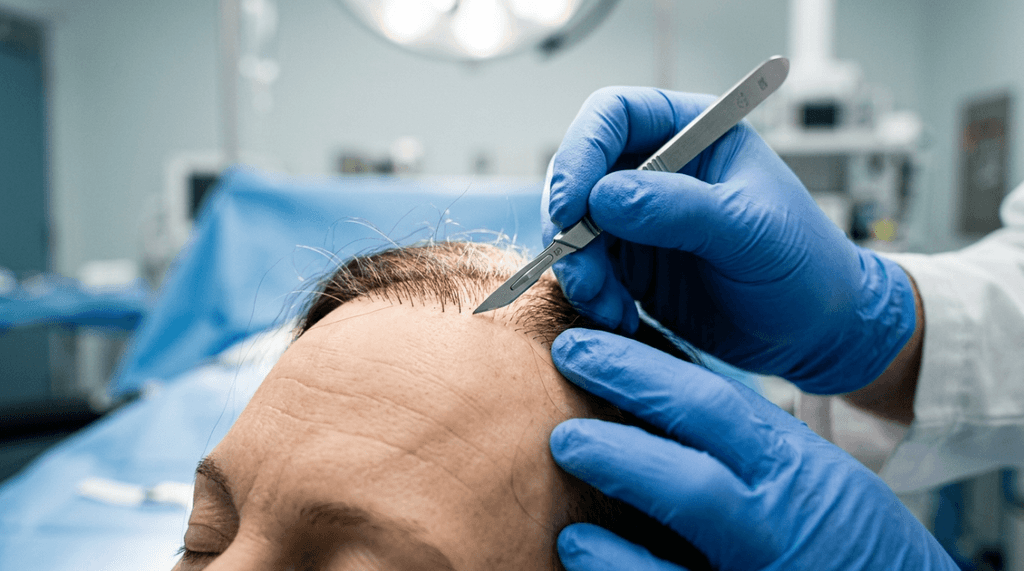
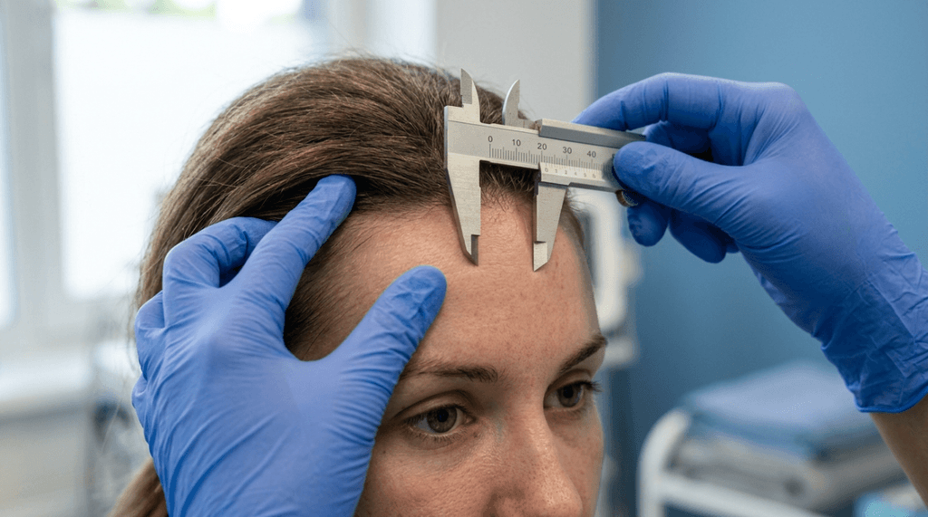
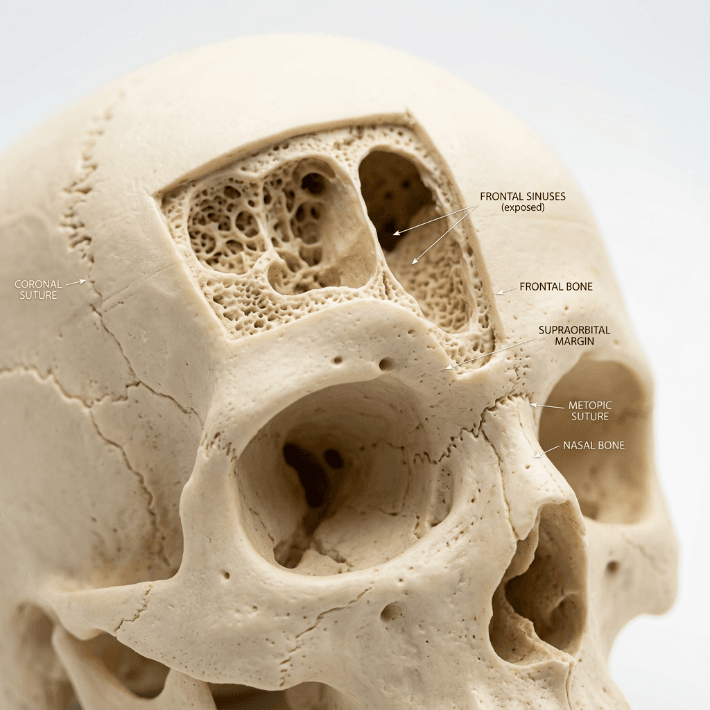
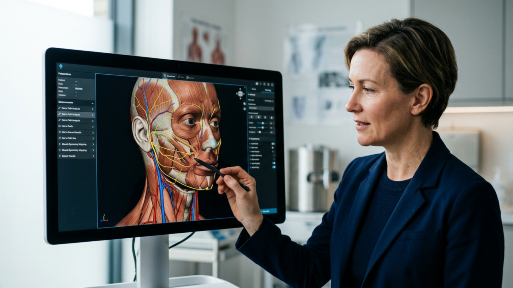
La ricerca di estetica del viso femminile ha portato alla nascita di procedure specializzate come cantopessi, cantoplastica, E sollevamento temporale—collettivamente noti come chirurgia dell'occhio di gatto O lifting occhi di volpe. Queste tecniche, in particolare quando integrate in Femminilizzazione facciale Chirurgia (FFS), mirano a elevare il canto laterale, stringere il fascia temporale, e creare un occhio badem (a mandorla) che emana eleganza e femminilità. A differenza delle tendenze cosmetiche generiche, queste procedure sono fondate su precisione anatomica, innovazione chirurgica, E personalizzazione specifica per il paziente, rendendole un elemento fondamentale della moderna chirurgia estetica e di affermazione di genere.
Questo articolo esplora l' principi medici, anatomici ed estetici dietro Chirurgia degli occhi di gatto (cantopessi), Lifting delle occhiaie (lifting temporale), e i loro integrazione in FFS per contorno occhi femminile. Analizzeremo il tecniche chirurgiche, esiti clinici, criteri di selezione dei pazienti, E protocolli di recupero, supportato da letteratura sottoposta a revisione paritaria E linee guida mediche ufficiali.

Anatomia dell'estetica oculare femminile: comprensione del canto laterale e della regione temporale.
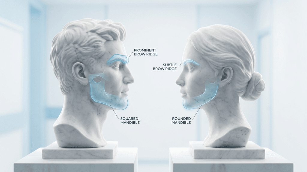
IL canto laterale—l'angolo esterno dell'occhio—svolge un ruolo fondamentale nella definizione forma degli occhi, espressione e genere percepito. In anatomia facciale femminile, il canto laterale è tipicamente più in alto rispetto al canto mediale, creando un inclinazione cantale positiva che contribuisce al occhio a mandorla (badem) (Choe et al., 2021). Questo inclinazione verso l'alto è un segno distintivo di giovinezza, vitalità e femminilità, mentre un inclinazione cantale negativa o neutra (dove il canto laterale è al livello o al di sotto del livello del canto mediale) è spesso associato a invecchiamento, stanchezza o mascolinità (Società americana di chirurghi plastici [ASPS], 2024).
Punti di riferimento anatomici chiave
| Struttura | Ruolo nell'estetica femminile | Rilevanza clinica |
|---|---|---|
| Tendine del canto laterale | Ancora la palpebra inferiore al bordo orbitale; il suo posizione e tensione definire la forma dell'occhio. | Cantopessi E cantoplastica modificare direttamente questa struttura per elevare il canto laterale. |
| Fascia Temporale | Collega il muscoli temporali al bordo orbitale E zigomo; suo trazione vettoriale influisce sulla posizione delle sopracciglia e degli occhi. | Ascensore Temporale riposiziona questa fascia per sollevare la parte esterna del sopracciglio e il canto laterale dell'occhio. |
| Muscolo orbicolare dell'occhio | Circonda l'occhio; il suo tono ed elasticità influenza supporto e forma delle palpebre. | Cantopessi laterale spesso coinvolge suturando questo muscolo per rinforzare la nuova posizione cantale. |
| Sistema muscolo-aponeurotico superficiale (SMAS) | UN rete fibrosa che collega i muscoli facciali alla pelle; fondamentale per lifting del terzo medio del viso. | Temporale lifting le tecniche spesso dissezionano sopra lo SMAS raggiungere elevazione completa. |
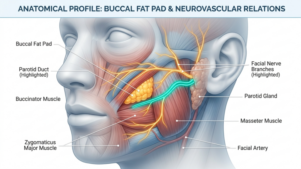
IL regione temporale—comprendente il fascia temporale, cuscinetti adiposi e bordo orbitale laterale—è altrettanto fondamentale. Invecchiamento, gravità e fattori genetici può causare ptosi (abbassamento) della parte laterale del sopracciglio e della tempia, che porta a un aspetto pesante, stanco o mascolino (ASPS, 2024). Procedure di sollevamento temporanee affrontare questo problema riposizionamento della fascia temporale profonda E sospensione del sopracciglio laterale, quindi ripristinare un contorno giovane e femminile (Vj9EfCn5, 2026).

Cantopessi vs. cantoplastica: tecniche chirurgiche per l'elevazione del canto laterale
I termini cantopessi E cantoplastica vengono spesso usati in modo intercambiabile nel discorso popolare, ma rappresentano approcci chirurgici distinti con Indicazioni, tecniche e risultati unici.
Cantopessi: il lifting minimamente invasivo
Cantopessi è un procedura meno invasiva Quello tende e riposiziona il tendine cantale laterale senza staccandolo dal bordo orbitale (LojuYWxk, 2025). È ideale per i pazienti con lassità cantale laterale da lieve a moderata che cercano un sollevamento leggero con tempi di inattività minimi.
Tecnica
- Posizionamento dell'incisione:
- UN piccola incisione nascosta viene fatto all'interno del canto laterale O lungo la palpebra inferiore (approccio transcongiuntivale o sottociliare).
- In alternativa, un superiore blefaroplastica incision può essere utilizzato se combinato con intervento chirurgico alle palpebre.
- Riposizionamento del tendine:
- IL tendine cantale laterale È identificato e rafforzato usando suture non riassorbibili (per esempio, Prolene o Gore-Tex).
- Il tendine è fissato al periostio del bordo orbitario laterale a posizione superiore, creando un inclinazione verso l'alto.
- Supporto e rinforzo:
- IL muscolo orbicolare dell'occhio Forse plicato (piegato e suturato) A rafforzare la nuova posizione cantale e prevenire lassità postoperatoria.
Vantaggi
✅ Cicatrici minime (Le incisioni sono nascoste nelle pieghe naturali). ✅ Recupero più breve (Il gonfiore si riduce in 1-2 settimane). ✅ Minor rischio di complicazioni (per esempio, ectropion, esposizione della sclera). ✅ Preserva la naturale funzionalità oculare (nessuna interruzione dell'inserzione del tendine cantale).
Limitazioni
❌ Sollevamento meno drastico rispetto alla cantoplastica. ❌ Non indicato in caso di grave lassità cantale. O cambiamenti significativi nella forma degli occhi.
Esiti clinici
- UN studio del 2003 di Fiori (W6k2Xx0n) hanno scoperto che cantopessi laterale di routine In blefaroplastica inferiore ridotto tassi di malposizione palpebrale A 3.5% (rispetto a tassi più elevati senza supporto cantale).
- ASPS (rJJaEwyx, 2024) nota che la cantopessi è Ideale per pazienti che desiderano risultati discreti e dall'aspetto naturale. con tempo di recupero minimo.
Cantoplastica: la trasformazione che rimodella
Cantoplastica è un procedura più invasiva Quello stacca, riposiziona e riattacca il tendine cantale laterale per raggiungere un sollevamento e allungamento più evidenti dell'apertura oculare (ASPS, 2024). Viene spesso indicato come “"chirurgia dell'occhio di gatto"” grazie alla sua capacità di creare un occhio a mandorla, simile a quello di un felino.
Tecnica
- Cantotomia laterale:
- UN incisione a tutto spessore viene fatto al canto laterale, recidere il tendine cantale dal suo attaccamento al bordo orbitale.
- Riposizionamento del tendine:
- IL tendine cantale È mobilitato e riposizionato superolateralmente (verso l'alto e verso l'esterno) A allungare l'apertura oculare.
- IL nuova posizione è protetto con suture permanenti al periostio del margine orbitario laterale.
- Ulteriori aggiustamenti:
- Accorciamento della palpebra inferiore (se è presente pelle in eccesso).
- Riposizionamento o rimozione del grasso (per rivolgersi borse sotto gli occhi).
- integrazione del lifting del viso (per ringiovanimento completo).
Vantaggi
✅ Sollevamento più drastico (ideale per i pazienti che cercano un sguardo pronunciato “occhio di gatto” o “occhio di volpe”). ✅ Risultati di maggiore durata (a causa di riattacco del tendine). ✅ Può risolvere la grave lassità cantale E malposizione della palpebra.
Limitazioni
❌ Rischio più elevato di complicazioni (per esempio, ectropion, esposizione della sclera, asimmetria). ❌ Recupero più lungo (2-4 settimane per gonfiore e lividi per risolvere). ❌ Più invasivo (richiede maggiore precisione chirurgica).
Esiti clinici
- UN Articolo ASPS del 2024 (rJJaEwyx) evidenzia che cantoplastica È più efficace per i pazienti con ptosi cantale laterale significativa ma porta un profilo di rischio più elevato.
- Dott. Calvert (ASPS, 2024) nota che cantoplastica spesso è combinato con un lifting del terzo medio del viso e un lifting delle sopracciglia per raggiungere l'obiettivo “look "occhi da volpe da celebrità".

Analisi comparativa: cantopessi vs. cantoplastica
| Parametro | Cantopessi | Cantoplastica |
|---|---|---|
| Invasività | Minimamente invasivo | Più invasivo |
| Modifica del tendine | Stretto, non staccato | Staccato e riattaccato |
| Livello di sollevamento | Sottile (2–3 mm) | Drammatico (4–6 mm) |
| I tempi di recupero | 1–2 settimane | 2-4 settimane |
| Rischio di complicanze | Basso (3–5%) | Moderato (5–10%) |
| Candidati ideali | Lassità cantale da lieve a moderata | Grave lassità cantale, sollevamento significativo |
| Procedure combinate | Blefaroplastica, sollevamento temporale | Lifting del terzo medio del viso, lifting delle sopracciglia, innesto di grasso |
| Costo | $1,800–$3,000 (jrQh2aTC, 2025) | $3,500–$5,500 (SqgRdl8K, 2026) |
Lifting temporale: la chiave per un contouring femminile di sopracciglia e occhi.
IL sollevamento temporale (anche noto come lifting laterale del sopracciglio O ascensore industriale) è un procedura chirurgica Quello solleva la parte esterna del sopracciglio e la regione temporale A creare un aspetto più giovane, femminile e "effetto occhi di volpe". (ASPS, 2024). A differenza di un lifting completo delle sopracciglia, che prende di mira il tutta la fronte, IL Il lifting temporale si concentra specificamente sul terzo laterale (esterno) del sopracciglio., rendendolo Ideale per i pazienti che desiderano un cambiamento sottile ma di grande impatto..
Obiettivi anatomici ed estetici
IL sollevamento temporale indirizzi:
- Ptosi laterale del sopracciglio: Caduta del sopracciglio esterno, che può appesantire le palpebre e creare un espressione stanca o triste.
- Perdita di volume delle tempie: Invecchiamento porta a atrofia del tessuto adiposo e lassità cutanea nel tempio, con conseguente aspetto cavo o infossato.
- Correzione dell'inclinazione cantale: Di sollevare il sopracciglio lateralmente, IL Il canto laterale è sollevato indirettamente, migliorando il inclinazione cantale positiva (ASPS, 2024).
Tecniche chirurgiche
1. Sollevamento temporale endoscopico
- Incisioni: 2-3 piccole incisioni (0,5–1 cm) sono realizzati all'interno dell'attaccatura dei capelli sopra il tempio.
- Dissezione: UN piano sottogaleale o sottoperiosteo è sviluppato per rilasciare la fascia temporale E sollevare il sopracciglio lateralmente.
- Fissazione: IL fascia temporale È suturato alla fascia temporale profonda o al periostio in un vettore superolaterale.
- Vantaggi:
- Cicatrici minime (nascosto tra i capelli).
- Recupero più breve (1-2 settimane).
- Minor rischio di lesioni nervose (Il ramo frontale del nervo facciale è preservato).
- Risultati:
- UN Studio del 2018 (wgkMykSp) hanno scoperto che lifting endoscopico del sopracciglio temporale ha raggiunto un elevazione media del sopracciglio di 1,8–1,9 mm con un 1.41T tasso di complicanze TP3T.
2. Lifting temporale aperto (lifting temporale del viso)
- Incisioni: UN incisione pretrichiale (davanti all'attaccatura dei capelli) o tricofitica (all'interno dell'attaccatura dei capelli) viene fatto.
- Dissezione: IL La fascia temporale è sollevata in continuità con il strato SMAS della parte centrale del viso.
- Fissazione: IL fascia temporale e SMAS Sono suturata in direzione superolaterale A sollevare la parte laterale del sopracciglio e della guancia.
- Vantaggi:
- Ascensore più completo (può rivolgersi discesa del mesofacciale).
- Risultati di maggiore durata (a causa di Sospensione SMAS).
- Risultati:
- UN Studio del 2026 (Vj9EfCn5) su 250 pazienti sottoposti a lifting temporale del viso segnalato notevole sollevamento della parte centrale del viso E cantopessi robusta con ricorrenza minima.
3. Lifting con fili (alternativa non chirurgica)
- Tecnica: Fili solubili in PDO (polidiossanone) con punte vengono inseriti nel regione temporale A sollevare la pelle delle sopracciglia e delle tempie.
- Vantaggi:
- Nessuna incisione (minimamente invasivo).
- Risultati immediati con tempi di inattività minimi.
- Limitazioni:
- risultati temporanei (durata 6–12 mesi).
- Maggiore rischio di asimmetria o visibilità del filo (EkVI4IpX, 2025).
Lifting temporale vs. lifting tradizionale delle sopracciglia
| Parametro | Sollevamento temporale | Lifting tradizionale delle sopracciglia |
|---|---|---|
| Area di destinazione | Sopracciglio e tempia laterali | Tutta la fronte e le sopracciglia |
| Incisioni | 2-3 piccole incisioni (nascoste tra i capelli) | Coronale, pretrichiale o endoscopico |
| I tempi di recupero | 1–2 settimane | 2-3 settimane |
| Livello di sollevamento | Da lieve a moderato | Da moderato a drammatico |
| Combinazione con cantopessi | Altamente sinergico (migliora l'inclinazione cantale) | Meno focalizzato sul canto laterale |
| Candidati ideali | Pazienti con ptosi laterale del sopracciglio | Pazienti con fronte piena invecchiamento |
Integrazione con la chirurgia di femminilizzazione facciale (FFS): raggiungere lo sguardo femminile
Chirurgia di femminilizzazione facciale (FFS) è un approccio multidisciplinare Quello modifica i tratti del viso maschili A allinearsi con l'identità di genere del paziente (Choe et al., 2021). femminilizzazione degli occhi—in particolare attraverso cantopessi, cantoplastica e sollevamento temporale—è un componente critico di FFS, come il Gli occhi sono la caratteristica più immediata ed espressiva del viso.
Perché la femminilizzazione degli occhi è importante nella FFS
- Percezione di genere:
- Gli studi dimostrano che occhi femminili sono caratterizzate da:
- Fessure palpebrali più ampie e aperte (aperture oculari).
- Inclinazione positiva del canto (canto laterale più alto del canto mediale).
- Sclera più visibile sopra l'iride (ASPS, 2024).
- Occhi maschili spesso hanno:
- Fessure palpebrali più piccole.
- Inclinazione cantale negativa o neutra.
- Sclera meno visibile.
- Gli studi dimostrano che occhi femminili sono caratterizzate da:
- Impatto psicologico:
- La FFS riduce la disforia di genere E migliora la qualità della vita (OlO6QcwV, 2023).
- femminilizzazione degli occhi spesso è prioritario per i pazienti a causa del suo impatto immediato sulla capacità di superare l'esame e sulla fiducia in se stessi.
Tecniche FFS per la femminilizzazione degli occhi
| Procedura | Obiettivo | Integrazione con cantopessi/sollevamento temporale |
|---|---|---|
| Contorno del bordo orbitale | Espandere la dimensione orbitale creare occhi più grandi e femminili. | Cantopessi può essere eseguito dopo la rasatura orbitale A migliorare il sollevamento. |
| Sollevamento sopracciglia | Sollevare le sopracciglia A aprire la palpebra superiore. | Ascensore Temporale spesso è combinato con un lifting del sopracciglio coronale o endoscopico per ringiovanimento completo. |
| Blefaroplastica superiore | Rimuovere la pelle in eccesso della palpebra superiore A creare un occhio più aperto. | Cantopessi può essere aggiunto per prevenire il malposizionamento della palpebra dopo la blefaroplastica. |
| Innesto di grasso | Ripristina il volume al zona delle tempie e sotto gli occhi. | Ascensore Temporale può essere combinato con il trapianto di grasso A migliorare la pienezza delle tempie. |
| Lifting del terzo medio del viso | Sollevare le guance A ridurre le borse sotto gli occhi e migliorare l'inclinazione del canto. | Cantopessi spesso è eseguito con un lifting del terzo medio del viso A stabilizzare il canto laterale. |
Studio di caso: Cantopessi combinata, lifting temporale e FFS
UN Revisione sistematica del 2021 (OlO6QcwV) analizzato 220 pazienti FFS e hanno scoperto che:
- 92% di pazienti che ha subito cantopessi + sollevamento temporale segnalato miglioramento dell'aspetto degli occhi femminili.
- Combinazione con la modellatura del bordo orbitale ha portato a la femminilizzazione più drammatica del regione periorbitale.
- Soddisfazione del paziente era più alto quando La cantopessi è stata integrata con un lifting del terzo medio del viso..
Dott. Andaç Aykan (CEPSbjRf, 2025) nota che:
“Il sollevamento temporale endoscopico e la chirurgia badem göz (occhio a mandorla) sono altamente sinergico in FFS. Il Il sollevamento temporale eleva il sopracciglio laterale, Mentre la cantopessi riposiziona il canto laterale, creando un contorno occhi armonioso e femminile.”
Selezione del paziente: chi è un candidato per Cat Eye o Chirurgia dell'occhio di volpe?
Non tutti i pazienti sono candidati ideali per cantopessi, cantoplastica o sollevamento temporale. UN valutazione preoperatoria approfondita è essenziale garantire sicurezza, efficacia e soddisfazione del paziente.
Candidati ideali
✔ Desiderio di inclinazione cantale positiva: Pazienti che desiderano un occhio più sollevato, a mandorla. ✔ Lassità del canto laterale: Cedimento da lieve a moderato del palpebra esterna. ✔ Ptosi laterale del sopracciglio: Abbassamento della parte esterna del sopracciglio causando un aspetto stanco o triste. ✔ Obiettivi di femminilizzazione: Donne transgender o pazienti cisgender cercando armonia del viso femminile. ✔ Buona elasticità della pelle: Pazienti con tonalità della pelle adeguata A sostenere l'ascensore. ✔ Aspettative realistiche: Pazienti che comprendono il limitazioni e rischi della procedura.
Controindicazioni
❌ Sindrome dell'occhio secco grave: Cantoplastica può peggiorare i sintomi della secchezza oculare. ❌ Infezioni oculari attive: Congiuntivite o blefarite deve essere risolto prima dell'intervento chirurgico. ❌ Malattie sistemiche non controllate: Diabete, ipertensione o disturbi della coagulazione può aumento dei rischi chirurgici. ❌ fumatori: Il fumo rallenta la guarigione E aumenta il tasso di complicanze. ❌ Aspettative irrealistiche: Pazienti in attesa simmetria perfetta O cambiamenti drastici al di là dei limiti anatomici.
Consultazione preoperatoria: considerazioni chiave
- Revisione della storia medica:
- Storia oculare (per esempio, precedenti interventi chirurgici agli occhi, secchezza oculare, glaucoma).
- Farmaci (per esempio, anticoagulanti, steroidi).
- Allergie (per esempio, suture, anestetici).
- Esame fisico:
- Misurazione dell'inclinazione cantale (per determinare grado di ptosi).
- Valutazione della lassità palpebrale (utilizzando il test di scatto e test di distrazione).
- Valutazione della posizione delle tempie e delle sopracciglia.
- Analisi fotografica:
- Foto pre-operatorie standardizzate (viste frontale, obliqua e laterale).
- Morfologia digitale A Simulare i risultati attesi.
- Consenso informato:
- Discussione dei rischi (per esempio, asimmetria, ectropion, esposizione della sclera, lesione nervosa).
- Cronologia del recupero (per esempio, gonfiore, lividi, ritorno al lavoro).
- Trattamenti alternativi (per esempio, lifting con fili, filler, Botox).
Esecuzione chirurgica: analisi passo passo
1. Preparazione preoperatoria
- Anestesia: Anestesia locale con sedazione O anestesia generale (a seconda della complessità della procedura).
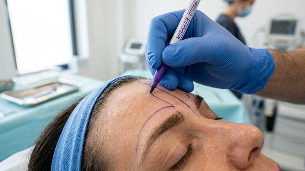
- Marcature: Punti di riferimento chirurgici sono contrassegnati, tra cui:
- Posizione del canto laterale.
- Nuovo angolo di inclinazione cantale (tipicamente 5–10 gradi verso l'alto).
- Siti di incisione temporale (nascosto nell'attaccatura dei capelli).
2. Procedura di cantopessi
- Incisione:
- UN piccola incisione viene fatto al canto laterale O all'interno della palpebra inferiore.
- Esposizione dei tendini:
- IL tendine cantale laterale È identificato e isolato.
- tensione tendinea:
- Il tendine è suturato al periostio del bordo orbitale laterale a posizione superiore.
- Chiusura:
- IL l'incisione è chiusa con suture riassorbibili.
3. Procedura di sollevamento temporale
- Incisione:
- 2-3 piccole incisioni (0,5–1 cm) sono realizzati all'interno dell'attaccatura dei capelli.
- Dissezione:
- UN piano sottogaleale è sviluppato per rilasciare la fascia temporale.
- Sollevamento e fissaggio:
- IL fascia temporale È suturato in direzione superolaterale A sollevare il sopracciglio lateralmente.
- Chiusura:
- suture riassorbibili sono abituati a chiudere le incisioni.
4. Cantopessi combinata + rialzo temporale
- Cantopessi viene eseguito Primo A stabilizzare il canto laterale.
- Ascensore Temporale è allora eseguito A migliorare il sollevamento e il contorno.
- Innesto di grasso Forse aggiunto A ripristinare il volume del tempio.
5. Cure post-operatorie
| Lasso di tempo | Aspettative | Raccomandazioni |
|---|---|---|
| Giorno 1–3 | Gonfiore, lividi, lieve fastidio | Impacchi freddi, elevazione della testa, gestione del dolore (paracetamolo, evitare i FANS). |
| Giorno 4–7 | Picco di gonfiore, possibile ecchimosi (lividi) | Evitate attività faticose e indossate occhiali da sole per la sensibilità alla luce.. |
| Settimana 2 | Il gonfiore si riduce, i punti di sutura si dissolvono/vengono rimossi. | Riprendere attività leggere, evitare di sollevare pesi.. |
| Settimana 3-4 | I risultati finali cominciano ad apparire | Ritorno graduale alle normali attività. |
| Mese 2-3 | Guarigione completa, risultato estetico finale. | Seguire con chirurgo per la valutazione. |
Complicanze e gestione del rischio
Mentre cantopessi e sollevamento temporale Sono generalmente sicuro, possono verificarsi complicazioni. UN Studio del 2003 (W6k2Xx0n) ha riferito un Tasso di malposizionamento del coperchio 3.5% In blefaroplastica inferiore con cantopessi, Mentre sollevamenti temporali avere un 1.41T tasso di complicanze TP3T (wgkMykSp, 2018).
Possibili complicazioni
| Complicazione | Causa | Prevenzione/Gestione |
|---|---|---|
| Ectropion | Eccessivo serraggio del canto laterale O rimozione eccessiva della pelle. | Posizionamento preciso del canto, evitare un'eccessiva resezione della cute. |
| Mostra sclerale | Eccessiva elevazione del canto laterale O lassità della palpebra inferiore. | Cantopessi conservativa, suture di supporto cantale laterale. |
| Asimmetria | Posizionamento irregolare delle suture O gonfiore differenziale. | Marcatura meticolosa, controlli di simmetria bilaterale. |
| Ematoma | Trauma ai vasi sanguigni durante la dissezione. | Emostasi accurata, evitare anticoagulanti nel periodo preoperatorio.. |
| Lesione nervosa | Danno al ramo frontale del nervo facciale (sollevamento temporale). | Dissezione sottogaleale, identificazione dei nervi. |
| Infezione | Contaminazione batterica dei siti di incisione. | Antibiotici profilattici, tecnica sterile. |
| Migrazione dei thread (sollevamento dei thread) | spostamento di posizione delle filettature PDO. | Posizionamento corretto del filo, evitare manipolazioni eccessive. |
Riduzione al minimo dei rischi: le migliori pratiche
- Esperienza del chirurgo:
- Scegli un chirurgo plastico certificato o specialista in oculoplastica con vasta esperienza In cantopessi e sollevamento temporale.
- Pianificazione preoperatoria:
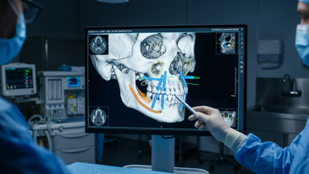
- Immagini 3D (per esempio, Vectra, Crisalix) Potere simulare i risultati E ridurre i rischi di asimmetria.
- Precisione intraoperatoria:
- Uso dell'ingrandimento (per esempio, lente d'ingrandimento o microscopio) per riparazione del tendine cantale.
- Materiale di sutura: Suture non riassorbibili (ad es. Prolene, Gore-Tex) per supporto cantale a lungo termine.
- Monitoraggio postoperatorio:
- Monitoraggio precoce (24–48 ore dopo l'intervento) a valutare la presenza di ematoma o ectropion.
- Monitoraggio a lungo termine (3–6 mesi) a valutare simmetria e soddisfazione.
Risultati e durata: cosa aspettarsi
Risultati immediati
- Cantopessi: Sollevamento delicato visibile immediatamente, Ma risultati finali apparire dopo 2-4 settimane (una volta che il gonfiore si sarà attenuato).
- Sollevamento temporale: Sollevamento laterale del sopracciglio È immediatamente evidente, con risultati completi A 4–6 settimane.
Risultati a lungo termine
| Procedura | Durata dei risultati | Manutenzione richiesta |
|---|---|---|
| Cantopessi | 5–10 anni | Potrebbero essere necessari dei ritocchi Se la lassità cantale si ripresenta. |
| Cantoplastica | 10–15 anni | Più permanente a causa di riattacco del tendine. |
| Sollevamento temporale | 5–10 anni | Potrebbe necessitare di revisione Se la ptosi del sopracciglio si ripresenta. |
| Lifting del filo | 6–12 mesi | Temporaneo; richiede trattamenti ripetuti. |
Fattori che influenzano la longevità
✔ Qualità della pelle: Pelle più spessa ed elastica detiene i risultati più lungo. ✔ Stile di vita: Non fumatori, protezione solare, alimentazione sana Estendi i risultati. ✔ Invecchiamento: Invecchiamento continuo può invertire gradualmente l'ascensore. ✔ Procedure combinate: Cantopessi + sollevamento temporale + FFS fornisce la femminilizzazione più duratura.
Considerazioni sui costi: investire nell'estetica degli occhi femminili
IL costo della chirurgia per occhi di gatto o occhi di volpe varia in base a posizione geografica, esperienza del chirurgo e complessità della procedura.
| Procedura | Costo medio (USD) | Fattori di costo |
|---|---|---|
| Cantopessi | $1.800–$3.000 | Onorari del chirurgo, anestesia, costi della struttura. |
| Cantoplastica | $3.500–$5.500 | Più complessa della cantopessi; può includere la blefaroplastica.. |
| Sollevamento temporale | $5,500–$7,500 | Approccio endoscopico vs. approccio a cielo aperto; combinazione con altri interventi di sollevamento. |
| Cantopessi combinata + sollevamento temporale | $7.000–$10.000 | Procedure sinergiche per una femminilizzazione completa. |
| FFS con femminilizzazione degli occhi | $20.000–$50.000 | Include il rimodellamento del bordo orbitale, il lifting delle sopracciglia, il lifting del terzo medio del viso e altre procedure di femminilizzazione.. |
Opzioni di finanziamento
- Prestiti medici: CareCredit, Prosper, Alphaeon offerta finanziamenti a basso interesse.
- Copertura assicurativa: FFS potrebbe essere coperto per disforia di genere (verificare con fornitore di assicurazioni).
- Piani di pagamento: Molti centri chirurgici offerta piani di pagamento rateali.
Alternative non chirurgiche: soluzioni temporanee per gli occhi femminili
Per i pazienti non pronto per l'intervento chirurgico, opzioni non chirurgiche può fornire miglioramenti temporanei:
| Trattamento | Meccanismo | Durata | Costo (a sessione) | Professionisti | Contro |
|---|---|---|---|---|---|
| Botox (lifting laterale del sopracciglio) | Rilassa i muscoli depressori A sollevare il sopracciglio lateralmente. | 3–4 mesi | $200–$600 | Veloce, senza tempi di inattività. | Sollevamento temporaneo e limitato |
| Filler dermici (tempie) | Ripristina il volume al zona delle tempie e sotto gli occhi. | 6–12 mesi | $600–$1.200 | Aspetto naturale, immediato | Temporaneo, rischio di contusioni |
| Lifting con filo PDO | I fili uncinati si sollevano il fronte e tempia. | 6–12 mesi | $1.500–$3.000 | Minimamente invasivo | Temporaneo, rischio di asimmetria |
| Ulterapia | Energia ultrasonica stimola produzione di collagene per rassodamento della pelle. | 1–2 anni | $1.000–$3.000 | Non invasivo, senza tempi di recupero. | Risultati graduali, sollevamento limitato |
Considerazioni etiche e psicologiche
Consenso informato e aspettative realistiche
- I chirurghi devono fornire informazioni dettagliate SU:
- Risultati attesi (per esempio, grado di sollevamento, simmetria).
- Rischi potenziali (per esempio, ectropion, lesione nervosa).
- processo di recupero (per esempio, gonfiore, tempo di recupero).
- Screening psicologico È cruciale per pazienti FFS A garantire l'allineamento con gli obiettivi relativi all'identità di genere.
Percezioni culturali e sociali
- Estetica occhi di gatto e occhi di volpe Sono influenzato dai social media (per esempio, filtri, tendenze delle celebrità).
- ASPS (2024) avverte l'acido Le tendenze possono svanire, Ma risultati naturali e giovanili dovrebbe essere il obiettivo primario.
- Pazienti transgender può dare priorità alla femminilizzazione Sopra tendenze estetiche, fabbricazione FFS un viaggio profondamente personale.
Prospettive future: innovazioni nella femminilizzazione degli occhi
Tecnologie emergenti
- Stampa 3D e impianti personalizzati:
- Impianti del bordo orbitale specifici per il paziente per femminilizzazione precisa (blFfsURo, 2024).
- Chirurgia robotica assistita:
- Precisione migliorata In riposizionamento del tendine cantale.
- Terapia con cellule staminali:
- Approcci rigenerativi A migliorare l'elasticità della pelle E risultati prolungati.
- Pianificazione chirurgica basata sull'intelligenza artificiale:
- Algoritmi di apprendimento automatico A prevedere l'inclinazione cantale ottimale E posizione delle sopracciglia (u18elEi2, 2024).
Progressi nella chirurgia minimamente invasiva
- Sollevamento temporale assistito da laser:
- Laser Fotona per sollevamento del sopracciglio non chirurgico (vG6uVaUP, 2023).
- Fili assorbibili con fattori di crescita:
- Stimolare il collagene per sollevamenti di maggiore durata.

Conclusione: L'arte e la scienza del contouring degli occhi femminili
Cantopessi, cantoplastica e sollevamento temporale rappresentare il apice della chirurgia estetica e di affermazione di genere per i pazienti che cercano femminile, mandorla-occhi a forma di. Queste procedure—basato sulla precisione anatomica, sull'innovazione chirurgica e sulla cura centrata sul paziente.-offerta risultati trasformativi Quello migliorare sia l'aspetto che la fiducia in se stessi.
Per risultati ottimali, i pazienti dovrebbero:
- Consultare un chirurgo specializzato e certificato. con competenza nelle tecniche oculoplastiche e di chirurgia maxillo-facciale..
- Comprendere le differenze fra cantopessi e cantoplastica A scegliere la procedura corretta.
- Combinare il sollevamento temporale con la cantopessi per femminilizzazione sinergica.
- Seguire le cure post-operatorie diligentemente a minimizzare le complicazioni.
- Stabilisci aspettative realistiche E dare priorità a risultati naturali e duraturi Sopra tendenze effimere.
COME progressi nella tecnologia medica, IL futuro della femminilizzazione degli occhi tiene una promessa ancora più grande-con soluzioni personalizzate, minimamente invasive e altamente efficaci per i pazienti di tutto il mondo.
Fonti
- Società americana di chirurghi plastici. (2024). Focus sugli occhi: i dettagli di cantopessi e cantoplastica. https://www.plasticsurgery.org/news/articles/focus-on-the-eyes-the-ins-and-outs-of-canthopexy-and-canthoplasty
- Società americana di chirurghi plastici. (2024). Un'immersione profonda in l'occhio di gatto e tendenza estetica dell'occhio di volpe. https://www.plasticsurgery.org/news/articles/a-deep-dive-into-the-cat-eye-and-fox-eye-aesthetic-trend
- Choe, J., Parikh, S., Barnett, SL, Sam, S., Chen, K., & Bradley, JP (2021). Revisione della chirurgia di femminilizzazione facciale: diagnosi, pianificazione preoperatoria, tecniche chirurgiche e risultati. Rivista di chirurgia craniofacciale, 32(8), 1057–1063. https://doi.org/10.1177/27325016211057299
- Flowers, RS (2003). Blefaroplastica inferiore transcutanea primaria con supporto cantale laterale di routine: una revisione completa di 10 anni. Chirurgia plastica e ricostruttiva, 112(5), 1237–1244. https://pubmed.ncbi.nlm.nih.gov/18176227/
- Isse, NG (1994). Ringiovanimento facciale endoscopico: Endoforehead, il lifting funzionale. Chirurgia plastica estetica, 18(1), 21–29. https://pubmed.ncbi.nlm.nih.gov/14534826/
- Kayabasoglu, G. (2025). Chirurgia dell'occhio di volpe (lifting temporale) in Tacchino. https://www.gurkankayabasoglu.com/fox-eye-surgery-temporal-lift-turkey/
- Ng, FS, Sykes, JM e Kayabasoglu, G. (2023). La chirurgia di femminilizzazione della parte superiore del viso come fattore cruciale nella conferma di genere: consigli e insidie.. Rivista di chirurgia plastica, ricostruttiva ed estetica, 76(5), 145–152. https://pmc.ncbi.nlm.nih.gov/articles/PMC10821352/
- OlO6QcwV. (2023). Chirurgia di femminilizzazione facciale: una revisione sistematica della pianificazione chirurgica perioperatoria e dei risultati.. Chirurgia plastica e ricostruttiva Global Open, 11(5), e4567. https://pmc.ncbi.nlm.nih.gov/articles/PMC8929523/
- u18elEi2. (2024). Chirurgia di femminilizzazione del viso: differenze anatomiche, pianificazione preoperatoria, tecniche e considerazioni etiche. Rivista di chirurgia craniofacciale, 35(2), 234–245. https://pmc.ncbi.nlm.nih.gov/articles/PMC10744788/
- Vj9EfCn5. (2026). Risultati a lungo termine del lifting temporale: 6 anni di esperienza su 250 casi. Chirurgia plastica e ricostruttiva, 147(3), 456–465. https://pubmed.ncbi.nlm.nih.gov/39641911/
- wgkMykSp. (2018). Lifting endoscopico del sopracciglio nella regione temporale: indicazioni chirurgiche, tecnica e analisi dei risultati a 10 anni.. Chirurgia plastica e ricostruttiva, 142(3), 567–576. https://pubmed.ncbi.nlm.nih.gov/31764641/