

ماذا لو كانت أقوى عملية شد للحواجب في تأنيث الوجه هل تتطلب الجراحة أي تثبيت، أو أجهزة تعليق، أو إجراء رفع مخصص؟ يقوم الجراحون عادةً بجدولة عملية رفع الحاجب بالمنظار بالتزامن مع الجراحة. شد الجبين, بافتراض أن الحاجب يحتاج إلى رفع مستقل. ومع ذلك، تكشف الملاحظات السريرية عن ظاهرة غير بديهية: تحديد محيط الحافة فوق الحجاجية وردود الفعل على موضع الحاجب يؤدي ذلك إلى رفع الحاجب هندسيًا بمقدار 2 إلى 4 مليمترات من خلال تقليل حجم العظام فقط. كما أن إزالة الحافة فوق الحجاجية البارزة تغير الهيكل الميكانيكي أسفل الحاجب، وتعاد توزيع الأنسجة الرخوة المغطية على طول المتجهات المتاحة حديثًا، مما يخلق رفعًا مرئيًا دون الحاجة إلى أي جراحة رفع منفصلة.
تُشكّل هذه الآلية تحدياً للنموذج الجمعي التقليدي لجماليات الحواجب في تأنيث الوجه.يعتقد المرضى والجراحون غالبًا أن إجراء المزيد من العمليات الجراحية يعني نتائج أفضل. ومع ذلك، عندما تصغير حافة الحجاج يُعيد هذا الإجراء تشكيل النتوء العظمي البارز أسفل الحاجب، حيث يتمدد غلاف الأنسجة الرخوة للأعلى على طول مستوى محيطي جديد. ويُنتج هذا التمدد ارتفاعًا ملحوظًا وقابلًا للقياس في الحاجب. تُقدم هذه المقالة شرحًا دقيقًا من الناحية البيوميكانيكية لكيفية رفع الحاجب عن طريق إزالة العظم، ومتى يصبح رفع الحاجب بالمنظار غير ضروري، ومتى يظل ضروريًا سريريًا.
جدول المحتويات
الميكانيكا الحيوية لتحديد محيط حافة الحجاج وردود فعل وضع الحاجب
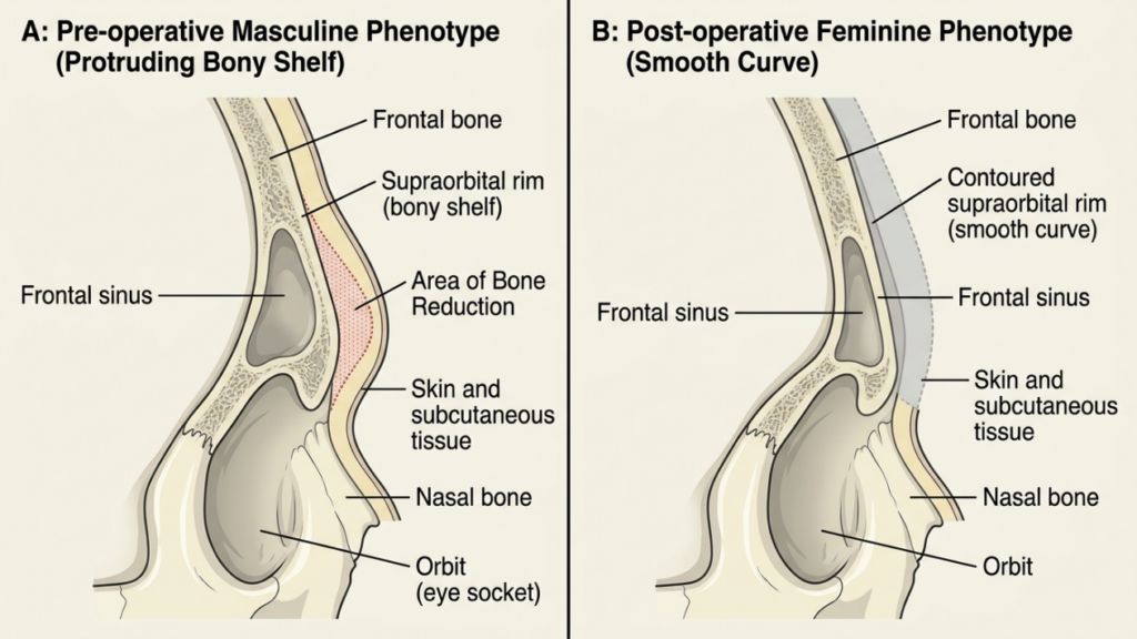
يبرز الحافة فوق الحجاجية في جبهة الذكور بيولوجيًا إلى الأمام والأسفل، مكونةً رفًا عظميًا يُظلل الجفن العلوي ويُخفض موضع الحاجب المرئي. يُشكل هذا البروز نقطة ارتكاز ميكانيكية لأنسجة الحاجب الرخوة. تتدلى العضلة الدويرية العينية، والعضلة المقطبة للحاجب، والوسادة الدهنية للحاجب فوق هذه الحافة كما يتدلى القماش فوق حافة رف. عندما يبرز الرف بقوة، يتدلى القماش إلى أسفل.
تحديد محيط حافة الحجاج يزيل ذلك البروز الأمامي السفلي. دكتور جراح يتم تقليل حجم العظم باستخدام المثاقب أو المبارد أو عمليات قطع العظم، مما يؤدي إلى نحت الحافة من نتوء بارز إلى منحنى أنثوي أكثر نعومة وتراجعًا للخلف. عندما يختفي هذا الرف العظمي، تنتقل نقطة تثبيت الأنسجة الرخوة للخلف وللأعلى قليلاً. يتبع غلاف الأنسجة الرخوة للحاجب، الذي كان ممتدًا سابقًا فوق نتوء بارز، الآن المحيط الجديد ويستقر في وضع رأسي أعلى بالنسبة لمقلة العين.
تخيل الأمر وكأنك تسحب عمود خيمة إلى الداخل. لا يسقط قماش الخيمة، بل يستقر ويرتفع على طول الاتجاه الجديد. وينطبق المبدأ نفسه على... إعادة تشكيل الأنسجة الرخوة فوق عظم الجبهة المُعاد تشكيله. يلاحظ الدكتور محمد فاتح أوكياي، أخصائي جراحة التجميل المعتمد من البورد الأوروبي والتركي، هذا الارتفاع الهندسي بشكل روتيني في عملياته قبل وبعد معرض FFS المرضى الذين يخضعون تم تحديد ملامح الجبهة بـإجراء عملية شد الحاجبين في نفس الوقت.
تحليل المتجهات: كيف يؤدي تقليل حجم العظام إلى رفع الحاجب هندسيًا
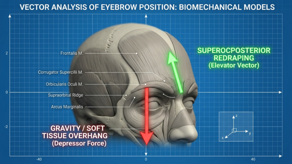
يتطلب فهم الفيزياء تحليل متجهات القوة. قبل الجراحة، تخلق الحافة فوق الحجاجية البارزة قوتين رئيسيتين على الحاجب. أولاً، متجه الدوران السفلي تتولد هذه الظاهرة بفعل الجاذبية المؤثرة على كتلة الأنسجة الرخوة المتدلية من حافة الحافة الأمامية. ثانيًا، متجه الإزاحة الخلفية يحدث ذلك بسبب وزن الأنسجة الضاغطة على النتوء العظمي.
عندما دكتور جراح عند إجراء عملية تصغير حافة الحجاج، يتغير كلا المتجهين بشكل كبير. يقل متجه الدوران السفلي لأن النسيج لم يعد يتدلى من حافة مائلة للأسفل. بدلاً من ذلك، يخلق المحيط الأنثوي الأملس الجديد متجه إعادة التغطية العلوي الخلفي.ينزلق غلاف الأنسجة الرخوة على طول هذا السطح الجديد، فيرتفع الحاجب بصريًا. وتؤكد القياسات السريرية ارتفاع الحاجب بمقدار 2 إلى 4 مليمترات بعد تحديد محيط الحافة المعزول، وهو ما تم توثيقه في الأدبيات الطبية المتعلقة بجراحة الوجه والجمجمة والمراجعة من قبل النظراء. (NCBI، 2018).
يحدث هذا الارتفاع من خلال ثلاث آليات مترابطة. أولاً، يؤدي إزالة العظم إلى التخلص من الرف السفلي الذي كان يسحب الحاجب إلى الأسفل ميكانيكياً. ثانياً، يؤدي تحرير السمحاق أثناء الوصول التاجي إلى تحرير عضلات خافضة الحاجب، مما يقلل من سحبها للأسفل على الحاجب. ثالثاً، يؤدي انكماش الندبة بعد العملية على طول العظم المعاد تشكيله إلى إنشاء تعليق طبيعي يثبت الأنسجة الرخوة في وضع أعلى.
قياس إزاحة المتجه
يستطيع الجراحون ذوو الخبرة تقدير حجم عملية رفع الحاجب المتوقعة بناءً على درجة تصغير حافة الحاجب. في عملية تحويل نموذجية من ذكر إلى أنثى شد الجبين, يقوم الجراح بإزالة ما بين 3 إلى 7 ملليمترات من النتوء العظمي الأمامي عند الحافة فوق الحجاجية. كل ملليمتر يتم إزالته من النتوء العظمي يُترجم إلى ما يقارب 0.5 إلى 0.7 ملليمتر من ارتفاع الحاجب عموديًا من خلال تغيير المتجه الهندسي. تعتمد هذه النسبة على سمك الأنسجة الرخوة، ومرونة الجلد، وزاوية العظم المستأصل.
ديناميكيات إعادة تشكيل الأنسجة الرخوة بعد تصغير حافة الحجاج
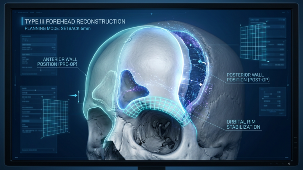
إعادة تشكيل الأنسجة الرخوة هي الآلية الأساسية وراء عملية رفع الحاجب الملحوظة بعد إعادة تشكيل العظام. يعكس هذا المفهوم ما يحدث أثناء عملية تجميل الحاجب التقليدية. شد الوجهعندما تعيد وضع الهيكل الأساسي، يتكيف الغلاف الخارجي مع المشهد الجديد. على عكس شد الوجه, ، لكن،, تقليل حافة الحجاج يُغير العظام بدلاً من إعادة تموضع الأنسجة الرخوة بشكل مباشر، ومع ذلك فإن النتيجة المرئية تشبه عملية شد العظام.
تؤثر عدة عوامل على مدى فعالية إعادة تشكيل الأنسجة الرخوة بعد عملية نحت الحواف. عادةً ما يشهد المرضى ذوو طبقات الدهون تحت الجلد الرقيقة إعادة تشكيل أكثر وضوحًا نظرًا لوجود كتلة نسيجية أقل لمقاومة التغيير الموضعي. في المقابل، قد يلاحظ المرضى ذوو الوسائد الدهنية السميكة في منطقة ما بين الحاجبين ارتفاعًا أقل وضوحًا، حيث يقاوم الغلاف النسيجي الأثقل الحركة.
يؤثر النهج الجراحي أيضًا على ديناميكية إعادة تشكيل الجبهة. يُحرر الشق التاجي المزدوج مع تشريح واسع تحت السمحاق كامل غلاف الأنسجة الرخوة للجبهة من ارتباطاتها العظمية. يسمح هذا التحرير للأنسجة بإعادة تشكيل نفسها بحرية فوق المحيط الجديد. في المقابل، قد لا يوفر الشق عند خط الشعر مع تشريح محدود تحريرًا كافيًا، مما يؤدي إلى إعادة تموضع أقل وضوحًا للحاجب. يؤكد الدكتور محمد فاتح أوكياي أن جودة تحرير السمحاق ترتبط ارتباطًا مباشرًا بدرجة رفع الحاجب السلبي التي يتم تحقيقها من خلال نحت الجبين وحيد.
يُخفي التورم بعد العملية الجراحية في البداية تأثير إعادة تشكيل الحاجب. خلال الأسابيع الثلاثة الأولى، يدفع التورم الحاجب إلى الأسفل، مما يُوهم بعدم حدوث عملية رفع. ومع انحسار التورم بين الأسبوعين الثالث والثامن، يرتفع الحاجب تدريجيًا إلى موضعه الهندسي الجديد. يجب على المرضى فهم هذا الجدول الزمني لتجنب القلق المبكر بشأن موضع حاجبيهم.
تأنيث الجبهة بدون عملية رفع منفصلة: الأدلة والنتائج
تدعم الأدلة السريرية بشكل متزايد الملاحظة القائلة بأن تحديد محيط الحافة فوق الحجاجية المعزول ينتج عنه ارتفاع ملحوظ في الحاجب. ولعل البيانات الأكثر إقناعًا تأتي من التحليلات الاسترجاعية لمرضى جراحة تجميل الوجه الذين خضعوا لتحديد محيط الجبهة دون إجراءات رفع الحاجب بالمنظار في نفس الوقت.
أظهرت دراسة رائدة حللت وضع الحاجب بعد عملية تحديد ملامح الجبهة من النوع الثالث أن متوسط ارتفاع الحاجب بلغ 2.8 مليمتر بعد ستة أشهر من الجراحة، وقد تحقق ذلك دون أي تثبيت أو تعليق للحاجب. (NCBI، 2018).هذه النتيجة رائعة لأنها تحدد كمياً ما لاحظه جراحو تجميل الوجه ذوو الخبرة منذ فترة طويلة: إن تقليل العظام وحده يرفع الحاجب.
التفسير البيوميكانيكي واضح. إزالة النتوء العظمي السفلي تُزيل البنية التشريحية التي كانت تُجبر الحاجب ميكانيكيًا على الانخفاض. بمجرد زوال هذا العائق، تعمل مرونة الأنسجة الطبيعية وقوى التئام الجروح بعد العملية على توجيه الحاجب للأعلى. كما أن شد السمحاق الذي يحدث أثناء نضج الندبة يُعزز هذا الارتفاع، مما يُؤدي إلى نتيجة مستقرة طويلة الأمد.
بيانات مقارنة: تحديد حواف الجنوط وحده مقابل الإجراءات المدمجة
يلخص الجدول التالي النتائج المقارنة بناءً على النهج الجراحي والملاحظات السريرية من حالات تجميل الوجه الأنثوي:
| النهج الجراحي | متوسط ارتفاع الحاجب (مم) | ندوب إضافية | زيادة وقت التشغيل | مخاطر المضاعفات |
|---|---|---|---|---|
| تحديد محيط الحافة المعزول | من 2.0 إلى 4.0 | لا شيء (شق تاجي فقط) | خط الأساس | قليل |
| تحديد محيط الحافة + رفع الحاجب بالمنظار | من 3.5 إلى 6.0 | شقوق صدغية | من 45 إلى 60 دقيقة | متوسطة (إصابة عصبية، عدم تناسق) |
| عملية رفع الحاجب بالمنظار فقط | من 1.5 إلى 3.0 | شقوق صدغية | من 30 إلى 45 دقيقة | متوسط (خطر التكرار) |
| تحديد محيط الحافة + رفع الصدغ بالمنظار | من 3.0 إلى 5.5 | شقوق صدغية | من 40 إلى 55 دقيقة | معتدل |
تُظهر البيانات أن تحديد محيط حافة الحاجب بشكل منفرد يحقق رفعًا ملحوظًا للحاجب دون الحاجة إلى وقت جراحي إضافي، أو ندوب، أو مخاطر مضاعفات مرتبطة بإجراءات التنظير الداخلي المتزامنة. يستخدم الدكتور محمد فاتح أوكياي، الحاصل على زمالة المجلس الأوروبي لجراحة التجميل والترميم منذ عام 2018، هذه الأدلة لتوجيه التخطيط الجراحي الفردي لكل مريض، كما هو موثق في من هو دكتور ام اف او؟.

شدة تدلي الحاجب: عندما لا يكفي تحديد شكل الحاجب وحده
لا تستجيب جميع الحواجب بنفس القدر للرفع الهندسي الناتج عن تحديد محيط الحافة. العامل الحاسم هو درجة تدلي الحاجب قبل الجراحة. المرضى الذين يعانون من تدلي طفيف، حيث يقع الحاجب عند أو بالقرب من الحافة العلوية للحجاج، يحققون باستمرار رفعًا مرضيًا من خلال تقليل حجم العظم فقط. تنشأ قوة الخفض الرئيسية لدى هؤلاء المرضى من بروز العظم وليس من ارتخاء الأنسجة الداخلي.
لكن، تدلي الحاجب أما الحالات التي تتجاوز الشدة المتوسطة فتشير إلى وضع مختلف. فعندما ينخفض الحاجب بشكل ملحوظ أسفل الحافة العلوية للحجاج نتيجة لترهل الأنسجة المرتبط بالتقدم في السن، أو هبوط الأنسجة الرخوة بفعل الجاذبية، أو ضعف عضلة الجبهة المزمن، فإن تصغير العظم لا يحل المشكلة بشكل كامل. صحيح أن عملية تحديد محيط الحافة تزيل النتوء العظمي، إلا أن غلاف الأنسجة الرخوة الممتد يفتقر إلى المرونة اللازمة للعودة إلى وضعه الطبيعي بشكل مستقل.
يتطلب التمييز بين انخفاض الحاجب البنيوي الناتج عن بروز الحافة وتدلي الجفن الحقيقي الناتج عن ارتخاء الأنسجة الرخوة تقييمًا دقيقًا قبل الجراحة. إن إجراء عملية تصغير الحافة لدى مريض يعاني من تدلي شديد في الأنسجة الرخوة ينتج عنه شكل جبهة أنثوي، ولكنه يترك الحاجب منخفضًا من الناحية الجمالية، مما يخلق عدم تناسق بين محيط العظم الأملس وموضع الأنسجة الرخوة المنخفض.

عملية رفع الحاجب بالمنظار: هل هي ضرورية حقاً أم زائدة عن الحاجة؟
يُعد هذا السؤال جوهر التخطيط الجراحي لـ عملية تجميل الحواجب.يعتمد الجواب كلياً على عوامل خاصة بكل مريض. إن تقديم توصية عامة لصالح أو ضد إجراء عملية رفع الحاجب بالمنظار في نفس الوقت يبسط الواقع السريري بشكل مفرط.
ان رفع الحاجب بالمنظار يصبح هذا الإجراء غير ضروري عند توافر ثلاثة شروط في آن واحد. أولاً، يعاني المريض من بروز واضح في الحافة فوق الحجاجية مما يُسبب بروزًا عظميًا كبيرًا. ثانيًا، يتمتع المريض بمرونة جيدة في الجلد مع حد أدنى من تدلي أنسجة الحاجب الرخوة. ثالثًا، يقل عمر المريض عن خمسين عامًا تقريبًا، حيث لم يصبح ارتخاء الأنسجة بعدُ القوة الخافضة الرئيسية.
في هؤلاء المرضى المختارين، يؤدي تحديد محيط الحافة وحده إلى رفع الحاجب بمقدار 2 إلى 4 ملليمترات من خلال تغيير المتجه الهندسي وإعادة تشكيل الأنسجة الرخوة. أما إضافة رفع الحاجب بالمنظار فلا يوفر سوى ارتفاع إضافي هامشي يتراوح بين 1 إلى 2 ملليمتر، مع وجود مخاطر تشمل إصابة العصب الصدغي، والتثبيت غير المتماثل، وتوسع ندبات الصدغ، وزيادة وقت العملية.
في المقابل، يصبح رفع الحاجب بالمنظار ضروريًا حقًا عندما يلاحظ الجراح أيًا مما يلي: عدم تناسق كبير بين الحاجبين يتجاوز 2 مليمتر بين الجانبين، أو تدلي حقيقي في الأنسجة الرخوة مع زيادة في جلد الجفن العلوي، أو هبوط جانبي في الحاجب لا يمكن معالجته بتحديد محيطه، أو المرضى الذين تزيد أعمارهم عن 50 عامًا والذين يعانون من ارتخاء واضح في الأنسجة. في هذه الحالات، لا يمكن للرفع الهندسي الناتج عن تقليل حجم العظم أن يعوض نقص الأنسجة الرخوة، ويصبح إجراء المنظار ضروريًا من الناحية الوظيفية.
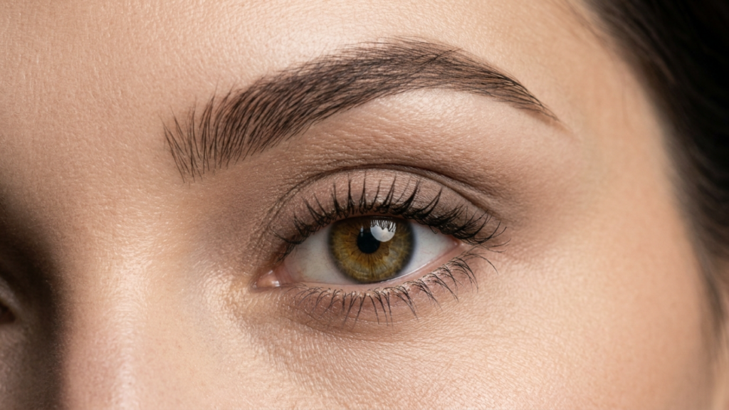
جماليات قوس الحاجب: كيف يُشكّل تحديد ملامح الحاجب بنية الحاجب الأنثوية
ما وراء الوضع الرأسي،, جماليات قوس الحاجب يعتمد ذلك بشكل أساسي على بنية العظام الأساسية. تخلق الحافة فوق الحجاجية الذكورية حاجبًا أكثر تسطحًا وثقلًا يكون منخفضًا ويبرز للأمام. يرتفع قوس الحاجب الأنثوي بشكل أكثر حدة من منتصف الحاجب إلى وسطه، ويبلغ ذروته تقريبًا عند الحافة الجانبية للقزحية، ويتناقص تدريجيًا نحو الجانب.
يؤثر تحديد محيط حافة الحاجب بشكل مباشر على هذا القوس من خلال تغيير الطبقة العظمية أسفل الحاجب. عندما يقوم الجراح بتقليل حافة الحاجب الوسطى والمركزية بشكل أكثر فعالية، يرتفع الحاجب بشكل ملحوظ في الجزء المركزي، مما يخلق بشكل طبيعي قمة قوس الحاجب الأنثوية. يعالج تقليل الحافة الوسطى محيط الحاجب البارز الذي يؤدي إلى تسطيح الحاجب من الداخل، بينما يحافظ الحفاظ على الحافة الجانبية على دعم الحاجب الجانبي.
تتيح تقنية التخفيض التفاضلي هذه للجراح نحت شكل قوس الحاجب من خلال إزالة العظم بشكل انتقائي. يؤدي التخفيض المركزي المكثف للحافة إلى رفع الجزء المركزي من الحاجب أكثر من الأجزاء الإنسية أو الوحشية، مما يُنتج قوسًا طبيعيًا. أما التخفيض المتناظر للجزء الإنسي والمركزي فيُنتج رفعًا أكثر دقة وتجانسًا، وهو مناسب للمرضى الذين يفضلون حاجبًا أقل تقوسًا بشكل ملحوظ.
فهم تحديد محيط الحافة فوق الحجاجية وردود الفعل على موضع الحاجب يُمكّن هذا الأسلوب الجراح من التحكم بشكل متوقع في شكل الحاجب من خلال استئصال العظام الموجه بدلاً من الاعتماد فقط على أجهزة التثبيت بالمنظار. وينتج عن هذا الأسلوب نتائج تبدو وتتحرك بشكل طبيعي، لأن الدعم الهيكلي الأساسي قد أعيد تشكيله بدلاً من تعليقه خارجياً.
ديناميكيات الأنسجة الرخوة حول الحجاج أثناء عملية تقليل حجم العظام
تحتوي المنطقة المحيطة بالعين على أكثر تفاعلات الأنسجة الرخوة تعقيدًا في الجزء العلوي من الوجه. إن فهم هذه الديناميكيات يوضح سبب رفع الحاجب عن طريق تحديد محيط العين وحده، ومتى يصبح التدخل الإضافي ضروريًا.
يتكون غلاف الأنسجة الرخوة للحاجب من خمس طبقات متميزة: الجلد، والدهون تحت الجلد، وعضلة العين الدائرية، والدهون خلف عضلة العين الدائرية، والسمحاق. تستجيب كل طبقة بشكل مختلف لعملية تقليل حجم العظم. يتكيف الجلد مع الشكل الجديد بشكل سلبي، مدفوعًا بالمرونة والشد. أما عضلة العين الدائرية، وهي عضلة خافضة للحاجب مهمة، فتتحرر جزئيًا أثناء تشريح ما تحت السمحاق المستخدم لتحديد شكل حافة الحاجب.
تتوزع وسادة الدهون خلف العضلة الدويرية للعين وفقًا للسطح العظمي الجديد. عندما يبرز حافة الحجاج، تتجمع وسادة الدهون هذه في الجزء الأمامي، مما يخلق امتلاءً للحاجب يوحي بصريًا بثقله. بعد تحديد محيط الحافة، يسمح سطح العظم الأملس لوسادة الدهون بالاستقرار بشكل أكبر في الجزء الخلفي، مما يقلل من امتلاء الجزء الأمامي من الحاجب ويخلق وهمًا بارتفاع إضافي.
يتم تحرير عضلات الحاجب المقطبة، التي تسحب الحاجب إلى الداخل والأسفل، جزئيًا أثناء النهج التاجي الثنائي القياسي لتحديد محيط الجبهة. ويساهم هذا التحرير في رفع الحاجب بمقدار 1 إلى 2 مليمتر إضافي بشكل مستقل عن تأثير تقليل العظام.
استقرار ارتفاع الحاجب الهندسي على المدى الطويل
يتساءل المرضى والجراحون على حد سواء عما إذا كان رفع الحاجب الهندسي الناتج عن تحديد محيط الحافة يدوم مع مرور الوقت. هذا القلق مشروع: فعمليات الأنسجة الرخوة مثل عمليات رفع الحاجب بالمنظار تعاني تاريخياً من معدلات انتكاس تصل إلى 30 بالمائة في غضون عامين بسبب فشل التثبيت وإعادة التوزيع بفعل الجاذبية.
مع ذلك، يُظهر الرفع الهندسي الناتج عن تحديد حواف الحاجب استقرارًا فائقًا على المدى الطويل، لأن آلية عمله تعتمد على تغيير هيكلي دائم بدلًا من تعليق مؤقت للأنسجة الرخوة. فبمجرد إزالة العظم، لا يمكنه النمو مجددًا. ويتكيف غلاف الأنسجة الرخوة بشكل دائم مع شكل الهيكل العظمي الجديد. وتؤكد بيانات المتابعة السريرية بعد اثني عشر شهرًا وما بعدها أن ارتفاع الحاجب بعد تحديد حواف الحاجب يظل مستقرًا، مع تسجيل انتكاس أقل من 0.5 مليمتر في معظم الدراسات.
يُساهم النسيج الندبي الذي يتشكل بين السمحاق والعظم المُعاد تشكيله خلال مرحلة الشفاء في زيادة استقرار وضع الحاجب. هذا التثبيت البيولوجي يُغني عن الحاجة إلى أدوات التثبيت الجراحية مثل البراغي أو المثبتات أو الخيوط الجراحية المستخدمة في عمليات رفع الحاجب بالمنظار. عمليًا، يُنشئ الجسم نظام تثبيت داخلي دائم خاص به.
التنبؤ بالنتائج: اختيار المرضى لرفع الحاجب بتقنية تحديد محيط الحاجب فقط
يتطلب اختيار المرشح المناسب لتحديد محيط حافة الأنف دون رفع الحاجب في الوقت نفسه تقييمًا منهجيًا. ويوجه الإطار التالي عملية اتخاذ القرارات السريرية بناءً على المتغيرات التشريحية والمتغيرات الخاصة بالمريض.
تقييم حجم النتوء العظمي
قم بتقييم التصوير المقطعي المحوسب أو إعادة البناء ثلاثي الأبعاد لتحديد مقدار بروز الحافة فوق الحجاجية الأمامية بالنسبة لمستوى القرنية. عادةً ما تُسبب الحواف البارزة لأكثر من 6 مليمترات انخفاضًا ملحوظًا في الحاجب، ويؤدي تقليلها إلى رفع الحاجب بشكل كبير. أما المرضى الذين يقل بروزهم عن 4 مليمترات، فإنهم يحصلون على رفع هندسي طفيف من خلال التقليل وحده.
قياس تدلي الحاجب الحقيقي
قِس المسافة من الحافة السفلية للحاجب إلى الحافة العلوية للحجاج في وضع الراحة. المرضى الذين يعانون من تدلي الجفن أقل من 3 ملليمترات يحققون باستمرار رفعًا مُرضيًا من خلال تحديد محيط الحافة فقط. أما المرضى الذين يعانون من تدلي الجفن الحقيقي الذي يزيد عن 5 ملليمترات، فيحتاجون دائمًا تقريبًا إلى إجراءات رفع إضافية بغض النظر عن مقدار تقليل حجم العظم الذي تم إجراؤه.
تقييم وضع الحاجب الجانبي
يقع ذيل الحاجب الجانبي عند خط الالتحام الصدغي، حيث تلتقي الحافة فوق الحجاجية باللفافة الصدغية. يؤثر تحديد محيط الحافة بشكل أساسي على الحاجب الإنسي والمركزي لأن تقليل العظم يتركز في هذه المنطقة. نادرًا ما يستجيب تدلي الحاجب الجانبي لتقليل العظم الإنسي والمركزي، وغالبًا ما يستدعي الأمر رفعًا صدغيًا عندما يتدلى الذيل الجانبي بشكل ملحوظ.
التقنية الجراحية: تحقيق أقصى قدر من الرفع الهندسي أثناء تشكيل حافة الإطار
تؤثر التقنية الجراحية لتحديد محيط حافة الحاجب بشكل مباشر على درجة رفع الحاجب السلبي التي يتم تحقيقها. يمكن للجراحين الذين يفهمون المبادئ الهندسية تعديل نمط استئصال العظام لتحقيق أقصى قدر من تأثير رفع الحاجب.
أولاً، أعطِ الأولوية لتقليل حافة الحافة السفلية. تُشكّل الحافة السفلية للحافة فوق الحجاجية أقوى قوة ضغط لأسفل على الحاجب. ويؤدي تقليل هذه الحافة السفلية بشكل كبير إلى رفع الحاجب هندسيًا بشكل ملحوظ. أما تقليل سطح الحافة العلوية فيُنعّم محيط الجبهة، ولكنه يُساهم بشكل أقل في رفع الحاجب مقارنةً بإزالة الحافة السفلية.
ثانيًا، يجب إنشاء منطقة انتقال سلسة بين الحافة المحددة وسقف الحجاج. قد يؤدي الانحدار الحاد عند حافة الحجاج إلى عدم انتظام الأنسجة الرخوة وتقليل تأثير إعادة التغطية. يضمن التناقص التدريجي لإزالة العظم في الحافة العلوية للحجاج انزلاق غلاف الأنسجة الرخوة بسلاسة على طول السطح الجديد.
ثالثًا، عالج بروز منطقة ما بين الحاجبين في الوقت نفسه. تبرز هذه المنطقة، التي تميز جبهة الرجل، بين الحاجبين الداخليين. إن تقليل هذه المنطقة يحرر تأثير خافض الحاجب الداخلي، مما يسمح للحاجب الداخلي بالارتفاع بشكل طبيعي.
الاعتبارات الفنية الهامة
يجب على الجراح مراعاة الطبقتين الأمامية والخلفية للجيوب الأنفية الأمامية. فالإفراط في تصغير الطبقة الأمامية يُعرّض تجويف الجيوب الأنفية للخطر، بينما يؤدي عدم كفاية التصغير إلى عدم تحقيق تحديد مناسب للجبهة. يوفر تحديد الجبهة من النوع الثالث مع تراجع جدار الجيوب الأنفية وإعادة بناء ترقيع العظام، كما هو موضح في أدبيات جراحة الوجه والجمجمة، النهج الأكثر شمولاً لبروز حافة الحجاج الشديد، ويُحقق أعلى ارتفاع هندسي للحاجب. (NCBI، 2018).
يؤثر إيقاف النزيف أثناء العملية أيضًا على إعادة تشكيل الحاجب. قد يؤدي الكي المفرط للأنسجة المحيطة بالعين إلى انكماش مفرط للندبة، مما يؤدي إلى سحب الحاجب في اتجاهات غير متوقعة. تحافظ تقنية الكي ثنائي القطب الدقيقة على صحة الأنسجة المحيطة وتعزز إعادة تشكيل الحاجب بشكل متوقع ومتناسق.
متى يتم الجمع بين الإجراءات: عملية رفع الصدغ بالمنظار كإجراء مساعد
بدلاً من اللجوء إلى عملية شد الحاجب بالمنظار بالكامل، فكّر في رفع الصدغ بالمنظار كإجراء مساعد مستهدف عندما يتطلب الحاجب الجانبي رفعًا. يعالج رفع الحاجب الصدغي ذيل الحاجب الخارجي دون التلاعب بالحاجب الداخلي، وهو ما يعالجه تحديد محيط الحافة بشكل فعال بالفعل.
يتجنب هذا النهج الانتقائي الإفراط في معالجة الجزء الأوسط والمركزي من الحاجب، حيث يؤدي تحديد الشكل وحده إلى رفع كافٍ، بينما يستهدف تحديدًا الجزء الجانبي حيث يكون لتقليل العظم تأثير ضئيل. يضيف رفع الحاجب الصدغي ما يقرب من 20 إلى 30 دقيقة من وقت العملية ويخلق شقوقًا صغيرة مخفية داخل خط الشعر، مما يجعله إجراءً مساعدًا أقل خطورة من رفع الحاجب الكامل بالمنظار.
الدكتور محمد فاتح أوكياي، بخبرته الواسعة في جراحة تجميل الوجه الأنثوي في عيادة الدكتور إم إف أو في أنطاليا, يقوم بشكل روتيني بتقييم ما إذا كان الحاجب الجانبي يتطلب علاجًا مستقلاً بناءً على التصوير الفوتوغرافي قبل الجراحة وتحليل التصوير المقطعي المحوسب ثلاثي الأبعاد. هذا النهج الفردي يتجنب الإجراءات غير الضرورية والمخاطر المصاحبة لها.
دليل خطوة بخطوة: رفع الحاجبين من خلال تحديد محيط الجفن
اتبع هذه الخطوات العملية لتحقيق أقصى ارتفاع هندسي للحاجب أثناء تحديد محيط الحافة فوق الحجاجية وردود الفعل على موضع الحاجب إجراءات:
- تحديد الكمية الإسقاط الأمامي للحافة فوق الحجاجية باستخدام إعادة بناء التصوير المقطعي المحوسب ثلاثي الأبعاد مع القياسات بالنسبة لمستوى القرنية قبل الجراحة.
- يقيس تحديد موضع الحاجب قبل الجراحة من الحافة السفلية للحاجب إلى الحافة العلوية للحجاج عند النقاط الوسطى والمركزية والجانبية لتحديد خط الأساس للمقارنة بعد الجراحة.
- تصميم منطقة تقليل العظام مع التركيز على الحافة السفلية، حيث يؤدي الإزالة إلى توليد أقوى متجه إعادة تشكيل علوي خلفي لرفع الحاجب هندسيًا.
- ينفذ تشريح واسع تحت السمحاق من خلال النهج الإكليلي، وتحرير ارتباطات العضلة المقطبة والعضلة المتقرنة لزيادة تأثير إعادة تشكيل الأنسجة الرخوة السلبية إلى أقصى حد.
- يقلل يتم تشكيل الحافة السفلية أولاً، ثم يتم تضييق المحيط باتجاه الأعلى وصولاً إلى العظم الجبهي، مما يخلق منحنى أنثويًا ناعمًا يشجع غلاف الأنسجة الرخوة على الانزلاق بشكل طبيعي.
- يقيم تحديد موضع الحاجب أثناء العملية بعد تحديد شكله وإغلاق السمحاق، ومقارنة الوضع المرئي بالصور الفوتوغرافية قبل العملية لتحديد ما إذا كانت هناك حاجة إلى تدخل تنظيري إضافي.
- وثيقة يتم تحديد موضع الحاجب بعد العملية الجراحية عند الشهر الأول، والثلاثة أشهر، والستة أشهر، والاثني عشر شهرًا لتتبع مسار الارتفاع الهندسي وتأكيد استقرار النتيجة على المدى الطويل.
هل أنتِ مستعدة لاكتشاف ما إذا كان وضع حاجبيكِ يمكن تحسينه من خلال تحديد محيط الحاجب فقط؟ قدّم طلبك احصل على تقييم جراحي شخصي اليوم.
أسئلة مكررة
ما هو تحديد محيط حافة الحجاج العلوي وردود الفعل على موضع الحاجب؟
يصف مصطلح تحديد محيط الحافة فوق الحجاجية وردود الفعل على موضع الحاجب الظاهرة البيوميكانيكية التي يؤدي فيها تقليل الحافة العظمية البارزة فوق العين إلى إعادة تشكيل الأنسجة الرخوة للحاجب على طول محيط جديد وأكثر سلاسة، مما ينتج عنه ارتفاع هندسي للحاجب من 2 إلى 4 ملليمترات دون إجراء عملية رفع منفصلة.
كيف يؤدي إزالة العظام إلى رفع الحاجب دون إجراء عملية شد؟
يؤدي إزالة الحافة العظمية السفلية للحافة فوق الحجاجية إلى التخلص من البروز الهيكلي الذي يضغط ميكانيكيًا على الحاجب. ثم يعود غلاف الأنسجة الرخوة ليغطي سطح العظم الأكثر نعومة والأكثر انحدارًا للخلف، متبعًا اتجاهًا علويًا خلفيًا يُحدث ارتفاعًا مرئيًا وقابلًا للقياس في الحاجب.
متى يكون إجراء عملية رفع الحاجب بالمنظار في نفس الوقت ضرورياً؟
يصبح رفع الحاجب بالمنظار ضرورياً عندما يعاني المريض من تدلي حقيقي كبير في الأنسجة الرخوة يتجاوز 5 ملليمترات، أو هبوط جانبي واضح للحاجب، أو عدم تناسق الحاجبين بأكثر من 2 ملليمتر بين الجانبين، أو ارتخاء الأنسجة الذي يُلاحظ عادةً لدى المرضى الذين تزيد أعمارهم عن 50 عامًا.
ما مقدار ارتفاع الحاجب الذي يمكن تحقيقه من خلال تحديد محيط الحاجب وحده؟
تُظهر القياسات السريرية أن تحديد محيط الحافة فوق الحجاجية بشكل منفرد يُنتج ارتفاعًا في الحاجب يتراوح بين 2 و 4 ملليمترات. كل ملليمتر يتم إزالته من بروز العظم يُترجم إلى ارتفاع رأسي في الحاجب يتراوح بين 0.5 و 0.7 ملليمتر تقريبًا من خلال آلية إزاحة المتجه الهندسي.
هل ارتفاع الحاجب الناتج عن تحديد محيط العين دائم؟
نعم، يُظهر رفع الحاجب الهندسي بتقنية تحديد محيط الحافة ثباتًا فائقًا على المدى الطويل مقارنةً برفع الحاجب بالمنظار. وبما أن العظم يُزال بشكل دائم، فإن غلاف الأنسجة الرخوة يتكيف بشكل دائم مع شكل الهيكل العظمي الجديد، مع تسجيل حالات انتكاس لا تتجاوز 0.5 مليمتر.
لماذا يبدو الحاجب منخفضاً مباشرة بعد جراحة تحديد محيط الحاجب؟
يؤدي التورم بعد العملية إلى انخفاض مؤقت في مستوى الحاجب خلال الأسابيع الثلاثة الأولى بعد الجراحة. ومع انحسار التورم بين الأسبوعين الثالث والثامن، يعود الحاجب تدريجيًا إلى وضعه الطبيعي الجديد. ينبغي على المرضى الانتظار ثلاثة أشهر على الأقل قبل تقييم الوضع النهائي للحاجب.
ما الفرق بين انخفاض الحاجب البنيوي وتدلي الحاجب الحقيقي؟
يحدث انخفاض الحاجب البنيوي عندما تدفع حافة الحجاج البارزة الحاجبَ إلى الأسفل ميكانيكيًا، ويستجيب جيدًا لتعديل شكل الحافة وحدها. أما تدلي الجفن الحقيقي فيتضمن ارتخاءً فعليًا في الأنسجة الرخوة وهبوطها، مما يتطلب إجراءات رفع إضافية لأن تقليل حجم العظم لا يُعيد مرونة الأنسجة المترهلة.







