

إعادة بناء الجبهة يُعدّ عنصرًا أساسيًا من الوجه جراحة التأنيث (FFS) وتشمل هذه الإجراءات عمليات تجميل الوجه والجمجمة، بهدف تحقيق ملامح وجه متناسقة وجميلة. ومن بين أكثر التقنيات التي تثير الجدل ما يلي: النوع الأول و رأب الجمجمة من النوع الثالث, حيث يقدم كل منها أساليب مميزة لإعادة تشكيل الجبهة. النوع الأول يتضمن كشط العظام أو تقشيرها, النوع 3 يتطلب الأمر أكثر تعقيدا قطع العظم والنكسة فيما يخص الجيوب الأنفية الأمامية. يُعدّ فهم الآثار التشريحية والوظيفية والجمالية لهذه التقنيات أمرًا بالغ الأهمية لكل من الجراحين والمرضى لاتخاذ قرارات مدروسة. تعرّف على المزيد حول تراجع الجيوب الأنفية وتصغير بروز الحاجب في دليلنا الشامل. شد الجبين.
يستكشف هذا الدليل الاختلافات الهيكلية بين رأب الجمجمة من النوع الأول والنوع الثالث، وتأثيرها على سمك العظم, تشريح الجيوب الأنفية الأمامية, وكيف يحدد الجراحون النهج الأنسب لكل مريض. في النهاية، ستتضح لك التقنية التي تتوافق بشكل أفضل مع احتياجاتك التشريحية وأهدافك الجمالية.
جدول المحتويات
الأساس التشريحي: العظم الجبهي والجيوب الأنفية
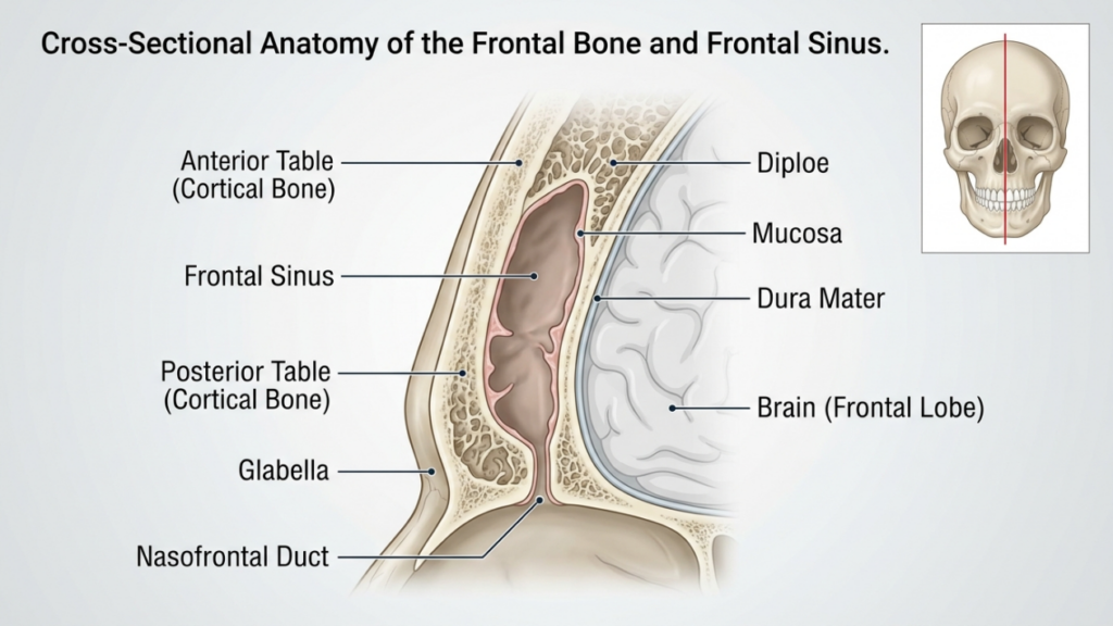
ال العظم الجبهي و الجيوب الأنفية الأمامية تلعب عظمة الجبهة دورًا محوريًا في إعادة بناء الجبهة. تشكل عظمة الجبهة الجزء العلوي من الوجه وتحتوي على الجيب الجبهي، وهو تجويف أجوف يختلف في حجمه وشكله بين الأفراد. الطاولة الأمامية يُعدّ العظم الخارجي للجيوب الأنفية الجبهية هو الذي يُساهم في تحديد شكل الجبهة، بينما الطاولة الخلفية يفصل هذا الإجراء الجيوب الأنفية عن الدماغ. ويؤثر سمك هذه الصفائح ودرجة تهوية الجيوب الأنفية (التمدد المملوء بالهواء) على اختيار نوع جراحة ترميم الجمجمة بين النوع الأول والنوع الثالث.
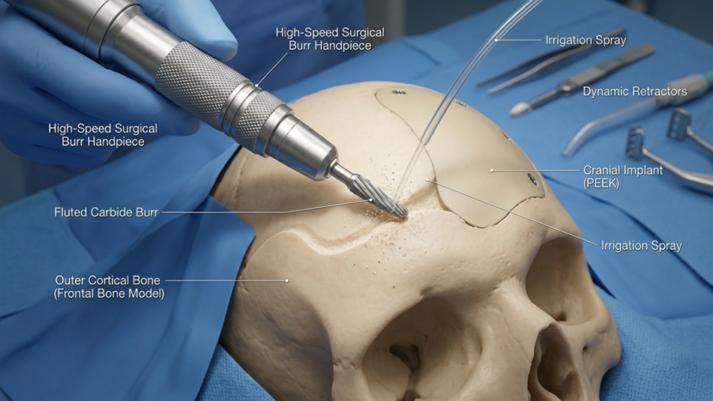
في رأب الجمجمة من النوع الأول، ال دكتور جراح تستخدم هذه التقنية مثقابًا عالي السرعة لتقشير الطبقة الخارجية من العظم القشري، مما يقلل من بروز العظم دون المساس بالجيوب الأنفية الأمامية. وتُعد هذه التقنية مثالية للمرضى الذين يعانون من عظمة جبهية رقيقة أو بروز طفيف، لأنه يحافظ على سلامة الجيوب الأنفية. ومع ذلك، قد لا يكون كافياً للمرضى الذين يعانون من بروز كبير في الحاجب أو سطح أمامي سميك، حيث أن التجريف المفرط قد يضر باستقرار العظم أو يفشل في تحقيق الشكل المطلوب (أوسترهوت، 2024).
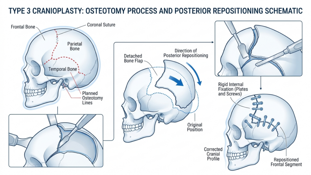
في المقابل،, رأب الجمجمة من النوع الثالث يتضمن قطع العظم— قطع دقيق في الطبقة الأمامية للجيوب الأنفية الجبهية. ثم يُعاد وضع الجزء العظمي للخلف (تراجع) لتقليل البروز والحصول على جبهة أكثر نعومة وأنوثة. تُستخدم هذه التقنية للمرضى الذين يعانون من بروز الحاجبين من متوسط إلى شديد أو في حالة وجود سطح أمامي سميك، حيث يكون التجريف وحده غير كافٍ. يسمح قطع العظم بإعادة تشكيل دقيقة مع الحفاظ على وظيفة الجيوب الأنفية الوقائية (تأنيث الجبهة: مراجعة شاملة للأدبيات، 2024).
رأب الجمجمة من النوع الأول: الحلاقة والتجريف
نظرة عامة على التقنية
يُعدّ رأب الجمجمة من النوع الأول الخيار الأقل توغلاً لإعادة بناء الجبهة. وهو يتضمن استخدام مثقب عالي السرعة لتقليل بروز عظم الجبهة تدريجياً. دكتور جراح يقوم الجراح بحلاقة الطبقة القشرية الخارجية بدقة متناهية، مع تجنب اختراق الجيب الجبهي. وتُعد هذه التقنية فعالة بشكل خاص للمرضى الذين يعانون من:
- بروز خفيف إلى متوسط للحواجب
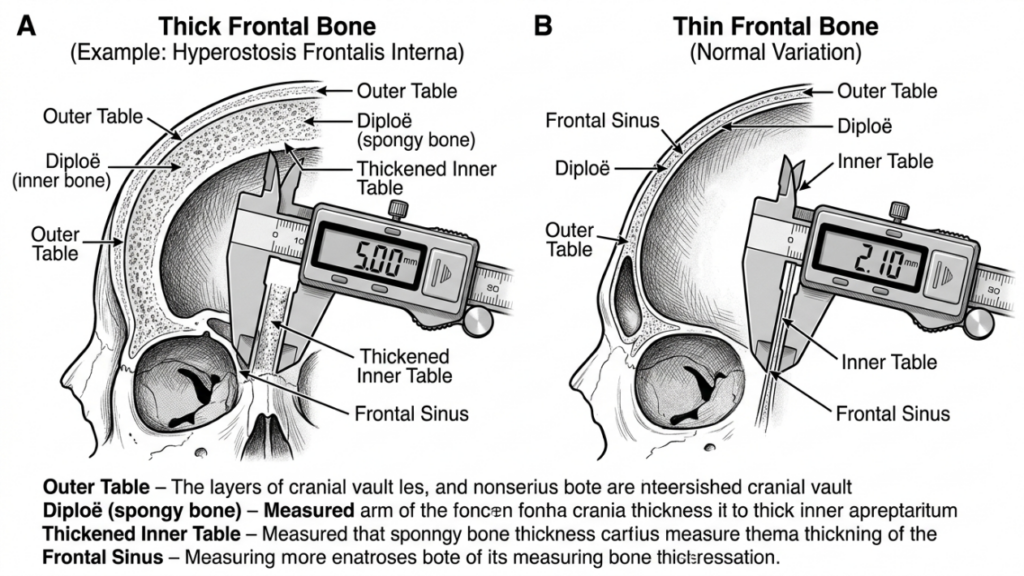
- عظم جبهي رقيق (أقل من 5 مم)
- غياب تضخم ملحوظ في الجيوب الأنفية الأمامية
يتم إجراء العملية من خلال شق تاجي, تتيح هذه التقنية الوصول إلى الجبهة مع تقليل الندوب الظاهرة إلى أدنى حد. يستخدم الجراح حاسة اللمس والإشارات البصرية لضمان تقليل حجم العظم بشكل متجانس دون ترقيقه بشكل مفرط، مما قد يؤدي إلى عدم استقراره أو عدم انتظام شكله.
مزايا عملية ترقيع الجمجمة من النوع الأول
يوفر النوع الأول من عملية ترقيع الجمجمة العديد من الفوائد:
- الحد الأدنى من التدخل الجراحي: إن عدم إجراء عملية قطع العظم أو إزالة العظام يقلل من الصدمة الجراحية ووقت التعافي.
- انخفاض خطر حدوث مضاعفات: إن الحفاظ على الجيوب الأنفية الأمامية يقلل من خطر الإصابة بالتهاب الجيوب الأنفية، أو تسرب السائل النخاعي، أو تكوين الكيس المخاطي.
- وقت تشغيل أقصر: يستغرق الإجراء عادةً من ساعة إلى ساعتين، مما يجعله خيارًا أسرع مقارنةً بالنوع 3.
- نتائج متوقعة: مثالي للمرضى الذين يعانون من بروز طفيف في الوجه، حيث يكون تحديد الملامح بشكل دقيق كافياً لتحقيق مظهر أنثوي.
محددات عملية ترقيع الجمجمة من النوع الأول
على الرغم من أن عملية ترقيع الجمجمة من النوع الأول أكثر أمانًا وأقل توغلاً، إلا أنها تنطوي على قيود ملحوظة:
- تخفيض محدود: غير كافٍ للمرضى الذين يعانون من بروز شديد في الحاجبين أو عظام جبهية سميكة.
- خطر الترقق المفرط: يمكن أن يؤدي الحفر العنيف إلى إضعاف العظم، مما يؤدي إلى عدم انتظام في الشكل أو كسور.
- التأنيث غير المكتمل: قد لا يحقق النتيجة الجمالية المرجوة للمرضى ذوي الملامح الذكورية البارزة.
رأب الجمجمة من النوع الثالث: قطع العظم والتراجع
نظرة عامة على التقنية
تُعد عملية ترقيع الجمجمة من النوع الثالث إجراءً أكثر تعقيدًا مصممًا للمرضى الذين يعانون من بروز الحاجبين من متوسط إلى شديد أو عظام الجبهة السميكة. يتضمن ذلك قطع العظم—قطع دقيق في الجدار الأمامي للجيوب الأنفية الجبهية—يتبعه إعادة وضع قطعة العظم للخلف (تراجع). تتيح هذه التقنية تقليل بروز الجبهة بشكل ملحوظ والحصول على مظهر أكثر نعومة وأنوثة.
يتم إجراء العملية عادةً من خلال شق تاجي, مما يتيح الوصول إلى عظم الجبهة والجيوب الأنفية. يستخدم الجراح منشار سهمي أو جهاز كهرضغطية لإجراء قطع العظم، مع التأكد من أن القطع يتبع الانحناء الطبيعي للجبهة. ثم يتم إعادة وضع قطعة العظم وتثبيتها بـ صفائح ومسامير من التيتانيوم أو الخيوط الجراحية القابلة للامتصاص. تُعد هذه التقنية مثالية للمرضى الذين يعانون من:
- بروز شديد في الحاجبين
- عظم جبهي سميك (أكبر من 5 مم)
- تضخم ملحوظ في الجيوب الأنفية الأمامية
تتطلب عملية ترقيع الجمجمة من النوع الثالث تخطيطًا دقيقًا، وغالبًا ما تتضمن التخطيط الجراحي الافتراضي ثلاثي الأبعاد (VSP) لمحاكاة عملية قطع العظم وتراجع العظم. وهذا يضمن الدقة ويقلل من خطر حدوث مضاعفات مثل اختراق الجيوب الأنفية أو تسرب السائل النخاعي (التخطيط الجراحي الافتراضي في تأنيث الوجه (من الجزء العلوي من الوجه، 2025).
مزايا عملية ترقيع الجمجمة من النوع الثالث
يوفر النوع الثالث من عملية ترقيع الجمجمة العديد من المزايا للمرضى ذوي السمات الذكورية البارزة:
- تحديد ملامح الوجه بشكل ملحوظ: يحقق انخفاضاً ملحوظاً في بروز الحاجبين، مما يخلق جبهة أكثر نعومة وأنوثة.
- براعه: مناسب للمرضى الذين يعانون من عظام جبهية سميكة أو تضخم الجيوب الأنفية بشكل كبير.
- الاستقرار على المدى الطويل: تندمج قطعة العظم المعاد وضعها بشكل جيد، مما يقلل من خطر عدم انتظام الشكل بمرور الوقت.
- التخصيص: تتيح عملية التخطيط الجراحي الافتراضي إجراء تعديلات دقيقة خاصة بكل مريض لتحقيق أفضل النتائج.
محددات عملية ترقيع الجمجمة من النوع الثالث
على الرغم من فعاليته، فإن عملية ترقيع الجمجمة من النوع الثالث تنطوي على مخاطر وتعقيدات أعلى:
- زيادة وقت الجراحة: تستغرق العملية عادةً من 3 إلى 5 ساعات، وتتطلب دقة وخبرة أكبر.
- ارتفاع خطر حدوث المضاعفات: تشمل المخاطر المحتملة التهاب الجيوب الأنفية، وتسرب السائل النخاعي، أو تكوين الكيس المخاطي في حالة انتهاك الجيوب الأنفية.
- فترة تعافي أطول: قد يعاني المرضى من تورم وعدم راحة لفترة طويلة مقارنة بجراحة تجميل الجمجمة من النوع الأول.
- يكلف: يؤدي استخدام تقنيات التصوير المتقدمة والأدوات الجراحية إلى زيادة التكلفة الإجمالية للعملية.

الاختلافات الرئيسية بين رأب الجمجمة من النوع الأول والنوع الثالث
| ميزة | رأب الجمجمة من النوع الأول | جراحة رأب الجمجمة من النوع الثالث |
|---|---|---|
| تقنية | كشط/تقشير العظام | قطع العظم والتراجع |
| الغزوية | الحد الأدنى | معتدلة إلى عالية |
| المرشحون المثاليون | بروز خفيف إلى متوسط في الحاجبين، وعظم جبهي رقيق | بروز شديد في الحاجبين، وعظم جبهي سميك |
| وقت الجراحة | من ساعة إلى ساعتين | 3-5 ساعات |
| وقت الانتعاش | 1-2 أسابيع | 3-6 أسابيع |
| خطر حدوث مضاعفات | قليل | معتدلة إلى عالية |
| يكلف | أدنى | أعلى |
| النتيجة الجمالية | تحديد ملامح الوجه بشكل دقيق | التأنيث الدرامي |
كيف يقرر الجراحون: النوع الأول مقابل النوع الثالث
يعتمد الاختيار بين النوع الأول والنوع الثالث من عملية ترقيع الجمجمة على عدة عوامل، بما في ذلك حالة المريض السمات التشريحية, الأهداف الجمالية، و مخاطر الجراحة. يعتمد الجراحون على مزيج من الفحص السريري, التصوير ثلاثي الأبعاد، و استشارة المريض لتحديد التقنية الأنسب.
1. التقييم التشريحي
الخطوة الأولى هي تقييم حالة المريض سُمك العظم الجبهي و تشريح الجيوب الأنفية الأمامية. أ التصوير المقطعي المحوسب أو إعادة بناء ثلاثية الأبعاد يقدم رؤى تفصيلية حول:
- سمك العظام: المرضى الذين يعانون من عظام أمامية رقيقة (أقل من 5 مم) هم الأنسب لعملية ترقيع الجمجمة من النوع 1، بينما قد يحتاج أولئك الذين لديهم عظام أكثر سمكًا إلى النوع 3.
- تهوية الجيوب الأنفية: قد يتطلب التوسع الكبير في الجيوب الأنفية إجراء عملية تجميل الجمجمة من النوع 3 لتجنب انتهاك الجيوب الأنفية أثناء عملية الحفر.
- درجة التسلط: غالباً ما يتطلب بروز الحاجب الشديد تقليلاً جذرياً لا يمكن تحقيقه إلا باستخدام تقنيات النوع 3.
2. الأهداف الجمالية
تلعب توقعات المرضى الجمالية دورًا حاسمًا في اختيار التقنية. أولئك الذين يسعون تأنيث دقيق قد يختار البعض إجراء عملية ترقيع الجمجمة من النوع الأول، بينما غالباً ما يحتاج الأفراد ذوو السمات الذكورية البارزة إلى نتائج تحويلية من النوع الثالث. يناقش الجراحون النتائج الواقعية بناءً على تشريح المريض والتغييرات المرغوبة.
3. المخاطر الجراحية والتعافي
تنطوي عملية ترقيع الجمجمة من النوع الثالث على مخاطر أعلى، بما في ذلك مضاعفات الجيوب الأنفية, تسرب السائل النخاعي، و تعافي مطول. يُقيّم الجراحون الحالة الصحية العامة للمريض، ومدى تحمله للجراحة، واستعداده للالتزام بالرعاية ما بعد الجراحة. وقد يُنصح المرضى الذين يعانون من حالات طبية تزيد من مخاطر الجراحة بالنظر في إجراء الجراحة من النوع الأول أو إجراءات بديلة.
4. التخطيط الجراحي الافتراضي (VSP)
التطورات في التخطيط الجراحي الافتراضي ثلاثي الأبعاد أحدثت تقنية الطباعة ثلاثية الأبعاد ثورة في جراحة ترميم الجمجمة. يستخدم الجراحون هذه التقنية لمحاكاة عمليات قطع العظم، والانتكاسات، والنتائج، مما يضمن الدقة ويقلل المخاطر. وتُعد هذه التقنية ذات قيمة خاصة في جراحة ترميم الجمجمة من النوع الثالث، حيث يُعد إعادة تموضع العظم بدقة أمرًا بالغ الأهمية (الطباعة ثلاثية الأبعاد والتخطيط الجراحي الافتراضي في جراحة الجمجمة والوجه وتقويم الفكين، 2025).
الرعاية والتعافي بعد الجراحة
تختلف فترة التعافي بشكل كبير بين النوع الأول والنوع الثالث من جراحة ترميم الجمجمة. يساعد فهم عملية ما بعد الجراحة المرضى على الاستعداد لرحلة شفاء سلسة.
التعافي من عملية ترقيع الجمجمة من النوع الأول
يعاني المرضى الذين يخضعون لعملية ترقيع الجمجمة من النوع الأول عادةً مما يلي:
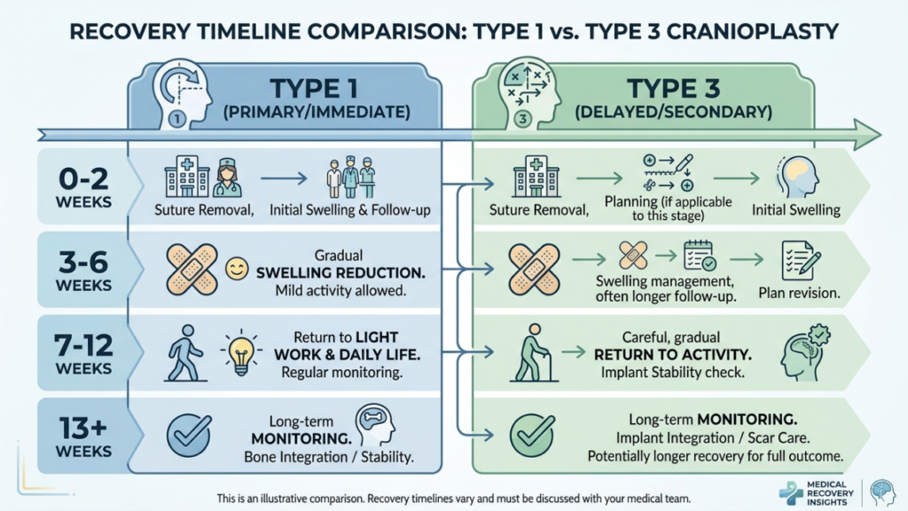
- تورم خفيف إلى متوسط: يزول في غضون أسبوع إلى أسبوعين.
- أقل قدر من الانزعاج: تمت السيطرة على الحالة باستخدام مسكنات الألم التي لا تستلزم وصفة طبية.
- العودة السريعة إلى الأنشطة: يستأنف معظم المرضى أنشطتهم الطبيعية في غضون أسبوعين.
التعافي من عملية ترقيع الجمجمة من النوع الثالث
التعافي من عملية ترقيع الجمجمة من النوع الثالث أكثر تعقيداً نظراً لتعقيد العملية:
- تورم كبير وكدمات: قد يستمر لمدة 3-4 أسابيع.
- ألم متوسط: قد يكون من الضروري تناول مسكنات الألم الموصوفة طبيًا خلال الأسبوع الأول.
- العودة التدريجية للأنشطة: يُمنع القيام بالأنشطة الشاقة لمدة تتراوح بين 4 و 6 أسابيع.
- زيارات المتابعة: المراقبة المنتظمة لضمان الشفاء السليم ومعالجة أي مضاعفات.
تتطلب كلتا التقنيتين من المرضى تجنب رفع الأحمال الثقيلة, تمرين شاق، و الضغط المباشر على الجبهة خلال مرحلة التعافي الأولية، يقدم الجراحون تعليمات مفصلة لما بعد الجراحة، بما في ذلك العناية بالجروح، والقيود المفروضة على النشاط، وعلامات المضاعفات التي يجب مراقبتها.
المضاعفات المحتملة وكيفية تجنبها
على الرغم من أن عملية ترقيع الجمجمة آمنة بشكل عام، إلا أنه قد تحدث مضاعفات. لذا، فإن الوعي بهذه المخاطر واتخاذ التدابير الوقائية أمر بالغ الأهمية لكل من الجراحين والمرضى.
المضاعفات الشائعة
- التهاب الجيوب الأنفية: التهاب أو عدوى الجيوب الأنفية الأمامية، وخاصة في عملية تجميل الجمجمة من النوع 3 إذا تم انتهاك الجيوب الأنفية.
- تسرب السائل النخاعي (CSF): مضاعفات نادرة ولكنها خطيرة في حالة اختراق الجدار الخلفي للجيوب الأنفية.
- المخالفات الكنتورية: يمكن أن يؤدي ترقق العظام المفرط في النوع 1 أو التراجع غير المناسب في النوع 3 إلى عدم التناسق أو ظهور نتوءات مرئية.
- تكوين الخصية المخاطية: يمكن أن يؤدي انسداد مسارات تصريف الجيوب الأنفية إلى تكوّن أكياس مليئة بالمخاط.
- عدوى: يزداد الخطر في النوع الثالث بسبب طول مدة العملية الجراحية واستخدام الغرسات.
التدابير الوقائية
يستخدم الجراحون عدة استراتيجيات لتقليل المضاعفات:
- التخطيط الجراحي الدقيق: يضمن التصوير ثلاثي الأبعاد والتخطيط الجراحي الافتراضي دقة عمليات قطع العظام والتراجع.
- الحفاظ على الجيوب الأنفية: تجنب انتهاك الجيوب الأنفية الأمامية أثناء عملية الحفر أو قطع العظم.
- الوقاية بالمضادات الحيوية: يتم إعطاؤه قبل وبعد العملية الجراحية لتقليل مخاطر العدوى.
- المراقبة بعد الجراحة: متابعة دورية للكشف المبكر عن أي علامات للمضاعفات.
- تثقيف المريض: توجيه المرضى بشأن العناية السليمة بالجروح، والقيود المفروضة على النشاط، وعلامات التحذير من المضاعفات.
بدائل لعملية ترقيع الجمجمة من النوع الأول والنوع الثالث
بالنسبة للمرضى الذين لا يُعتبرون مرشحين مثاليين لعملية ترقيع الجمجمة من النوع الأول أو النوع الثالث، يمكن النظر في تقنيات بديلة:
- تطعيم الدهون: يمكن لعملية نقل الدهون الذاتية أن تُخفف من حدة ملامح الجبهة دون تغيير بنية العظام. وهذا خيار مثالي للمرضى الذين يعانون من عدم انتظام طفيف في ملامح الوجه أو الذين يبحثون عن خيارات غير جراحية.
- زراعة الأعضاء المخصصة: الغرسات الجاهزة، مثل PEEK (بولي إيثر إيثر كيتون) أو التيتانيوم, يمكن استخدام هذه التقنية لتكبير أو إعادة تشكيل الجبهة دون الحاجة إلى قطع العظم. وهي مفيدة بشكل خاص للمرضى الذين يعانون من عظام رقيقة أو مضاعفات في الجيوب الأنفية.
- جراحة تقويم الفكين: في الحالات التي شد الجبين جزء من نطاق أوسع تأنيث الوجه قد يتم الجمع بين إجراءات تقويم الفكين (مثل قطع عظم الفك Le Fort I) لمعالجة محاذاة منتصف الوجه والفك.
- تقنيات التنظير الداخلي: يمكن للأساليب التنظيرية طفيفة التوغل أن تقلل من بروز الحاجبين من خلال شقوق أصغر وأوقات تعافي أسرع.
شهادات المرضى والنتائج الواقعية
تُقدّم نتائج التجارب العملية رؤى قيّمة حول فعالية عمليات ترقيع الجمجمة من النوع الأول والثالث، ومعدلات رضا المرضى عنها. وتُبرز شهادات المرضى الأثر التحويلي لهذه العمليات.
دراسة حالة 1: رأب الجمجمة من النوع 1
سعت امرأة متحولة جنسيًا تبلغ من العمر 28 عامًا إلى إضفاء لمسة أنثوية رقيقة على جبهتها. ونظرًا لرقة عظمة جبهتها وقلة بروزها، اختارت عملية تجميل الجمجمة من النوع الأول. وقد حققت العملية مظهرًا أكثر نعومة مع فترة نقاهة قصيرة. وأعربت عن رضاها التام، مشيرةً إلى أن النتائج توافقت مع توقعاتها للحصول على مظهر طبيعي وأنثوي.
دراسة حالة ٢: رأب الجمجمة من النوع ٣
حضرت امرأة متحولة جنسيًا تبلغ من العمر 35 عامًا تعاني من بروز شديد في الحاجبين وسماكة في عظم الجبهة. خضعت لعملية تجميل الجمجمة من النوع الثالث، والتي تضمنت قطع العظم وتراجعه. أدى الانخفاض الكبير في بروز الجبهة إلى تأنيث ملامح وجهها بشكل ملحوظ. على الرغم من أن فترة التعافي استغرقت وقتًا أطول، إلا أنها أعربت عن رضاها التام عن النتائج، قائلةً إن العملية "غيرت حياتها".“
دراسة حالة 3: جراحة المراجعة
خضعت مريضة تبلغ من العمر 40 عامًا في البداية لعملية تجميل الجمجمة من النوع الأول، لكنها لم تكن راضية عن النتائج الطفيفة. لاحقًا، اختارت عملية تجميل الجمجمة من النوع الثالث لتحقيق مظهر أنثوي أكثر وضوحًا. نجحت الجراحة التصحيحية في معالجة مخاوفها، مما يُظهر أهمية اختيار التقنية المناسبة بناءً على الاحتياجات التشريحية والأهداف الجمالية.
دور التخطيط الجراحي الافتراضي في جراحة تجميل الجمجمة
أصبح التخطيط الجراحي الافتراضي (VSP) نقلة نوعية في جراحة ترميم الجمجمة، وخاصةً في عمليات النوع الثالث. يتيح التخطيط الجراحي الافتراضي للجراحين ما يلي:
- محاكاة عمليات قطع العظم: قم بالتخطيط بدقة لعمليات قطع العظام وإعادة وضعها لتحقيق أفضل شكل ممكن.
- توقع النتائج: تصور نتائج ما بعد الجراحة وقم بتعديل الخطة الجراحية وفقًا لذلك.
- تقليل المخاطر: تجنب الهياكل الحيوية مثل الجيوب الأنفية الأمامية والأعصاب فوق الحجاجية.
- تحسين التواصل: شارك النماذج ثلاثية الأبعاد مع المرضى لوضع توقعات واقعية وتحسين الموافقة المستنيرة.
أظهرت الدراسات أن تقنية التخطيط الجراحي الافتراضي (VSP) تقلل من وقت العملية، وتحسن الدقة، وتعزز رضا المرضى (الطباعة ثلاثية الأبعاد والتخطيط الجراحي الافتراضي في جراحة الجمجمة والوجه وتقويم الفكين، 2025). وتُعتبر الآن المعيار الذهبي لإجراءات رأب الجمجمة المعقدة.

أسئلة مكررة
ما هو الفرق الرئيسي بين رأب الجمجمة من النوع الأول والنوع الثالث؟
تتضمن عملية تجميل الجمجمة من النوع الأول كشط أو برد الطبقة الخارجية لعظم الجبهة لتقليل بروزها، بينما تتطلب عملية تجميل الجمجمة من النوع الثالث قطع العظم (استئصال العظم) وإرجاع جزء من عظم الجبهة إلى الخلف لتحقيق شكل أكثر وضوحًا. يُعد النوع الأول أقل توغلاً ومناسبًا للبروز البسيط إلى المتوسط، بينما يُستخدم النوع الثالث للبروز الشديد أو عظام الجبهة السميكة.
كيف يختار الجراحون بين النوع الأول والنوع الثالث من عملية ترقيع الجمجمة؟
يُقيّم الجراحون سُمك عظم الجبهة، ودرجة بروز الحاجب، وتشريح الجيوب الأنفية الأمامية للمريض باستخدام التصوير المقطعي المحوسب والتصوير ثلاثي الأبعاد. يُختار النوع الأول للعظام الرقيقة والبروز الطفيف، بينما يُفضّل النوع الثالث للعظام السميكة، والبروز الشديد، أو التوسع الكبير في الجيوب الأنفية. كما تؤثر أهداف المريض ومدى تحمّله للمخاطر الجراحية على القرار.
ما هي المخاطر المرتبطة بجراحة ترقيع الجمجمة من النوع الثالث؟
تُعدّ جراحة ترميم الجمجمة من النوع الثالث أكثر خطورةً نظرًا لتعقيدها، وتشمل هذه المخاطر التهاب الجيوب الأنفية، وتسرب السائل النخاعي، وعدم انتظام شكل الجمجمة، وتكوّن الكيس المخاطي، والعدوى. ويتم تقليل هذه المخاطر إلى أدنى حدّ من خلال التخطيط الجراحي الدقيق، والحفاظ على الجيوب الأنفية، والمتابعة بعد العملية. كما يتم توعية المرضى بالعلامات التحذيرية والرعاية اللاحقة لضمان التعافي السلس.
ما هي مدة فترة التعافي لعملية ترقيع الجمجمة من النوع الأول مقارنة بالنوع الثالث؟
يستغرق التعافي من عملية ترقيع الجمجمة من النوع الأول عادةً من أسبوع إلى أسبوعين، مع تورم طفيف وشعور بعدم الراحة. أما عملية ترقيع الجمجمة من النوع الثالث فتتطلب فترة نقاهة أطول تتراوح بين ثلاثة وستة أسابيع نظرًا لتعقيد الجراحة واحتمالية حدوث مضاعفات. يُنصح المرضى بتجنب الأنشطة المجهدة واتباع تعليمات ما بعد الجراحة بدقة.
هل يمكن لعملية ترقيع الجمجمة من النوع الأول أن تحقق نفس نتائج النوع الثالث؟
لا، يقتصر النوع الأول من جراحة تجميل الجمجمة على تعديل بسيط في شكل الوجه، وهو مثالي لحالات بروز الجمجمة البسيط إلى المتوسط. أما النوع الثالث، فيحقق نتائج أكثر وضوحًا في تجميل الوجه من خلال إعادة تموضع عظم الجبهة، مما يجعله مناسبًا لحالات بروز الجمجمة الشديد أو عظام الجبهة السميكة. ويعتمد اختيار النوع المناسب على احتياجات المريض التشريحية وأهدافه الجمالية.
ما هو دور التخطيط الجراحي الافتراضي في عملية ترقيع الجمجمة؟
يُمكّن التخطيط الجراحي الافتراضي الجراحين من محاكاة عمليات قطع العظم، والتنبؤ بالنتائج، وتقليل المخاطر عن طريق تجنب المناطق الحساسة كالجيوب الأنفية الأمامية. كما يُحسّن هذا التخطيط الدقة، ويُقلل وقت العملية، ويرفع مستوى رضا المريض من خلال توفير رؤية واضحة للخطة الجراحية والنتائج المتوقعة.
هل توجد بدائل غير جراحية لعملية تجميل الجمجمة لتأنيث الجبهة؟
نعم، تشمل البدائل غير الجراحية تطعيم الدهون لتنعيم ملامح الوجه، تُستخدم غرسات مخصصة (مثل مادة PEEK أو التيتانيوم) لإعادة تشكيل الجبهة دون جراحة. تُعد هذه الخيارات مثالية للمرضى الذين يعانون من عدم انتظام طفيف في ملامح الوجه أو الذين يفضلون تجنب الجراحة. مع ذلك، قد لا تُحقق هذه الخيارات نفس مستوى التأنيث الذي تُحققه التقنيات الجراحية.
ما الذي يجب أن أتوقعه خلال الاستشارة الخاصة بإعادة بناء الجبهة؟
خلال الاستشارة، سيقوم الجراح بتقييم سُمك عظم الجبهة، وتشريح الجيوب الأنفية، ودرجة بروز الجمجمة باستخدام الفحص السريري والتصوير ثلاثي الأبعاد. سيناقش معك أهدافك التجميلية، ويشرح لك الفرق بين جراحة تجميل الجمجمة من النوع الأول والنوع الثالث، ويوصي بالتقنية الأنسب بناءً على تشريحك وتوقعاتك.







