

Ricostruzione della fronte è una componente critica di Facciale Chirurgia della femminilizzazione (FFS) e procedure craniofacciali, volte a ottenere un contorno del viso armonioso ed esteticamente gradevole. Tra le tecniche più dibattute ci sono Tipo 1 E cranioplastica di tipo 3, ognuno dei quali offre approcci distinti per rimodellare la fronte. Mentre Tipo 1 coinvolge fresatura o rasatura dell'osso, Digitare 3 richiede un approccio più complesso osteotomia e arretramento del seno frontale. Comprendere le implicazioni anatomiche, funzionali ed estetiche di queste tecniche è essenziale sia per i chirurghi che per i pazienti per prendere decisioni informate. Scopri di più sulla riduzione del seno frontale e della protuberanza sopracciliare nella nostra guida completa a rimodellamento della fronte.
Questa guida esplora le differenze strutturali tra cranioplastica di tipo 1 e di tipo 3, il loro impatto su spessore osseo, anatomia del seno frontale, e come i chirurghi determinano l'approccio più adatto a ciascun paziente. Al termine, avrai le idee chiare su quale tecnica si allinei al meglio alle tue esigenze anatomiche e ai tuoi obiettivi estetici.
Sommario
Le basi anatomiche: osso frontale e seno mascellare
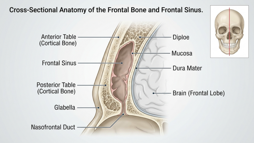
IL osso frontale E seno frontale svolgono un ruolo fondamentale nella ricostruzione della fronte. L'osso frontale forma la parte superiore del viso e ospita il seno frontale, una cavità cava che varia in dimensioni e forma tra gli individui. tavolo anteriore del seno frontale è lo strato osseo esterno che contribuisce al contorno della fronte, mentre il tavolo posteriore separa il seno mascellare dal cervello. Lo spessore di queste lamine e il grado di pneumatizzazione del seno (espansione con aria) influenzano la scelta tra cranioplastica di tipo 1 e di tipo 3.
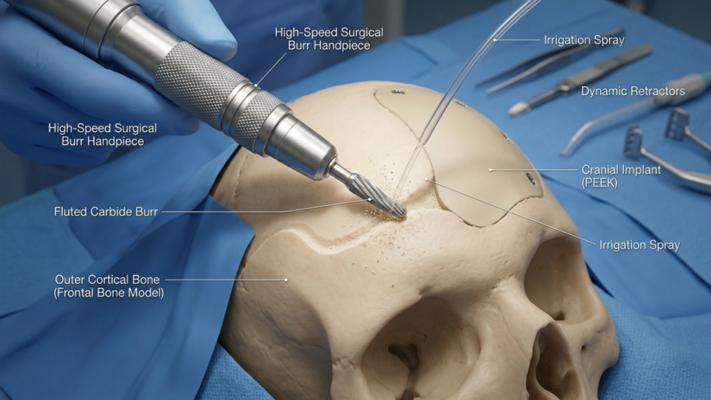
In cranioplastica di tipo 1, IL chirurgo utilizza una fresa ad alta velocità per limare l'osso corticale esterno, riducendo la prominenza senza violare il seno frontale. La tecnica è ideale per i pazienti con osso frontale sottile o una minima sporgenza, poiché preserva l'integrità del seno. Tuttavia, potrebbe non essere sufficiente per i pazienti con una sporgenza significativa del sopracciglio o una spessa tavola anteriore, poiché una fresatura eccessiva può compromettere la stabilità ossea o non riuscire a ottenere il contorno desiderato (Ousterhout, 2024).
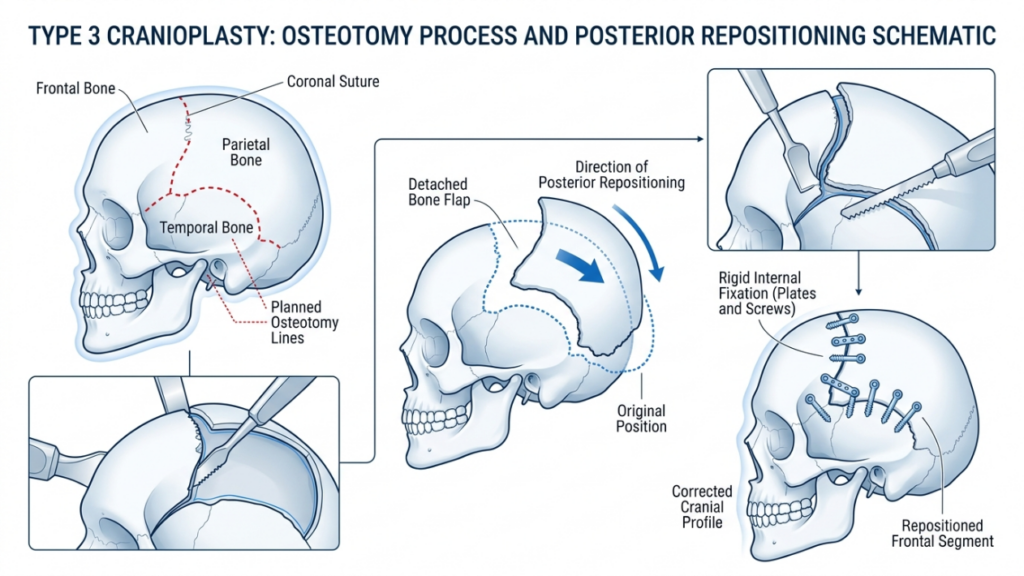
Al contrario, cranioplastica di tipo 3 coinvolge un osteotomia—un taglio controllato attraverso la tavola anteriore del seno frontale. Il segmento osseo viene quindi riposizionato all'indietro (arretramento) per ridurre la proiezione e creare una fronte più liscia e femminile. Questa tecnica è riservata ai pazienti con protrusione delle sopracciglia da moderata a grave o una spessa tavola anteriore, dove la sola fresatura sarebbe inadeguata. L'osteotomia consente un rimodellamento preciso mantenendo la funzione protettiva del seno (Femminilizzazione della fronte: una revisione della letteratura, 2024).
Cranioplastica di tipo 1: rasatura e fresatura
Panoramica tecnica
La cranioplastica di tipo 1 è l'opzione meno invasiva per la ricostruzione della fronte. Prevede l'utilizzo di un fresa ad alta velocità per ridurre gradualmente la prominenza dell'osso frontale. chirurgo Rasatura meticolosa dello strato corticale esterno, evitando la penetrazione nel seno frontale. Questa tecnica è particolarmente efficace per i pazienti con:
- Lieve o moderata sporgenza delle sopracciglia
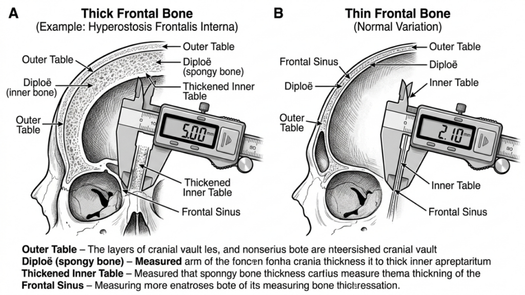
- Osso frontale sottile (meno di 5 mm)
- Assenza di pneumatizzazione significativa del seno frontale
La procedura viene eseguita tramite un incisione coronale, che consente l'accesso alla fronte riducendo al minimo le cicatrici visibili. Il chirurgo utilizza il feedback tattile e gli indizi visivi per garantire una riduzione uniforme senza assottigliare eccessivamente l'osso, il che potrebbe portare a instabilità o irregolarità del contorno.
Vantaggi della cranioplastica di tipo 1
La cranioplastica di tipo 1 offre diversi vantaggi:
- Minima invasività: L'assenza di osteotomia o rimozione ossea riduce il trauma chirurgico e i tempi di recupero.
- Minor rischio di complicazioni: Preservare il seno frontale riduce al minimo il rischio di sinusite, perdite di liquido cerebrospinale o formazione di mucoceli.
- Tempo operatorio più breve: La procedura richiede in genere da 1 a 2 ore, risultando quindi un'opzione più rapida rispetto al tipo 3.
- Risultati prevedibili: Ideale per pazienti con lieve protrusione palpabile, dove un leggero rimodellamento è sufficiente per ottenere un aspetto femminile.
Limitazioni della cranioplastica di tipo 1
Sebbene la cranioplastica di tipo 1 sia più sicura e meno invasiva, presenta alcune limitazioni significative:
- Riduzione limitata: Non adatto a pazienti con sopracciglia prominenti o ossa frontali spesse.
- Rischio di eccessivo diradamento: Una fresatura aggressiva può indebolire l'osso, causando irregolarità del contorno o fratture.
- Femminilizzazione incompleta: Potrebbe non garantire il risultato estetico desiderato per i pazienti con tratti somatici marcatamente maschili.
Cranioplastica di tipo 3: osteotomia e arretramento
Panoramica tecnica
La cranioplastica di tipo 3 è una procedura più complessa progettata per pazienti con protrusione delle sopracciglia da moderata a grave o ossa frontali spesse. Coinvolge un osteotomia—Un taglio preciso attraverso la parete anteriore del seno frontale— seguito dal riposizionamento all'indietro del segmento osseo (arretramento). Questa tecnica consente una significativa riduzione della proiezione della fronte e un contorno più armonioso e femminile.
La procedura viene in genere eseguita tramite un incisione coronale, fornendo accesso all'osso frontale e al seno. Il chirurgo utilizza un sega sagittale o dispositivo piezoelettrico per creare l'osteotomia, assicurandosi che il taglio segua la curvatura naturale della fronte. Il segmento osseo viene quindi riposizionato e fissato con piastre e viti in titanio o suture riassorbibili. Questa tecnica è ideale per pazienti con:
- Forte protrusione delle sopracciglia
- Osso frontale spesso (maggiore di 5 mm)
- Significativa pneumatizzazione del seno frontale
La cranioplastica di tipo 3 richiede una pianificazione meticolosa, che spesso coinvolge Pianificazione chirurgica virtuale 3D (VSP) per simulare l'osteotomia e l'arretramento. Ciò garantisce precisione e riduce al minimo il rischio di complicazioni quali violazione del seno o perdite di liquido cerebrospinale (Pianificazione chirurgica virtuale in Femminilizzazione facciale della parete superiore, 2025).
Vantaggi della cranioplastica di tipo 3
La cranioplastica di tipo 3 offre diversi vantaggi ai pazienti con tratti maschili pronunciati:
- Contouring significativo: Riduce drasticamente le sporgenze delle sopracciglia, creando una fronte più liscia e femminile.
- Versatilità: Adatto a pazienti con ossa frontali spesse o pneumatizzazione estesa dei seni paranasali.
- Stabilità a lungo termine: Il segmento osseo riposizionato si integra bene, riducendo nel tempo il rischio di irregolarità del contorno.
- Personalizzazione: La pianificazione chirurgica virtuale consente di apportare modifiche precise e personalizzate al paziente per ottenere risultati ottimali.
Limitazioni della cranioplastica di tipo 3
Nonostante la sua efficacia, la cranioplastica di tipo 3 comporta rischi e complessità maggiori:
- Aumento dei tempi chirurgici: La procedura richiede in genere dalle 3 alle 5 ore, e necessita di maggiore precisione e competenza.
- Rischio più elevato di complicazioni: Tra i potenziali rischi si annoverano sinusite, perdite di liquido cerebrospinale o formazione di mucocele in caso di lesione del seno paranasale.
- Recupero più lungo: Rispetto alla cranioplastica di tipo 1, i pazienti potrebbero avvertire gonfiore e fastidio prolungati.
- Costo: L'utilizzo di strumenti di imaging e chirurgici avanzati aumenta il costo complessivo della procedura.

Principali differenze tra cranioplastica di tipo 1 e di tipo 3
| Caratteristica | Cranioplastica di tipo 1 | Cranioplastica di tipo 3 |
|---|---|---|
| Tecnica | Bruciatura/rasatura delle ossa | Osteotomia e arretramento |
| Invasività | Minimo | Da moderato ad alto |
| Candidati ideali | Sporgenze sopracciliari da lievi a moderate, osso frontale sottile | Forte sporgenza delle sopracciglia, osso frontale spesso |
| Tempo chirurgico | 1-2 ore | 3–5 ore |
| I tempi di recupero | 1–2 settimane | 3-6 settimane |
| Rischio di complicazioni | Basso | Da moderato ad alto |
| Costo | Inferiore | Più alto |
| Risultato estetico | Contornatura delicata | Femminilizzazione drammatica |
Come i chirurghi decidono: tipo 1 vs. tipo 3
La scelta tra cranioplastica di tipo 1 e di tipo 3 dipende da diversi fattori, tra cui le condizioni del paziente. caratteristiche anatomiche, obiettivi estetici, E classi chirurgiche. I chirurghi si affidano a una combinazione di esame clinico, Immagini 3D, E consultazione del paziente per determinare la tecnica più appropriata.
1. Valutazione anatomica
Il primo passo consiste nella valutazione del paziente spessore dell'osso frontale E anatomia del seno frontale. UN TAC O Ricostruzione 3D fornisce approfondimenti dettagliati su:
- Spessore osseo: I pazienti con ossa frontali sottili (meno di 5 mm) sono più adatti alla cranioplastica di tipo 1, mentre quelli con ossa più spesse potrebbero necessitare di quella di tipo 3.
- Pneumatizzazione dei seni paranasali: Una pneumatizzazione estesa dei seni paranasali può rendere necessaria una cranioplastica di tipo 3 per evitare di danneggiare i seni durante la fresatura.
- Grado di autorità: Un grave avvallamento delle sopracciglia spesso richiede una drastica riduzione, ottenibile solo con le tecniche di Tipo 3.
2. Obiettivi estetici
Le aspettative estetiche dei pazienti giocano un ruolo cruciale nella scelta della tecnica. Coloro che cercano sottile femminilizzazione possono optare per la cranioplastica di tipo 1, mentre gli individui con caratteristiche maschili pronunciate spesso richiedono la risultati trasformativi di tipo 3. I chirurghi discutono i risultati realistici in base all'anatomia del paziente e ai cambiamenti desiderati.
3. Rischi chirurgici e recupero
La cranioplastica di tipo 3 comporta rischi più elevati, tra cui: complicazioni dei seni paranasali, perdite di liquido cerebrospinale, E recupero prolungato. I chirurghi valutano lo stato di salute generale del paziente, la sua tolleranza all'intervento chirurgico e la sua disponibilità ad aderire alle cure post-operatorie. Ai pazienti con patologie che aumentano i rischi chirurgici può essere consigliato di prendere in considerazione la chirurgia di tipo 1 o procedure alternative.
4. Pianificazione chirurgica virtuale (VSP)
Progressi in pianificazione chirurgica virtuale 3D hanno rivoluzionato la cranioplastica. I chirurghi utilizzano la pianificazione chirurgica virtuale (VSP) per simulare osteotomie, regressioni e risultati, garantendo precisione e minimizzando i rischi. Questa tecnologia è particolarmente preziosa per la cranioplastica di tipo 3, dove un accurato riposizionamento osseo è fondamentale (3D Printing and Virtual Surgical Planning in Craniofacial and Orthognathic Surgery, 2025).
Cura e recupero postoperatorio
Il periodo di recupero varia significativamente tra la cranioplastica di tipo 1 e quella di tipo 3. Comprendere il processo post-operatorio aiuta i pazienti a prepararsi per una guarigione senza intoppi.
Recupero dopo cranioplastica di tipo 1
I pazienti sottoposti a cranioplastica di tipo 1 in genere manifestano:
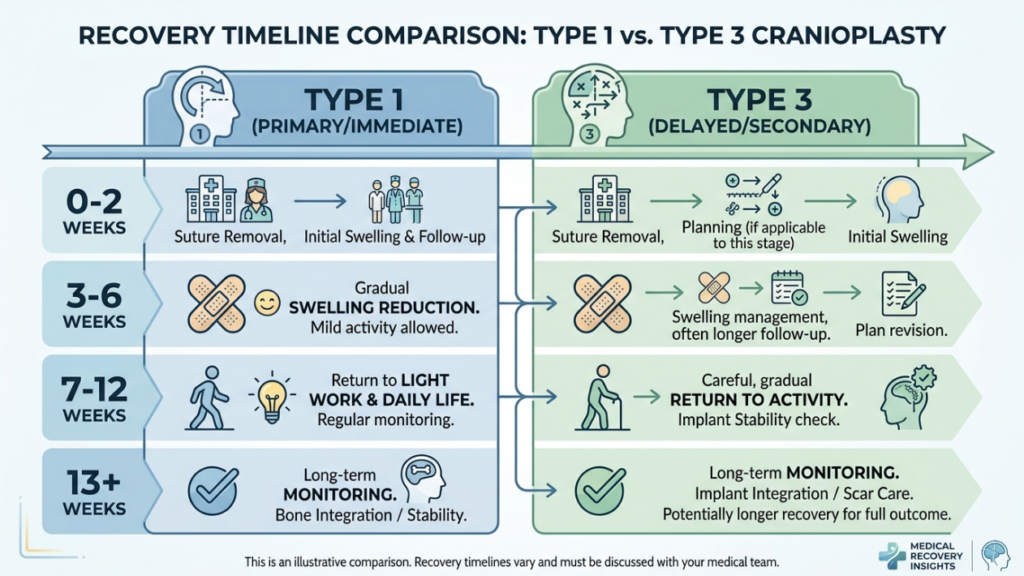
- Gonfiore da lieve a moderato: Si risolve entro 1-2 settimane.
- Disagio minimo: Gestito con farmaci antidolorifici da banco.
- Ritorno rapido alle attività: La maggior parte dei pazienti riprende le normali attività entro 2 settimane.
Recupero dopo cranioplastica di tipo 3
Il recupero dopo una cranioplastica di tipo 3 è più complesso a causa della complessità della procedura:
- Gonfiore e lividi significativi: Può persistere per 3-4 settimane.
- Dolore moderato: Potrebbe essere necessario assumere farmaci antidolorifici su prescrizione medica durante la prima settimana.
- Ritorno graduale alle attività: Le attività faticose sono vietate per 4-6 settimane.
- Visite di controllo: Monitoraggio regolare per garantire una corretta guarigione e affrontare eventuali complicazioni.
Entrambe le tecniche richiedono ai pazienti di evitare sollevamento di carichi pesanti, esercizio fisico intenso, E pressione diretta sulla fronte Durante la fase iniziale di recupero, i chirurghi forniscono istruzioni post-operatorie dettagliate, tra cui la cura della ferita, le restrizioni all'attività fisica e i segni di possibili complicazioni a cui prestare attenzione.
Potenziali complicazioni e come evitarle
Sebbene la cranioplastica sia generalmente sicura, possono insorgere complicazioni. La consapevolezza di questi rischi e delle misure preventive è fondamentale sia per i chirurghi che per i pazienti.
Complicazioni comuni
- Sinusite: Infiammazione o infezione del seno frontale, in particolare nella cranioplastica di tipo 3 se il seno viene lesionato.
- Perdita di liquido cerebrospinale (CSF): Complicazione rara ma grave in caso di perforazione della parete posteriore del seno mascellare.
- Irregolarità del contorno: Un eccessivo assottigliamento dell'osso nel tipo 1 o un arretramento improprio nel tipo 3 possono portare ad asimmetria o alla presenza di creste visibili.
- Formazione di mucocele: L'ostruzione delle vie di drenaggio dei seni paranasali può provocare la formazione di cisti piene di muco.
- Infezione: Il rischio è maggiore nel tipo 3 a causa dei tempi operatori più lunghi e dell'utilizzo di impianti.
Misure preventive
I chirurghi impiegano diverse strategie per ridurre al minimo le complicanze:
- Pianificazione chirurgica precisa: L'imaging 3D e la pianificazione chirurgica virtuale garantiscono osteotomie e reinterventi accurati.
- Preservazione dei seni paranasali: Evitare di lesionare il seno frontale durante la fresatura o l'osteotomia.
- Profilassi antibiotica: Somministrato prima e dopo l'intervento chirurgico per ridurre il rischio di infezioni.
- Monitoraggio postoperatorio: Controlli periodici per individuare precocemente eventuali complicazioni.
- Educazione del paziente: Fornire ai pazienti istruzioni sulla corretta cura delle ferite, sulle restrizioni relative all'attività fisica e sui segnali di allarme di possibili complicazioni.
Alternative alla cranioplastica di tipo 1 e di tipo 3
Per i pazienti che non sono candidati ideali per la cranioplastica di tipo 1 o di tipo 3, si possono prendere in considerazione tecniche alternative:
- Innesto di grasso: Il trapianto di grasso autologo può ammorbidire i contorni della fronte senza alterare la struttura ossea. Questa soluzione è ideale per i pazienti con lievi irregolarità o per coloro che cercano opzioni non chirurgiche.
- Impianti personalizzati: Impianti prefabbricati, come PEEK (polietereterchetone) O titanio, Possono essere utilizzati per aumentare o rimodellare la fronte senza osteotomia. Sono particolarmente utili per i pazienti con ossa sottili o problemi ai seni paranasali.
- Chirurgia ortognatica: Nei casi in cui rimodellamento della fronte fa parte di un più ampio femminilizzazione facciale In fase di pianificazione, le procedure ortognatiche (ad esempio, l'osteotomia di Le Fort I) possono essere combinate per correggere l'allineamento del massiccio facciale e della mandibola.
- Tecniche endoscopiche: Le tecniche endoscopiche minimamente invasive possono ridurre la protuberanza delle sopracciglia grazie a incisioni più piccole e tempi di recupero più rapidi.
Testimonianze dei pazienti e risultati concreti
I risultati ottenuti nella pratica clinica reale forniscono preziose informazioni sull'efficacia e sui tassi di soddisfazione della cranioplastica di tipo 1 e di tipo 3. Le testimonianze dei pazienti evidenziano l'impatto trasformativo di queste procedure:
Caso di studio 1: Cranioplastica di tipo 1
Una donna transgender di 28 anni desiderava un lieve intervento di femminilizzazione della fronte. Avendo un osso frontale sottile e una protuberanza ossea minima, ha optato per una cranioplastica di tipo 1. L'intervento ha permesso di ottenere un contorno più armonioso con tempi di recupero minimi. La paziente si è dichiarata molto soddisfatta, affermando che i risultati corrispondevano alle sue aspettative di un aspetto naturale e femminile.
Caso di studio 2: Cranioplastica di tipo 3
Una donna transgender di 35 anni presentava una marcata sporgenza delle sopracciglia e un osso frontale spesso. È stata sottoposta a cranioplastica di tipo 3, con osteotomia e arretramento. La drastica riduzione della proiezione frontale ha femminilizzato significativamente i suoi lineamenti. Sebbene il periodo di recupero sia stato più lungo del previsto, la paziente si è detta estremamente soddisfatta dei risultati, affermando che l'intervento le ha "cambiato la vita".“
Caso di studio 3: Chirurgia di revisione
Una paziente di 40 anni si era inizialmente sottoposta a cranioplastica di tipo 1, ma non era soddisfatta dei risultati, che erano troppo lievi. In seguito, ha optato per una cranioplastica di tipo 3 per ottenere una femminilizzazione più marcata. L'intervento di revisione ha risolto con successo le sue problematiche, dimostrando l'importanza di scegliere la tecnica più appropriata in base alle esigenze anatomiche e agli obiettivi estetici.
Il ruolo della pianificazione chirurgica virtuale nella cranioplastica
La pianificazione chirurgica virtuale (VSP) ha rivoluzionato la cranioplastica, in particolare per gli interventi di tipo 3. La VSP consente ai chirurghi di:
- Simulare osteotomie: Pianificare con precisione i tagli ossei e il riposizionamento per ottenere un contorno ottimale.
- Prevedere i risultati: Visualizzare i risultati post-operatori e adattare il piano chirurgico di conseguenza.
- Ridurre al minimo i rischi: Evitare strutture critiche come il seno frontale e i nervi sovraorbitali.
- Migliorare la comunicazione: Condividere i modelli 3D con i pazienti permette di stabilire aspettative realistiche e migliorare il consenso informato.
Gli studi hanno dimostrato che la pianificazione chirurgica virtuale (VSP) riduce i tempi operatori, migliora la precisione e aumenta la soddisfazione del paziente (3D Printing and Virtual Surgical Planning in Craniofacial and Orthognathic Surgery, 2025). È ormai considerata il gold standard per le procedure di cranioplastica complesse.

Domande frequenti
Qual è la principale differenza tra la cranioplastica di tipo 1 e quella di tipo 3?
La cranioplastica di tipo 1 prevede la limatura o la fresatura dello strato esterno dell'osso frontale per ridurne la prominenza, mentre la cranioplastica di tipo 3 richiede un'osteotomia (taglio osseo) e l'arretramento del segmento dell'osso frontale per ottenere un rimodellamento più marcato. Il tipo 1 è meno invasivo ed è indicato per prominenze lievi o moderate, mentre il tipo 3 è riservato a prominenze gravi o ossa frontali spesse.
Come fanno i chirurghi a decidere tra cranioplastica di tipo 1 e di tipo 3?
I chirurghi valutano lo spessore dell'osso frontale del paziente, il grado di prominenza delle sopracciglia e l'anatomia del seno frontale mediante tomografia computerizzata (TC) e immagini 3D. Il tipo 1 viene scelto per ossa più sottili e prominenza lieve, mentre il tipo 3 è preferibile per ossa più spesse, prominenza grave o pneumatizzazione estesa del seno. Anche gli obiettivi del paziente e la sua tolleranza ai rischi chirurgici influenzano la decisione.
Quali sono i rischi associati alla cranioplastica di tipo 3?
La cranioplastica di tipo 3 comporta rischi maggiori a causa della sua complessità, tra cui sinusite, perdite di liquido cerebrospinale, irregolarità del contorno, formazione di mucocele e infezioni. Questi rischi vengono minimizzati attraverso un'accurata pianificazione chirurgica, la preservazione dei seni paranasali e il monitoraggio postoperatorio. I pazienti vengono informati sui segnali di allarme e sulle cure di follow-up per garantire una pronta guarigione.
Quanto dura il periodo di recupero per una cranioplastica di tipo 1 rispetto a una di tipo 3?
Il recupero da una cranioplastica di tipo 1 richiede in genere 1-2 settimane, con lieve gonfiore e fastidio. La cranioplastica di tipo 3 richiede un recupero più lungo, da 3 a 6 settimane, a causa dell'intervento chirurgico più esteso e delle potenziali complicazioni. Si consiglia ai pazienti di evitare attività faticose e di seguire attentamente le istruzioni post-operatorie.
La cranioplastica di tipo 1 può ottenere gli stessi risultati della cranioplastica di tipo 3?
No, la cranioplastica di tipo 1 si limita a un rimodellamento sottile ed è ideale per bozze frontali da lievi a moderate. La cranioplastica di tipo 3 ottiene una femminilizzazione più marcata riposizionando il segmento osseo frontale, risultando quindi adatta a bozze frontali gravi o ossa frontali spesse. La scelta dipende dalle esigenze anatomiche e dagli obiettivi estetici della paziente.
Che ruolo svolge la pianificazione chirurgica virtuale nella cranioplastica?
La pianificazione chirurgica virtuale (VSP) consente ai chirurghi di simulare le osteotomie, prevedere i risultati e minimizzare i rischi evitando strutture critiche come il seno frontale. Migliora la precisione, riduce i tempi operatori e aumenta la soddisfazione del paziente fornendo una chiara visualizzazione del piano chirurgico e dei risultati attesi.
Esistono alternative non chirurgiche alla cranioplastica per la femminilizzazione della fronte?
Sì, le alternative non chirurgiche includono innesto di grasso Per ammorbidire i contorni e rimodellare la fronte con impianti personalizzati (ad esempio in PEEK o titanio) senza osteotomia, queste opzioni sono ideali per pazienti con lievi irregolarità o per chi preferisce evitare la chirurgia. Tuttavia, potrebbero non garantire lo stesso livello di femminilizzazione delle tecniche chirurgiche.
Cosa devo aspettarmi durante la visita per la ricostruzione della fronte?
Durante la visita, il chirurgo valuterà lo spessore dell'osso frontale, l'anatomia dei seni paranasali e il grado di prominenza ossea mediante esame clinico e immagini 3D. Discuterà i vostri obiettivi estetici, spiegherà le differenze tra cranioplastica di tipo 1 e di tipo 3 e vi consiglierà la tecnica più adatta in base alla vostra anatomia e alle vostre aspettative.







