

Alın rekonstrüksiyonu kritik bir bileşenidir Yüz Feminizasyon Cerrahisi (FFS) Yüz ve çene cerrahisi, uyumlu ve estetik açıdan hoş bir yüz konturu elde etmeyi amaçlayan kraniyofasiyal işlemlerdir. En çok tartışılan teknikler arasında şunlar yer almaktadır: Tip 1 Ve Tip 3 kranioplasti, Her biri alın bölgesini yeniden şekillendirmek için farklı yaklaşımlar sunuyor. Tip 1 içerir kemik törpüleme veya tıraşlama, Tip 3 daha karmaşık bir işlem gerektirir osteotomi ve gerileme Alın sinüsünün küçültülmesi. Bu tekniklerin anatomik, fonksiyonel ve estetik etkilerini anlamak, hem cerrahlar hem de hastalar için bilinçli kararlar alabilmek açısından çok önemlidir. Sinüs geri çekme ve kaş çıkıntısı azaltma hakkında daha fazla bilgi edinmek için kapsamlı kılavuzumuza göz atın. alın şekillendirme.
Bu kılavuz, Tip 1 ve Tip 3 kranioplasti arasındaki yapısal farklılıkları ve bunların etkilerini incelemektedir. kemik kalınlığı, frontal sinüs anatomisi, ve cerrahların her hasta için en uygun yaklaşımı nasıl belirlediğini öğreneceksiniz. Sonunda, hangi tekniğin anatomik ihtiyaçlarınıza ve estetik hedeflerinize en uygun olduğunu anlayacaksınız.
İçindekiler
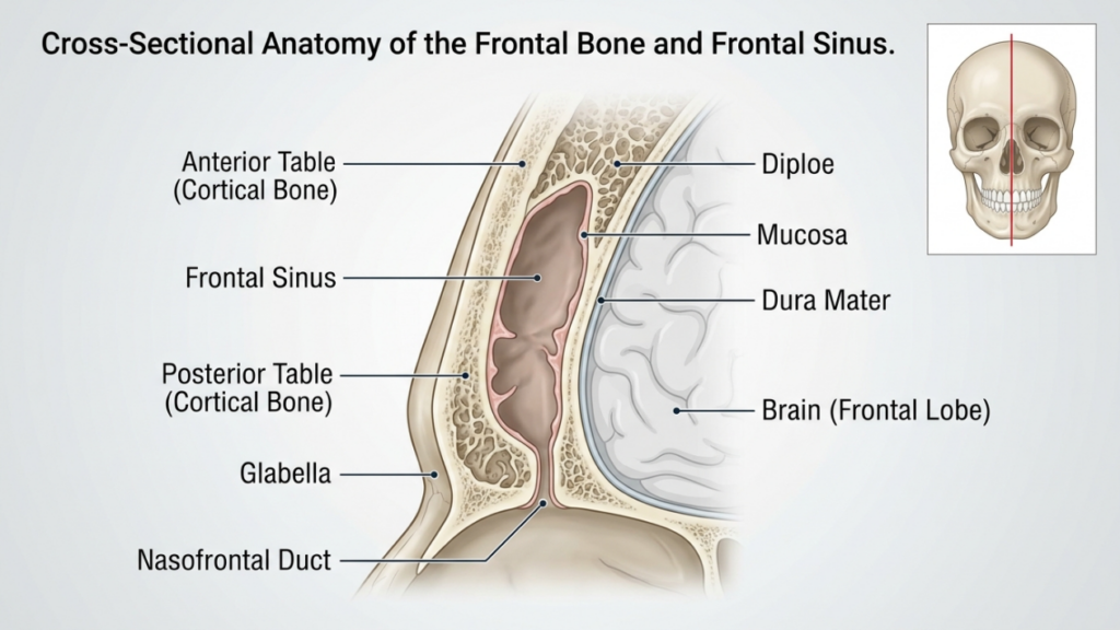
Anatomik Temel: Alın Kemiği ve Sinüs
The ön kemik Ve frontal sinüs Alın rekonstrüksiyonunda çok önemli bir rol oynarlar. Alın kemiği, yüzün üst kısmını oluşturur ve bireyler arasında boyut ve şekil bakımından farklılık gösteren içi boş bir boşluk olan frontal sinüsü barındırır. ön tablo Alın sinüsünün dış katmanı, alnın konturuna katkıda bulunan kemiğin dış tabakasıdır. arka tablo Sinüsü beyinden ayırır. Bu tabakaların kalınlığı ve sinüs pnömatizasyonunun (hava dolu genişleme) derecesi, Tip 1 ve Tip 3 kranioplasti arasında seçim yapmayı etkiler.
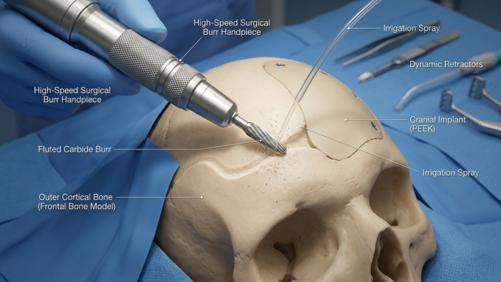
İçinde Tip 1 kranioplasti, Cerrah Bu teknik, frontal sinüse zarar vermeden dış kortikal kemiği tıraşlamak için yüksek hızlı bir freze kullanır ve çıkıntıyı azaltır. Bu teknik, aşağıdaki durumlardaki hastalar için idealdir: ince alın kemiği Veya minimal çıkıntı durumunda sinüsün bütünlüğünü korur. Bununla birlikte, aşırı törpüleme kemik stabilitesini tehlikeye atabileceğinden veya istenen konturu elde edemeyebileceğinden, belirgin kaş çıkıntısı veya kalın ön tabakaya sahip hastalar için yeterli olmayabilir (Ousterhout, 2024).
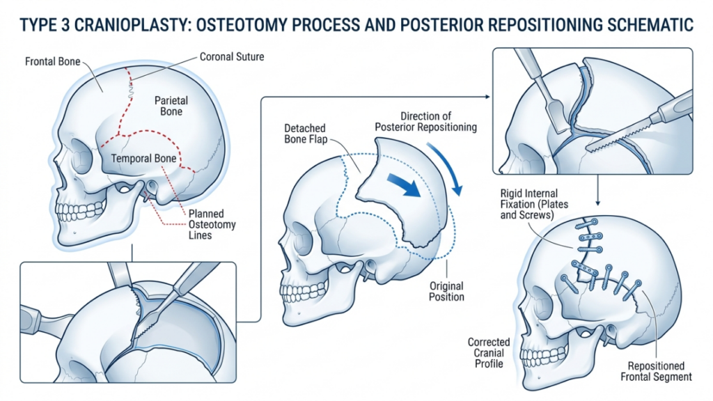
Tersine, Tip 3 kranioplasti bir osteotomi—frontal sinüsün ön tabakasında kontrollü bir kesi yapılır. Kemik parçası daha sonra geriye doğru yeniden konumlandırılır (geri çekme), böylece çıkıntı azaltılır ve daha pürüzsüz, daha kadınsı bir alın oluşturulur. Bu teknik, aşağıdaki durumlardaki hastalar için ayrılmıştır: orta ila şiddetli kaş çıkıntısı veya kalın bir ön tabaka söz konusu olduğunda, sadece törpüleme yetersiz kalır. Osteotomi, sinüsün koruyucu işlevini korurken hassas bir şekilde yeniden şekillendirmeye olanak tanır (Alın Feminizasyonu: Kapsamlı Bir Literatür İncelemesi, 2024).
Tip 1 Kranioplasti: Tıraşlama ve Frezeleme
Teknik Genel Bakış
Tip 1 kranioplasti, alın rekonstrüksiyonu için en az invaziv seçenektir. Bu işlemde bir cerrahi alet kullanılır. yüksek hızlı çapak Alın kemiğinin belirginliğini kademeli olarak azaltmak. Cerrah Bu teknik, frontal sinüse nüfuz etmekten kaçınarak, dış kortikal tabakayı titizlikle tıraş eder. Bu teknik özellikle aşağıdaki hastalar için etkilidir:
- Hafif ila orta derecede kaş çıkıntısı
- İnce alın kemiği (5 mm'den az)
- Ön sinüslerde belirgin pnömatizasyonun yokluğu
Bu işlem bir yöntemle gerçekleştirilir. koronal kesi, Bu yöntem, görünür yara izini en aza indirirken alın bölgesine erişimi sağlar. Cerrah, kemiği aşırı inceltmeden, yani instabiliteye veya kontur düzensizliklerine yol açmadan, düzgün bir küçültme sağlamak için dokunsal geri bildirim ve görsel ipuçlarından yararlanır.
Tip 1 Kranioplastinin Avantajları
Tip 1 kranioplasti çeşitli avantajlar sunar:
- Minimal İnvazivlik: Osteotomi veya kemik çıkarılmasının yapılmaması, cerrahi travmayı ve iyileşme süresini azaltır.
- Komplikasyon Riski Daha Düşük: Alın sinüsünün korunması, sinüzit, beyin omurilik sıvısı sızıntısı veya mukosel oluşumu riskini en aza indirir.
- Daha Kısa Çalışma Süresi: Bu işlem genellikle 1-2 saat sürer, bu da onu Tip 3'e kıyasla daha hızlı bir seçenek haline getirir.
- Öngörülebilir Sonuçlar: Hafif karın çıkıntısı olan ve kadınsı bir görünüm elde etmek için ince kontürlemenin yeterli olduğu hastalar için idealdir.
Tip 1 Kranioplastinin Sınırlamaları
Tip 1 kranioplasti daha güvenli ve daha az invaziv olmakla birlikte, önemli sınırlamaları da vardır:
- Sınırlı İndirim: Kaş çıkıntısı çok fazla olan veya alın kemikleri kalın olan hastalar için yetersizdir.
- Aşırı Seyreltme Riski: Aşırı törpüleme kemiği zayıflatabilir ve bu da kontur düzensizliklerine veya kırıklara yol açabilir.
- Eksik Feminizasyon: Belirgin erkeksi yüz hatlarına sahip hastalarda istenen estetik sonucu vermeyebilir.
Tip 3 Kranioplasti: Osteotomi ve Geriye Çekme
Teknik Genel Bakış
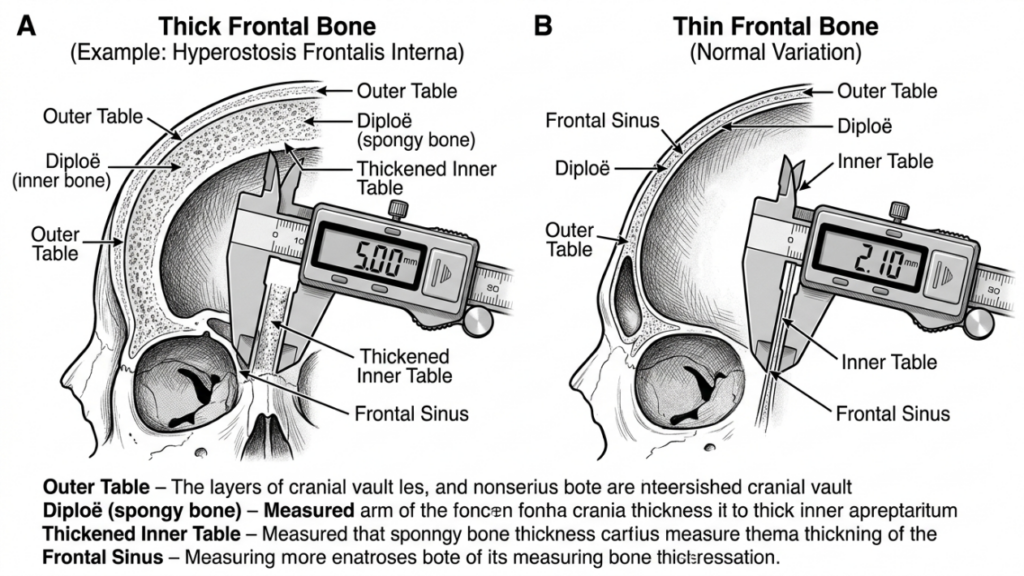
Tip 3 kranioplasti, aşağıdaki durumlardaki hastalar için tasarlanmış daha karmaşık bir işlemdir: orta ila şiddetli kaş çıkıntısı veya kalın alın kemikleri. Bu, bir osteotomi—frontal sinüsün ön tabakasında yapılan hassas bir kesi ve ardından kemik parçasının geriye doğru yeniden konumlandırılması (geri çekme). Bu teknik, alın çıkıntısında önemli bir azalma ve daha pürüzsüz, daha kadınsı bir kontur sağlar.
Bu işlem genellikle şu yolla gerçekleştirilir: koronal kesi, Bu işlem, alın kemiğine ve sinüse erişim sağlar. Cerrah, bir alet kullanır. sagital testere veya piezoelektrik cihaz Osteotomi oluşturulurken, kesimin alnın doğal kıvrımını takip etmesi sağlanır. Daha sonra kemik parçası yeniden konumlandırılır ve sabitlenir. titanyum plakalar ve vidalar veya emilebilir dikişler. Bu teknik aşağıdaki hastalar için idealdir:
- Şiddetli kaş çıkıntısı
- Kalın alın kemiği (5 mm'den fazla)
- Önemli frontal sinüs pnömatizasyonu
Tip 3 kranioplasti, genellikle titiz bir planlama gerektirir ve şunları içerir: 3 boyutlu sanal cerrahi planlama (VSP) Osteotomi ve geri çekme işlemini simüle etmek için. Bu, hassasiyeti sağlar ve sinüs ihlali veya beyin omurilik sıvısı sızıntısı gibi komplikasyon riskini en aza indirir (Sanal Cerrahi Planlama). Yüz Feminizasyonu Üst Yüzün, 2025).
Tip 3 Kranioplastinin Avantajları
Tip 3 kranioplasti, belirgin erkeksi özelliklere sahip hastalar için çeşitli avantajlar sunar:
- Önemli Kontürleme: Kaş çıkıntısını belirgin şekilde azaltarak daha pürüzsüz, daha kadınsı bir alın oluşturur.
- Çok yönlülük: Kalın alın kemikleri veya yaygın sinüs pnömatizasyonu olan hastalar için uygundur.
- Uzun Vadeli İstikrar: Yeniden konumlandırılan kemik parçası iyi bir şekilde bütünleşerek zamanla kontur düzensizlikleri riskini azaltır.
- Özelleştirme: Sanal cerrahi planlama, en iyi sonuçları elde etmek için hastaya özel hassas ayarlamalar yapılmasına olanak tanır.
Tip 3 Kranioplastinin Sınırlamaları
Etkinliğine rağmen, Tip 3 kranioplasti daha yüksek riskler ve karmaşıklıklar taşır:
- Ameliyat Süresinin Uzaması: Bu işlem genellikle 3-5 saat sürer ve daha fazla hassasiyet ve uzmanlık gerektirir.
- Komplikasyon Riski Daha Yüksek: Olası riskler arasında sinüzit, beyin omurilik sıvısı sızıntısı veya sinüsün zarar görmesi durumunda mukosel oluşumu yer almaktadır.
- Daha Uzun İyileşme: Hastalar, Tip 1 kranioplastiye kıyasla daha uzun süreli şişlik ve rahatsızlık yaşayabilirler.
- Maliyet: Gelişmiş görüntüleme ve cerrahi aletlerin kullanımı, işlemin toplam maliyetini artırmaktadır.

Tip 1 ve Tip 3 Kranioplasti Arasındaki Temel Farklar
| Özellik | Tip 1 Kranioplasti | Tip 3 Kraniyoplasti |
|---|---|---|
| Teknik | Kemik törpüleme/tıraşlama | Osteotomi ve geriye çekme |
| İstilacılık | En az | Orta ila yüksek |
| İdeal Adaylar | Hafif ila orta derecede kaş çıkıntısı, ince alın kemiği | Belirgin kaş çıkıntısı, kalın alın kemiği |
| Cerrahi Zaman | 1-2 saat | 3-5 saat |
| İyileşme süresi | 1–2 hafta | 3-6 hafta |
| Komplikasyon Riski | Düşük | Orta ila yüksek |
| Maliyet | Daha düşük | Daha yüksek |
| Estetik Sonuç | İnce kontürleme | Dramatik kadınlaşma |
Cerrahlar Nasıl Karar Veriyor: Tip 1 mi, Tip 3 mü?
Tip 1 ve Tip 3 kranioplasti arasındaki seçim, hastanın durumu da dahil olmak üzere çeşitli faktörlere bağlıdır. anatomik özellikler, estetik hedefler, Ve cerrahi riskler. Cerrahlar çeşitli yöntemlerin bir kombinasyonuna güvenirler. klinik muayene, 3 boyutlu görüntüleme, Ve hasta konsültasyonu En uygun tekniği belirlemek için.
1. Anatomik Değerlendirme
İlk adım hastanın değerlendirilmesidir. alın kemiği kalınlığı Ve frontal sinüs anatomisi. A BT taraması veya 3 boyutlu yeniden yapılandırma Aşağıdaki konularda detaylı bilgiler sunmaktadır:
- Kemik Kalınlığı: Alın kemikleri ince olan (5 mm'den az) hastalar Tip 1 kranioplasti için daha uygundur, daha kalın kemiklere sahip olanlar ise Tip 3'e ihtiyaç duyabilir.
- Sinüs Pnömatizasyonu: Geniş çaplı sinüs pnömatizasyonu, törpüleme sırasında sinüsün zarar görmesini önlemek için Tip 3 kranioplasti gerektirebilir.
- Patronluk Derecesi: Aşırı kaş çıkıntısı genellikle yalnızca Tip 3 teknikleriyle elde edilebilen dramatik bir küçültmeyi gerektirir.
2. Estetik Hedefler
Hastaların estetik beklentileri, teknik seçiminde çok önemli bir rol oynar. incelikli kadınlaştırma Bazı kişiler Tip 1 kranioplastiyi tercih edebilirken, belirgin erkeksi özelliklere sahip kişiler genellikle Tip 1 kranioplastiye ihtiyaç duyar. dönüştürücü sonuçlar Tip 3'ün bir örneği. Cerrahlar, hastanın anatomisine ve istenen değişikliklere dayanarak gerçekçi sonuçları tartışırlar.
3. Cerrahi Riskler ve İyileşme
Tip 3 kranioplasti daha yüksek riskler taşır, bunlar arasında şunlar yer alır: sinüs komplikasyonları, beyin omurilik sıvısı sızıntıları, Ve uzun süreli iyileşme. Cerrahlar hastanın genel sağlığını, ameliyata toleransını ve ameliyat sonrası bakıma uyma isteğini değerlendirir. Ameliyat riskini artıran tıbbi rahatsızlıkları olan hastalara Tip 1 veya alternatif prosedürleri düşünmeleri önerilebilir.
4. Sanal Cerrahi Planlama (VSP)
Gelişmeler 3 boyutlu sanal cerrahi planlama Kranioplasti alanında devrim yarattı. Cerrahlar, osteotomileri, geri çekmeleri ve sonuçları simüle etmek için VSP'yi kullanarak hassasiyeti sağlıyor ve riskleri en aza indiriyor. Bu teknoloji, özellikle doğru kemik yeniden konumlandırmasının kritik olduğu Tip 3 kranioplasti için çok değerlidir (3D Printing and Virtual Surgical Planning in Craniofacial and Orthognathic Surgery, 2025).
Ameliyat Sonrası Bakım ve İyileşme
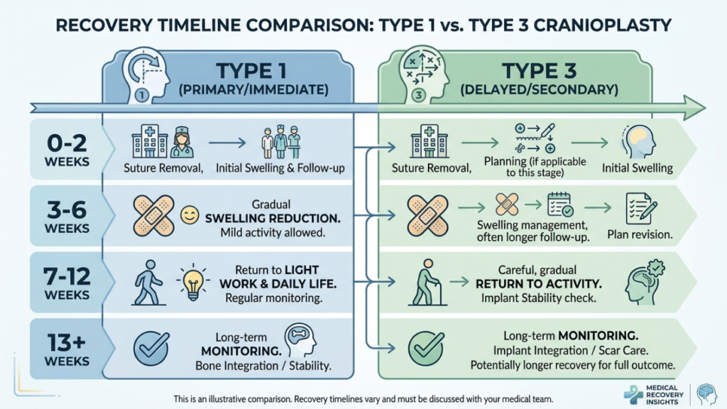
Tip 1 ve Tip 3 kranioplasti arasında iyileşme süreci önemli ölçüde farklılık gösterir. Ameliyat sonrası süreci anlamak, hastaların sorunsuz bir iyileşme yolculuğuna hazırlanmalarına yardımcı olur.
Tip 1 Kranioplasti Sonrası İyileşme
Tip 1 kranioplasti ameliyatı geçiren hastalar genellikle şu durumları yaşarlar:
- Hafif ila orta şiddette şişlik: 1-2 hafta içinde geçer.
- Minimum Rahatsızlık: Reçetesiz satılan ağrı kesicilerle kontrol altına alındı.
- Aktivitelere Hızlı Dönüş: Hastaların çoğu 2 hafta içinde normal aktivitelerine döner.
Tip 3 Kranioplasti İyileşme Süreci
Tip 3 kranioplasti ameliyatından sonra iyileşme süreci, işlemin karmaşıklığı nedeniyle daha uzundur:
- Belirgin Şişlik ve Morarma: 3-4 hafta sürebilir.
- Orta Derecede Ağrı: İlk hafta boyunca reçeteli ağrı kesici ilaçlara ihtiyaç duyulabilir.
- Aktivitelere Kademeli Dönüş: Yoğun fiziksel aktiviteler 4-6 hafta süreyle kısıtlanmıştır.
- Takip Ziyaretleri: Doğru iyileşmeyi sağlamak ve olası komplikasyonları gidermek için düzenli izleme yapılmalıdır.
Her iki teknik de hastaların kaçınmasını gerektirir. ağır kaldırma, yorucu egzersiz, Ve alına doğrudan baskı İlk iyileşme evresinde cerrahlar, yara bakımı, aktivite kısıtlamaları ve dikkat edilmesi gereken komplikasyon belirtileri de dahil olmak üzere ayrıntılı ameliyat sonrası talimatlar verirler.
Olası Komplikasyonlar ve Bunlardan Nasıl Kaçınılır?
Kranioplasti genellikle güvenli bir işlem olsa da, komplikasyonlar ortaya çıkabilir. Bu risklerin ve önleyici tedbirlerin farkında olmak hem cerrahlar hem de hastalar için çok önemlidir.
Sık Görülen Komplikasyonlar
- Sinüzit: Özellikle Tip 3 kranioplasti ameliyatında sinüsün hasar görmesi durumunda frontal sinüsün iltihaplanması veya enfeksiyonu.
- Beyin Omurilik Sıvısı (BOS) Kaçağı: Sinüsün arka tabakasının delinmesi durumunda nadir ancak ciddi bir komplikasyon ortaya çıkar.
- Kontur Düzensizlikleri: Tip 1'deki aşırı kemik incelmesi veya Tip 3'teki uygunsuz geriye çekilme, asimetriye veya görünür sırtlara yol açabilir.
- Mukosel Oluşumu: Sinüs drenaj yollarının tıkanması, mukus dolu kistlere yol açabilir.
- Enfeksiyon: Tip 3'te ameliyat süresinin daha uzun olması ve implant kullanımı nedeniyle risk daha yüksektir.
Önleyici Tedbirler
Cerrahlar komplikasyonları en aza indirmek için çeşitli stratejiler kullanırlar:
- Hassas Cerrahi Planlama: 3 boyutlu görüntüleme ve sanal cerrahi planlama, osteotomilerin ve geri çekmelerin doğru yapılmasını sağlar.
- Sinüslerin Korunması: Frezeleme veya osteotomi sırasında frontal sinüsün zarar görmesinden kaçınılmalıdır.
- Antibiyotik Profilaksisi: Enfeksiyon riskini azaltmak için ameliyat öncesi ve sonrası uygulanır.
- Ameliyat Sonrası İzleme: Komplikasyonların erken belirtilerini tespit etmek için düzenli kontroller.
- Hasta Eğitimi: Hastaları doğru yara bakımı, aktivite kısıtlamaları ve komplikasyon belirtileri konusunda bilgilendirmek.
Tip 1 ve Tip 3 Kranioplastiye Alternatifler
Tip 1 veya Tip 3 kranioplasti için ideal aday olmayan hastalar için alternatif teknikler düşünülebilir:
- Yağ Greftleme: Otojen yağ transferi, kemik yapısını değiştirmeden alın konturlarını yumuşatabilir. Bu yöntem, hafif düzensizlikleri olan veya cerrahi olmayan seçenekler arayan hastalar için idealdir.
- Kişiye Özel İmplantlar: Önceden üretilmiş implantlar, örneğin; PEEK (polieter eter keton) veya titanyum, Bu yöntem, osteotomi yapılmadan alın bölgesini büyütmek veya yeniden şekillendirmek için kullanılabilir. Özellikle ince kemik yapısına sahip veya sinüs sorunları yaşayan hastalar için faydalıdır.
- Ortognatik Cerrahi: Şu durumlarda alın şekillendirme daha geniş bir yapının parçasıdır yüz feminizasyonu Plan kapsamında, orta yüz ve çene hizalamasını ele almak için ortognatik prosedürler (örneğin, Le Fort I osteotomisi) birleştirilebilir.
- Endoskopik Teknikler: Minimal invaziv endoskopik yaklaşımlar, daha küçük kesiler ve daha hızlı iyileşme süreleri ile kaş çıkıntısını azaltabilir.
Hasta Görüşleri ve Gerçek Dünya Sonuçları
Gerçek dünya sonuçları, Tip 1 ve Tip 3 kranioplastinin etkinliği ve memnuniyet oranları hakkında değerli bilgiler sunmaktadır. Hasta yorumları, bu prosedürlerin dönüştürücü etkisini vurgulamaktadır:
Vaka Çalışması 1: Tip 1 Kranioplasti
28 yaşında trans bir kadın, alnında hafif bir kadınsılaştırma istedi. İnce alın kemiği ve minimal çıkıntısı nedeniyle Tip 1 kranioplasti yöntemini tercih etti. İşlem, minimal iyileşme süresiyle daha pürüzsüz bir kontur sağladı. Sonuçlardan son derece memnun olduğunu ve doğal, kadınsı bir görünüm beklentileriyle örtüştüğünü belirtti.
Vaka Çalışması 2: Tip 3 Kranioplasti
35 yaşında trans bir kadın, şiddetli kaş çıkıntısı ve kalın alın kemiği şikayetiyle başvurdu. Osteotomi ve geriye çekme işlemlerini içeren Tip 3 kranioplasti uygulandı. Alın çıkıntısındaki dramatik azalma, yüz hatlarını önemli ölçüde kadınsılaştırdı. İyileşme süreci daha uzun sürse de, sonuçlardan son derece memnun olduğunu ve işlemin "hayatını değiştirdiğini" belirtti.“
Vaka Çalışması 3: Revizyon Ameliyatı
40 yaşında bir hasta başlangıçta Tip 1 kranioplasti ameliyatı geçirdi ancak sonuçlardan memnun kalmadı. Daha sonra daha belirgin bir kadınsı görünüm elde etmek için Tip 3 kranioplasti ameliyatını tercih etti. Revizyon ameliyatı, hastanın endişelerini başarıyla giderdi ve anatomik ihtiyaçlara ve estetik hedeflere göre doğru tekniğin seçilmesinin önemini gösterdi.
Kranioplastide Sanal Cerrahi Planlamanın Rolü
Sanal cerrahi planlama (VSP), özellikle Tip 3 prosedürler için kranioplastide çığır açan bir gelişme haline geldi. VSP, cerrahlara şunları yapma olanağı sağlar:
- Osteotomileri Simüle Etme: En uygun konturlamayı elde etmek için kemik kesimlerini ve yeniden konumlandırmayı hassas bir şekilde planlayın.
- Sonuçları Tahmin Et: Ameliyat sonrası sonuçları görselleştirin ve cerrahi planı buna göre ayarlayın.
- Riskleri En Aza İndirin: Alın sinüsü ve supraorbital sinirler gibi kritik yapılardan kaçının.
- İletişimi Geliştirin: Hastalarla 3 boyutlu modelleri paylaşarak gerçekçi beklentiler oluşturun ve bilgilendirilmiş onam sürecini iyileştirin.
Çalışmalar, VSP'nin ameliyat süresini kısalttığını, doğruluğu artırdığını ve hasta memnuniyetini yükselttiğini göstermiştir (Kraniyofasiyal ve Ortognatik Cerrahide 3B Baskı ve Sanal Cerrahi Planlama, 2025). Şu anda karmaşık kranioplasti prosedürleri için altın standart olarak kabul edilmektedir.

Sıkça Sorulan Sorular
Tip 1 ve Tip 3 kranioplasti arasındaki temel fark nedir?
Tip 1 kranioplasti, çıkıntıyı azaltmak için frontal kemiğin dış tabakasının tıraşlanmasını veya törpülenmesini içerirken, Tip 3 kranioplasti daha belirgin bir konturlama elde etmek için osteotomi (kemik kesimi) ve frontal kemik segmentinin geriye çekilmesini gerektirir. Tip 1 daha az invazivdir ve hafif ila orta dereceli çıkıntılar için uygundur, Tip 3 ise şiddetli çıkıntılar veya kalın frontal kemikler için ayrılmıştır.
Cerrahlar Tip 1 ve Tip 3 kranioplasti arasında nasıl karar veriyor?
Cerrahlar, BT taramaları ve 3 boyutlu görüntüleme kullanarak hastanın alın kemiği kalınlığını, kaş çıkıntısının derecesini ve alın sinüs anatomisini değerlendirir. Daha ince kemikler ve hafif çıkıntılar için Tip 1, daha kalın kemikler, şiddetli çıkıntı veya yaygın sinüs pnömatizasyonu için ise Tip 3 tercih edilir. Hastanın hedefleri ve cerrahi risklere toleransı da kararı etkiler.
Tip 3 kranioplasti ile ilişkili riskler nelerdir?
Tip 3 kranioplasti, sinüzit, beyin omurilik sıvısı sızıntısı, kontur düzensizlikleri, mukosel oluşumu ve enfeksiyon gibi karmaşıklığı nedeniyle daha yüksek riskler taşır. Bu riskler, hassas cerrahi planlama, sinüslerin korunması ve ameliyat sonrası izleme ile en aza indirilir. Hastalar, sorunsuz bir iyileşme sağlamak için uyarı işaretleri ve takip bakımı konusunda bilgilendirilir.
Tip 1 ve Tip 3 kranioplasti ameliyatlarının iyileşme süreleri ne kadardır?
Tip 1 kranioplasti sonrası iyileşme genellikle 1-2 hafta sürer ve hafif şişlik ve rahatsızlık hissedilir. Tip 3 kranioplasti ise daha kapsamlı bir ameliyat ve olası komplikasyonlar nedeniyle 3-6 hafta süren daha uzun bir iyileşme süreci gerektirir. Hastaların yorucu aktivitelerden kaçınmaları ve ameliyat sonrası talimatlara yakından uymaları önerilir.
Tip 1 kranioplasti, Tip 3 ile aynı sonuçları verebilir mi?
Hayır, Tip 1 kranioplasti, hafif konturlama ile sınırlıdır ve hafif ila orta dereceli alın çıkıntıları için idealdir. Tip 3 kranioplasti, alın kemiği segmentinin yeniden konumlandırılmasıyla daha belirgin bir kadınsılaştırma sağlar ve bu nedenle şiddetli alın çıkıntıları veya kalın alın kemikleri için uygundur. Seçim, hastanın anatomik ihtiyaçlarına ve estetik hedeflerine bağlıdır.
Kranioplastide sanal cerrahi planlamanın rolü nedir?
Sanal cerrahi planlama (VSP), cerrahların osteotomileri simüle etmelerine, sonuçları tahmin etmelerine ve frontal sinüs gibi kritik yapılardan kaçınarak riskleri en aza indirmelerine olanak tanır. Cerrahi planın ve beklenen sonuçların net bir şekilde görselleştirilmesini sağlayarak hassasiyeti artırır, ameliyat süresini kısaltır ve hasta memnuniyetini iyileştirir.
Alın bölgesinin kadınsılaştırılması için kranioplastiye cerrahi olmayan alternatifler var mı?
Evet, cerrahi olmayan alternatifler şunları içerir: yağ aşısı Alın bölgesini osteotomi gerektirmeden yeniden şekillendirmek için konturları yumuşatmak ve özel implantlar (örneğin, PEEK veya titanyum) kullanmak mümkündür. Bu seçenekler, hafif düzensizlikleri olan veya ameliyattan kaçınmayı tercih eden hastalar için idealdir. Bununla birlikte, cerrahi tekniklerle elde edilen aynı düzeyde kadınsılaştırmayı sağlamayabilirler.
Alın rekonstrüksiyonu görüşmesi sırasında neler beklemeliyim?
Konsültasyon sırasında cerrahınız, klinik muayene ve 3 boyutlu görüntüleme yöntemlerini kullanarak frontal kemik kalınlığınızı, sinüs anatomisini ve çıkıntı derecesini değerlendirecektir. Estetik hedeflerinizi görüşecek, Tip 1 ve Tip 3 kranioplasti arasındaki farkları açıklayacak ve anatomik yapınıza ve beklentilerinize göre en uygun tekniği önerecektir.







