

Voorhoofdreconstructie is een cruciaal onderdeel van Gezichtsbehandeling Feminisatiechirurgie (VFF) en craniofaciale ingrepen, gericht op het bereiken van een harmonieuze en esthetisch aantrekkelijke gezichtscontour. Tot de meest besproken technieken behoren: Type 1 En Type 3 cranioplastie, waarbij elk een eigen aanpak biedt voor het vormgeven van het voorhoofd. Type 1 houdt in botfrezen of -schaven, Soort 3 vereist een complexere aanpak osteotomie en tegenslag van de frontale sinus. Inzicht in de anatomische, functionele en esthetische implicaties van deze technieken is essentieel voor zowel chirurgen als patiënten om weloverwogen beslissingen te kunnen nemen. Lees meer over sinus-terugplaatsing en wenkbrauwversteviging in onze complete gids. voorhoofd contouren.
Deze gids onderzoekt de structurele verschillen tussen cranioplastiek type 1 en type 3, en de impact daarvan op botdikte, anatomie van de frontale sinus, En hoe chirurgen de meest geschikte aanpak voor elke patiënt bepalen. Aan het einde van deze cursus weet u precies welke techniek het beste aansluit bij uw anatomische behoeften en esthetische doelen.
Inhoudsopgave
De anatomische basis: het voorhoofdsbeen en de sinussen
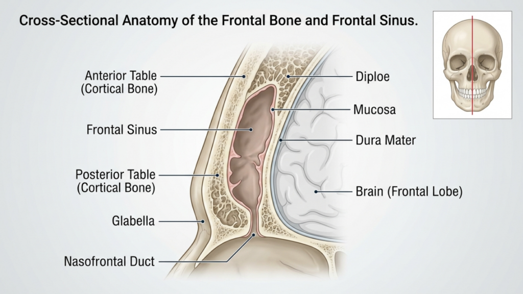
De voorhoofdsbeen En frontale sinus Het voorhoofdsbeen speelt een cruciale rol bij de reconstructie van het voorhoofd. Het voorhoofdsbeen vormt het bovenste deel van het gezicht en bevat de voorhoofdsholte, een holle ruimte waarvan de grootte en vorm per persoon verschillen. voorste tafel van de frontale sinus is de buitenste botlaag die bijdraagt aan de contouren van het voorhoofd, terwijl de achterste tafel Het scheidt de sinussen van de hersenen. De dikte van deze platen en de mate van sinuspneumatisatie (uitzetting met lucht) beïnvloeden de keuze tussen cranioplastiek type 1 en type 3.
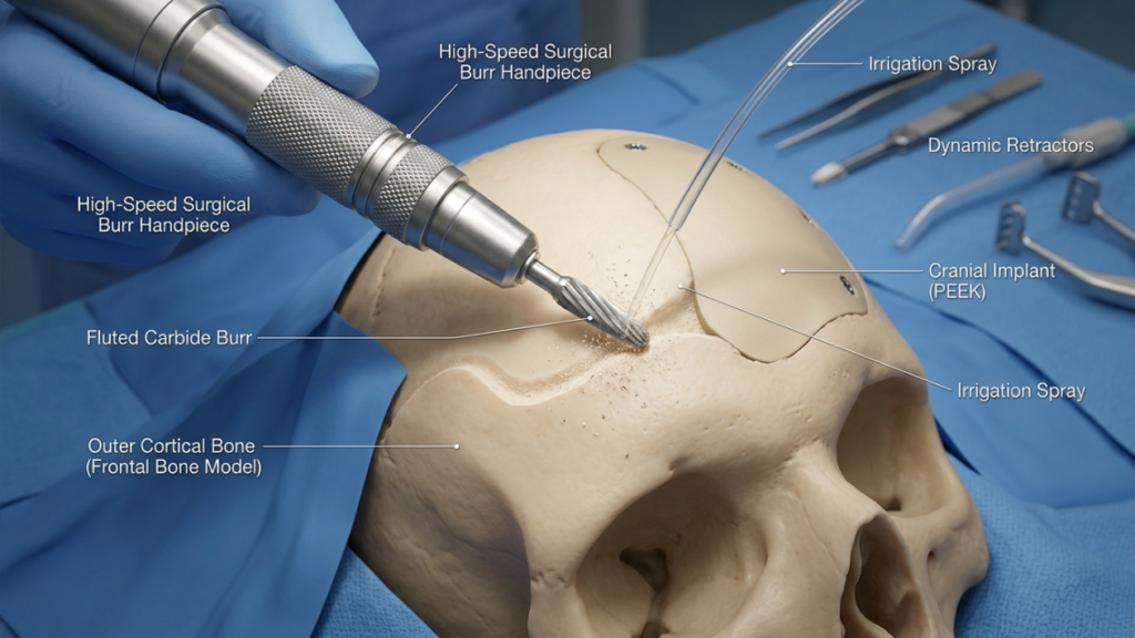
In Cranioplastie type 1, de chirurg Hierbij wordt een sneldraaiende frees gebruikt om het buitenste botweefsel weg te schaven, waardoor de prominentie wordt verminderd zonder de frontale sinus te beschadigen. Deze techniek is ideaal voor patiënten met dun voorhoofdsbeen of minimale bultvorming, omdat dit de integriteit van de sinus behoudt. Het is echter mogelijk niet voldoende voor patiënten met een aanzienlijke wenkbrauwbult of een dikke voorste botlaag, omdat overmatig frezen de botstabiliteit kan aantasten of het gewenste contour niet kan bereiken (Ousterhout, 2024).
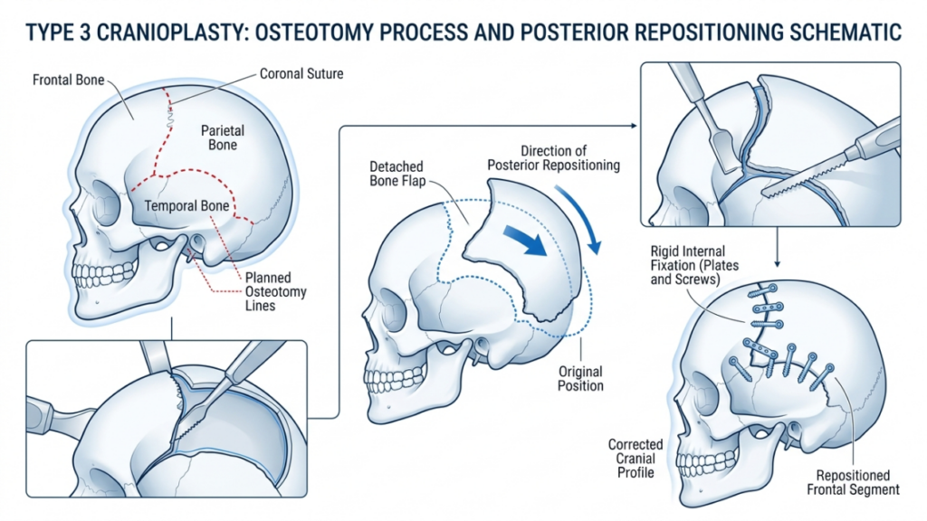
Daarentegen, Type 3 cranioplastie houdt een in osteotomie—een gecontroleerde snede door de voorste wand van de frontale sinus. Het botsegment wordt vervolgens naar achteren verplaatst (teruggezet) om de projectie te verminderen en een gladder, vrouwelijker voorhoofd te creëren. Deze techniek is voorbehouden aan patiënten met matige tot ernstige wenkbrauwuitstulping of een dikke voorste botlaag, waar alleen frezen onvoldoende zou zijn. De osteotomie maakt een precieze vormgeving mogelijk met behoud van de beschermende functie van de sinus (Feminization of the Forehead: A Scoping Literature Review, 2024).
Cranioplastiek type 1: afschaven en frezen
Techniekoverzicht
Type 1 cranioplastiek is de minst invasieve optie voor voorhoofdreconstructie. Hierbij wordt gebruik gemaakt van een hogesnelheidsfrees om de prominentie van het voorhoofdsbeen geleidelijk te verminderen. chirurg De buitenste cortexlaag wordt zorgvuldig afgeschoren, waarbij penetratie in de frontale sinus wordt vermeden. Deze techniek is met name effectief voor patiënten met:
- Lichte tot matige wenkbrauwuitstulping
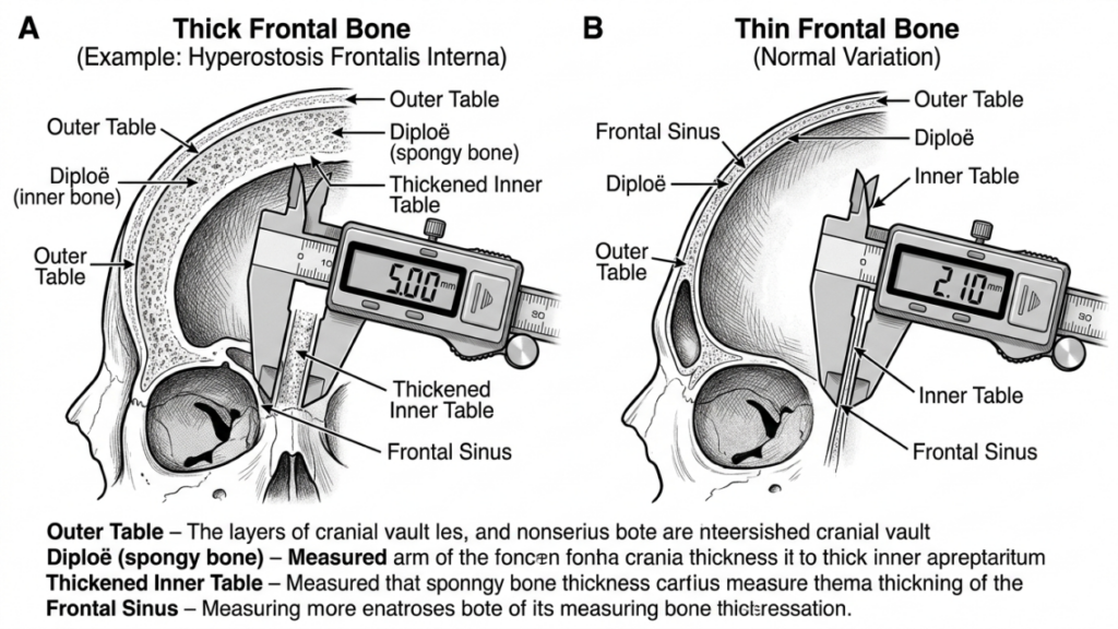
- Dun voorhoofdsbeen (minder dan 5 mm)
- Afwezigheid van significante pneumatisatie van de frontale sinus
De procedure wordt uitgevoerd via een coronale insnijding, Dit maakt toegang tot het voorhoofd mogelijk en minimaliseert tegelijkertijd zichtbare littekens. De chirurg gebruikt tactiele feedback en visuele aanwijzingen om een gelijkmatige reductie te garanderen zonder het bot te veel te verdunnen, wat zou kunnen leiden tot instabiliteit of onregelmatigheden in de contouren.
Voordelen van cranioplastie type 1
Cranioplastiek type 1 biedt diverse voordelen:
- Minimale invasiviteit: Door geen osteotomie of botverwijdering uit te voeren, wordt de chirurgische belasting en de hersteltijd verminderd.
- Lager risico op complicaties: Het behoud van de voorhoofdsholte minimaliseert het risico op sinusitis, lekkage van hersenvocht of de vorming van een mucocele.
- Kortere operatietijd: De procedure duurt doorgaans 1-2 uur, waardoor het een snellere optie is in vergelijking met type 3.
- Voorspelbare resultaten: Ideaal voor patiënten met een lichte botuitstulping, waarbij subtiele contourcorrectie voldoende is om een vrouwelijke uitstraling te creëren.
Beperkingen van cranioplastie type 1
Hoewel cranioplastiek type 1 veiliger en minder ingrijpend is, kent deze wel enkele belangrijke beperkingen:
- Beperkte reductie: Niet geschikt voor patiënten met ernstig uitpuilende wenkbrauwen of dikke voorhoofdsbeenderen.
- Risico op overmatige uitdunning: Door agressief frezen kan het bot verzwakken, wat kan leiden tot onregelmatigheden in de contouren of breuken.
- Onvolledige feminisering: Het gewenste esthetische resultaat wordt mogelijk niet bereikt bij patiënten met uitgesproken mannelijke gelaatstrekken.
Cranioplastiek type 3: Osteotomie en terugplaatsing
Techniekoverzicht
Type 3 cranioplastiek is een complexere procedure, bedoeld voor patiënten met matige tot ernstige wenkbrauwuitstulping of dikke voorhoofdsbeenderen. Het betreft een osteotomie—een precieze snede door de voorste wand van de frontale sinus—gevolgd door het naar achteren verplaatsen van het botsegment (terugplaatsing). Deze techniek zorgt voor een aanzienlijke vermindering van de voorhoofdsprojectie en een vloeiendere, meer vrouwelijke contour.
De procedure wordt doorgaans uitgevoerd via een coronale insnijding, waardoor toegang wordt verkregen tot het voorhoofdsbeen en de sinussen. De chirurg gebruikt een sagittale zaag of piëzo-elektrisch apparaat Om de osteotomie te creëren, wordt ervoor gezorgd dat de snede de natuurlijke kromming van het voorhoofd volgt. Het botsegment wordt vervolgens teruggeplaatst en vastgezet met titanium platen en schroeven of resorbeerbare hechtingen. Deze techniek is ideaal voor patiënten met:
- Ernstige wenkbrauwuitstulping
- Dik voorhoofdsbeen (groter dan 5 mm)
- Aanzienlijke pneumatisatie van de frontale sinus
Een cranioplastie van type 3 vereist nauwgezette planning, vaak inclusief 3D virtuele chirurgische planning (VSP) om de osteotomie en terugplaatsing te simuleren. Dit garandeert precisie en minimaliseert het risico op complicaties zoals sinusbeschadiging of lekkage van hersenvocht (Virtual Surgical Planning in Gezichtsfeminisering van het bovenste deel van het gezicht, 2025).
Voordelen van cranioplastie type 3
Cranioplastiek type 3 biedt verschillende voordelen voor patiënten met uitgesproken mannelijke gelaatstrekken:
- Aanzienlijke contourvorming: Vermindert opvallend veel wenkbrauwbultjes, waardoor een gladder en vrouwelijker voorhoofd ontstaat.
- Veelzijdigheid: Geschikt voor patiënten met dikke voorhoofdsbeenderen of uitgebreide sinuspneumatisatie.
- Stabiliteit op lange termijn: Het verplaatste botsegment integreert goed, waardoor het risico op contourafwijkingen op lange termijn wordt verminderd.
- Aanpassing: Virtuele chirurgische planning maakt nauwkeurige, patiëntspecifieke aanpassingen mogelijk om optimale resultaten te bereiken.
Beperkingen van cranioplastie type 3
Ondanks de effectiviteit ervan, brengt cranioplastiek type 3 hogere risico's en complexiteit met zich mee:
- Langere operatietijd: De procedure duurt doorgaans 3 tot 5 uur en vereist meer precisie en expertise.
- Hoger risico op complicaties: Mogelijke risico's zijn onder andere sinusitis, lekkage van hersenvocht of de vorming van een mucocele als de sinus beschadigd raakt.
- Langer herstel: Patiënten kunnen na een cranioplastie van type 1 langer last hebben van zwelling en ongemak.
- Kosten: Het gebruik van geavanceerde beeldvormingstechnieken en chirurgische instrumenten verhoogt de totale kosten van de ingreep.

Belangrijkste verschillen tussen cranioplastiek type 1 en type 3
| Functie | Cranioplastie type 1 | Type 3 Cranioplastie |
|---|---|---|
| Techniek | Botten afschuinen/schaven | Osteotomie en terugslag |
| Invasiviteit | Minimaal | Matig tot hoog |
| Ideale kandidaten | Lichte tot matige wenkbrauwuitstulping, dun voorhoofdsbeen | Ernstige wenkbrauwuitstulping, dik voorhoofdsbeen |
| Operatietijd | 1-2 uur | 3-5 uur |
| Hersteltijd | 1–2 weken | 3-6 weken |
| Risico op complicaties | Laag | Matig tot hoog |
| Kosten | Onder | Hoger |
| Esthetisch resultaat | Subtiele contouren | Dramatische feminisering |
Hoe chirurgen beslissen: Type 1 versus Type 3
De keuze tussen een cranioplastie van type 1 en type 3 hangt af van verschillende factoren, waaronder de patiënt. anatomische kenmerken, esthetische doelen, En chirurgische risico's. Chirurgen maken gebruik van een combinatie van klinisch onderzoek, 3D-beeldvorming, En patiëntconsultatie om de meest geschikte techniek te bepalen.
1. Anatomische beoordeling
De eerste stap is het evalueren van de patiënt. dikte van het voorhoofdsbeen En anatomie van de frontale sinus. A CT-scan of 3D-reconstructie biedt gedetailleerde inzichten in:
- Botdikte: Patiënten met dunne voorhoofdsbeenderen (minder dan 5 mm) zijn beter geschikt voor cranioplastiek type 1, terwijl patiënten met dikkere botten mogelijk een cranioplastiek type 3 nodig hebben.
- Sinuspneumatisatie: Uitgebreide sinuspneumatisatie kan een cranioplastiek van type 3 noodzakelijk maken om te voorkomen dat de sinus tijdens het frezen wordt beschadigd.
- Mate van leiderschap: Bij een ernstige wenkbrauwuitstulping is vaak een drastische vermindering nodig, die alleen mogelijk is met technieken van type 3.
2. Esthetische doelen
De esthetische verwachtingen van de patiënt spelen een cruciale rol bij de keuze van de techniek. Patiënten die op zoek zijn naar een dergelijke behandeling, hebben hier een specifieke voorkeur voor. subtiele feminisering Sommige personen kiezen mogelijk voor een cranioplastie van type 1, terwijl personen met uitgesproken mannelijke gelaatstrekken vaak een cranioplastie van type 1 nodig hebben. transformatieve resultaten van type 3. Chirurgen bespreken realistische resultaten op basis van de anatomie van de patiënt en de gewenste veranderingen.
3. Chirurgische risico's en herstel
Cranioplastiek type 3 brengt hogere risico's met zich mee, waaronder sinuscomplicaties, lekkage van hersenvocht, En langdurig herstel. Chirurgen beoordelen de algehele gezondheid van de patiënt, diens tolerantie voor de operatie en de bereidheid om de nazorg na te leven. Patiënten met medische aandoeningen die de risico's van de operatie verhogen, kunnen worden geadviseerd om een Type 1-procedure of een alternatieve procedure te overwegen.
4. Virtuele chirurgische planning (VSP)
Vooruitgang in 3D virtuele chirurgische planning hebben een revolutie teweeggebracht in de cranioplastiek. Chirurgen gebruiken VSP om osteotomieën, terugplaatsingen en resultaten te simuleren, waardoor precisie wordt gewaarborgd en risico's worden geminimaliseerd. Deze technologie is met name waardevol voor cranioplastiek type 3, waarbij nauwkeurige botrepositionering cruciaal is (3D Printing and Virtual Surgical Planning in Craniofacial and Orthognathic Surgery, 2025).
Postoperatieve zorg en herstel
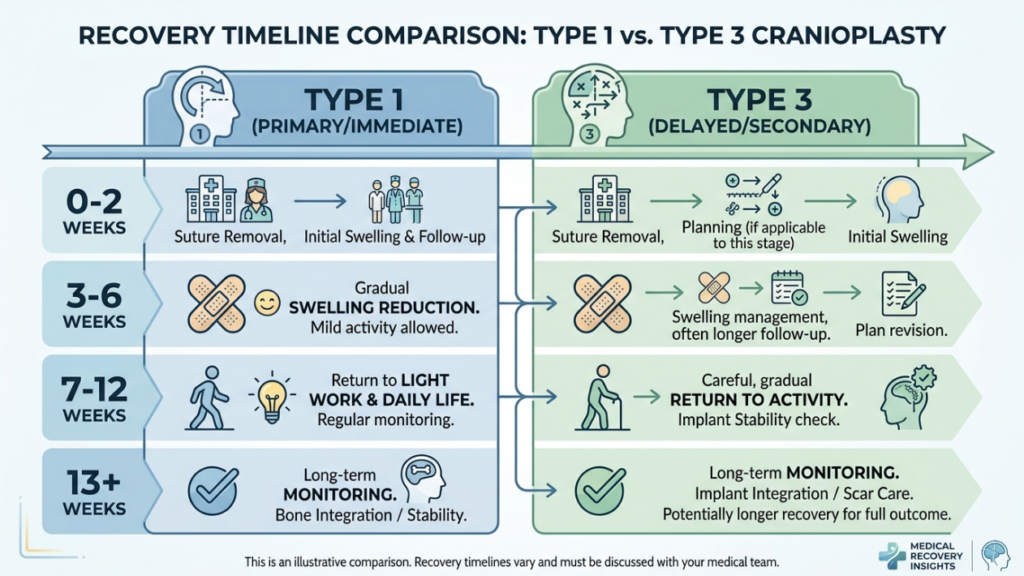
Het herstel verschilt aanzienlijk tussen cranioplastiek type 1 en type 3. Inzicht in het postoperatieve proces helpt patiënten zich voor te bereiden op een voorspoedig herstel.
Herstel na cranioplastie type 1
Patiënten die een cranioplastie van type 1 ondergaan, ervaren doorgaans het volgende:
- Lichte tot matige zwelling: Het probleem is binnen 1-2 weken opgelost.
- Minimaal ongemak: Te behandelen met vrij verkrijgbare pijnstillers.
- Snelle terugkeer naar activiteiten: De meeste patiënten hervatten binnen twee weken hun normale activiteiten.
Herstel na cranioplastie type 3
Het herstel na een cranioplastie van type 3 is complexer vanwege de ingewikkeldheid van de ingreep:
- Significante zwelling en kneuzing: Kan 3-4 weken aanhouden.
- Matige pijn: Mogelijk zijn pijnstillers op recept nodig gedurende de eerste week.
- Geleidelijke terugkeer naar activiteiten: Zware inspanningen zijn gedurende 4 tot 6 weken beperkt.
- Vervolgbezoeken: Regelmatige controle is essentieel voor een goede genezing en om eventuele complicaties tijdig aan te pakken.
Bij beide technieken moeten patiënten het volgende vermijden: zwaar tillen, zware inspanning, En directe druk op het voorhoofd Tijdens de eerste herstelfase geven chirurgen gedetailleerde postoperatieve instructies, waaronder wondverzorging, beperkingen ten aanzien van activiteiten en tekenen van complicaties waar men op moet letten.
Mogelijke complicaties en hoe u ze kunt vermijden
Hoewel cranioplastiek over het algemeen veilig is, kunnen er complicaties optreden. Kennis van deze risico's en preventieve maatregelen is cruciaal voor zowel chirurgen als patiënten.
Veelvoorkomende complicaties
- Sinusitis: Ontsteking of infectie van de frontale sinus, met name bij cranioplastiek type 3 als de sinus beschadigd raakt.
- Lekkage van hersenvocht (CSF): Een zeldzame maar ernstige complicatie als de achterste wand van de sinus wordt doorbroken.
- Onregelmatigheden in contouren: Overmatige botontkalking bij type 1 of onjuiste terugplaatsing bij type 3 kan leiden tot asymmetrie of zichtbare richels.
- Mucocele-vorming: Een blokkade van de afvoerwegen van de sinussen kan leiden tot met slijm gevulde cysten.
- Infectie: Het risico is hoger bij type 3 vanwege de langere operatietijd en het gebruik van implantaten.
Preventieve maatregelen
Chirurgen hanteren verschillende strategieën om complicaties te minimaliseren:
- Nauwkeurige chirurgische planning: 3D-beeldvorming en virtuele chirurgische planning zorgen voor nauwkeurige osteotomieën en terugplaatsingen.
- Behoud van de sinussen: Het is belangrijk om beschadiging van de voorhoofdsholte te voorkomen tijdens het frezen of osteotomiseren.
- Antibiotische profylaxe: Wordt vóór en na de operatie toegediend om het risico op infecties te verminderen.
- Postoperatieve monitoring: Regelmatige controles om vroegtijdige tekenen van complicaties op te sporen.
- Patiënteneducatie: Patiënten instrueren over de juiste wondverzorging, beperkingen in activiteiten en waarschuwingssignalen voor complicaties.
Alternatieven voor cranioplastiek type 1 en type 3
Voor patiënten die niet de ideale kandidaten zijn voor een cranioplastiek van type 1 of type 3, kunnen alternatieve technieken worden overwogen:
- Vettransplantatie: Autologe vettransplantatie kan de contouren van het voorhoofd verzachten zonder de botstructuur te veranderen. Dit is ideaal voor patiënten met lichte onregelmatigheden of voor diegenen die een niet-chirurgische oplossing zoeken.
- Implantaten op maat: Geprefabriceerde implantaten, zoals PEEK (polyetheretherketon) of titanium, Deze instrumenten kunnen worden gebruikt om het voorhoofd te vergroten of te hervormen zonder osteotomie. Ze zijn met name nuttig voor patiënten met dunne botten of sinusproblemen.
- Orthognatische chirurgie: In gevallen waarin voorhoofd contouren maakt deel uit van een breder geheel gezichtsfeminisering In het kader van een kaakcorrectie kunnen orthognatische procedures (zoals een Le Fort I osteotomie) worden gecombineerd om de uitlijning van het middengezicht en de kaak te verbeteren.
- Endoscopische technieken: Minimaal invasieve endoscopische methoden kunnen een uitstulping van de wenkbrauwen verminderen door middel van kleinere incisies en een sneller herstel.
Patiëntgetuigenissen en resultaten uit de praktijk
Resultaten uit de praktijk bieden waardevolle inzichten in de effectiviteit en tevredenheidscijfers van cranioplastiek type 1 en type 3. Patiëntgetuigenissen benadrukken de transformerende impact van deze ingrepen:
Casestudie 1: Cranioplastie type 1
Een 28-jarige transgender vrouw wilde haar voorhoofd subtiel feminiseren. Met een dun voorhoofdsbeen en minimale uitstulping koos ze voor een cranioplastiek type 1. De ingreep resulteerde in een gladdere contour met minimale hersteltijd. Ze gaf aan zeer tevreden te zijn en dat het resultaat overeenkwam met haar verwachtingen van een natuurlijke, vrouwelijke uitstraling.
Casestudie 2: Cranioplastie type 3
Een 35-jarige transgender vrouw presenteerde zich met een ernstig uitstekende wenkbrauw en een dik voorhoofdsbeen. Er werd een cranioplastiek type 3 uitgevoerd, waarbij een osteotomie en terugplaatsing plaatsvonden. De drastische vermindering van de voorhoofdsprojectie gaf haar gelaatstrekken een aanzienlijk vrouwelijker uiterlijk. Hoewel het herstel langer duurde, was ze zeer tevreden met het resultaat en verklaarde ze dat de ingreep "haar leven had veranderd".“
Casestudie 3: Revisiechirurgie
Een 40-jarige patiënte onderging aanvankelijk een cranioplastiek type 1, maar was ontevreden over het subtiele resultaat. Later koos ze voor een cranioplastiek type 3 om een meer uitgesproken feminisering te bereiken. De revisieoperatie verhielp haar bezwaren met succes en toonde het belang aan van het kiezen van de juiste techniek op basis van anatomische behoeften en esthetische doelen.
De rol van virtuele chirurgische planning bij cranioplastiek
Virtuele chirurgische planning (VSP) is een revolutionaire ontwikkeling in de cranioplastiek, met name voor type 3-procedures. VSP stelt chirurgen in staat om:
- Osteotomieën simuleren: Plan de botdoorsnijdingen en -herpositionering nauwkeurig om een optimale contour te bereiken.
- Voorspel de uitkomst: Visualiseer de resultaten na de operatie en pas het operatieplan daarop aan.
- Minimaliseer risico's: Vermijd kritieke structuren zoals de voorhoofdsholte en de bovenoogzenuwen.
- Verbeter de communicatie: Deel 3D-modellen met patiënten om realistische verwachtingen te scheppen en de geïnformeerde toestemming te verbeteren.
Uit onderzoek is gebleken dat VSP de operatietijd verkort, de nauwkeurigheid verbetert en de patiënttevredenheid verhoogt (3D Printing and Virtual Surgical Planning in Craniofacial and Orthognathic Surgery, 2025). Het wordt nu beschouwd als de gouden standaard voor complexe cranioplastiekprocedures.

Veelgestelde vragen
Wat is het belangrijkste verschil tussen een cranioplastie van type 1 en type 3?
Bij een cranioplastie type 1 wordt de buitenste laag van het voorhoofdsbeen afgeschaafd of afgevlakt om de prominentie te verminderen, terwijl bij een cranioplastie type 3 een osteotomie (botdoorsnijding) en terugplaatsing van het voorhoofdsbeensegment nodig zijn om een meer uitgesproken contour te bereiken. Type 1 is minder ingrijpend en geschikt voor milde tot matige bulten, terwijl type 3 is voorbehouden aan ernstige bulten of dikke voorhoofdsbeenderen.
Hoe maken chirurgen de keuze tussen een cranioplastie van type 1 en type 3?
Chirurgen beoordelen de dikte van het voorhoofdsbeen, de mate van wenkbrauwuitstulping en de anatomie van de voorhoofdsholten van de patiënt met behulp van CT-scans en 3D-beeldvorming. Type 1 wordt gekozen bij dunnere botten en een lichte uitstulping, terwijl Type 3 de voorkeur heeft bij dikkere botten, een ernstige uitstulping of uitgebreide pneumatisatie van de voorhoofdsholten. De doelen van de patiënt en de bereidheid tot het nemen van chirurgische risico's spelen ook een rol bij de beslissing.
Welke risico's zijn verbonden aan een cranioplastie type 3?
Een cranioplastie type 3 brengt vanwege de complexiteit hogere risico's met zich mee, waaronder sinusitis, lekkage van hersenvocht, contourafwijkingen, mucocelevorming en infectie. Deze risico's worden geminimaliseerd door nauwkeurige chirurgische planning, behoud van de sinussen en postoperatieve monitoring. Patiënten worden geïnformeerd over waarschuwingssignalen en nazorg om een voorspoedig herstel te garanderen.
Hoe lang duurt het herstel na een cranioplastie van type 1 vergeleken met type 3?
Het herstel na een cranioplastie van type 1 duurt doorgaans 1-2 weken, met lichte zwelling en ongemak. Een cranioplastie van type 3 vereist een langer herstel van 3-6 weken vanwege de uitgebreidere ingreep en mogelijke complicaties. Patiënten wordt geadviseerd zware inspanningen te vermijden en de postoperatieve instructies nauwgezet op te volgen.
Kan een cranioplastie van type 1 dezelfde resultaten opleveren als een cranioplastie van type 3?
Nee, een cranioplastie type 1 beperkt zich tot subtiele contourcorrecties en is ideaal bij een lichte tot matige bult op het voorhoofd. Cranioplastie type 3 bereikt een meer dramatische feminisering door het herpositioneren van het voorhoofdsbeensegment, waardoor het geschikt is voor een ernstige bult op het voorhoofd of een dik voorhoofdsbeen. De keuze hangt af van de anatomische behoeften en esthetische doelen van de patiënt.
Welke rol speelt virtuele chirurgische planning bij cranioplastiek?
Virtuele chirurgische planning (VSP) stelt chirurgen in staat osteotomieën te simuleren, de uitkomst te voorspellen en risico's te minimaliseren door kritieke structuren zoals de voorhoofdsholte te vermijden. Het verbetert de precisie, verkort de operatietijd en verhoogt de patiënttevredenheid door een duidelijke visualisatie van het chirurgische plan en de verwachte resultaten te bieden.
Zijn er niet-chirurgische alternatieven voor cranioplastiek om het voorhoofd te feminiseren?
Ja, niet-chirurgische alternatieven zijn onder andere: vet enten Om de contouren te verzachten en op maat gemaakte implantaten (bijvoorbeeld PEEK of titanium) te gebruiken om het voorhoofd te hervormen zonder osteotomie. Deze opties zijn ideaal voor patiënten met lichte onregelmatigheden of voor degenen die een operatie liever vermijden. Ze bereiken echter mogelijk niet hetzelfde niveau van feminisering als chirurgische technieken.
Wat kan ik verwachten tijdens het consult voor een voorhoofdreconstructie?
Tijdens het consult beoordeelt uw chirurg de dikte van uw voorhoofdsbeen, de anatomie van uw sinussen en de mate van botuitstulping aan de hand van een klinisch onderzoek en 3D-beeldvorming. Hij of zij bespreekt uw esthetische wensen, legt de verschillen tussen cranioplastiek type 1 en type 3 uit en adviseert de meest geschikte techniek op basis van uw anatomie en verwachtingen.







