
Et si la décision la plus importante concernant votre intervention de féminisation du front n'avait rien à voir avec le remodelage osseux, mais tout à voir avec l'emplacement de la zone à traiter ? chirurgien Quelle incision choisir ? La plupart des patients passent des heures à se renseigner sur les différents types de fraises, les classifications du front de type III et les techniques de réduction du sinus frontal. Pourtant, l'incision choisie pour accéder à l'arcade sourcilière détermine la visibilité de la cicatrice, la trajectoire de la ligne des cheveux, la récupération sensorielle et même si… chirurgien peut atteindre correctement les rebords orbitaires latéraux. Accès coronal vs trichophytique vs endoscopique au front La décision prise constitue le fondement architectural sur lequel repose toute autre manœuvre chirurgicale. Un mauvais choix, et même une intervention osseuse impeccable peut laisser une cicatrice visible, une calvitie naissante ou une insensibilité permanente du cuir chevelu.
Après avoir réalisé des centaines de féminisation faciale procédures à Dr MFO Clinique à Antalya, J'ai constaté à quel point le choix de l'incision influence la satisfaction à long terme. Les patients arrivent obsédés par la réduction de quelques millimètres au niveau de l'arcade sourcilière. Ils repartent dévastés par une cicatrice qu'ils n'avaient jamais imaginée. Ce guide offre ce qu'aucune autre ressource ne propose : une comparaison détaillée, sous forme d'arbre de décision, des trois incisions d'accès au front. Vous pourrez ainsi aborder votre consultation avec des connaissances précises et concrètes. Vous découvrirez précisément les performances de chaque approche selon sept critères essentiels : qualité de l'exposition, préservation de la ligne capillaire, la visibilité de la cicatrice, les troubles sensoriels, le risque de perte de cheveux, la vitesse de récupération et l'accès du chirurgien aux rebords orbitaires — et vous utiliserez l'organigramme d'auto-évaluation inclus pour identifier votre incision idéale avant même de parler à un chirurgien.
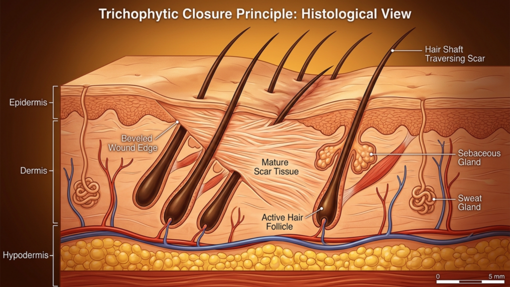
Table des matières
Anatomie de l'accès frontal : pourquoi le choix de l'incision détermine le résultat de votre chirurgie de féminisation faciale
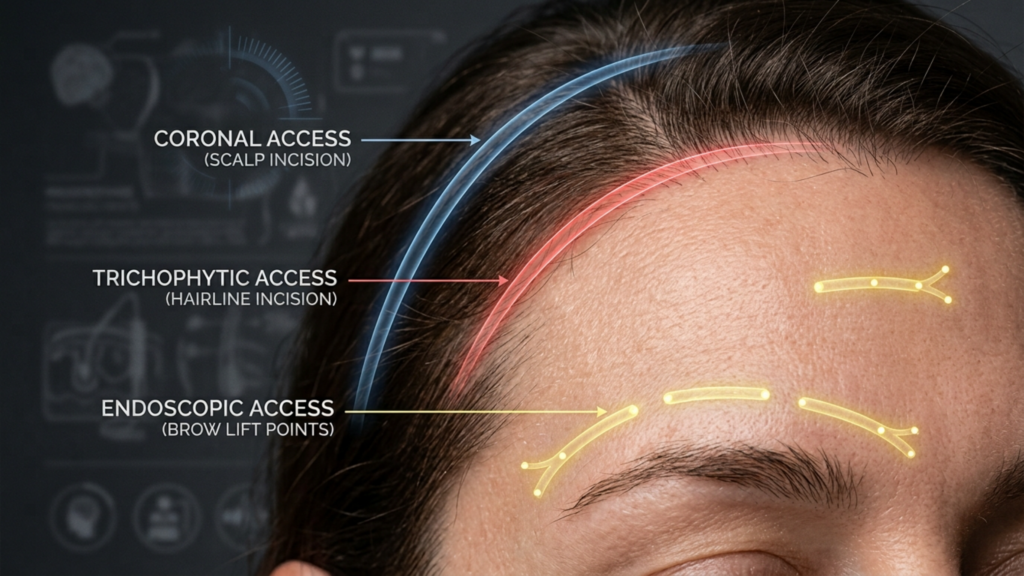
La chirurgie de féminisation du front nécessite un accès physique direct à l'os frontal, aux rebords orbitaires et aux sinus frontaux. Le chirurgien doit visualiser, manipuler et remodeler ces structures. Chaque type d'incision représente un compromis différent entre la surface du front visible et manipulable et les dommages collatéraux potentiels au cuir chevelu et à la ligne capillaire. Aucune technique n'offre une exposition maximale sans aucun impact esthétique. Incision coronale FFS Elle offre une exposition maximale, mais au prix d'une longue cicatrice et d'une importante altération de la sensibilité. L'approche endoscopique dissimule parfaitement les cicatrices, mais limite la zone accessible au chirurgien. L'incision trichophytique suit un trajet médian qui décale la cicatrice directement au niveau de la ligne des cheveux.
Un fait souvent surprenant pour les patients : les nerfs supra-orbitaire et supratrochléaire, responsables de la sensibilité du front et du cuir chevelu, traversent directement la zone opérée. Une incision coronale sectionne inévitablement les branches postérieures de ces nerfs sur toute la largeur du cuir chevelu. Les incisions endoscopiques préservent la plupart des trajets nerveux, car la dissection se fait sous le cuir chevelu au lieu de le couper. Cette simple réalité anatomique explique pourquoi les patients ayant subi une incision coronale rapportent une perte de sensibilité durant douze à dix-huit mois, tandis que ceux ayant bénéficié d’une incision endoscopique retrouvent souvent une sensibilité complète en six semaines. L’incision n’est pas qu’un simple point d’accès ; elle est le principal facteur déterminant de votre rétablissement fonctionnel.
Incision coronale FFS : Exposition maximale, compromis maximaux
L'incision coronale, également appelée approche bicoronale ou par lambeau coronal, consiste en une incision continue unique, d'une oreille à l'autre, au sommet du cuir chevelu, généralement quatre à six centimètres en arrière de la ligne des cheveux. Le chirurgien soulève ensuite l'ensemble du lambeau frontal vers le bas afin d'exposer l'os frontal, de la ligne des cheveux jusqu'aux rebords orbitaires. Cette technique est utilisée en chirurgie craniofaciale depuis des décennies car elle offre une visualisation exceptionnelle du champ opératoire.
Qualité de l'exposition et accès au bord orbitaire
L'approche coronale constitue la référence absolue pour exposition de l'arcade sourcilière. Lorsque le lambeau frontal est entièrement abaissé, le chirurgien bénéficie d'une visualisation directe et continue des deux rebords orbitaires latéraux, de la glabelle, du sinus frontal et des fosses temporales. Pour les fronts de type III nécessitant un recul important de la paroi sinusale et une réduction osseuse significative, ce niveau d'exposition est irremplaçable. Le chirurgien peut positionner les instruments sous n'importe quel angle, vérifier la symétrie visuellement et en temps réel, et corriger les anomalies anatomiques inattendues sans être gêné par une visibilité réduite.
Préservation de la ligne frontale et risque de chute de cheveux
C’est ici que l’incision coronale révèle son aspect le plus tranchant. Comme elle se situe derrière la ligne frontale, cette dernière reste anatomiquement inchangée. Cependant, l’incision crée une zone de calvitie permanente le long de la suture. La fermeture sous tension, l’électrocoagulation et l’élargissement postopératoire de la cicatrice peuvent engendrer une bande de calvitie visible quatre à six centimètres derrière la ligne frontale. Chez les patients aux cheveux fins ou ayant une ligne frontale naturellement haute, cette zone glabre devient particulièrement visible. risque de lésion nerveuse du cuir chevelu elle est importante car l'incision coronale coupe bilatéralement les branches postérieures du complexe nerveux supra-orbitaire, provoquant un engourdissement de toute la partie supérieure du cuir chevelu qui peut persister pendant dix-huit mois ou plus.
Visibilité et cicatrisation
La cicatrice coronale se situe dans le cuir chevelu, la rendant invisible lorsque les cheveux sont longs et denses. Les patients aux cheveux épais peuvent la dissimuler facilement en quelques semaines. En revanche, ceux qui ont les cheveux fins, clairsemés ou dégarnis rencontrent plus de difficultés. L'approche coronale exige la convalescence la plus longue car l'ensemble du lambeau frontal doit être soulevé et redrapé. La dissection étendue engendre un œdème et des ecchymoses importants qui mettent généralement trois à quatre semaines à se résorber visiblement. La récupération complète de la sensibilité est nettement plus longue, certains patients rapportant des zones de sensibilité diminuée permanentes le long de la ligne d'incision.
Ligne capillaire par incision trichophytique : le compromis de la frontière
L'incision trichophytique consiste à pratiquer l'incision précisément au niveau de la ligne frontale, à la limite anatomique où la peau du front rencontre le cuir chevelu. Le chirurgien biseaute l'incision, sectionnant les follicules pileux de sorte que les cheveux repoussent à travers et devant la cicatrice. Maîtrisée, cette technique permet d'obtenir une cicatrice si bien camouflée qu'elle devient pratiquement invisible, même avec les cheveux tirés en arrière.
Qualité de l'exposition et accès au bord orbitaire
L'approche trichophytique offre une exposition quasi identique à celle de la technique coronale. L'incision étant pratiquée au niveau de la ligne d'implantation des cheveux et non en arrière, le chirurgien rabat le lambeau frontal de la même manière, permettant une large visualisation de l'os frontal et des deux rebords orbitaires. La principale différence réside dans le positionnement : l'incision trichophytique déplace la cicatrice d'une zone intracuir chevelu dissimulée vers le bord de la ligne d'implantation des cheveux. Pour les patients qui nécessitent simultanément modelage du front et réduction du front— ce qui signifie que la ligne frontale doit être avancée — l'incision trichophytique n'est pas simplement un choix raisonnable. C'est la seule option logique car elle permet au chirurgien d'enlever l'excès de peau du front et de déplacer la ligne frontale vers l'avant tout en accédant à l'os sous-jacent.
Préservation de la ligne frontale et risque de chute de cheveux
préservation de la ligne capillaire FFS La méthode trichophytique donne d'excellents résultats lorsqu'elle est pratiquée par un chirurgien expérimenté. L'incision biseautée permet aux follicules pileux situés sur le bord supérieur de continuer à faire pousser les cheveux vers l'avant, à travers la cicatrice. Cependant, les incisions trichophytiques présentent un risque particulier et souvent sous-estimé : si la plaie est refermée sous tension – ce qui est fréquent lors d'une avancée importante de la ligne frontale – une nécrose folliculaire peut survenir de part et d'autre de l'incision. Il en résulte une bande de calvitie étroite précisément au niveau de la ligne frontale, qui devient paradoxalement la zone la plus visible. J'ai repris le traitement de nombreux patients provenant d'autres cliniques chez lesquels une avancée trop importante avait provoqué une alopécie cicatricielle le long de la ligne d'incision, nécessitant une greffe de cheveux ultérieure pour restaurer la ligne frontale.
Visibilité des cicatrices et troubles sensoriels
La cicatrice trichophytique se situe à la jonction des cheveux. Lorsque les cheveux repoussent à travers la cicatrice comme prévu, celle-ci devient extrêmement bien dissimulée. Cependant, elle reste visible pendant les trois à quatre premiers mois de cicatrisation, avant que les cheveux ne pénètrent le tissu cicatriciel. Les patients doivent comprendre ce délai et se préparer à une période durant laquelle dissimuler l'incision nécessitera des ajustements de coiffure. La perte de sensibilité est similaire à celle observée lors de l'abord coronal au niveau du front, car les mêmes branches nerveuses sont sectionnées lors du soulèvement du lambeau. Toutefois, la partie postérieure du cuir chevelu conserve sa sensibilité, car l'incision ne s'étend pas jusqu'à cette zone. Les patients décrivent généralement un engourdissement limité au front, le cuir chevelu situé derrière l'incision restant sensible.
Accès endoscopique au front : la voie minimaliste aux contraintes maximales
L'abord endoscopique du front nécessite deux à cinq petites incisions, de un à deux centimètres chacune, pratiquées dans le cuir chevelu. Le chirurgien insère un endoscope et des instruments par ces orifices, disséquant sous le périoste frontal pour accéder à l'os frontal et aux rebords supra-orbitaires. Cette technique est devenue populaire car elle élimine la longue cicatrice et réduit considérablement les lésions nerveuses.
Qualité de l'exposition : la limite critique
Accès endoscopique au front Cette technique offre une visualisation fondamentalement différente des approches chirurgicales ouvertes. Le chirurgien visualise le champ opératoire à travers une caméra, travaillant dans un tunnel sous-périosté qui limite la mobilité des instruments et restreint l'arc de dissection. Si l'endoscope offre une excellente visibilité du front et de la glabelle, l'accès aux rebords orbitaires latéraux – les arcades osseuses encadrant les coins externes des yeux – est limité. Pour les fronts de type I et II nécessitant un remodelage sans intervention majeure sur les sinus, l'accès endoscopique est souvent suffisant. En revanche, pour les fronts de type III exigeant un recul de la paroi du sinus frontal avec une résection osseuse importante, l'approche endoscopique contraint le chirurgien à opérer par de minuscules orifices, ce qui allonge la durée de l'intervention et limite la précision du remodelage osseux au niveau des rebords orbitaires.
Une nuance essentielle : l’accès endoscopique est plus efficace lorsqu’il est combiné à d’autres procédures endoscopiques. Les patients subissant simultanément lifting temporal endoscopique Les interventions sur la partie médiane du visage bénéficient de l'utilisation de ports d'accès partagés. La stratégie d'incision devient efficace plutôt que contraignante lorsque plusieurs régions anatomiques nécessitent un traitement par le même champ opératoire.
Préservation de la ligne frontale, risque de chute de cheveux et impact sensoriel
L'approche endoscopique excelle dans la préservation de la ligne frontale. Les petites incisions pratiquées dans le cuir chevelu entraînent une chute de cheveux négligeable, généralement moins de vingt follicules par port. La position et la forme de la ligne frontale restent inchangées. risque de lésion nerveuse du cuir chevelu Le risque est considérablement réduit car la dissection se déroule sous le périoste sans sectionner les principaux troncs nerveux. La plupart des patients opérés par endoscopie rapportent un léger engourdissement du front qui disparaît en quatre à six semaines. Cette récupération sensorielle rapide constitue le principal avantage fonctionnel de cette approche et fait de l'accès endoscopique le choix privilégié pour les patients dont la préoccupation première est de minimiser les perturbations de leur anatomie.
Visibilité des cicatrices et vitesse de cicatrisation
Les cicatrices endoscopiques sont totalement invisibles sous le cuir chevelu. Chaque incision d'un centimètre cicatrise en une minuscule marque, imperceptible même sous les cheveux courts. La récupération est plus rapide qu'avec les approches coronale et trichophytique, car la dissection est limitée. La plupart des patients reprennent des activités non physiques en sept à dix jours. L'œdème se résorbe plus vite, car l'importante perturbation sous-cutanée des approches ouvertes est absente. Le compromis demeure : on bénéficie d'une récupération rapide et de cicatrices invisibles, mais l'accès chirurgical est restreint, ce qui peut compromettre la précision du remodelage osseux, notamment au niveau de la structure latérale du sourcil.
Comparaison des incisions de féminisation du front : La matrice à sept dimensions
Aucune ressource existante ne compare les trois types d'incision en fonction de toutes les variables critiques. Vous trouverez ci-dessous le cadre comparatif que j'utilise dans ma pratique lorsque je discute de la stratégie d'incision avec mes patients. Chaque dimension est évaluée sur une échelle de cinq points, cinq représentant le résultat le plus favorable et un le résultat le moins favorable pour le patient.
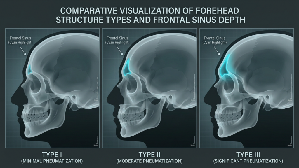
Tableau comparatif : Sept dimensions cliniques
| Dimension clinique | Incision coronale | Incision trichophytique | Incision endoscopique |
|---|---|---|---|
| Qualité de l'exposition | ★★★★★ (Vue panoramique complète) | ★★★★☆ (Quasi identique à la coronaire) | ★★★☆☆ (Tunnelé, dépend de la caméra) |
| Préservation de la ligne capillaire | ★★★★☆ (Position stable ; risque de calvitie) | ★★★☆☆ (Avance la ligne frontale ; risque d’alopécie de tension) | ★★★★★ (Ligne des cheveux totalement intacte) |
| Visibilité des cicatrices | ★★★★☆ (Masqué si les cheveux sont denses) | ★★★★☆ (Les cheveux repoussent à travers la cicatrice) | ★★★★★ (Incisions minuscules, totalement invisibles) |
| Troubles sensoriels | ★★☆☆☆ (Engourdissement pendant 12 à 18 mois et plus) | ★★★☆☆ (Engourdissement du front pendant 6 à 12 mois) | ★★★★☆ (Engourdissement léger pendant 4 à 6 semaines) |
| Risque de chute de cheveux | ★★★☆☆ (Bande chauve le long de l'incision) | ★★★☆☆ (Possibilité de perte de follicules périphériques) | ★★★★★ (Sauvetage folliculaire minimal) |
| Vitesse de récupération | ★★☆☆☆ (Gonflement visible pendant 3 à 4 semaines) | ★★★☆☆ (Gonflement visible pendant 2 à 3 semaines) | ★★★★☆ (gonflement visible pendant 1 à 2 semaines) |
| Accès au bord orbitaire | ★★★★★ (Direct, sans restriction) | ★★★★☆ (Direct, légère restriction du bord) | ★★☆☆☆ (Portée latérale limitée) |
Cette comparaison révèle pourquoi aucune incision ne domine toutes les dimensions. Les approches coronale et trichophytique excellent en termes d'exposition et d'accès au rebord orbitaire – priorités chirurgicales. L'approche endoscopique excelle quant à elle en termes de préservation de la ligne d'implantation des cheveux, de protection sensorielle et de rapidité de récupération – priorités liées à l'expérience du patient. vous choisissez doit correspondre à la dimension qui a le plus d'importance pour votre anatomie spécifique et vos priorités personnelles.
Exposition de l'arcade sourcilière : ce que votre chirurgien doit réellement atteindre
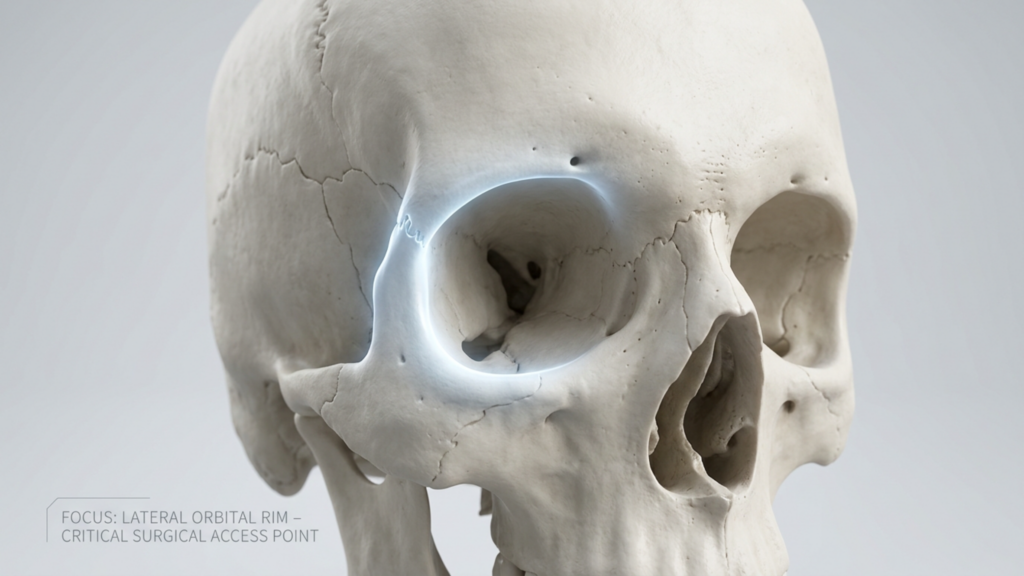
Les patients comprennent rarement l'importance cruciale de l'accès aux rebords orbitaires. Ces rebords latéraux, ces arcades osseuses situées aux angles externes des orbites, représentent la partie la plus large du front masculin. Lors d'une féminisation du front, il est essentiel de réduire ou de redessiner ces rebords pour obtenir un contour des sourcils plus doux et plus arrondi. Un accès insuffisant à cette zone entraîne un résultat malheureusement trop fréquent : un front féminisé au centre, avec des rebords orbitaires latéraux toujours proéminents, qui trahissent la structure osseuse masculine.

Par une incision coronale ou trichophytique, le chirurgien peut accéder aux rebords orbitaires latéraux grâce à une visualisation directe et une manipulation instrumentale dans tous les plans. Par voie endoscopique, les rebords latéraux se situent à la périphérie du champ opératoire. L'endoscope et les instruments doivent emprunter des tunnels étroits, et l'angle d'approche devient tangentiel plutôt que perpendiculaire. Pour des réductions mineures – arrondir le bord du rebord latéral plutôt qu'un remodelage osseux important – cet accès limité convient. En revanche, pour un remodelage orbitaire latéral conséquent, l'approche endoscopique implique des compromis en termes de précision, ce qui peut rendre la structure externe du sourcil insuffisamment féminisée. Demandez directement à votre chirurgien : « Quelle intervention sur les rebords orbitaires latéraux est nécessaire, et est-elle réalisable par voie endoscopique ? » Sa réponse honnête déterminera si l'accès endoscopique est approprié dans votre cas.
Risque de lésions nerveuses du cuir chevelu : un examen approfondi des conséquences sensorielles
Le nerf supra-orbitaire sort du crâne par le foramen ou l'échancrure supra-orbitaire, puis se ramifie au niveau du front et de la partie antérieure du cuir chevelu. Le nerf supratrochléaire se dirige médialement, innervant la partie centrale du front et la glabelle. Ces nerfs constituent l'axe sensitif essentiel du front. Une incision coronale au niveau du vertex du cuir chevelu sectionne les branches ascendantes de ces nerfs sur toute leur largeur. Les manuels d'anatomie décrivent cette lésion comme une atteinte nerveuse temporaire et attendue. Cependant, en pratique clinique, la récupération n'est ni garantie ni complète.
Environ 15 % des patients ayant subi une incision coronale présentent une altération sensorielle permanente : engourdissement persistant, hypersensibilité ou dysesthésie (picotements désagréables) au niveau du cuir chevelu fronto-pariétal. L’incision trichophytique présente également cette atteinte nerveuse centrale, car le soulèvement du lambeau sectionne des branches nerveuses au niveau de la ligne d’implantation des cheveux. L’accès endoscopique change radicalement la donne. La dissection sous-périostée préserve les troncs nerveux supra-orbitaire et supratrochléaire, car ces nerfs sont superficiels par rapport au plan de dissection. Les petites incisions pratiquées provoquent une irritation nerveuse mineure et localisée, plutôt qu’une atteinte étendue. Pour les patients qui privilégient l’intégrité sensorielle – et beaucoup n’en perçoivent l’importance qu’une fois perdue – l’approche endoscopique offre un profil de sécurité nettement supérieur.
Préservation de la ligne capillaire FFS : Le dilemme des cheveux clairsemés
Les patients aux cheveux épais et denses bénéficient d'une grande liberté dans le choix de l'incision, car toute cicatrice se camoufle efficacement. En revanche, les patients aux cheveux fins, clairsemés ou dégarnis sont confrontés à un choix beaucoup plus restreint. Pour ces derniers, la bande dégarnie laissée par l'incision coronale devient un véritable problème, la densité capillaire étant insuffisante pour la dissimuler. Le mécanisme de repousse des cheveux à travers la cicatrice, propre à l'incision trichophytique, dépend de la densité folliculaire existante : si la ligne frontale est déjà clairsemée, le nombre de follicules capables de pousser à travers la cicatrice est réduit. C'est précisément dans ce cas que l'approche endoscopique excelle : les petites incisions préservent quasiment tous les follicules et la ligne frontale reste parfaitement intacte.

Prenons l'exemple suivant : un patient présentant une calvitie de classe II selon l'échelle de Norwood subit une incision trichophytique avec une avancée de la ligne frontale de trois centimètres. La tension exercée lors de la fermeture étire davantage la ligne frontale déjà clairsemée, entraînant une perte folliculaire supplémentaire. Six mois après l'intervention, la ligne frontale a avancé, mais la cicatrice est désormais visible. Le patient nécessite alors une greffe de cheveux pour restaurer la densité capillaire — une seconde intervention qui aurait pu être évitée par une approche endoscopique. Je souligne ce cas de figure non pas pour dissuader la technique trichophytique, mais pour mettre en évidence que… préservation de la ligne capillaire FFS Cela nécessite d'évaluer non seulement l'emplacement de l'incision, mais aussi la façon dont les forces de fermeture affectent votre densité capillaire spécifique.
Arbre décisionnel relatif à l'incision FFS : un organigramme clinique pour l'auto-évaluation du patient
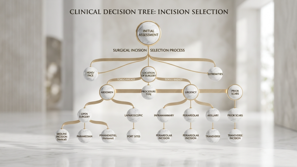
Ci-dessous figure l'arbre de décision que j'ai élaboré pour mon cabinet. Dr MFO Ce service permet de traduire l'interaction complexe des variables anatomiques en une auto-évaluation progressive qui guide les patients vers le type d'incision le plus approprié avant la consultation formelle. Répondez honnêtement aux questions, et la voie à suivre se dessinera d'elle-même.

Organigramme d'auto-évaluation étape par étape
Question 1 : Avez-vous besoin d'une réduction de la peau du front ou d'un avancement de la ligne des cheveux ?
- OUI → Passez à la question 2A.
- NON → Passez à la question 2B.
Question 2A : Vous avez besoin d’une avancée de la ligne frontale. Votre densité capillaire est-elle suffisante pour supporter la tension au niveau de la ligne de fermeture ?
- OUI → Incision trichophytique C'est votre option principale. Le chirurgien avancera la ligne des cheveux tout en accédant à l'os par la même incision.
- NON → Considérez un incision coronale avec un séparé greffe de cheveux séance pour abaisser la ligne des cheveux, évitant ainsi la chute folliculaire liée à la tension au niveau du bord de la ligne des cheveux.
Question 2B : Vous n’avez pas besoin d’une avancée de la ligne des cheveux. Votre front est-il de type III avec atteinte importante des sinus frontaux nécessitant un recul majeur ?
- OUI → Votre densité capillaire est-elle suffisante pour camoufler une cicatrice postérieure du cuir chevelu ?
- NON (Votre front est de type I ou de type II, nécessitant un remodelage sans recul majeur des sinus) → Passez à la question 3.
Question 3 : Vous avez un front de type I ou II. Avez-vous besoin d’un remodelage important du rebord orbitaire latéral ?
- OUI → Incision trichophytique ou coronale Elle permet au chirurgien un accès direct au bord latéral. L'accès endoscopique peut s'avérer insuffisant pour des interventions latérales importantes.
- NON → Un rétablissement rapide et une perturbation sensorielle minimale figurent-ils parmi vos trois principales priorités ?
Cet arbre décisionnel permet de faire le tri parmi les arguments marketing qui entourent chaque type d'incision. Chaque approche présente des avantages et des inconvénients réels. La solution idéale dépend de votre anatomie, de la forme de votre front, de la densité de vos cheveux et de vos priorités personnelles, et non des préférences d'un chirurgien.
Délai et rapidité de la reprise : à quoi s’attendre selon chaque approche
Le délai de convalescence influe directement sur votre capacité à reprendre le travail, à socialiser et à retrouver une vie normale. Comprendre le temps de convalescence requis pour chaque type d'incision vous aide à planifier de manière réaliste.

Récupération endoscopique : La plupart des patients reprennent leur travail de bureau dans les sept à dix jours. L'œdème atteint son maximum à quarante-huit heures et diminue sensiblement au bout de dix jours. La récupération sensorielle est complète en quatre à six semaines. La reprise d'une activité physique complète, sans restriction, est possible en trois semaines.
Récupération des trichophytes : La reprise du travail de bureau se fait généralement entre dix et quatorze jours après l'intervention. L'œdème atteint son maximum vers le troisième jour et diminue significativement en deux à trois semaines. La cicatrice au niveau de la ligne des cheveux devient la plus visible entre la deuxième et la sixième semaine, avant la repousse des cheveux. La récupération de la sensibilité s'étend sur six à douze mois pour la zone centrale du front. Le retour à une activité normale est possible en quatre semaines.
Rétablissement post-Covid : La reprise du travail de bureau nécessite deux à trois semaines en raison d'un œdème important du front et des troubles sensitifs étendus. L'œdème atteint son maximum entre le troisième et le cinquième jour et peut mettre quatre à six semaines à se résorber complètement. La récupération sensorielle s'étend sur douze à dix-huit mois, avec la possibilité de séquelles permanentes. La reprise d'une activité normale se fait généralement au bout de six semaines après validation par l'équipe chirurgicale.
Interventions combinées : lorsqu’une seule incision remplit plusieurs objectifs
L'efficacité chirurgicale est importante. Lorsqu'un patient a besoin modelage du front Lors d'un lifting temporal, d'un lifting frontal ou d'un rajeunissement du tiers supérieur du visage, la stratégie d'incision doit répondre simultanément à plusieurs objectifs. L'incision coronale facilite naturellement un lifting frontal, le décollement du lambeau frontal étant déjà réalisé. L'incision trichophytique s'associe parfaitement à l'avancement de la ligne capillaire, les deux objectifs – l'amélioration esthétique et l'accès chirurgical – partageant la même ligne d'incision. L'accès endoscopique se combine aisément aux interventions endoscopiques temporales et médiofaciales grâce à des incisions communes, permettant au chirurgien de traiter le front, les tempes et le tiers moyen du visage sans créer de cicatrices supplémentaires.
À Clinique Dr MFO, J'évalue systématiquement si la combinaison de plusieurs interventions par une incision unique permet d'obtenir de meilleurs résultats globaux que le traitement de chaque zone séparément. Un patient nécessitant un remodelage du front, une légère ptose des sourcils et un creux temporal peut tirer le meilleur parti d'une approche endoscopique qui traite les trois zones par les mêmes petits orifices. Un patient présentant une bosse frontale, une implantation haute des cheveux et une ptose importante des sourcils obtient les meilleurs résultats avec une incision trichophytique qui permet de remonter l'implantation des cheveux, d'accéder à l'os et de lifter le sourcil en une seule manœuvre coordonnée.
Maturation des cicatrices : la réalité de douze mois dont personne ne parle
Chaque cicatrice suit une trajectoire de maturation prévisible. La phase inflammatoire dure une à deux semaines, durant lesquelles la cicatrice apparaît rouge, en relief et ferme. La phase proliférative s'étend de la deuxième à la douzième semaine, période où la production de collagène atteint son maximum et où la cicatrice peut paraître plus épaisse et plus visible qu'au cours de la phase initiale de cicatrisation. La phase de remodelage se poursuit de trois à douze mois, durant laquelle la cicatrice s'assouplit, s'aplatit et s'estompe progressivement. Les patients qui évaluent leur cicatrice à six semaines – alors qu'elle est encore en pleine phase proliférative – s'inquiètent souvent inutilement. L'aspect réel d'une cicatrice du front ne peut être évalué qu'au bout de douze mois.
Cette réalité a des implications pratiques sur le choix de l'incision. Si vous optez pour une incision trichophytique, la cicatrice au niveau de la ligne des cheveux peut paraître préoccupante huit semaines après l'intervention. C'est normal. Les cheveux n'ont pas encore repoussé à travers la cicatrice et les tissus sont toujours en cours de remodelage. Attendre douze mois avant de se prononcer permet d'éviter une anxiété inutile et des retouches prématurées. Un patient qui comprend ce délai aborde l'opération avec des attentes réalistes, plutôt qu'avec le faux espoir d'une cicatrice à peine visible un mois plus tard.
Application pratique : Votre guide de choix d’incision en cinq étapes
Mettez ces connaissances en pratique. Suivez ces cinq étapes pour identifier et défendre le type d'incision idéal pour vous :
- Déterminez votre type de front. Obtenez une téléradiographie de profil ou un scanner 3D. Un radiologue ou un chirurgien doit déterminer si votre front présente un sinus de type I (bosse minime, paroi sinusale intacte), de type II (bosse avec atteinte sinusale superficielle) ou de type III (sinus frontal profond nécessitant un recul). Le type III requiert presque toujours une approche chirurgicale ouverte (coronale ou trichophytique). Les types I et II peuvent être traités par voie endoscopique ou ouverte selon d'autres facteurs.
- Mesurez la hauteur et la densité de votre ligne capillaire. Mesurez la distance entre le point le plus saillant de vos sourcils et votre ligne capillaire à l'aide d'une règle. Une mesure supérieure à six centimètres indique généralement un front haut nécessitant une avancée de la ligne capillaire, suggérant un front trichophytique. Évaluez la densité de vos cheveux en vérifiant si votre cuir chevelu est visible à travers vos cheveux au niveau du vertex et des tempes. Un cuir chevelu visible indique une faible densité capillaire, ce qui limite les options de camouflage des cicatrices et favorise l'accès endoscopique.
- Classez vos priorités personnelles par ordre de priorité. Notez les sept dimensions du tableau comparatif ci-dessus : exposition, préservation de la ligne frontale, visibilité de la cicatrice, troubles sensitifs, risque de chute de cheveux, rapidité de la récupération et accès au rebord orbitaire. Classez-les par ordre d’importance décroissante. Vos deux ou trois priorités principales vous indiqueront le type d’incision qui correspond le mieux à vos valeurs. Les patients qui accordent une importance primordiale à la préservation de la sensibilité et à la rapidité de la récupération privilégient généralement l’endoscopie. Ceux qui privilégient l’exposition et l’accès au rebord orbitaire privilégient quant à eux les techniques chirurgicales ouvertes.
- Parcourez l'arbre de décision ci-dessus. Répondez honnêtement à chaque question du diagramme en vous basant sur vos observations cliniques. Notez le résultat. Comparez-le ensuite à votre classement par ordre de priorité établi à l'étape trois. Si les deux résultats concordent, vous avez votre réponse. En cas de divergence, cette divergence fera l'objet de votre consultation chirurgicale.
- Présentez votre analyse lors de la consultation. Apportez à votre rendez-vous chez le chirurgien la classification de votre front, les mesures de votre ligne capillaire, votre classement par ordre de priorité et le résultat de votre arbre de décision. Cette préparation transforme l'échange d'informations passives en une prise de décision partagée et active. Un chirurgien respectueux de l'autonomie du patient appréciera ce niveau d'implication et adaptera sa recommandation en conséquence.
Choisir votre Accès coronal vs trichophytique vs endoscopique au front Le choix de l'incision ne relève pas d'une décision à déléguer entièrement. Votre anatomie définit les paramètres. Vos priorités orientent le choix. L'expertise de votre chirurgien assure la mise en œuvre du plan. Prenez la décision en main et vous serez responsable du résultat. Prêt(e) à déterminer quelle incision convient le mieux à votre anatomie ? Postulez dès maintenant pour une consultation personnalisée à la clinique du Dr MFO et bénéficiez d'une évaluation complète du front avec une recommandation d'incision personnalisée.
Questions fréquemment posées
Pourquoi le type d'incision est-il plus important que la technique de remodelage osseux dans la féminisation du front ?
L'incision détermine la qualité de l'accès chirurgical, la visibilité de la cicatrice, le risque de lésion nerveuse, le risque de chute de cheveux et le délai de convalescence. Même un remodelage osseux parfait ne peut compenser une incision mal choisie laissant une cicatrice visible ou une insensibilité permanente. L'incision est la base même du résultat final.
Comment savoir si j'ai besoin d'une incision coronale, trichophytique ou endoscopique pour une féminisation faciale ?
La classification de votre type de front, la hauteur de votre ligne frontale, la densité de vos cheveux et vos priorités personnelles déterminent la meilleure approche. Les fronts de type III avec atteinte des sinus profonds nécessitent généralement un abord coronal ou trichophytique. Les patients nécessitant un avancement de la ligne frontale bénéficient d'une incision trichophytique. Les fronts de type I ou II avec une bonne densité capillaire peuvent être candidats à un abord endoscopique.
Vais-je perdre des cheveux au niveau de l'incision après une féminisation du front ?
La chute de cheveux varie selon le type d'incision. Les incisions coronales présentent un risque de calvitie permanente le long de la suture. Les incisions trichophytiques peuvent entraîner la perte des follicules périphériques en cas de tension excessive lors de la fermeture. Les incisions endoscopiques sacrifient moins de vingt follicules par voie, rendant la chute de cheveux pratiquement négligeable chez la plupart des patients.
Combien de temps dure l'engourdissement du cuir chevelu après chaque type d'incision ?
Les patients ayant subi une endoscopie retrouvent généralement la sensibilité en quatre à six semaines. Ceux ayant subi une intervention trichophytique présentent un engourdissement du front pendant six à douze mois. Les patients ayant subi une intervention coronale souffrent d'un engourdissement du cuir chevelu pendant douze à dix-huit mois, et environ quinze pour cent d'entre eux présentent une altération sensorielle permanente.
Est-il possible de bénéficier d'une élévation de la ligne capillaire par une incision endoscopique ?
Non. Les incisions endoscopiques ne permettent pas d'avancer la ligne des cheveux car elles ne retirent pas de peau du front. L'avancement de la ligne des cheveux nécessite l'excision d'un excès de tissu frontal, ce qui requiert une incision trichophytique ou coronale permettant au chirurgien de retirer la peau et de repositionner la ligne des cheveux vers l'avant.
La cicatrice trichophytique est-elle visible lorsque les cheveux sont tirés en arrière ?
Durant les trois à quatre premiers mois, la cicatrice reste visible à la jonction des cheveux. Après la repousse des cheveux, environ quatre à six mois plus tard, sa visibilité diminue considérablement. Au bout de douze mois, la plupart des cicatrices trichophytiques bien réalisées deviennent très difficiles à détecter, même avec les cheveux tirés en arrière.
Que se passe-t-il si mon chirurgien ne peut pas atteindre les rebords orbitaires latéraux par voie endoscopique ?
Un remodelage insuffisant du rebord orbitaire latéral laisse apparaître des arcades sourcilières externes proéminentes, conservant une structure masculine malgré une féminisation du front central. Si un travail important sur le rebord latéral est nécessaire, votre chirurgien devrait recommander une approche ouverte (coronale ou trichophytique) plutôt que de compromettre le résultat du remodelage par un accès endoscopique limité.







