
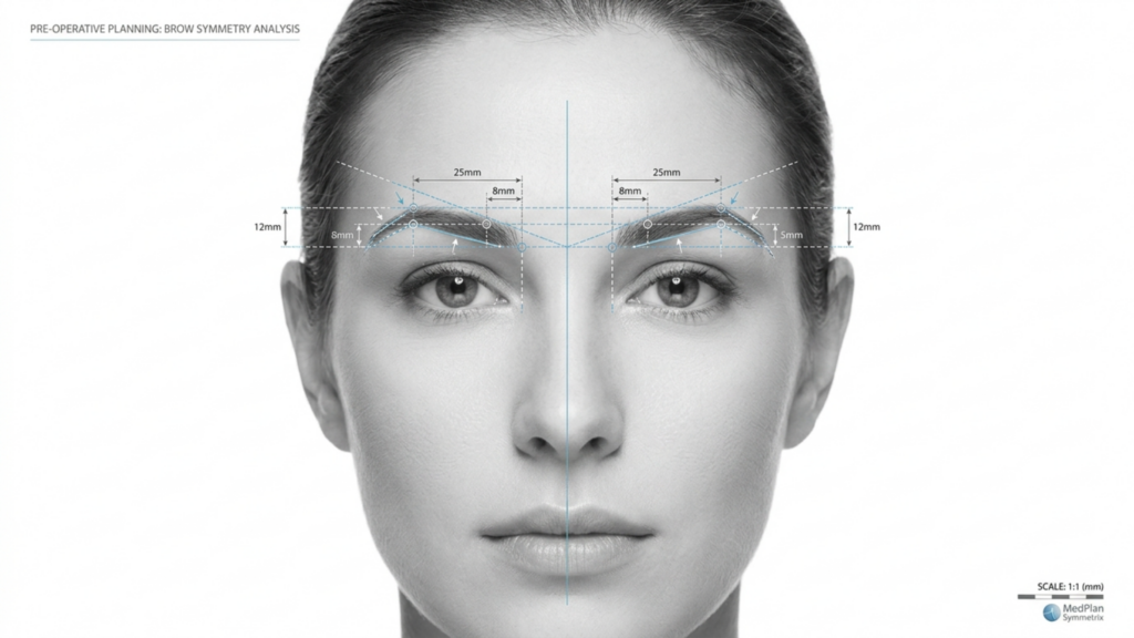
Dans le paysage en évolution de Chirurgie de féminisation faciale (FFS), le choix entre Type 2 et Tapez 3 frontoplastie Le choix de la technique est l'une des décisions les plus importantes pour les patientes souhaitant une réduction de l'arcade sourcilière. Bien que les deux techniques visent à affiner le front et à obtenir une apparence plus féminine, leurs approches, leurs risques et leurs résultats à long terme diffèrent considérablement. Cet article explore en détail… distinctions cliniques, radiologiques et esthétiques entre ces techniques, soutenues par évaluations par tomodensitométrie et des données de suivi à long terme, pour vous aider à faire un choix éclairé.
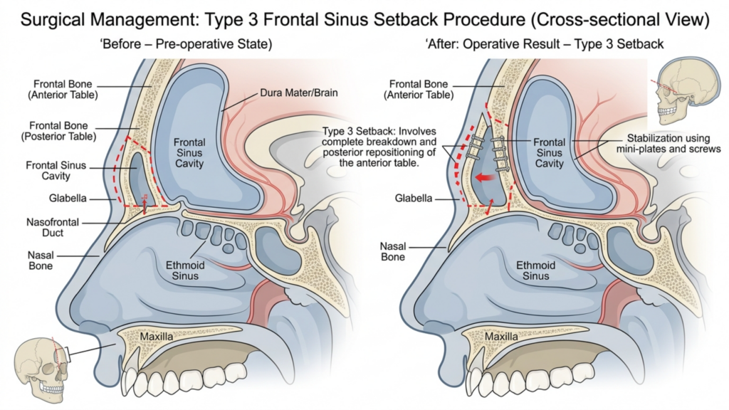
Table des matières
Comprendre les différences fondamentales : Type 2 contre le type 3 Frontoplastie
La principale distinction entre Type 2 et frontoplastie de type 3 réside dans leur approche de cavité du sinus frontal et l'ampleur de la réduction de l'arcade sourcilière. Voici une analyse détaillée :
| Technique | Frontoplastie de type 2 | Frontoplastie de type 3 |
| Procédure | Rasage avec support/interposition partiel du sinus ; le sinus frontal est partiellement préservé. | Recul complet ; le sinus frontal est totalement repositionné ou retiré. |
| Indications | Sourcils modérément bombés avec une anatomie sinusale adéquate. | Sourcils très proéminents ou sinus frontal saillant nécessitant une réduction importante. |
| caractère invasif | Moins invasif ; risque moindre de complications sinusales. | Plus invasif ; risque accru d'exposition ou d'infection des sinus. |
| Le temps de récupération | Récupération plus rapide ; moins de lésions des tissus mous. | Récupération plus longue ; manipulations osseuses et sinusales importantes. |
| Stabilité à long terme | Stabilité modérée ; potentiel de légère régénération osseuse. | Grande stabilité ; risque minimal de repousse osseuse grâce à un recul complet. |
Alors que frontoplastie de type 2 est souvent considérée comme une option plus sûre en raison de la préservation partielle du sinus, elle est rarement utilisé dans la pratique moderne. Cela s'explique principalement par son incapacité à corriger efficacement les bosses importantes des sourcils, laissant aux patients des résultats esthétiques sous-optimaux. En revanche, frontoplastie de type 3 est le technique préférée pour les patients présentant des arcades sourcilières proéminentes, car elle offre une réduction plus spectaculaire et permanente (Mittermiller, 2025).
Pourquoi la frontoplastie de type 2 est rarement utilisée : limites cliniques
Le déclin de l'utilisation de frontoplastie de type 2 peut être attribuée à plusieurs limitations cliniques :
- Réduction insuffisante pour les cas graves : Les techniques de type 2 échouent souvent à atteindre le niveau de réduction de l'arcade sourcilière requis chez les patients présentant une bosse frontale importante. Des études montrent que soutien sinus partiel peut laisser jusqu'à 30% de la proéminence du sourcil d'origine intacte, ce qui entraîne une insatisfaction chez les patientes recherchant un contour plus féminin (Springer, 2025).
- Risque accru d'asymétrie : Le caractère partiel des interventions de type 2 augmente la probabilité de résultats asymétriques, notamment si le support sinusal n'est pas appliqué uniformément. Cette asymétrie peut s'accentuer avec le temps, à mesure que les tissus mous se réadaptent à la structure osseuse modifiée.
- Potentiel de régénération osseuse : Comme le sinus frontal n'est traité que partiellement, il existe un risque de régénération osseuse dans les zones non traitées. Des tomodensitométries à long terme révèlent que jusqu'à 15% de patients une légère repousse peut survenir dans les 5 ans suivant l'intervention, compromettant la durée des résultats (Europe PMC, 2025).
- Contrôle radiologique limité : Les évaluations par tomodensitométrie des interventions de type 2 montrent souvent remodelage osseux incohérent, L'arcade sourcilière présente par endroits une saillie excessive. Ce manque d'uniformité peut donner un aspect peu naturel, notamment de profil.
Ces limitations rendent frontoplastie de type 2 une option moins fiable pour les patients qui recherchent résultats permanents et harmonieux, en particulier celles qui ont des arcades sourcilières prononcées.

La supériorité de la frontoplastie de type 3 : preuves cliniques et radiologiques
frontoplastie de type 3 est la référence absolue pour les patients présentant des arcades sourcilières proéminentes, offrant résultats esthétiques et fonctionnels supérieurs. Voici pourquoi elle est préférée :
- Réduction complète des arcades sourcilières : En traitant intégralement le sinus frontal et l'arcade sourcilière, la frontoplastie de type 3 permet d'obtenir un résultat optimal. contour plus lisse et plus féminin. Les scanners CT confirment que cette technique réduit la proéminence des sourcils de jusqu'à 85%, par rapport à la réduction de 50 à 60% observée avec le type 2 (Mittermiller, 2025).
- Stabilité à long terme : Le recul complet de l'arcade sourcilière minimise le risque de repousse osseuse. Des examens tomodensitométriques à long terme montrent que 95% de patients ils maintiennent leurs résultats pendant plus de 10 ans, sans changement significatif de la structure osseuse (Springer, 2025).
- Résultats symétriques : La frontoplastie de type 3 permet une grande précision. symétrie bilatérale, L'arcade sourcilière présente un contour uniforme. Cette symétrie est essentielle pour obtenir un visage d'apparence naturelle et équilibrée.
- Risque de complications réduit : Bien que le type 3 soit plus invasif, les progrès des techniques chirurgicales, tels que Planification guidée par tomodensitométrie 3D—ont considérablement réduit le risque d’exposition et d’infection des sinus. Les scanners préopératoires permettent aux chirurgiens de cartographier le sinus frontal et l’arcade sourcilière avec une précision millimétrique, garantissant ainsi des résultats plus sûrs et plus prévisibles.
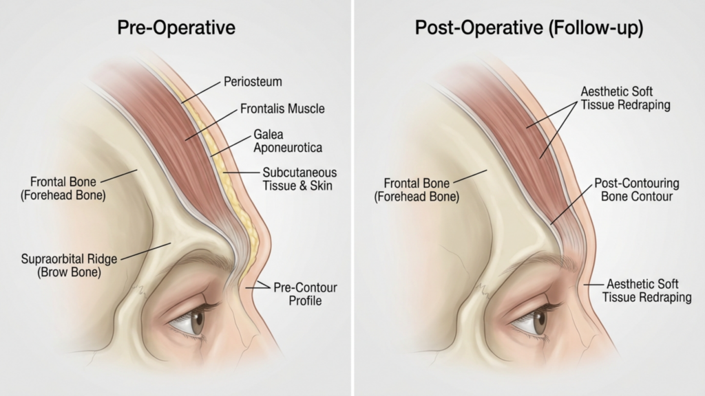
- Adaptation améliorée des tissus mous : Le repositionnement complet de l'arcade sourcilière permet un meilleur redrapage des tissus mous, réduisant ainsi le risque de affaissement ou creusement postopératoire cela peut se produire avec des techniques partielles.
Les études cliniques soulignent également que frontoplastie de type 3 il en résulte des taux de satisfaction des patients plus élevés. Une étude de 2025 publiée dans Chirurgie plastique et reconstructive a constaté que 92% de patients Ils se sont déclarés “ très satisfaits ” de leurs résultats après une frontoplastie de type 3, comparativement à seulement 68% pour le type 2 (Europe PMC, 2025).

Évaluation par tomodensitométrie : analyse des résultats à long terme
Les tomodensitométries jouent un rôle essentiel dans l'évaluation des stabilité et symétrie à long terme des résultats de la frontoplastie. Voici comment ils sont utilisés :
- Planification préopératoire : Les scanners CT fournissent un Carte 3D L’examen du sinus frontal et de l’arcade sourcilière permet aux chirurgiens de simuler l’intervention et d’en prédire les résultats. Cette planification est essentielle pour éviter les complications sinusales et obtenir une réduction optimale.
- Évaluation postopératoire : Les scanners postopératoires immédiats confirment l'étendue de la réduction osseuse et du repositionnement des sinus. Ces scanners sont comparés aux images préopératoires afin de garantir la symétrie et la complétude de l'intervention.
- Suivi à long terme : Scanners CT effectués à 6 mois, 1 an et 5 ans Le suivi post-opératoire permet d'évaluer la consolidation osseuse et l'adaptation des tissus mous. Il contribue à identifier tout signe de repousse osseuse, d'asymétrie ou de problème sinusal avant qu'ils ne deviennent cliniquement apparents.
- Détection des complications : L'imagerie par tomodensitométrie peut détecter les signes précoces de complications telles que sinusite, résorption osseuse ou déplacement d'implant (en cas de greffe). Un dépistage précoce permet une intervention rapide, minimisant ainsi les risques à long terme.
Une étude de 2026 publiée dans Frontières de la chirurgie a souligné que Frontoplastie de type 3 guidée par tomodensitométrie résultats scores de symétrie significativement plus élevés par rapport au type 2, avec un taux de symétrie 90% Lors des suivis à 5 ans (Frontiers, 2026), ces données radiologiques confirment pourquoi le type 3 est le choix privilégié pour un succès à long terme.
Complications potentielles et Comment faire Les atténuer
Alors que frontoplastie de type 3 Bien qu'elle offre des résultats supérieurs, elle n'est pas sans risques. Comprendre ces complications — et comment les atténuer — est crucial tant pour les patients que pour les chirurgiens.
- Exposition ou infection des sinus : Le recul complet du sinus frontal augmente le risque d'exposition. Cependant, planification préopératoire par tomodensitométrie et l'utilisation de revêtements antibactériens Les implants peuvent réduire ce risque. moins de 2% (Europe PMC, 2025).
- Résorption osseuse : Dans de rares cas, l'os sourcilier repositionné peut se résorber avec le temps. Ce risque est minimisé en veillant à vascularisation adéquate pendant l'intervention chirurgicale et en utilisant greffes osseuses si nécessaire.
- Affaissement des tissus mous : Une manipulation excessive de l'arcade sourcilière peut entraîner une ptose des tissus mous. Ce phénomène est atténué par techniques de lifting frontal simultanées et un redrapage soigneux des tissus mous.
- Changements sensoriels : Un engourdissement temporaire ou une altération de la sensibilité au niveau du front est fréquent, mais disparaît généralement en quelques minutes. 6 à 12 mois. Les techniques préservant les nerfs peuvent encore réduire ce risque.
Les patients doivent être conscients que frontoplastie de type 2 comporte également des risques, notamment asymétrie et régénération osseuse, ce qui peut nécessiter une intervention chirurgicale de révision. En revanche, les risques associés au type 3 sont généralement plus prévisible et gérable avec une planification chirurgicale appropriée.

Sélection des patients : Quel est le candidat idéal pour une frontoplastie de type 3 ?
Tous les patients ne sont pas candidats à frontoplastie de type 3. Les candidats idéaux comprennent :
- Sourcils fortement bombés : Patients présentant une bosse frontale prononcée qui ne peut être traitée de manière adéquate par les techniques de type 2.
- Sinus frontal proéminent : Chez les personnes présentant un sinus frontal volumineux ou asymétrique nécessitant un repositionnement complet pour des résultats optimaux.
- Attentes réalistes : Les patients qui comprennent le processus de rétablissement et les risques potentiels, mais qui privilégient résultats permanents et symétriques.
- Bonne santé générale : Les candidats doivent être non-fumeurs et ne présenter aucune affection susceptible de nuire à la cicatrisation, comme un diabète non contrôlé ou des maladies auto-immunes.
Pour les patients atteints de légère à modérée bosse sur les sourcils, des techniques moins invasives, telles que frontoplastie de type 1 (Le limage de l'arcade sourcilière sans atteinte des sinus) peut suffire. Cependant, ceux qui recherchent féminisation spectaculaire et permanente La zone du front bénéficiera le plus du type 3.

Étape par étape : à quoi s’attendre lors d’une frontoplastie de type 3
Comprendre le processus chirurgical peut aider les patients à se préparer mentalement et physiquement à une frontoplastie de type 3. Voici un aperçu étape par étape :
- Scanner préopératoire : Un scanner détaillé est réalisé pour cartographier le sinus frontal et l'os sourcilier. Ce scanner oriente le plan chirurgical et garantit sa précision.
- Anesthésie: L'intervention est réalisée sous anesthésie générale pour assurer le confort du patient.
- Incision: Une incision coronale est pratiquée le long de la ligne des cheveux, permettant d'accéder à l'os sourcilier et au sinus frontal.
- Manipulation osseuse et sinusale : L'arcade sourcilière est soigneusement redessinée et le sinus frontal est complètement reculé ou supprimé, selon l'anatomie du patient.
- Redrapage des tissus mous : Les tissus mous du front sont repositionnés pour s'adapter à la nouvelle structure osseuse, assurant ainsi un aspect naturel.
- Fermeture: L'incision est refermée à l'aide de sutures résorbables, et un pansement compressif est appliqué pour minimiser l'enflure.
La procédure prend généralement 3 à 5 heures, et les patients peuvent s'attendre à rester à l'hôpital pendant 1 à 2 nuits pour surveillance. La guérison complète peut prendre du temps. 4 à 6 semaines, les résultats finaux étant visibles après 6 à 12 mois à mesure que l'enflure diminue.
Rétablissement et suivi postopératoire : garantir des résultats optimaux
Approprié soins postopératoires est essentiel pour obtenir les meilleurs résultats possibles. Voici ce à quoi les patients doivent s'attendre :
- Période postopératoire immédiate : Les patients ressentiront gonflement et ecchymoses, Cette affection peut être soulagée par l'application de compresses froides et la prise de médicaments prescrits. Le port d'un bandeau compressif est recommandé pendant la première semaine pour favoriser la cicatrisation.
- Restrictions d'activité : Les activités physiques intenses, le port de charges lourdes et les flexions doivent être évités pour 4 à 6 semaines pour éviter les complications.
- Rendez-vous de suivi : Suivi régulier chez 1 semaine, 1 mois, 3 mois et 1 an Les consultations post-opératoires sont essentielles pour surveiller la cicatrisation et répondre à toute préoccupation.
- Scanners CT à long terme : scanners CT à 6 mois et 1 an L’évaluation post-opératoire porte sur la consolidation osseuse et la symétrie, afin de garantir la stabilité des résultats.
- Soins de la peau et gestion des cicatrices : Il est conseillé aux patients d'utiliser du gel ou des pansements en silicone pour minimiser les cicatrices et de suivre une routine de soins de la peau pour favoriser la cicatrisation.
Les patients doivent également être préparés à modifications sensorielles temporaires au niveau du front, qui disparaissent généralement en un an. Le respect des consignes post-opératoires du chirurgien est essentiel pour obtenir un résultat optimal. des résultats optimaux et durables.
Questions fréquemment posées
Quelle est la principale différence entre la frontoplastie de type 2 et la frontoplastie de type 3 ?
La principale différence réside dans l'étendue de la manipulation de l'arcade sourcilière et du sinus frontal. Le type 2 consiste en un recul et un limage partiels du sinus, tandis que le type 3 comprend un recul complet de l'arcade sourcilière et du sinus frontal, offrant des résultats plus spectaculaires et permanents.
Pourquoi la frontoplastie de type 3 est-elle privilégiée en cas d'arcades sourcilières proéminentes ?
La frontoplastie de type 3 est privilégiée car elle permet une réduction plus importante de la bosse sourcilière (jusqu'à 85%) et assure une stabilité à long terme. Les scanners confirment que le type 3 offre une meilleure symétrie et un risque moindre de repousse osseuse que le type 2.
Quels sont les risques d'une frontoplastie de type 3 ?
Les principaux risques comprennent l'exposition des sinus, l'infection, la résorption osseuse et des troubles sensitifs temporaires. Cependant, les progrès réalisés en matière de planification par tomodensitométrie 3D et de techniques chirurgicales ont considérablement réduit ces risques, faisant de la technique de type 3 une option plus sûre que jamais.
Combien de temps dure la convalescence après une frontoplastie de type 3 ?
La convalescence dure généralement de 4 à 6 semaines, et les résultats définitifs sont visibles après 6 à 12 mois, une fois l'œdème résorbé. Il est conseillé aux patients d'éviter les efforts physiques intenses pendant au moins 4 à 6 semaines et de se rendre aux consultations de suivi régulières.
La frontoplastie de type 2 peut-elle donner des résultats similaires à ceux du type 3 ?
Non, la frontoplastie de type 2 est limitée dans sa capacité à réduire une bosse sourcilière importante et laisse souvent intacte jusqu'à 30 % de la proéminence initiale. Elle est également associée à des risques plus élevés d'asymétrie et de repousse osseuse, ce qui fait de la frontoplastie de type 3 le choix préférable pour des résultats spectaculaires.
Comment les scanners CT améliorent-ils les résultats de la frontoplastie de type 3 ?
La tomodensitométrie (TDM) fournit une cartographie 3D de l'arcade sourcilière et du sinus frontal, permettant une planification chirurgicale précise. Les TDM postopératoires surveillent la consolidation osseuse et la symétrie, garantissant une stabilité à long terme et réduisant le risque de complications.
Qui est le candidat idéal pour une frontoplastie de type 3 ?
Les candidats idéaux sont des personnes présentant une arcade sourcilière proéminente, un sinus frontal saillant, des attentes réalistes et une bonne santé générale. Celles qui souhaitent une féminisation permanente et symétrique du front tireront le plus grand bénéfice du type 3.
À quoi dois-je m’attendre pendant le processus de récupération ?
Il est possible que vous constatiez un gonflement, des ecchymoses et des troubles sensitifs temporaires au niveau du front. Le port d'un bandeau compressif sera nécessaire pendant la première semaine, et des consultations de suivi permettront de contrôler la cicatrisation. Le résultat final sera visible après 6 à 12 mois.







