

Реконструкция лба является важнейшим компонентом лица Операция по феминизации (ФФС) а также черепно-лицевые процедуры, направленные на достижение гармоничного и эстетически привлекательного контура лица. Среди наиболее обсуждаемых методик: Тип 1 и краниопластика 3-го типа, Каждый из них предлагает свой собственный подход к изменению формы лба. Тип 1 включает в себя зачистка или шлифовка костей, Тип 3 требуется более сложный остеотомия и регресс лобной пазухи. Понимание анатомических, функциональных и эстетических последствий этих методик имеет важное значение как для хирургов, так и для пациентов, чтобы принимать обоснованные решения. Узнайте больше о смещении пазухи назад и уменьшении выступа надбровной дуги в нашем полном руководстве. контурирование лба.
В этом руководстве рассматриваются структурные различия между краниопластикой 1-го и 3-го типов, а также их влияние на толщина кости, Анатомия лобной пазухи, и о том, как хирурги определяют наиболее подходящий подход для каждого пациента. В итоге вы поймете, какая методика лучше всего соответствует вашим анатомическим особенностям и эстетическим целям.
Оглавление
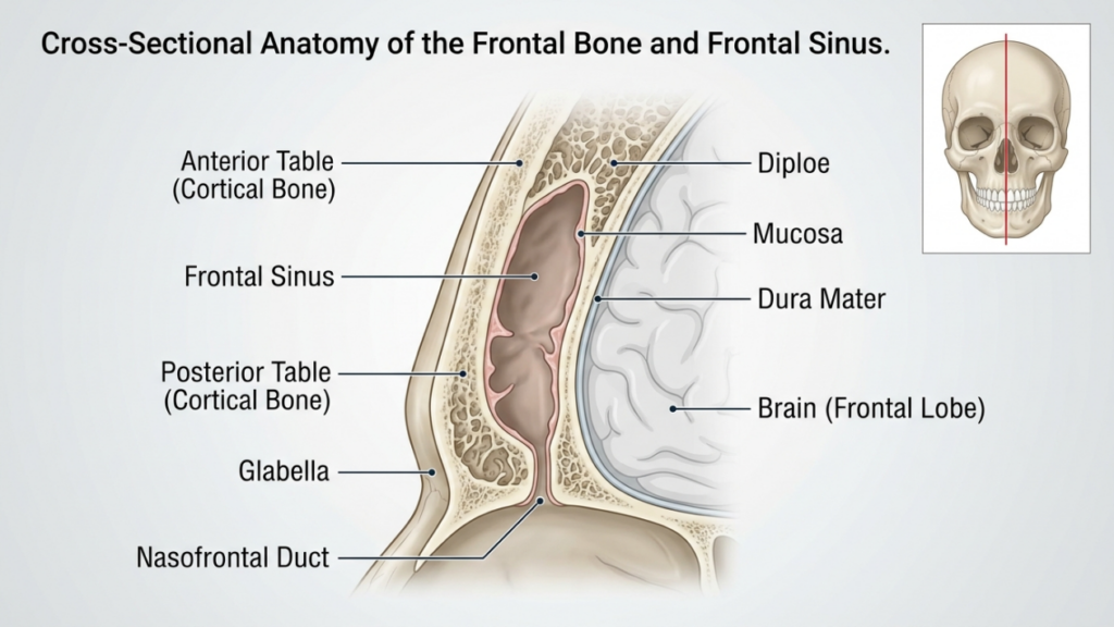
Анатомические основы: лобная кость и пазуха.
The лобная кость и лобная пазуха Лобная кость играет ключевую роль в реконструкции лба. Лобная кость образует верхнюю часть лица и вмещает лобную пазуху — полость, размер и форма которой варьируются у разных людей. передняя стол Лобная пазуха — это наружный слой кости, который формирует контур лба, в то время как задняя таблетка отделяет пазуху от головного мозга. Толщина этих пластин и степень пневматизации пазухи (расширения за счет заполнения воздухом) влияют на выбор между краниопластикой 1-го и 3-го типов.
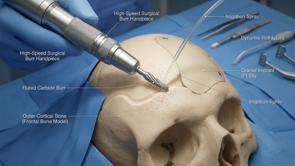
В краниопластика 1-го типа, врач хирург Используется высокоскоростная бормашина для шлифовки наружной кортикальной кости, уменьшая выступание без повреждения лобной пазухи. Эта методика идеально подходит для пациентов с тонкая лобная кость или минимальное выпячивание, поскольку это сохраняет целостность пазухи. Однако этого может быть недостаточно для пациентов со значительным выпячиванием надбровной дуги или толстой передней стенкой, поскольку чрезмерное бормашинирование может нарушить стабильность кости или не обеспечить желаемый контур (Остерхаут, 2024).
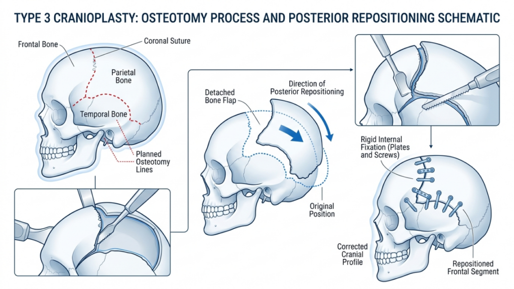
В отличие, краниопластика 3-го типа включает в себя остеотомия—Контролируемый разрез передней стенки лобной пазухи. Затем костный сегмент перемещается назад (смещается назад), чтобы уменьшить выступание и создать более гладкий, женственный лоб. Эта техника применяется у пациентов с умеренное или сильное выпячивание надбровных дуг или толстой передней стенки, где одной лишь шлифовки было бы недостаточно. Остеотомия позволяет точно изменить форму, сохраняя при этом защитную функцию пазухи (Феминизация лба: обзор литературы, 2024).
Краниопластика 1-го типа: шлифовка и зачистка.
Обзор техники
Краниопластика 1-го типа — наименее инвазивный вариант реконструкции лба. Она включает в себя использование высокоскоростная бормашина постепенно уменьшать выступание лобной кости. врач хирург Тщательно сбривает наружный корковый слой, избегая проникновения в лобную пазуху. Эта техника особенно эффективна для пациентов со следующими заболеваниями:
- Слабое или умеренное выпячивание надбровных дуг
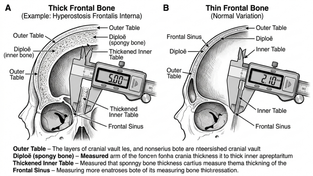
- Тонкая лобная кость (менее 5 мм)
- Отсутствие значительной пневматизации лобной пазухи
Процедура выполняется через коронарный разрез, Это позволяет получить доступ к лбу, минимизируя при этом видимые рубцы. Хирург использует тактильную обратную связь и визуальные подсказки для обеспечения равномерного уменьшения объема кости без чрезмерного истончения, что может привести к нестабильности или неровностям контура.
Преимущества краниопластики 1-го типа
Краниопластика 1-го типа имеет ряд преимуществ:
- Минимальная инвазивность: Отсутствие остеотомии или удаления костной ткани снижает хирургическую травму и сокращает время восстановления.
- Снижение риска осложнений: Сохранение лобной пазухи минимизирует риск синусита, утечки спинномозговой жидкости или образования мукоцеле.
- Более короткое время операции: Процедура обычно занимает 1–2 часа, что делает ее более быстрым вариантом по сравнению с типом 3.
- Предсказуемые результаты: Идеально подходит для пациенток с легкой выпуклостью в области паха, где для достижения женственного вида достаточно легкой коррекции контуров.
Ограничения краниопластики 1-го типа
Хотя краниопластика 1-го типа является более безопасной и менее инвазивной, она имеет существенные ограничения:
- Ограниченное сокращение: Недостаточно для пациентов с выраженным выпячиванием надбровных дуг или утолщением лобных костей.
- Риск чрезмерного истончения: Агрессивная обработка бормашиной может ослабить кость, что приведет к неровностям контура или переломам.
- Неполная феминизация: Может не обеспечить желаемый эстетический результат у пациентов с выраженными мужскими чертами лица.
Краниопластика 3-го типа: остеотомия и смещение назад.
Обзор техники
Краниопластика 3-го типа — это более сложная процедура, предназначенная для пациентов с умеренное или сильное выпячивание надбровных дуг или толстые лобные кости. Это включает в себя остеотомия—Точный разрез передней стенки лобной пазухи — с последующим перемещением костного сегмента назад (смещением назад). Эта техника позволяет значительно уменьшить выступание лба и создать более плавный, женственный контур.
Процедура обычно выполняется через коронарный разрез, обеспечивая доступ к лобной кости и пазухе. Хирург использует сагиттальная пила или пьезоэлектрическое устройство Для выполнения остеотомии необходимо убедиться, что разрез повторяет естественную кривизну лба. Затем сегмент кости возвращается на место и фиксируется с помощью... титановые пластины и винты или рассасывающимися швами. Этот метод идеально подходит для пациентов со следующими заболеваниями:
- Сильное выпячивание надбровных дуг
- Толстая лобная кость (более 5 мм)
- Значительная пневматизация лобной пазухи
Краниопластика 3-го типа требует тщательного планирования, часто включающего в себя Виртуальное 3D-планирование хирургических операций (VSP) для имитации остеотомии и смещения. Это обеспечивает точность и минимизирует риск осложнений, таких как повреждение синуса или утечка спинномозговой жидкости (виртуальное хирургическое планирование в Феминизация лица Верхней части лица, 2025).
Преимущества краниопластики 3-го типа
Краниопластика 3-го типа предлагает ряд преимуществ для пациентов с выраженными мужскими чертами лица:
- Значительная коррекция контуров: Достигается значительного уменьшения выступа над бровями, что создает более гладкий и женственный лоб.
- Универсальность: Подходит для пациентов с утолщенными лобными костями или выраженной пневматизацией пазух носа.
- Долгосрочная стабильность: Перемещенный костный сегмент хорошо срастается, снижая риск возникновения неровностей контура с течением времени.
- Настройка: Виртуальное планирование хирургических операций позволяет вносить точные, индивидуальные корректировки для достижения оптимальных результатов.
Ограничения краниопластики 3-го типа
Несмотря на свою эффективность, краниопластика 3-го типа сопряжена с более высокими рисками и сложностями:
- Увеличение времени операции: Процедура обычно занимает 3–5 часов и требует высокой точности и квалификации.
- Повышенный риск осложнений: К потенциальным рискам относятся синусит, утечка спинномозговой жидкости или образование мукоцеле в случае повреждения пазухи.
- Более длительное восстановление: По сравнению с краниопластикой 1-го типа, у пациентов могут наблюдаться более длительные отеки и дискомфорт.
- Расходы: Использование современных методов визуализации и хирургических инструментов увеличивает общую стоимость процедуры.

Основные различия между краниопластикой 1-го и 3-го типов.
| Особенность | Краниопластика 1-го типа | Краниопластика 3 типа |
|---|---|---|
| Техника | Обработка/шлифовка костей | Остеотомия и репозиция |
| Инвазивность | Минимальный | От умеренного до высокого |
| Идеальные кандидаты | Слабое или умеренное выпячивание надбровных дуг, тонкая лобная кость. | Выраженное выпячивание надбровных дуг, толстая лобная кость |
| Время операции | 1–2 часа | 3–5 часов |
| Время восстановления | 1–2 недели | 3–6 недель |
| Риск осложнений | Низкий | От умеренного до высокого |
| Расходы | Ниже | Выше |
| Эстетический результат | Тонкое контурирование | Драматическая феминизация |
Как хирурги принимают решение: тип 1 или тип 3
Выбор между краниопластикой 1-го и 3-го типа зависит от нескольких факторов, включая состояние пациента. анатомические особенности, эстетические цели, и хирургические риски. Хирурги полагаются на сочетание следующих методов: клиническое обследование, 3D-изображение, и консультация пациента определить наиболее подходящую методику.
1. Анатомическая оценка
Первый шаг — оценка состояния пациента. толщина лобной кости и Анатомия лобной пазухи. А КТ-сканирование или 3D реконструкция Предоставляет подробную информацию о:
- Толщина кости: Пациентам с тонкими лобными костями (менее 5 мм) краниопластика 1-го типа подходит лучше, тогда как пациентам с более толстыми костями может потребоваться краниопластика 3-го типа.
- Пневматизация пазух: Обширная пневматизация пазух может потребовать краниопластики 3-го типа, чтобы избежать повреждения пазух во время обработки бором.
- Степень влияния босса: Сильно выраженное выпячивание бровей часто требует существенного уменьшения, достижимого только с помощью техник 3-го типа.
2. Эстетические цели
Эстетические ожидания пациентов играют решающую роль в выборе методики. Те, кто ищет... тонкая феминизация Люди с выраженными мужскими чертами лица могут выбрать краниопластику 1-го типа, в то время как лицам с более выраженными мужскими чертами часто требуется краниопластика. трансформационные результаты Типа 3. Хирурги обсуждают реалистичные результаты, исходя из анатомии пациента и желаемых изменений.
3. Хирургические риски и восстановление
Краниопластика 3-го типа сопряжена с более высокими рисками, в том числе: осложнения со стороны пазух, утечки спинномозговой жидкости, и длительное восстановление. Хирурги оценивают общее состояние здоровья пациента, его переносимость операции и готовность соблюдать послеоперационный уход. Пациентам с заболеваниями, повышающими хирургический риск, может быть рекомендовано рассмотреть возможность проведения операций 1-го типа или альтернативных процедур.
4. Виртуальное хирургическое планирование (ВХП)
Достижения в Виртуальное 3D-планирование хирургических операций Виртуальная хирургическая печать (VSP) произвела революцию в краниопластике. Хирурги используют VSP для моделирования остеотомий, смещений и результатов, обеспечивая точность и минимизируя риски. Эта технология особенно ценна для краниопластики 3-го типа, где точное перемещение кости имеет решающее значение (3D-печать и виртуальное хирургическое планирование в черепно-лицевой и ортогнатической хирургии, 2025).
Послеоперационный уход и восстановление
Процесс восстановления значительно различается между краниопластикой 1-го и 3-го типов. Понимание послеоперационного процесса помогает пациентам подготовиться к плавному заживлению.
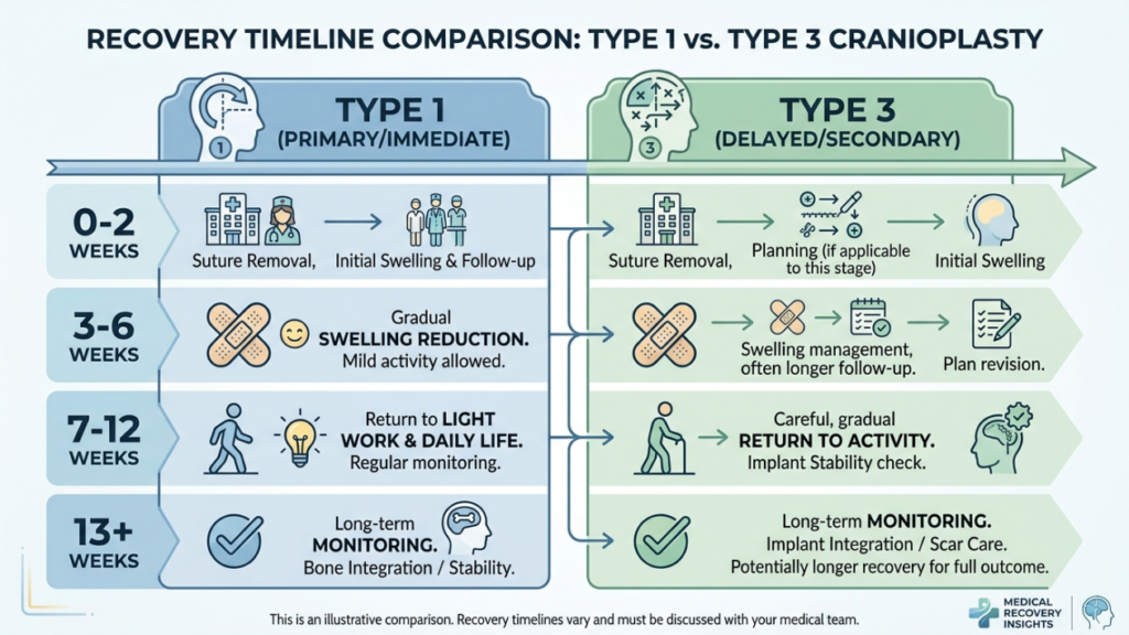
Восстановление после краниопластики 1 типа
У пациентов, перенесших краниопластику 1-го типа, обычно наблюдаются следующие симптомы:
- Отек от легкой до умеренной степени: Проходит в течение 1–2 недель.
- Минимальный дискомфорт: Лечение проводилось безрецептурными обезболивающими препаратами.
- Быстрый возврат к действиям: Большинство пациентов возвращаются к нормальной активности в течение 2 недель.
Восстановление после краниопластики 3-го типа
Восстановление после краниопластики 3-го типа более длительное из-за сложности процедуры:
- Значительные отеки и синяки: Может сохраняться в течение 3–4 недель.
- Умеренная боль: В течение первой недели могут потребоваться обезболивающие препараты, отпускаемые по рецепту.
- Постепенное возвращение к занятиям: Интенсивные физические нагрузки ограничиваются на 4–6 недель.
- Последующие визиты: Регулярный мониторинг для обеспечения правильного заживления и выявления любых осложнений.
Обе методики требуют от пациентов избегать подъем тяжестей, интенсивные упражнения, и прямое давление на лоб На начальном этапе восстановления хирурги предоставляют подробные послеоперационные инструкции, включая уход за раной, ограничения активности и признаки осложнений, на которые следует обращать внимание.
Возможные осложнения и как их избежать
Хотя краниопластика в целом безопасна, могут возникнуть осложнения. Осведомленность об этих рисках и профилактических мерах крайне важна как для хирургов, так и для пациентов.
Распространенные осложнения
- Синусит: Воспаление или инфекция лобной пазухи, особенно при краниопластике 3-го типа, если пазуха повреждена.
- Утечка спинномозговой жидкости (СМЖ): Редкое, но серьезное осложнение при повреждении задней стенки пазухи.
- Неровности контура: Чрезмерное истончение костной ткани при типе 1 или неправильное смещение при типе 3 может привести к асимметрии или видимым гребням.
- Формирование мукоцеле: Закупорка путей оттока слизи из пазух может привести к образованию кист, заполненных слизью.
- Инфекционное заболевание: Риск выше при 3-м типе из-за более длительного времени операции и использования имплантатов.
Профилактические меры
Хирурги используют несколько стратегий для минимизации осложнений:
- Точное хирургическое планирование: Трехмерная визуализация и виртуальное планирование хирургического вмешательства обеспечивают точность остеотомий и смещения костей.
- Сохранение здоровья пазух носа: Во избежание повреждения лобной пазухи во время обработки бором или остеотомии.
- Профилактика антибиотиками: Применяется до и после операции для снижения риска инфекций.
- Послеоперационный мониторинг: Регулярные контрольные осмотры для выявления ранних признаков осложнений.
- Обучение пациентов: Обучение пациентов правильному уходу за ранами, ограничениям активности и признакам осложнений.
Альтернативы краниопластике 1-го и 3-го типов
Для пациентов, которым не подходят краниопластика 1-го или 3-го типа, могут быть рассмотрены альтернативные методики:
- Пересадка жира: Аутологичная пересадка жировой ткани позволяет смягчить контуры лба без изменения структуры костей. Это идеально подходит для пациентов с незначительными неровностями или для тех, кто ищет нехирургические варианты лечения.
- Индивидуальные имплантаты: Готовые имплантаты, такие как PEEK (полиэфирэфиркетон) или титан, Эти методы могут быть использованы для увеличения или изменения формы лба без остеотомии. Они особенно полезны для пациентов с тонкими костями или осложнениями со стороны пазух носа.
- Ортогнатическая хирургия: В случаях, когда контурирование лба является частью более широкой феминизация лица В рамках плана лечения могут быть объединены ортогнатические процедуры (например, остеотомия Ле Фор I) для коррекции средней части лица и выравнивания челюсти.
- Эндоскопические методы: Минимально инвазивные эндоскопические методы позволяют уменьшить выступание надбровных дуг благодаря меньшим разрезам и более быстрому восстановлению.
Отзывы пациентов и результаты лечения в реальной практике
Результаты, полученные в реальных условиях, позволяют получить ценную информацию об эффективности и уровне удовлетворенности пациентов краниопластикой 1-го и 3-го типов. Отзывы пациентов подчеркивают преобразующее воздействие этих процедур:
Пример из практики 1: Краниопластика 1 типа
28-летняя трансгендерная женщина хотела добиться деликатной феминизации лба. Имея тонкую лобную кость и минимальное выпячивание лба, она выбрала краниопластику 1-го типа. Процедура позволила получить более гладкий контур с минимальным периодом восстановления. Она выразила высокую степень удовлетворенности, отметив, что результаты соответствуют ее ожиданиям естественного, женственного внешнего вида.
Пример из практики 2: Краниопластика 3-го типа
35-летняя трансгендерная женщина обратилась с выраженным выпячиванием надбровных дуг и утолщением лобной кости. Была выполнена краниопластика 3-го типа, включающая остеотомию и смещение лба назад. Значительное уменьшение выступа лба существенно феминизировало черты ее лица. Хотя период восстановления занял больше времени, пациентка выразила огромную удовлетворенность результатами, заявив, что процедура “изменила ее жизнь”.”
Клинический случай 3: Повторная операция
40-летней пациентке первоначально была проведена краниопластика 1-го типа, но она осталась недовольна незначительными результатами. Позже она выбрала краниопластику 3-го типа для достижения более выраженной феминизации. Повторная операция успешно устранила ее опасения, продемонстрировав важность выбора правильной методики, основанной на анатомических потребностях и эстетических целях.
Роль виртуального хирургического планирования в краниопластике
Виртуальное хирургическое планирование (ВХП) стало настоящим прорывом в краниопластике, особенно для операций 3-го типа. ВХП позволяет хирургам:
- Имитация остеотомии: Точно спланируйте разрезы костей и их перемещение для достижения оптимального контура.
- Прогнозирование результатов: Визуализируйте послеоперационные результаты и скорректируйте хирургический план соответствующим образом.
- Минимизируйте риски: Избегайте контакта с важными структурами, такими как лобная пазуха и надглазничные нервы.
- Улучшение коммуникации: Демонстрируйте пациентам 3D-модели, чтобы сформировать реалистичные ожидания и улучшить информированное согласие.
Исследования показали, что виртуальное хирургическое планирование сокращает время операции, повышает точность и улучшает удовлетворенность пациентов (3D-печать и виртуальное хирургическое планирование в черепно-лицевой и ортогнатической хирургии, 2025). В настоящее время оно считается золотым стандартом для сложных краниопластических процедур.

Часто задаваемые вопросы
В чём основное различие между краниопластикой 1-го и 3-го типов?
Краниопластика 1-го типа включает в себя удаление или шлифовку наружного слоя лобной кости для уменьшения ее выступа, в то время как краниопластика 3-го типа требует остеотомии (рассечения кости) и смещения сегмента лобной кости назад для достижения более выраженного контура. Краниопластика 1-го типа менее инвазивна и подходит для умеренного выступания лобной кости, тогда как краниопластика 3-го типа применяется при сильном выступлении или утолщении лобной кости.
Как хирурги выбирают между краниопластикой 1-го и 3-го типа?
Хирурги оценивают толщину лобной кости пациента, степень выступания надбровных дуг и анатомию лобной пазухи с помощью компьютерной томографии и 3D-визуализации. Тип 1 выбирается при более тонких костях и умеренном выступлении надбровных дуг, тогда как тип 3 предпочтителен при более толстых костях, выраженном выступлении надбровных дуг или обширной пневматизации пазухи. На решение также влияют цели пациента и его готовность к хирургическим рискам.
Какие риски связаны с краниопластикой 3-го типа?
Краниопластика 3-го типа сопряжена с более высокими рисками из-за своей сложности, включая синусит, утечку спинномозговой жидкости, неровности контура, образование мукоцеле и инфекцию. Эти риски минимизируются за счет точного планирования операции, сохранения пазух и послеоперационного наблюдения. Пациентов информируют о тревожных признаках и последующем уходе для обеспечения беспроблемного выздоровления.
Какова продолжительность восстановительного периода после краниопластики 1-го типа по сравнению с краниопластикой 3-го типа?
Восстановление после краниопластики 1-го типа обычно занимает 1–2 недели и сопровождается небольшим отеком и дискомфортом. Краниопластика 3-го типа требует более длительного периода восстановления – 3–6 недель – из-за более обширного хирургического вмешательства и потенциальных осложнений. Пациентам рекомендуется избегать интенсивных физических нагрузок и строго следовать послеоперационным инструкциям.
Может ли краниопластика 1-го типа дать такие же результаты, как и краниопластика 3-го типа?
Нет, краниопластика 1-го типа ограничивается лишь незначительной коррекцией контуров и идеально подходит для умеренного выступания лобной кости. Краниопластика 3-го типа позволяет добиться более выраженной феминизации за счет перемещения сегмента лобной кости, что делает ее подходящей для сильного выступания или утолщения лобной кости. Выбор зависит от анатомических потребностей пациента и его эстетических целей.
Какова роль виртуального планирования хирургического вмешательства при краниопластике?
Виртуальное хирургическое планирование (ВХП) позволяет хирургам моделировать остеотомии, прогнозировать результаты и минимизировать риски, избегая критически важных структур, таких как лобная пазуха. Оно повышает точность, сокращает время операции и улучшает удовлетворенность пациентов, обеспечивая четкую визуализацию хирургического плана и ожидаемых результатов.
Существуют ли нехирургические альтернативы краниопластике для феминизации лба?
Да, к нехирургическим альтернативам относятся: пересадка жира Для смягчения контуров и использования индивидуальных имплантатов (например, из PEEK или титана) для изменения формы лба без остеотомии. Эти варианты идеально подходят для пациентов с незначительными неровностями или для тех, кто предпочитает избегать хирургического вмешательства. Однако они могут не обеспечить такой же уровень феминизации, как хирургические методы.
Чего мне следует ожидать во время консультации по реконструкции лба?
В ходе консультации ваш хирург оценит толщину лобной кости, анатомию пазух и степень выступания десны, используя клиническое обследование и 3D-визуализацию. Он обсудит ваши эстетические цели, объяснит различия между краниопластикой 1-го и 3-го типов и порекомендует наиболее подходящую методику, исходя из вашей анатомии и ожиданий.







