في المشهد المتطور تأنيث الوجه الجراحة (FFS), الخيار بين النوع الثاني و عملية تجميل الجبهة من النوع الثالث يُعدّ قرار تصغير عظمة الحاجب من أهم القرارات بالنسبة للمرضى الذين يسعون إلى ذلك. فبينما تهدف كلتا التقنيتين إلى تحسين شكل الجبهة وإضفاء مظهر أكثر أنوثة، إلا أن أساليبهما ومخاطرهما ونتائجهما على المدى الطويل تختلف اختلافًا كبيرًا. تتناول هذه المقالة بالتفصيل... الفروقات السريرية والشعاعية والجمالية بين هذه التقنيات، المدعومة بـ تقييمات التصوير المقطعي المحوسب وبيانات المتابعة طويلة الأجل، لمساعدتك على اتخاذ خيار مستنير.
جدول المحتويات
فهم الاختلافات الأساسية: جراحة تجميل الجبهة من النوع الثاني مقابل النوع الثالث
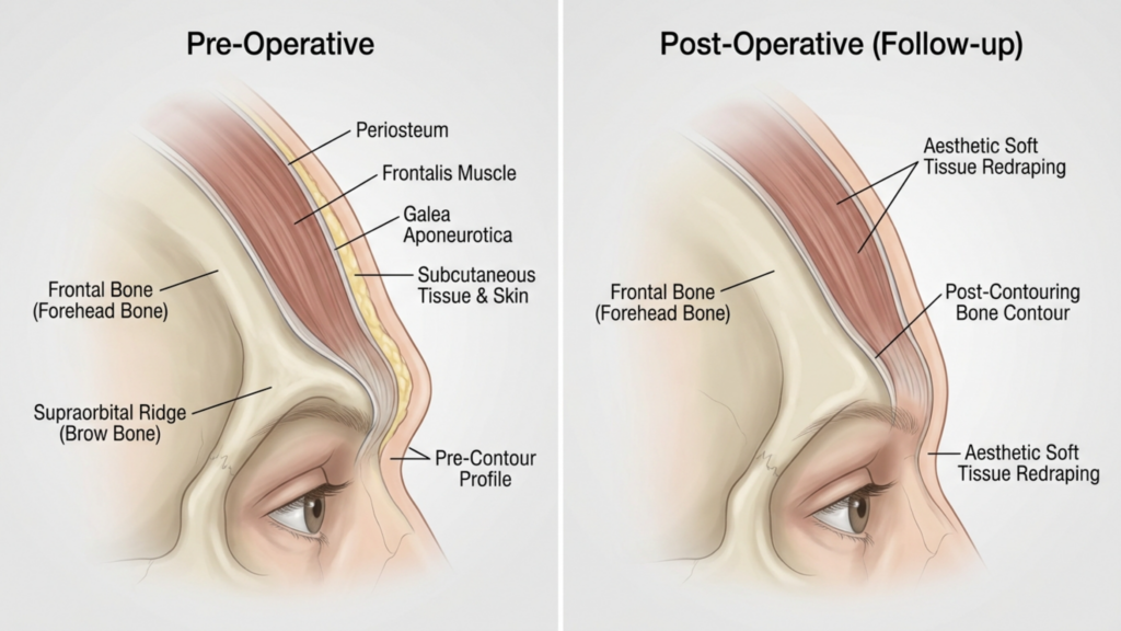
الفرق الأساسي بين النوع الثاني و عملية تجميل الجبهة من النوع الثالث يكمن ذلك في نهجهم تجاه تجويف الجيوب الأنفية الأمامية ومدى انخفاض عظم الحاجب. إليكم التفاصيل:
| تقنية | عملية تجميل الجبهة من النوع الثاني | عملية تجميل الجبهة من النوع الثالث |
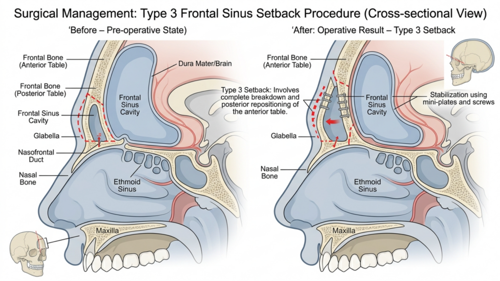
| إجراء | الحلاقة مع دعم/تداخل جزئي للجيوب الأنفية؛ يتم الحفاظ على الجيوب الأنفية الأمامية جزئيًا. | تراجع كامل؛ يتم إعادة وضع الجيب الجبهي بالكامل أو إزالته. |
| دواعي الاستعمال | بروز معتدل للحاجبين مع تشريح جيوب أنفية مناسب. | بروز شديد في الحاجب أو بروز الجيوب الأنفية الأمامية يتطلب تصغيراً جذرياً. |
| الغزوية | أقل توغلاً؛ خطر أقل لحدوث مضاعفات في الجيوب الأنفية. | أكثر توغلاً؛ خطر أكبر للتعرض للجيوب الأنفية أو الإصابة بالعدوى. |
| وقت الانتعاش | فترة نقاهة أقصر؛ واضطراب أقل في الأنسجة الرخوة. | فترة نقاهة أطول؛ معالجة واسعة النطاق للعظام والجيوب الأنفية. |
| الاستقرار على المدى الطويل | استقرار معتدل؛ إمكانية نمو العظام بشكل طفيف. | استقرار عالٍ؛ خطر ضئيل لإعادة نمو العظام بسبب التراجع الكامل. |
بينما عملية تجميل الجبهة من النوع الثاني يُعتبر هذا الخيار أكثر أمانًا نظرًا للحفاظ الجزئي على الجيوب الأنفية، وهو نادر الاستخدام في الممارسة الحديثة. ويرجع ذلك أساسًا إلى فشلها في معالجة بروز الحاجب الشديد بفعالية، مما يترك المرضى بنتائج تجميلية دون المستوى الأمثل. على النقيض من ذلك،, عملية تجميل الجبهة من النوع الثالث هو التقنية المفضلة بالنسبة للمرضى الذين يعانون من عظام حاجب بارزة، حيث يوفر ذلك انخفاضًا أكثر دراماتيكية ودائمة (ميترميلر، 2025).
لماذا نادراً ما يُستخدم النوع الثاني من جراحة تجميل الجبهة: القيود السريرية
انخفاض استخدام عملية تجميل الجبهة من النوع الثاني يمكن أن يُعزى ذلك إلى العديد من القيود السريرية:
- تخفيض غير كافٍ للحالات الشديدة: غالباً ما تفشل تقنيات النوع الثاني في تحقيق مستوى تقليل عظم الحاجب المطلوب للمرضى الذين يعانون من بروز جبهي ملحوظ. تشير الدراسات إلى أن دعم الجيوب الأنفية الجزئي يمكن أن يترك ما يصل إلى 30% من بروز الحاجب الأصلي سليم, مما يؤدي إلى عدم الرضا بين المرضى الذين يسعون للحصول على شكل أكثر أنوثة (سبرينغر، 2025).
- ارتفاع خطر عدم التناسق: تزيد الطبيعة الجزئية لإجراءات النوع الثاني من احتمالية نتائج غير متماثلة, وخاصةً إذا لم يتم تطبيق دعامة الجيوب الأنفية بشكل متجانس. وقد يصبح هذا التباين أكثر وضوحًا بمرور الوقت، حيث تتكيف الأنسجة الرخوة مع بنية العظام المتغيرة.
- إمكانية إعادة نمو العظام: نظراً لأن الجيب الجبهي لا يُعالج إلا جزئياً، فهناك خطر من إعادة نمو العظام في المناطق غير المعالجة. تكشف فحوصات التصوير المقطعي المحوسب طويلة الأمد أن ما يصل إلى 15% من المرضى قد يحدث نمو طفيف في غضون 5 سنوات بعد الجراحة، مما يؤثر على استمرار النتائج (Europe PMC، 2025).
- التحكم الإشعاعي المحدود: غالباً ما تُظهر تقييمات التصوير المقطعي المحوسب لإجراءات النوع الثاني إعادة تشكيل العظام غير المتناسقة, مع بقاء بعض مناطق عظم الحاجب بارزة بشكل مفرط. قد يؤدي هذا التباين إلى مظهر غير طبيعي، خاصة في الصور الجانبية.
هذه القيود تجعل عملية تجميل الجبهة من النوع الثاني خيار أقل موثوقية للمرضى الذين يبحثون نتائج دائمة ومتناغمة, وخاصة أولئك الذين لديهم عظام حاجب بارزة.

تفوق عملية تجميل الجبهة من النوع الثالث: الأدلة السريرية والشعاعية
عملية تجميل الجبهة من النوع الثالث يُعدّ هذا الإجراء المعيار الذهبي للمرضى الذين يعانون من بروز عظام الحاجب، حيث يوفر نتائج جمالية ووظيفية فائقة. إليكم سبب تفضيله:
- تصغير عظم الحاجب بالكامل: من خلال معالجة الجيوب الأنفية الأمامية وعظم الحاجب بشكل كامل، تحقق عملية تجميل الجبهة من النوع الثالث ما يلي: ملامح أكثر نعومة وأنوثة. تؤكد فحوصات التصوير المقطعي المحوسب أن هذه التقنية تقلل من بروز الحاجب عن طريق حتى 85%, ، مقارنة بانخفاض 50-60% الذي لوحظ مع النوع 2 (Mittermiller، 2025).
- الاستقرار على المدى الطويل: يقلل التراجع الكامل لعظم الحاجب من خطر إعادة نمو العظم. وتُظهر تقييمات التصوير المقطعي المحوسب طويلة الأمد أن 95% من المرضى يحافظون على نتائجهم لأكثر من 10 سنوات، دون حدوث تغييرات كبيرة في بنية العظام (Springer، 2025).
- نتائج متناظرة: تتيح عملية تجميل الجبهة من النوع الثالث دقة عالية التناظر الثنائي, حيث أن عظمة الحاجب بأكملها ذات شكل متناسق. هذا التناسق ضروري لتحقيق مظهر وجه طبيعي ومتوازن.
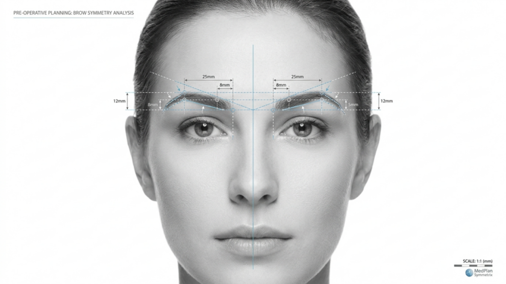
- انخفاض خطر حدوث مضاعفات: على الرغم من أن النوع الثالث أكثر توغلاً، إلا أن التطورات في التقنيات الجراحية - مثل التخطيط الموجه بالتصوير المقطعي المحوسب ثلاثي الأبعادلقد ساهمت هذه التقنيات بشكل كبير في تقليل خطر تعرض الجيوب الأنفية للعدوى. تُمكّن فحوصات التصوير المقطعي المحوسب قبل الجراحة الجراحين من رسم خريطة للجيوب الأنفية الأمامية وعظم الحاجب بدقة متناهية تصل إلى الملليمتر، مما يضمن نتائج أكثر أمانًا وقابلية للتنبؤ.
- تحسين تكيف الأنسجة الرخوة: تتيح إعادة تموضع عظم الحاجب بالكامل إعادة توزيع أفضل للأنسجة الرخوة، مما يقلل من خطر الترهل أو التجويف بعد العملية الجراحية وهذا ما يمكن أن يحدث مع التقنيات الجزئية.
كما تُبرز الدراسات السريرية أن عملية تجميل الجبهة من النوع الثالث تؤدي إلى ارتفاع معدلات رضا المرضى. دراسة نُشرت عام 2025 في جراحة التجميل والترميم وجد أن 92% من المرضى أفادوا بأنهم "راضون جداً" عن نتائجهم بعد عملية تجميل الجبهة من النوع الثالث، مقارنةً بـ 68% للنوع 2 (Europe PMC، 2025).

تقييم التصوير المقطعي المحوسب: تقييم النتائج طويلة المدى
تلعب الأشعة المقطعية دورًا محوريًا في تقييم الاستقرار والتناظر على المدى الطويل نتائج عملية تجميل الجبهة. إليك كيفية استخدامها:
- التخطيط قبل الجراحة: توفر الأشعة المقطعية خريطة ثلاثية الأبعاد يُتيح هذا التخطيط للجيوب الأنفية الأمامية وعظم الحاجب للجراحين محاكاة العملية وتوقع النتائج. ويُعدّ هذا التخطيط ضروريًا لتجنب مضاعفات الجيوب الأنفية وتحقيق أفضل النتائج المرجوة.
- التقييم بعد العملية الجراحية: تؤكد فحوصات التصوير المقطعي المحوسب التي تُجرى مباشرة بعد العملية مدى تقليل حجم العظم وإعادة تموضع الجيوب الأنفية. وتُقارن هذه الفحوصات بصور ما قبل العملية لضمان التناسق واكتمال العملية.
- المتابعة طويلة الأمد: تم إجراء فحوصات التصوير المقطعي المحوسب في ستة أشهر، وسنة واحدة، وخمس سنوات تُستخدم الفحوصات بعد الجراحة لمراقبة التئام العظام وتكيف الأنسجة الرخوة. وتساعد هذه الفحوصات في تحديد أي علامات لنمو العظام من جديد، أو عدم التناسق، أو مشاكل الجيوب الأنفية قبل أن تصبح ظاهرة سريريًا.
- الكشف عن المضاعفات: يمكن للتصوير المقطعي المحوسب الكشف عن العلامات المبكرة للمضاعفات مثل التهاب الجيوب الأنفية، أو ارتشاف العظام، أو انزياح الزرعات (في حال استخدام الطعوم). يسمح الكشف المبكر بالتدخل في الوقت المناسب، مما يقلل من المخاطر طويلة المدى.
دراسة نُشرت عام 2026 في مجلة فرونتيرز إن سيرجري أكد على ذلك عملية تجميل الجبهة من النوع الثالث الموجهة بالتصوير المقطعي المحوسب النتائج في درجات تناظر أعلى بشكل ملحوظ مقارنةً بالنوع الثاني، مع معدل التناظر 90% في متابعة لمدة 5 سنوات (فرونتيرز، 2026). تؤكد هذه الأدلة الإشعاعية سبب كون النوع 3 هو الخيار المفضل لتحقيق النجاح على المدى الطويل.
المضاعفات المحتملة وكيفية التخفيف منها
بينما عملية تجميل الجبهة من النوع الثالث على الرغم من أنها تقدم نتائج فائقة، إلا أنها لا تخلو من المخاطر. إن فهم هذه المضاعفات - وكيفية التخفيف منها - أمر بالغ الأهمية لكل من المرضى والجراحين.
- التعرض للجيوب الأنفية أو الإصابة بالعدوى: يؤدي التراجع الكامل للجيوب الأنفية الأمامية إلى زيادة خطر التعرض. ومع ذلك،, التخطيط قبل الجراحة باستخدام التصوير المقطعي المحوسب واستخدام الطلاءات المضادة للبكتيريا يمكن أن يقلل استخدام الغرسات من هذا الخطر إلى أقل من 2% (Europe PMC، 2025).
- ارتشاف العظام: في حالات نادرة، قد يمتص عظم الحاجب الذي أعيد وضعه بمرور الوقت. ويتم تقليل هذا الخطر عن طريق ضمان التروية الدموية السليمة أثناء الجراحة واستخدامها طعوم العظام إذا لزم الأمر.
- ترهل الأنسجة الرخوة: قد يؤدي التلاعب المفرط بعظم الحاجب إلى تدلي الأنسجة الرخوة. ويتم التخفيف من ذلك من خلال تقنيات رفع الحاجب المتزامنة وإعادة توزيع الأنسجة الرخوة بعناية.
- التغييرات الحسية: يُعدّ الشعور المؤقت بالخدر أو تغير الإحساس في الجبهة أمراً شائعاً، ولكنه عادةً ما يزول في غضون 6-12 شهرًا. يمكن لتقنيات الحفاظ على الأعصاب أن تقلل من هذا الخطر بشكل أكبر.
ينبغي على المرضى أن يكونوا على دراية بأن عملية تجميل الجبهة من النوع الثاني كما أنها تنطوي على مخاطر، لا سيما عدم التناسق وإعادة نمو العظام, مما قد يستدعي إجراء جراحة تصحيحية. في المقابل، فإن المخاطر المرتبطة بالنوع 3 تكون عمومًا أكثر قابلية للتنبؤ والإدارة مع التخطيط الجراحي المناسب.

اختيار المريض: من هو المرشح المثالي لعملية تجميل الجبهة من النوع الثالث؟
ليس كل المرضى مرشحين لـ عملية تجميل الجبهة من النوع الثالث. تشمل قائمة المرشحين المثاليين ما يلي:
- بروز شديد في الحاجبين: المرضى الذين يعانون من بروز أمامي واضح لا يمكن معالجته بشكل كافٍ باستخدام تقنيات النوع الثاني.
- الجيب الجبهي البارز: الأفراد الذين يعانون من جيوب أنفية أمامية كبيرة أو غير متناظرة تتطلب إعادة تموضع كاملة للحصول على أفضل النتائج.
- توقعات واقعية: المرضى الذين يفهمون عملية التعافي والمخاطر المحتملة ولكنهم يعطون الأولوية نتائج دائمة ومتماثلة.
- الصحة العامة جيدة: ينبغي أن يكون المرشحون غير مدخنين وخالين من الحالات التي قد تعيق الشفاء، مثل مرض السكري غير المنضبط أو اضطرابات المناعة الذاتية.
للمرضى الذين يعانون بروز خفيف إلى متوسط للحواجب, تقنيات أقل توغلاً - مثل عملية تجميل الجبهة من النوع الأول (كشط عظم الحاجب دون إصابة الجيوب الأنفية) قد يكون كافياً. ومع ذلك، فإن أولئك الذين يسعون إلى تأنيث جذري ودائم ستستفيد منطقة الجبهة بشكل أكبر من النوع الثالث.
خطوة بخطوة: ما يمكن توقعه أثناء عملية تجميل الجبهة من النوع الثالث
فهم العملية الجراحية يمكن أن يساعد ذلك المرضى على الاستعداد نفسياً وجسدياً لعملية تجميل الجبهة من النوع الثالث. إليكم نظرة عامة خطوة بخطوة:
- التصوير المقطعي المحوسب قبل الجراحة: يُجرى فحص بالأشعة المقطعية مفصل لرسم خريطة للجيوب الأنفية الأمامية وعظم الحاجب. يوجه هذا الفحص الخطة الجراحية، مما يضمن الدقة.
- تخدير: يتم إجراء العملية تحت التخدير العام لضمان راحة المريض.
- شق: يتم إجراء شق تاجي على طول خط الشعر، مما يسمح بالوصول إلى عظم الحاجب والجيوب الأنفية الأمامية.
- معالجة العظام والجيوب الأنفية: يتم تشكيل عظم الحاجب بعناية، ويتم إرجاع الجيب الجبهي بالكامل أو إزالته، وذلك حسب تشريح المريض.
- إعادة تغطية الأنسجة الرخوة: يتم إعادة وضع الأنسجة الرخوة في الجبهة لتتكيف مع بنية العظام الجديدة، مما يضمن مظهراً طبيعياً.
- إنهاء: يتم إغلاق الشق الجراحي بخيوط قابلة للذوبان، ويتم وضع ضمادة ضاغطة لتقليل التورم.
يتم إجراء العملية عادة 3-5 ساعات, ويمكن للمرضى أن يتوقعوا البقاء في المستشفى لفترة ليلة أو ليلتين للمراقبة. قد يستغرق التعافي التام بعض الوقت. 4-6 أسابيع, ، مع ظهور النتائج النهائية بعد ذلك 6-12 شهرًا مع انحسار التورم.
التعافي والرعاية اللاحقة: ضمان أفضل النتائج
سليم الرعاية بعد العملية الجراحية يُعدّ هذا الأمر ضروريًا لتحقيق أفضل النتائج الممكنة. إليكم ما يجب أن يتوقعه المرضى:
- الفترة التالية للجراحة مباشرة: سيعاني المرضى تورم وكدمات, ويمكن السيطرة على هذه الحالة باستخدام الكمادات الباردة والأدوية الموصوفة. ويتم ارتداء عصابة رأس ضاغطة خلال الأسبوع الأول لدعم عملية الشفاء.
- قيود النشاط: ينبغي تجنب الأنشطة الشاقة ورفع الأحمال الثقيلة والانحناء من أجل 4-6 أسابيع لتجنب المضاعفات.
- مواعيد المتابعة: متابعة منتظمة في أسبوع واحد، شهر واحد، ثلاثة أشهر، وسنة واحدة تُعد المتابعة بعد الجراحة أمراً بالغ الأهمية لمراقبة عملية الشفاء ومعالجة أي مخاوف.
- التصوير المقطعي المحوسب طويل الأمد: الأشعة المقطعية في ستة أشهر وسنة واحدة بعد الجراحة، يتم تقييم التئام العظام وتناسقها، لضمان استقرار النتائج.
- العناية بالبشرة وإدارة الندبات: يُنصح المرضى باستخدام جل أو صفائح السيليكون لتقليل الندبات واتباع روتين للعناية بالبشرة لدعم الشفاء.
ينبغي أيضاً إعداد المرضى لـ تغيرات حسية مؤقتة في الجبهة، والتي عادةً ما تزول في غضون عام. يُعدّ الالتزام بتعليمات الجراح للعناية اللاحقة أمرًا أساسيًا لتحقيق النتائج المرجوة. نتائج مثالية وطويلة الأمد.
أسئلة مكررة
ما هو الفرق الرئيسي بين النوع الثاني والنوع الثالث من عملية تجميل الجبهة؟
يكمن الاختلاف الرئيسي في مدى التلاعب بعظم الحاجب والجيوب الأنفية الأمامية. يتضمن النوع الثاني تراجعًا جزئيًا للجيوب الأنفية الأمامية وحلاقتها، بينما يشمل النوع الثالث تراجعًا كاملًا لعظم الحاجب والجيوب الأنفية الأمامية، مما يوفر نتائج أكثر وضوحًا ودائمة.
لماذا يُفضل إجراء عملية تجميل الجبهة من النوع الثالث في حالة بروز عظام الحاجب؟
يُفضّل إجراء عملية تجميل الجبهة من النوع الثالث لأنها تُحقق انخفاضًا ملحوظًا في بروز الحاجبين (يصل إلى 85%) وتوفر استقرارًا طويل الأمد. وتؤكد فحوصات التصوير المقطعي المحوسب أن النوع الثالث يُحقق تناسقًا أكبر ومخاطر أقل لنمو العظام مجددًا مقارنةً بالنوع الثاني.
ما هي مخاطر عملية تجميل الجبهة من النوع الثالث؟
تشمل المخاطر الرئيسية انكشاف الجيوب الأنفية، والعدوى، وارتشاف العظام، وتغيرات حسية مؤقتة. ومع ذلك، فقد قللت التطورات في تخطيط التصوير المقطعي المحوسب ثلاثي الأبعاد والتقنيات الجراحية هذه المخاطر بشكل كبير، مما يجعل النوع الثالث خيارًا أكثر أمانًا من أي وقت مضى.
كم تستغرق فترة التعافي بعد عملية تجميل الجبهة من النوع الثالث؟
يستغرق التعافي عادةً من 4 إلى 6 أسابيع، وتظهر النتائج الكاملة بعد 6 إلى 12 شهرًا مع انحسار التورم. ينبغي على المرضى تجنب الأنشطة المجهدة لمدة 4 إلى 6 أسابيع على الأقل، والالتزام بمواعيد المتابعة الدورية.
هل يمكن أن تحقق عملية تجميل الجبهة من النوع الثاني نتائج مماثلة للنوع الثالث؟
لا، إن عملية تجميل الجبهة من النوع الثاني محدودة في قدرتها على تقليل بروز الحاجب الشديد، وغالبًا ما تُبقي على ما يصل إلى 30% من البروز الأصلي. كما أنها مرتبطة بمخاطر أعلى لعدم التناسق ونمو العظام، مما يجعل النوع الثالث الخيار الأمثل للحصول على نتائج مذهلة.
كيف تُحسّن فحوصات التصوير المقطعي المحوسب نتائج عملية تجميل الجبهة من النوع الثالث؟
توفر الأشعة المقطعية خريطة ثلاثية الأبعاد لعظم الحاجب والجيوب الأنفية الأمامية، مما يسمح بالتخطيط الجراحي الدقيق. وتراقب الأشعة المقطعية بعد العملية التئام العظام وتناسقها، مما يضمن استقرارًا طويل الأمد ويقلل من خطر حدوث مضاعفات.
من هو المرشح المثالي لعملية تجميل الجبهة من النوع الثالث؟
المرشحون المثاليون هم الأفراد الذين يعانون من بروز شديد في الحاجبين، وجيوب أنفية أمامية بارزة، وتوقعات واقعية، وصحة عامة جيدة. أما أولئك الذين يسعون إلى تجميل دائم ومتناسق للجبهة، فسوف يستفيدون أكثر من النوع الثالث.
ماذا يجب أن أتوقع أثناء عملية التعافي؟
من المتوقع حدوث تورم وكدمات وتغيرات حسية مؤقتة في الجبهة. سيتم ارتداء رباط ضاغط للرأس خلال الأسبوع الأول، وستُجرى مواعيد متابعة لمراقبة عملية الشفاء. تظهر النتائج الكاملة بعد 6 إلى 12 شهرًا.