
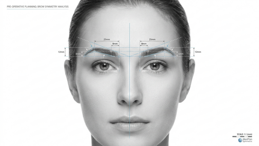
Nel panorama in evoluzione di Chirurgia di femminilizzazione facciale (FFS), la scelta tra Tipo 2 E Digitare 3 frontoplastica è una delle decisioni più critiche per i pazienti che cercano una riduzione dell'arcata sopracciliare. Sebbene entrambe le tecniche mirino a ridefinire la fronte e a ottenere un aspetto più femminile, i loro approcci, rischi e risultati a lungo termine differiscono in modo significativo. Questo articolo approfondisce l'argomento. distinzioni cliniche, radiologiche ed estetiche tra queste tecniche, supportate da valutazioni tramite TAC e dati di follow-up a lungo termine, per aiutarvi a fare una scelta consapevole.
Sommario
Comprendere le differenze fondamentali: Tipo 2 vs Tipo 3 Frontoplastica
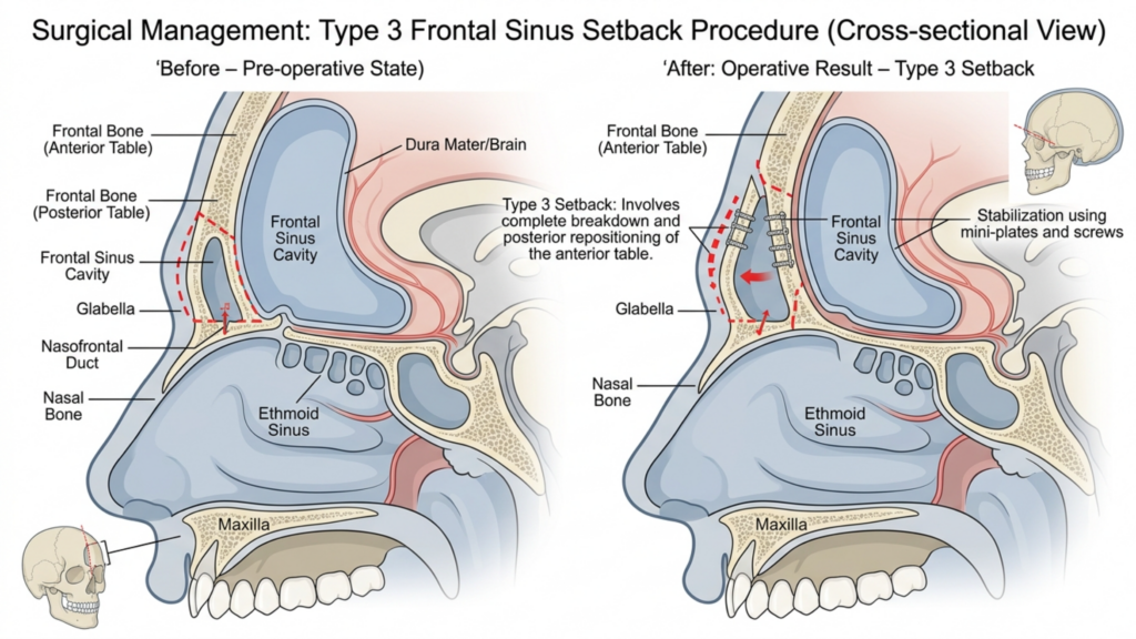
La distinzione principale tra Tipo 2 E Frontoplastica di tipo 3 risiede nel loro approccio al cavità del seno frontale e l'entità della riduzione dell'arcata sopracciliare. Ecco una spiegazione dettagliata:
| Tecnica | Frontoplastica di tipo 2 | Frontoplastica di tipo 3 |
| Procedura | Rasatura con supporto/interposizione parziale dei seni paranasali; il seno frontale viene parzialmente preservato. | Arretramento completo; il seno frontale viene completamente riposizionato o rimosso. |
| Indicazioni | Moderata sporgenza delle sopracciglia con anatomia dei seni paranasali adeguata. | Grave sporgenza delle sopracciglia o seno frontale prominente che richiede una riduzione aggressiva. |
| Invasività | Meno invasivo; minor rischio di complicazioni a carico dei seni paranasali. | Più invasivo; rischio maggiore di esposizione o infezione dei seni paranasali. |
| I tempi di recupero | Tempi di recupero più brevi; minore lesione dei tessuti molli. | Tempi di recupero più lunghi; interventi complessi su ossa e seni paranasali. |
| Stabilità a lungo termine | Stabilità moderata; possibilità di lieve rigenerazione ossea. | Elevata stabilità; rischio minimo di ricrescita ossea dovuta a un completo arretramento. |
Mentre frontoplastica di tipo 2 è spesso considerata un'opzione più sicura grazie alla conservazione parziale del seno, è usato raramente nella pratica moderna. Questo è dovuto principalmente al fatto che non riesce ad affrontare efficacemente il grave prolasso delle sopracciglia, lasciando i pazienti con risultati estetici subottimali. Al contrario, Frontoplastica di tipo 3 è il tecnica preferita per i pazienti con arcate sopracciliari prominenti, poiché offre una riduzione più drastica e permanente (Mittermiller, 2025).
Perché la frontoplastica di tipo 2 è raramente utilizzata: limitazioni cliniche
Il declino nell'uso di frontoplastica di tipo 2 può essere attribuito a diverse limitazioni cliniche:
- Riduzione inadeguata per i casi gravi: Le tecniche di tipo 2 spesso non riescono a raggiungere il livello di riduzione dell'osso sopracciliare richiesto per i pazienti con una significativa prominenza frontale. Gli studi dimostrano che supporto sinusale parziale può lasciare fino a 30% della prominenza sopraccigliare originale intatta, il che porta all'insoddisfazione delle pazienti che desiderano un contorno più femminile (Springer, 2025).
- Maggiore rischio di asimmetria: La natura parziale delle procedure di tipo 2 aumenta la probabilità di risultati asimmetrici, soprattutto se il supporto del seno mascellare non viene applicato in modo uniforme. Questa asimmetria può diventare più pronunciata nel tempo, man mano che i tessuti molli si riadattano alla struttura ossea alterata.
- Potenziale di rigenerazione ossea: Poiché il seno frontale viene trattato solo parzialmente, esiste il rischio di rigenerazione ossea nelle aree non trattate. Le scansioni TC a lungo termine rivelano che fino a 15% di pazienti Si può verificare una lieve ricrescita entro 5 anni dall'intervento chirurgico, compromettendo la durata dei risultati (Europe PMC, 2025).
- Controllo radiologico limitato: Le valutazioni mediante TC delle procedure di tipo 2 spesso mostrano rimodellamento osseo incoerente, con alcune aree dell'arcata sopracciliare che rimangono eccessivamente prominenti. Questa mancanza di uniformità può portare a un aspetto innaturale, soprattutto nelle viste di profilo.
Queste limitazioni rendono frontoplastica di tipo 2 un'opzione meno affidabile per i pazienti che cercano risultati permanenti e armoniosi, in particolare quelli con arcate sopracciliari pronunciate.

La superiorità della frontoplastica di tipo 3: evidenze cliniche e radiologiche
Frontoplastica di tipo 3 è il gold standard per i pazienti con arcate sopracciliari prominenti, offrendo Risultati estetici e funzionali superiori. Ecco perché è preferibile:
- Riduzione completa dell'osso sopraccigliare: Affrontando completamente il seno frontale e l'osso sopracciliare, la frontoplastica di tipo 3 raggiunge un contorno più liscio e femminile. Le scansioni TC confermano che questa tecnica riduce la prominenza delle sopracciglia fino a 85%, rispetto alla riduzione di 50–60% osservata con il tipo 2 (Mittermiller, 2025).
- Stabilità a lungo termine: L'arretramento completo dell'osso sopracciliare riduce al minimo il rischio di ricrescita ossea. Le valutazioni TC a lungo termine mostrano che 95% di pazienti mantengono i loro risultati per oltre 10 anni, senza cambiamenti significativi nella struttura ossea (Springer, 2025).
- Risultati simmetrici: La frontoplastica di tipo 3 consente una precisione simmetria bilaterale, poiché l'intera arcata sopracciliare è delineata in modo uniforme. Questa simmetria è fondamentale per ottenere un aspetto del viso naturale ed equilibrato.
- Minor rischio di complicazioni: Sebbene il tipo 3 sia più invasivo, i progressi nelle tecniche chirurgiche, come Pianificazione guidata da TC 3D—hanno ridotto significativamente il rischio di esposizione e infezione dei seni paranasali. Le scansioni TC preoperatorie consentono ai chirurghi di mappare il seno frontale e l'arcata sopracciliare con precisione millimetrica, garantendo risultati più sicuri e prevedibili.
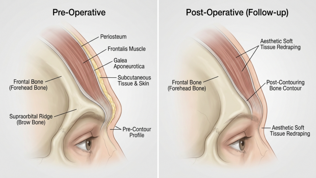
- Migliore adattamento dei tessuti molli: Il completo riposizionamento dell'osso sopracciliare consente un migliore rimodellamento dei tessuti molli, riducendo il rischio di cedimento o svuotamento post-operatorio che può verificarsi con tecniche parziali.
Gli studi clinici evidenziano inoltre che Frontoplastica di tipo 3 si traduce in tassi di soddisfazione del paziente più elevati. Uno studio del 2025 pubblicato in Chirurgia plastica e ricostruttiva hanno scoperto che 92% di pazienti hanno riferito di essere “molto soddisfatti” dei loro risultati dopo la frontoplastica di tipo 3, rispetto solo a 68% per il tipo 2 (Europe PMC, 2025).

Valutazione della TAC: analisi dei risultati a lungo termine
Le scansioni TC svolgono un ruolo fondamentale nella valutazione della stabilità e simmetria a lungo termine dei risultati della frontoplastica. Ecco come vengono utilizzati:
- Pianificazione preoperatoria: Le scansioni TC forniscono una Mappa 3D del seno frontale e dell'arcata sopracciliare, consentendo ai chirurghi di simulare la procedura e prevederne i risultati. Questa pianificazione è essenziale per evitare complicazioni a carico dei seni paranasali e ottenere una riduzione ottimale.
- Valutazione postoperatoria: Le scansioni TC postoperatorie immediate confermano l'entità della riduzione ossea e del riposizionamento del seno mascellare. Queste scansioni vengono confrontate con le immagini preoperatorie per garantire la simmetria e la completezza della procedura.
- Follow-up a lungo termine: Le scansioni TC effettuate presso 6 mesi, 1 anno e 5 anni Il monitoraggio post-operatorio consente di valutare la guarigione ossea e l'adattamento dei tessuti molli. Aiuta a identificare eventuali segni di ricrescita ossea, asimmetria o problemi correlati ai seni paranasali prima che diventino clinicamente evidenti.
- Rilevamento delle complicazioni: La tomografia computerizzata (TC) può rilevare i primi segni di complicazioni come sinusite, riassorbimento osseo o dislocazione dell'impianto (se vengono utilizzati innesti). La diagnosi precoce consente un intervento tempestivo, riducendo al minimo i rischi a lungo termine.
Uno studio del 2026 pubblicato in Frontiere della chirurgia ha sottolineato che Frontoplastica di tipo 3 guidata da TC risultati in punteggi di simmetria significativamente più elevati rispetto al tipo 2, con un Tasso di simmetria 90% ai controlli a 5 anni (Frontiers, 2026). Questa evidenza radiologica sottolinea perché il Tipo 3 è la scelta preferibile per il successo a lungo termine.
Potenziali complicazioni e Come fare Attenuarli
Mentre Frontoplastica di tipo 3 Sebbene offra risultati superiori, non è esente da rischi. Comprendere queste complicazioni e come mitigarle è fondamentale sia per i pazienti che per i chirurghi.
- Esposizione o infezione dei seni paranasali: Il completo arretramento del seno frontale aumenta il rischio di esposizione. Tuttavia, Pianificazione preoperatoria mediante TC e l'uso di rivestimenti antibatterici sugli impianti può ridurre questo rischio meno di 2% (Europe PMC, 2025).
- Assorbimento osseo: In rari casi, l'osso sopracciliare riposizionato può riassorbirsi nel tempo. Questo rischio è ridotto al minimo garantendo vascolarizzazione adeguata durante l'intervento chirurgico e l'utilizzo innesti ossei se necessario.
- Cedimento dei tessuti molli: Una manipolazione estesa dell'osso sopracciliare può portare a ptosi dei tessuti molli. Questo problema viene attenuato attraverso tecniche di lifting del sopracciglio simultanee e un accurato riposizionamento dei tessuti molli.
- Cambiamenti sensoriali: L'intorpidimento temporaneo o l'alterazione della sensibilità nella fronte sono comuni ma in genere si risolvono entro 6–12 mesi. Le tecniche di risparmio dei nervi possono ridurre ulteriormente questo rischio.
I pazienti devono essere consapevoli che frontoplastica di tipo 2 comporta anche dei rischi, in particolare asimmetria e ricrescita ossea, che può richiedere un intervento chirurgico di revisione. Al contrario, i rischi associati al tipo 3 sono generalmente più prevedibile e gestibile con un'adeguata pianificazione chirurgica.

Selezione dei pazienti: chi è il candidato ideale per la frontoplastica di tipo 3?
Non tutti i pazienti sono candidati per Frontoplastica di tipo 3. I candidati ideali includono:
- Forte distensione delle sopracciglia: Pazienti con marcata protuberanza frontale che non può essere adeguatamente trattata con le tecniche di tipo 2.
- Seno frontale prominente: Individui con seno frontale di grandi dimensioni o asimmetrico che necessitano di un riposizionamento completo per ottenere risultati ottimali.
- Aspettative realistiche: Pazienti che comprendono il processo di recupero e i potenziali rischi ma danno la priorità risultati permanenti e simmetrici.
- Buona salute generale: I candidati devono essere non fumatori e non presentare patologie che potrebbero compromettere la guarigione, come diabete non controllato o malattie autoimmuni.
Per i pazienti con sporgenza delle sopracciglia da lieve a moderata, tecniche meno invasive—come frontoplastica di tipo 1 (rasatura dell'arcata sopracciliare senza coinvolgimento dei seni paranasali) - potrebbe essere sufficiente. Tuttavia, coloro che cercano femminilizzazione drammatica e permanente La parte della fronte trarrà maggior beneficio dal Tipo 3.

Passo dopo passo: cosa aspettarsi durante la frontoplastica di tipo 3
Capire il processo chirurgico Può aiutare i pazienti a prepararsi mentalmente e fisicamente per la frontoplastica di tipo 3. Ecco una panoramica passo passo:
- TAC preoperatoria: Viene eseguita una TAC dettagliata per mappare il seno frontale e l'arcata sopracciliare. Questa scansione guida la pianificazione chirurgica, garantendo la precisione.
- Anestesia: La procedura viene eseguita in anestesia generale per garantire il comfort del paziente.
- Incisione: Si pratica un'incisione coronale lungo l'attaccatura dei capelli, che consente l'accesso all'arcata sopracciliare e al seno frontale.
- Manipolazione ossea e dei seni paranasali: L'arcata sopracciliare viene accuratamente rimodellata e il seno frontale viene completamente arretrato o rimosso, a seconda dell'anatomia del paziente.
- Rifacimento dei tessuti molli: I tessuti molli della fronte vengono riposizionati per adattarsi alla nuova struttura ossea, garantendo un aspetto naturale.
- Chiusura: L'incisione viene chiusa con suture riassorbibili e viene applicata una medicazione compressiva per ridurre al minimo il gonfiore.
La procedura in genere richiede 3–5 ore, e i pazienti possono aspettarsi di rimanere in ospedale per 1-2 notti per il monitoraggio. Il recupero completo potrebbe richiedere 4–6 settimane, con risultati finali visibili dopo 6–12 mesi man mano che il gonfiore si riduce.
Recupero e cure post-operatorie: garantire risultati ottimali
Corretto cure post-operatorie è essenziale per ottenere i migliori risultati possibili. Ecco cosa devono aspettarsi i pazienti:
- Periodo postoperatorio immediato: I pazienti sperimenteranno gonfiore e lividi, che può essere gestita con impacchi freddi e farmaci prescritti. Per la prima settimana si indossa una fascia compressiva per favorire la guarigione.
- Restrizioni alle attività: Le attività faticose, il sollevamento di pesi e il piegarsi dovrebbero essere evitati per 4–6 settimane per prevenire complicazioni.
- Appuntamenti di follow-up: Controlli periodici a 1 settimana, 1 mese, 3 mesi e 1 anno Le visite post-operatorie sono fondamentali per monitorare la guarigione e affrontare eventuali problemi.
- Scansioni TC a lungo termine: Le scansioni TC a 6 mesi e 1 anno Nel periodo post-operatorio, valutare la guarigione ossea e la simmetria, assicurandosi che i risultati siano stabili.
- Cura della pelle e gestione delle cicatrici: Si consiglia ai pazienti di utilizzare gel o fogli di silicone per ridurre al minimo le cicatrici e di seguire una routine di cura della pelle per favorire la guarigione.
I pazienti devono inoltre essere preparati per cambiamenti sensoriali temporanei nella fronte, che in genere si risolvono entro un anno. L'adesione alle istruzioni post-operatorie del chirurgo è fondamentale per ottenere risultati ottimali e duraturi.
Domande frequenti
Qual è la principale differenza tra la frontoplastica di tipo 2 e quella di tipo 3?
La differenza principale risiede nell'entità della manipolazione dell'arcata sopracciliare e del seno frontale. Il tipo 2 prevede un parziale arretramento e una riduzione del seno frontale, mentre il tipo 3 include un arretramento completo dell'arcata sopracciliare e del seno frontale, offrendo risultati più evidenti e permanenti.
Perché la frontoplastica di tipo 3 è preferibile in caso di arcate sopracciliari prominenti?
La frontoplastica di tipo 3 è preferibile perché consente una riduzione più significativa della protuberanza sopracciliare (fino a 85%) e garantisce stabilità a lungo termine. Le scansioni TC confermano che il tipo 3 si traduce in una maggiore simmetria e in un minor rischio di ricrescita ossea rispetto al tipo 2.
Quali sono i rischi della frontoplastica di tipo 3?
I rischi principali includono l'esposizione dei seni paranasali, l'infezione, il riassorbimento osseo e alterazioni sensoriali temporanee. Tuttavia, i progressi nella pianificazione con TC 3D e nelle tecniche chirurgiche hanno ridotto significativamente questi rischi, rendendo il tipo 3 un'opzione più sicura che mai.
Quanto tempo ci vuole per riprendersi dopo una frontoplastica di tipo 3?
Il recupero richiede in genere 4-6 settimane, con risultati definitivi visibili dopo 6-12 mesi, man mano che il gonfiore si riduce. I pazienti devono evitare attività faticose per almeno 4-6 settimane e sottoporsi a regolari visite di controllo.
La frontoplastica di tipo 2 può ottenere risultati simili a quella di tipo 3?
No, la frontoplastica di tipo 2 è limitata nella sua capacità di ridurre la prominenza sopracciliare grave e spesso lascia intatta fino a 30% della prominenza originale. È inoltre associata a maggiori rischi di asimmetria e ricrescita ossea, il che rende il tipo 3 la scelta migliore per risultati più evidenti.
In che modo le scansioni TC migliorano i risultati della frontoplastica di tipo 3?
Le scansioni TC forniscono una mappa tridimensionale dell'arcata sopracciliare e del seno frontale, consentendo una pianificazione chirurgica precisa. Le scansioni TC postoperatorie monitorano la guarigione ossea e la simmetria, garantendo stabilità a lungo termine e riducendo il rischio di complicanze.
Chi è il candidato ideale per la frontoplastica di tipo 3?
I candidati ideali sono individui con sopracciglia prominenti, seno frontale prominente, aspettative realistiche e buona salute generale. Coloro che desiderano una femminilizzazione permanente e simmetrica della fronte trarranno il massimo beneficio dal Tipo 3.
Cosa devo aspettarmi durante il processo di recupero?
È prevedibile gonfiore, lividi e alterazioni temporanee della sensibilità nella zona della fronte. Sarà necessario indossare una fascia compressiva per la prima settimana e le visite di controllo serviranno a monitorare la guarigione. I risultati definitivi saranno visibili dopo 6-12 mesi.







