
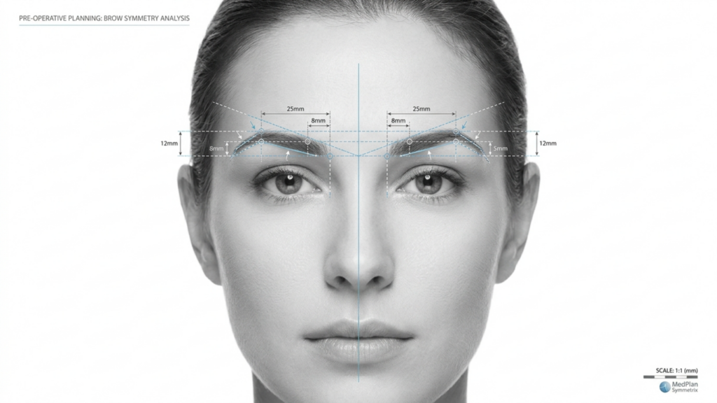
In het veranderende landschap van Gezichtsfeminisatiechirurgie (VFF), de keuze tussen Type 2 En Soort 3 frontoplastie De keuze voor een wenkbrauwverkleining is een van de meest cruciale beslissingen voor patiënten die een wenkbrauwverkleining overwegen. Hoewel beide technieken gericht zijn op het verfijnen van het voorhoofd en het bereiken van een meer vrouwelijke uitstraling, verschillen hun aanpak, risico's en resultaten op lange termijn aanzienlijk. Dit artikel gaat dieper in op de verschillende aspecten. klinische, radiologische en esthetische verschillen tussen deze technieken, ondersteund door CT-scanbeoordelingen en langetermijngegevens, zodat u een weloverwogen keuze kunt maken.
Inhoudsopgave
De kernverschillen begrijpen: Type 2 versus Type 3 Frontoplastie
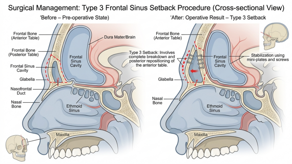
Het voornaamste onderscheid tussen Type 2 En Type 3 frontoplastie ligt in hun benadering van de frontale sinusholte en de mate van vermindering van het wenkbrauwbot. Hier volgt een overzicht:
| Techniek | Type 2 frontoplastie | Type 3 frontoplastie |
| Procedure | Scheertechniek met gedeeltelijke ondersteuning/interpositie van de sinus; de frontale sinus blijft gedeeltelijk behouden. | Volledige terugplaatsing; de frontale sinus wordt volledig verplaatst of verwijderd. |
| Indicaties | Matige wenkbrauwuitstulping met adequate sinusanatomie. | Ernstige wenkbrauwuitstulping of prominente voorhoofdsholte die een agressieve correctie vereist. |
| Invasiviteit | Minder ingrijpend; lager risico op complicaties aan de sinussen. | Invasiever; hoger risico op blootstelling van de sinussen of infectie. |
| Hersteltijd | Kortere herstelperiode; minder beschadiging van zacht weefsel. | Langer herstel; uitgebreide manipulatie van botten en sinussen. |
| Stabiliteit op lange termijn | Matige stabiliteit; mogelijkheid tot geringe botgroei. | Hoge stabiliteit; minimaal risico op botregeneratie als gevolg van volledige terugval. |
Terwijl Type 2 frontoplastie wordt vaak als een veiligere optie beschouwd vanwege de gedeeltelijke instandhouding van de sinussen, het is zelden gebruikt in de moderne praktijk. Dit komt voornamelijk doordat het ernstige wenkbrauwuitstulpingen niet effectief aanpakt, waardoor patiënten met suboptimale esthetische resultaten achterblijven. Daarentegen, Type 3 frontoplastie is de voorkeurstechniek voor patiënten met prominente wenkbrauwbotten, omdat het een meer dramatische en permanente vermindering biedt (Mittermiller, 2025).
Waarom frontoplastiek type 2 zelden wordt toegepast: klinische beperkingen
De afname in het gebruik van Type 2 frontoplastie Dit kan worden toegeschreven aan verschillende klinische beperkingen:
- Onvoldoende reductie bij ernstige gevallen: Type 2-technieken slagen er vaak niet in om de vereiste mate van wenkbrauwbotreductie te bereiken bij patiënten met een aanzienlijke frontale bult. Studies tonen aan dat gedeeltelijke sinusverstopping kan tot 30% van de oorspronkelijke wenkbrauwprominentie intact, wat leidt tot ontevredenheid bij patiënten die een meer vrouwelijke lichaamscontour wensen (Springer, 2025).
- Verhoogd risico op asymmetrie: Het gedeeltelijke karakter van procedures van type 2 vergroot de kans op asymmetrische resultaten, Vooral als de sinusvulling niet gelijkmatig is aangebracht. Deze asymmetrie kan in de loop der tijd verergeren, doordat het zachte weefsel zich aanpast aan de veranderde botstructuur.
- Potentieel voor botregeneratie: Omdat de frontale sinus slechts gedeeltelijk wordt behandeld, bestaat er een risico op botregeneratie in de onbehandelde gebieden. CT-scans op lange termijn laten zien dat tot 15% van patiënten binnen 5 jaar na de operatie treedt er een lichte hergroei op, waardoor de duurzaamheid van de resultaten in gevaar komt (Europe PMC, 2025).
- Beperkte radiologische controle: CT-scans bij type 2-procedures laten vaak zien dat inconsistente botremodellering, waarbij sommige delen van het wenkbrauwbot te prominent aanwezig blijven. Dit gebrek aan uniformiteit kan leiden tot een onnatuurlijk uiterlijk, met name in profielaanzichten.
Deze beperkingen maken Type 2 frontoplastie een minder betrouwbare optie voor patiënten die op zoek zijn naar duurzame en harmonieuze resultaten, met name mensen met uitgesproken wenkbrauwbotten.

De superioriteit van frontoplastiek type 3: klinisch en radiologisch bewijs
Type 3 frontoplastie is de gouden standaard voor patiënten met prominente wenkbrauwbotten en biedt superieure esthetische en functionele resultaten. Dit is waarom het de voorkeur geniet:
- Volledige wenkbrauwbotreductie: Door de frontale sinus en het wenkbrauwbot volledig aan te pakken, bereikt een frontoplastiek type 3 een een gladdere, meer vrouwelijke contour. CT-scans bevestigen dat deze techniek de prominentie van de wenkbrauwen vermindert door tot 85%, vergeleken met de 50–60%-reductie die wordt gezien bij type 2 (Mittermiller, 2025).
- Stabiliteit op lange termijn: De volledige terugplaatsing van het wenkbrauwbot minimaliseert het risico op botregeneratie. CT-onderzoeken op lange termijn tonen aan dat 95% van patiënten Ze behouden hun resultaten gedurende meer dan 10 jaar, zonder significante veranderingen in de botstructuur (Springer, 2025).
- Symmetrische uitkomsten: Type 3 frontoplastiek maakt nauwkeurige resultaten mogelijk. bilaterale symmetrie, Doordat het gehele wenkbrauwbot gelijkmatig is gevormd. Deze symmetrie is cruciaal voor een natuurlijke en evenwichtige gezichtsuitstraling.
- Lager risico op complicaties: Hoewel type 3 invasiever is, hebben verbeteringen in chirurgische technieken, zoals 3D CT-geleide planning—hebben het risico op blootstelling van de sinussen en infecties aanzienlijk verminderd. Preoperatieve CT-scans stellen chirurgen in staat om de voorhoofdsholte en het wenkbrauwbot met millimeterprecisie in kaart te brengen, wat zorgt voor veiligere en voorspelbaardere resultaten.
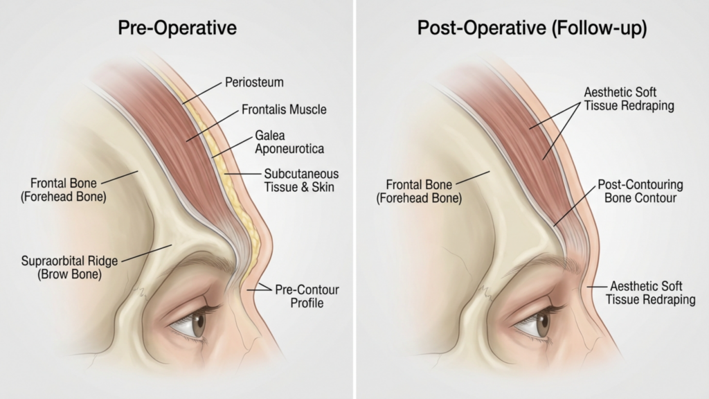
- Verbeterde aanpassing van zacht weefsel: Door de volledige herpositionering van het wenkbrauwbot kan het zachte weefsel beter worden gedrapeerd, waardoor het risico op postoperatieve verzakking of inzinking Dat kan gebeuren bij gedeeltelijke technieken.
Klinische studies tonen ook aan dat Type 3 frontoplastie Dit leidt tot hogere patiënttevredenheid. Een studie uit 2025, gepubliceerd in Plastische en reconstructieve chirurgie ontdekte dat 92% van patiënten gaven aan “zeer tevreden” te zijn met hun resultaten na een frontoplastie van type 3, vergeleken met slechts 68% voor Type 2 (Europe PMC, 2025).

CT-scanbeoordeling: evaluatie van de resultaten op lange termijn
CT-scans spelen een cruciale rol bij de beoordeling van de stabiliteit en symmetrie op lange termijn van de resultaten van een frontoplastie. Zo worden ze gebruikt:
- Preoperatieve planning: CT-scans bieden een 3D-kaart van de voorhoofdsholte en het wenkbrauwbot, waardoor chirurgen de procedure kunnen simuleren en de resultaten kunnen voorspellen. Deze planning is essentieel om complicaties aan de sinussen te voorkomen en een optimale reductie te bereiken.
- Postoperatieve evaluatie: Direct na de operatie worden CT-scans gemaakt om de mate van botreductie en sinusrepositionering te bevestigen. Deze scans worden vergeleken met preoperatieve beelden om symmetrie en volledigheid van de ingreep te waarborgen.
- Langetermijnfollow-up: CT-scans uitgevoerd bij 6 maanden, 1 jaar en 5 jaar Ze controleren na de operatie de botgenezing en de aanpassing van het zachte weefsel. Ze helpen bij het opsporen van tekenen van botgroei, asymmetrie of problemen met de sinussen voordat deze klinisch zichtbaar worden.
- Complicatiedetectie: CT-scans kunnen vroege tekenen van complicaties opsporen, zoals sinusitis, botresorptie of verplaatsing van het implantaat (indien er gebruik wordt gemaakt van transplantaten). Vroegtijdige opsporing maakt tijdige interventie mogelijk, waardoor de risico's op lange termijn worden geminimaliseerd.
Een studie uit 2026 gepubliceerd in Grenzen in de chirurgie benadrukte dat CT-geleide frontoplastiek type 3 resulteert in significant hogere symmetriescores vergeleken met type 2, met een 90% symmetriesnelheid bij follow-ups na 5 jaar (Frontiers, 2026). Dit radiologische bewijs onderstreept waarom type 3 de voorkeur geniet voor succes op de lange termijn.
Mogelijke complicaties en Hoe te Verzacht ze
Terwijl Type 3 frontoplastie Hoewel het superieure resultaten oplevert, is het niet zonder risico's. Inzicht in deze complicaties – en hoe ze te beperken – is cruciaal voor zowel patiënten als chirurgen.
- Blootstelling aan of infectie van de sinussen: De volledige terugtrekking van de voorhoofdsholte vergroot het risico op blootstelling. Echter, preoperatieve CT-planning en het gebruik van antibacteriële coatings op implantaten kan dit risico verminderen minder dan 2% (Europe PMC, 2025).
- Botafbraak: In zeldzame gevallen kan het verplaatste wenkbrauwbot na verloop van tijd resorberen. Dit risico wordt geminimaliseerd door ervoor te zorgen dat... goede doorbloeding tijdens de operatie en met gebruik van bottransplantaten indien nodig.
- Verslapping van het weke weefsel: Uitgebreide manipulatie van het wenkbrauwbot kan leiden tot ptosis van het weke weefsel. Dit wordt verholpen door gelijktijdige wenkbrauwlifttechnieken en zorgvuldige herpositionering van het weke weefsel.
- Sensorische veranderingen: Tijdelijke gevoelloosheid of een veranderd gevoel in het voorhoofd komt vaak voor, maar verdwijnt doorgaans vanzelf. 6–12 maanden. Zenuwsparende technieken kunnen dit risico verder verlagen.
Patiënten moeten zich ervan bewust zijn dat Type 2 frontoplastie brengt ook risico's met zich mee, met name asymmetrie en botregeneratie, wat een revisieoperatie noodzakelijk kan maken. Daarentegen zijn de risico's verbonden aan type 3 over het algemeen voorspelbaarder en beter beheersbaar met een goede chirurgische planning.

Patiëntselectie: Wie is de ideale kandidaat voor een frontoplastiek type 3?
Niet alle patiënten komen in aanmerking voor Type 3 frontoplastie. Ideale kandidaten zijn onder andere:
- Ernstige wenkbrauwuitstulping: Patiënten met een uitgesproken frontale bult die niet adequaat kan worden behandeld met Type 2-technieken.
- Opvallende frontale sinus: Personen met een grote of asymmetrische frontale sinus die een volledige herpositionering vereist voor optimale resultaten.
- Realistische verwachtingen: Patiënten die het herstelproces en de mogelijke risico's begrijpen, maar prioriteit geven aan... permanente, symmetrische resultaten.
- Goede algemene gezondheid: Kandidaten dienen niet-rokers te zijn en geen aandoeningen te hebben die de genezing kunnen belemmeren, zoals ongecontroleerde diabetes of auto-immuunziekten.
Voor patiënten met milde tot matige wenkbrauwuitstulping, minder invasieve technieken, zoals Type 1 frontoplastie (Het schaven van het wenkbrauwbot zonder betrokkenheid van de sinussen) kan voldoende zijn. Degenen die echter op zoek zijn naar een oplossing, kunnen hier wellicht meer informatie vinden. dramatische en permanente feminisering Het voorhoofd zal het meest baat hebben bij type 3.

Stap voor stap: Wat u kunt verwachten tijdens een frontoplastie type 3
Begrijpen van de chirurgisch proces Dit kan patiënten helpen zich mentaal en fysiek voor te bereiden op een frontoplastie type 3. Hier volgt een stapsgewijs overzicht:
- Preoperatieve CT-scan: Er wordt een gedetailleerde CT-scan gemaakt om de voorhoofdsholte en het wenkbrauwbot in kaart te brengen. Deze scan dient als leidraad voor het chirurgische plan en zorgt voor precisie.
- Anesthesie: De ingreep wordt uitgevoerd onder algehele narcose om het comfort van de patiënt te garanderen.
- Insnijding: Er wordt een coronale incisie gemaakt langs de haarlijn, waardoor toegang wordt verkregen tot het wenkbrauwbot en de voorhoofdsholte.
- Bot- en sinusmanipulatie: Het wenkbrauwbot wordt zorgvuldig gevormd en de voorhoofdsholte wordt volledig teruggeplaatst of verwijderd, afhankelijk van de anatomie van de patiënt.
- Herstructurering van zacht weefsel: Het zachte weefsel van het voorhoofd wordt verplaatst om zich aan te passen aan de nieuwe botstructuur, waardoor een natuurlijk uiterlijk wordt gegarandeerd.
- Sluiting: De incisie wordt gesloten met oplosbare hechtingen en er wordt een drukverband aangebracht om zwelling te minimaliseren.
De procedure duurt doorgaans 3-5 uur, en patiënten kunnen verwachten dat ze gedurende een bepaalde periode in het ziekenhuis zullen verblijven. 1-2 nachten voor monitoring. Volledig herstel kan langer duren. 4–6 weken, waarbij de uiteindelijke resultaten na afloop zichtbaar zijn. 6–12 maanden naarmate de zwelling afneemt.
Herstel en nazorg: zorgen voor optimale resultaten
Juist postoperatieve zorg is essentieel voor het behalen van de best mogelijke resultaten. Dit is wat patiënten kunnen verwachten:
- Directe postoperatieve periode: Patiënten zullen het volgende ervaren: zwelling en blauwe plekken, Dit kan worden verholpen met koude kompressen en voorgeschreven medicijnen. Gedurende de eerste week wordt een compressiehoofdband gedragen ter ondersteuning van de genezing.
- Activiteitsbeperkingen: Inspannende activiteiten, zwaar tillen en bukken moeten worden vermeden. 4–6 weken om complicaties te voorkomen.
- Vervolgafspraken: Regelmatige vervolgbezoeken bij 1 week, 1 maand, 3 maanden en 1 jaar Controles na de operatie zijn cruciaal voor het bewaken van het genezingsproces en het aanpakken van eventuele problemen.
- CT-scans voor de lange termijn: CT-scans bij 6 maanden en 1 jaar Na de operatie wordt de botgenezing en symmetrie beoordeeld om er zeker van te zijn dat de resultaten stabiel zijn.
- Huidverzorging en littekenbeheer: Patiënten wordt aangeraden siliconengel of -pleisters te gebruiken om littekenvorming te minimaliseren en een huidverzorgingsroutine te volgen ter ondersteuning van de genezing.
Patiënten moeten ook voorbereid zijn op tijdelijke zintuiglijke veranderingen in het voorhoofd, die doorgaans binnen een jaar verdwijnen. Het opvolgen van de nazorginstructies van de chirurg is essentieel voor een succesvol resultaat. optimale, langdurige resultaten.
Veelgestelde vragen
Wat is het belangrijkste verschil tussen een frontoplastie type 2 en type 3?
Het belangrijkste verschil zit hem in de mate waarin het wenkbrauwbot en de voorhoofdsholte worden gemanipuleerd. Bij type 2 wordt de voorhoofdsholte gedeeltelijk teruggeplaatst en afgeschraapt, terwijl bij type 3 het wenkbrauwbot en de voorhoofdsholte volledig worden teruggeplaatst, wat een dramatischer en permanenter resultaat oplevert.
Waarom wordt frontoplastiek type 3 verkozen bij prominente wenkbrauwbotten?
Frontoplastiek type 3 heeft de voorkeur omdat hiermee een significantere vermindering van de wenkbrauwuitstulping wordt bereikt (tot 85%) en stabiliteit op lange termijn wordt geboden. CT-scans bevestigen dat type 3 resulteert in een hogere symmetrie en een lager risico op botregroei in vergelijking met type 2.
Wat zijn de risico's van een frontoplastie type 3?
De belangrijkste risico's zijn blootstelling van de sinussen, infectie, botresorptie en tijdelijke sensorische veranderingen. Dankzij vooruitgang in 3D CT-planning en chirurgische technieken zijn deze risico's echter aanzienlijk verminderd, waardoor type 3 een veiligere optie is dan ooit tevoren.
Hoe lang duurt het herstel na een frontoplastie type 3?
Het herstel duurt doorgaans 4-6 weken, waarbij het volledige resultaat na 6-12 maanden zichtbaar is, wanneer de zwelling afneemt. Patiënten dienen gedurende ten minste 4-6 weken inspannende activiteiten te vermijden en regelmatig op controle te komen.
Kan een frontoplastie van type 2 vergelijkbare resultaten opleveren als een frontoplastie van type 3?
Nee, een frontoplastie type 2 is beperkt in zijn mogelijkheden om ernstige wenkbrauwuitstulpingen te verminderen en laat vaak tot wel 30% van de oorspronkelijke prominentie intact. Het is ook geassocieerd met een hoger risico op asymmetrie en botregroei, waardoor type 3 de betere keuze is voor een spectaculair resultaat.
Hoe verbeteren CT-scans de resultaten van een frontoplastiek type 3?
CT-scans leveren een 3D-kaart van het wenkbrauwbot en de voorhoofdsholte, waardoor een nauwkeurige chirurgische planning mogelijk is. Postoperatieve CT-scans controleren de botgenezing en symmetrie, wat zorgt voor stabiliteit op lange termijn en het risico op complicaties verkleint.
Wie is de ideale kandidaat voor een frontoplastiek type 3?
Ideale kandidaten zijn personen met een sterk uitstekende wenkbrauw, een prominente voorhoofdsholte, realistische verwachtingen en een goede algehele gezondheid. Degenen die een permanente en symmetrische feminisering van het voorhoofd wensen, zullen het meest baat hebben bij type 3.
Wat kan ik verwachten tijdens het herstelproces?
Verwacht zwelling, blauwe plekken en tijdelijke veranderingen in het gevoel in het voorhoofd. Gedurende de eerste week zal een compressieverband gedragen worden en tijdens vervolgafspraken zal het herstel in de gaten gehouden worden. Het volledige resultaat is zichtbaar na 6-12 maanden.







