
Gelişen manzarada Yüz Dişileştirme Ameliyatı (FFS), aralarındaki seçim Tip 2 Ve Tip 3 önplasti Kaş kemiği küçültme ameliyatı arayan hastalar için en kritik kararlardan biridir. Her iki teknik de alnı inceltmeyi ve daha kadınsı bir görünüm elde etmeyi amaçlasa da, yaklaşımları, riskleri ve uzun vadeli sonuçları önemli ölçüde farklılık gösterir. Bu makale, bu iki tekniğin derinlemesine incelenmesine odaklanmaktadır. klinik, radyolojik ve estetik farklılıklar bu teknikler arasında, desteklenen BT tarama değerlendirmeleri ve uzun vadeli takip verileri, bilinçli bir seçim yapmanıza yardımcı olacaktır.
İçindekiler
Temel Farklılıkları Anlamak: Tip 2 Tip 3'e karşı Frontoplasti
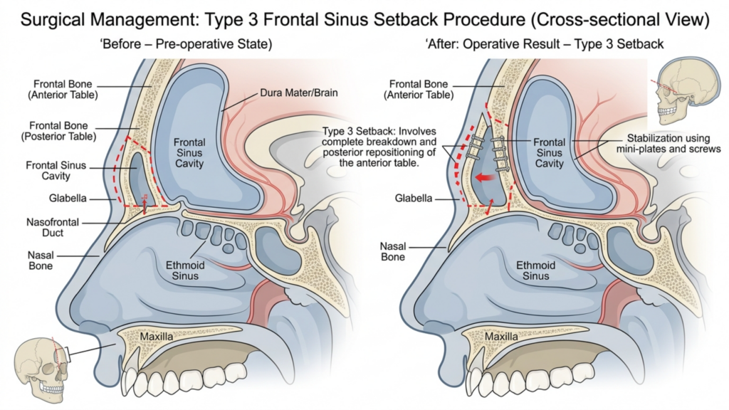
Temel fark şudur: Tip 2 Ve Tip 3 frontoplasti Bu, onların yaklaşım biçiminde yatmaktadır. frontal sinüs boşluğu ve kaş kemiği küçültme işleminin derecesi. İşte ayrıntılar:
| Teknik | Tip 2 Frontoplasti | Tip 3 Frontoplasti |
| Prosedür | Kısmi sinüs desteği/ara yerleştirme ile tıraşlama; frontal sinüs kısmen korunmuştur. | Tam bir gerileme; frontal sinüs tamamen yeniden konumlandırılır veya çıkarılır. |
| Endikasyonlar | Orta derecede kaş çıkıntısı ve yeterli sinüs anatomisi. | Agresif bir şekilde küçültülmesi gereken ciddi kaş çıkıntısı veya belirgin alın sinüsü. |
| İstilacılık | Daha az invaziv; sinüs komplikasyonları riski daha düşük. | Daha invaziv; sinüslerin açığa çıkması veya enfeksiyon riski daha yüksek. |
| İyileşme süresi | Daha kısa iyileşme süresi; daha az yumuşak doku hasarı. | Daha uzun iyileşme süreci; kapsamlı kemik ve sinüs manipülasyonu. |
| Uzun Vadeli İstikrar | Orta düzeyde stabilite; küçük çaplı kemik yeniden büyüme potansiyeli. | Yüksek stabilite; tam geri çekilme nedeniyle kemik yeniden büyüme riski minimum düzeydedir. |
Sırasında Tip 2 frontoplasti Sinüsün kısmen korunması nedeniyle genellikle daha güvenli bir seçenek olarak kabul edilir. nadiren kullanılır Modern uygulamada bunun başlıca nedeni, ciddi kaş çıkıntılarını etkili bir şekilde ele alamaması ve hastaları optimal olmayan estetik sonuçlarla baş başa bırakmasıdır. Buna karşılık, Tip 3 frontoplasti dır tercih edilen teknik Kaş kemikleri belirgin olan hastalar için daha çarpıcı ve kalıcı bir azalma sağladığı için tercih edilir (Mittermiller, 2025).
Tip 2 Frontoplastinin Nadiren Kullanılmasının Nedenleri: Klinik Sınırlamalar
Kullanımındaki azalma Tip 2 frontoplasti Bu durum çeşitli klinik sınırlamalara bağlanabilir:
- Ağır vakalarda yetersiz azalma: Tip 2 teknikler, belirgin alın çıkıntısı olan hastalarda gereken kaş kemiği küçültme seviyesine ulaşmada genellikle başarısız olur. Çalışmalar şunu göstermektedir ki kısmi sinüs desteği en fazla bırakabilir Orijinal kaş çıkıntısının 30%'si bozulmamış halde., Bu durum, daha kadınsı bir görünüm arayan hastalar arasında memnuniyetsizliğe yol açmaktadır (Springer, 2025).
- Asimetri Riski Daha Yüksek: Tip 2 prosedürlerinin kısmi niteliği, olasılığını artırır. asimetrik sonuçlar, Özellikle sinüs desteği düzgün bir şekilde uygulanmadığında bu durum daha da belirginleşir. Yumuşak doku değişen kemik yapısına yeniden uyum sağladıkça bu asimetri zamanla daha da belirgin hale gelebilir.
- Kemik Yeniden Büyüme Potansiyeli: Alın sinüsüne yalnızca kısmen müdahale edildiği için şu risk mevcuttur: kemik yeniden büyümesi Tedavi edilmemiş bölgelerde. Uzun süreli BT taramaları, 'ye kadar hasar olduğunu ortaya koymaktadır. 15% hastası Ameliyat sonrası 5 yıl içinde küçük çaplı yeniden büyüme görülebilir ve bu durum sonuçların kalıcılığını tehlikeye atabilir (Europe PMC, 2025).
- Sınırlı Radyolojik Kontrol: Tip 2 prosedürlerin BT taraması değerlendirmeleri genellikle şunu gösterir: tutarsız kemik yeniden şekillenmesi, Kaş kemiğinin bazı bölgeleri aşırı belirgin kalır. Bu düzensizlik, özellikle profilden bakıldığında doğallıktan uzak bir görünüme yol açabilir.
Bu sınırlamalar, Tip 2 frontoplasti hastalar için daha az güvenilir bir seçenek kalıcı ve uyumlu sonuçlar, özellikle belirgin kaş kemiklerine sahip olanlar.

Tip 3 Frontoplastinin Üstünlüğü: Klinik ve Radyolojik Kanıtlar
Tip 3 frontoplasti Kaş kemikleri belirgin olan hastalar için altın standarttır ve şu avantajları sunar: üstün estetik ve fonksiyonel sonuçlar. İşte tercih edilmesinin nedenleri:
- Kaş Kemiği Küçültme İşleminin Tamamı: Tip 3 frontoplasti, frontal sinüs ve kaş kemiğini tamamen ele alarak şu sonucu elde eder: daha pürüzsüz, daha kadınsı bir kontür. BT taramaları, bu tekniğin kaş çıkıntısını azalttığını doğrulamaktadır. 85%'ye kadar, Tip 2'de görülen 50–60% azalmasına kıyasla (Mittermiller, 2025).
- Uzun Vadeli İstikrar: Kaş kemiğinin tamamen geriye çekilmesi, kemik yeniden büyüme riskini en aza indirir. Uzun vadeli BT değerlendirmeleri şunu göstermektedir: 95% hastanın Kemik yapısında önemli bir değişiklik olmaksızın sonuçlarını 10 yıldan fazla süreyle koruyabiliyorlar (Springer, 2025).
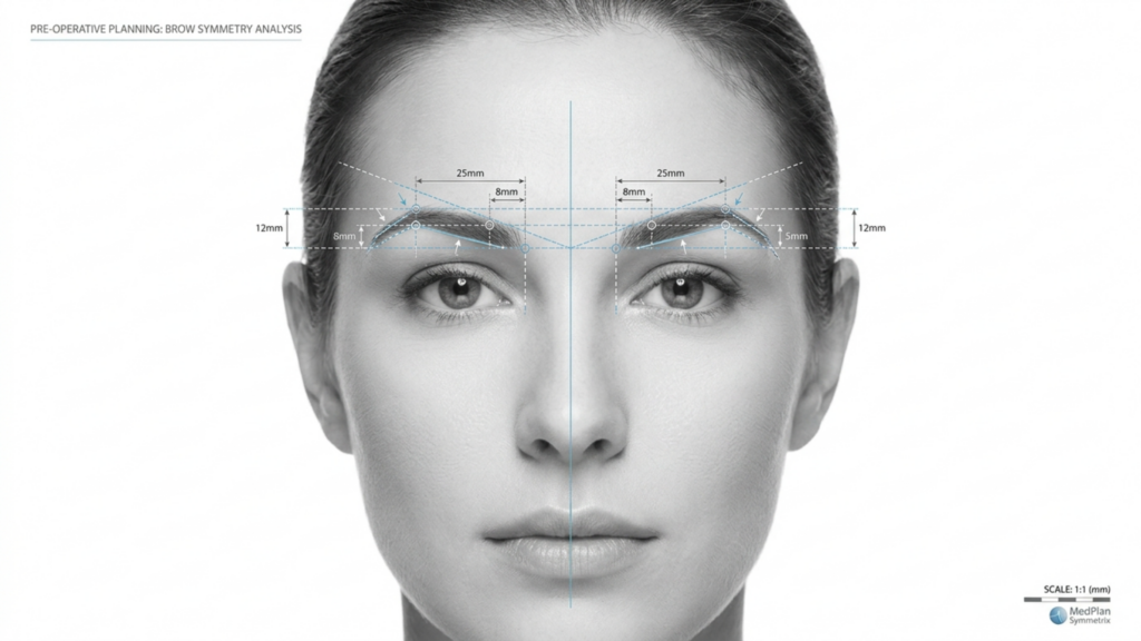
- Simetrik Sonuçlar: Tip 3 frontoplasti, hassas bir uygulama sağlar. iki taraflı simetri, Çünkü kaş kemiğinin tamamı eşit şekilde şekillendirilmiştir. Bu simetri, doğal ve dengeli bir yüz görünümü elde etmek için çok önemlidir.
- Komplikasyon Riski Daha Düşük: Tip 3 daha invaziv olmakla birlikte, cerrahi tekniklerdeki gelişmeler—örneğin, 3 boyutlu BT rehberliğinde planlama—sinüs maruziyeti ve enfeksiyon riskini önemli ölçüde azaltmıştır. Ameliyat öncesi BT taramaları, cerrahların frontal sinüsü ve kaş kemiğini milimetre hassasiyetinde haritalandırmasını sağlayarak daha güvenli ve daha öngörülebilir sonuçlar elde edilmesini sağlar.
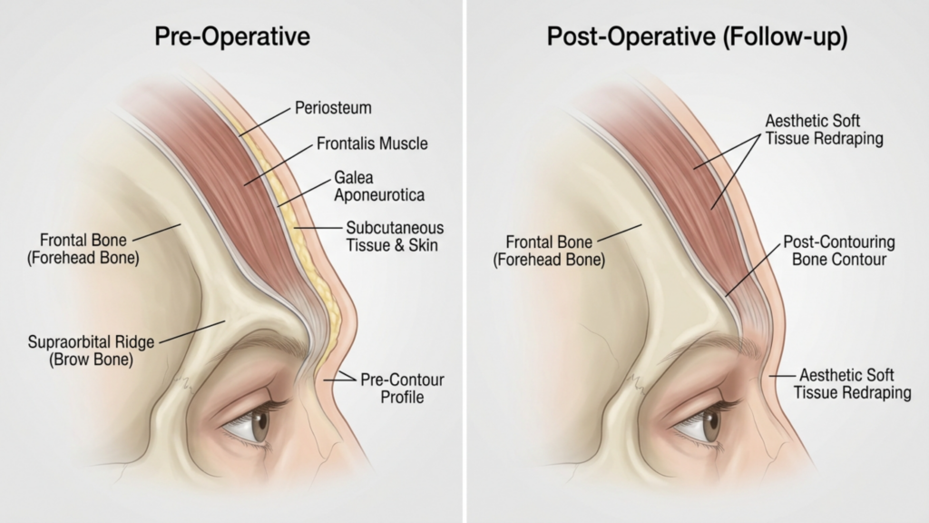
- Yumuşak Doku Adaptasyonunun Geliştirilmesi: Kaş kemiğinin tamamen yeniden konumlandırılması, yumuşak dokuların daha iyi yeniden şekillenmesini sağlayarak riskini azaltır. ameliyat sonrası sarkma veya çökme Bu durum kısmi tekniklerde ortaya çıkabilir.
Klinik çalışmalar ayrıca şunu da vurgulamaktadır: Tip 3 frontoplasti Bu durum, hasta memnuniyet oranlarının artmasına yol açar. 2025 yılında yayınlanan bir çalışmada bu durum belirtilmiştir. Plastik ve Rekonstrüktif Cerrahi şunu buldu ki 92% hastanın Tip 3 frontoplasti sonrası sonuçlarından "çok memnun" olduklarını bildirenlerin sayısı, yalnızca diğerlerine kıyasla daha yüksekti. 68% Tip 2 için (Avrupa PMC, 2025).

BT Tarama Değerlendirmesi: Uzun Vadeli Sonuçların Değerlendirilmesi
BT taramaları değerlendirmede çok önemli bir rol oynar. uzun vadeli istikrar ve simetri Ön yüz germe ameliyatının sonuçları. İşte bunların nasıl kullanıldığı:
- Ameliyat Öncesi Planlama: BT taramaları şunları sağlar: 3 boyutlu harita Alın sinüsü ve kaş kemiğinin modellenmesi, cerrahların prosedürü simüle etmelerine ve sonuçları tahmin etmelerine olanak tanır. Bu planlama, sinüs komplikasyonlarından kaçınmak ve optimum küçülmeyi sağlamak için çok önemlidir.
- Ameliyat Sonrası Değerlendirme: Ameliyat sonrası hemen çekilen BT taramaları, kemik küçültme ve sinüs yeniden konumlandırma işleminin kapsamını doğrular. Bu taramalar, işlemin simetrisini ve eksiksizliğini sağlamak için ameliyat öncesi görüntülerle karşılaştırılır.
- Uzun Süreli Takip: BT taramaları şu adreste gerçekleştirildi: 6 ay, 1 yıl ve 5 yıl Ameliyat sonrası kemik iyileşmesini ve yumuşak doku adaptasyonunu izlerler. Kemik yeniden büyümesi, asimetri veya sinüsle ilgili sorunların klinik olarak belirgin hale gelmeden önce tespit edilmesine yardımcı olurlar.
- Komplikasyon Tespiti: BT görüntüleme, aşağıdakiler gibi komplikasyonların erken belirtilerini tespit edebilir: sinüzit, kemik erimesi veya implant yer değiştirmesi (Eğer greft kullanılıyorsa). Erken teşhis, zamanında müdahale edilmesini sağlayarak uzun vadeli riskleri en aza indirir.
2026 yılında yayınlanan bir çalışma Cerrahi Alanındaki Sınırlar vurguladı ki BT kılavuzluğunda Tip 3 frontoplasti sonuçlar önemli ölçüde daha yüksek simetri puanları Tip 2 ile karşılaştırıldığında, 90% simetri oranı 5 yıllık takip çalışmalarında (Frontiers, 2026). Bu radyolojik kanıt, Tip 3'ün uzun vadeli başarı için neden tercih edilen seçenek olduğunu vurgulamaktadır.
Olası Komplikasyonlar ve Nasıl yapılır Bunları hafifletin
Sırasında Tip 3 frontoplasti Üstün sonuçlar sunsa da, risksiz değildir. Bu komplikasyonları ve bunların nasıl azaltılacağını anlamak hem hastalar hem de cerrahlar için çok önemlidir.
- Sinüs Maruziyeti veya Enfeksiyonu: Alın sinüsünün tamamen geriye çekilmesi, maruz kalma riskini artırır. Ancak, ameliyat öncesi BT planlaması ve kullanımı antibakteriyel kaplamalar implantlar bu riski azaltabilir 2%'den az (Avrupa PMC, 2025).
- Kemik Erimesi: Nadir durumlarda, yeniden konumlandırılan kaş kemiği zamanla eriyebilir. Bu risk, aşağıdaki önlemler alınarak en aza indirilir: uygun vaskülarizasyon ameliyat sırasında ve kullanırken kemik greftleri Gerekirse.
- Yumuşak Doku Sarkması: Kaş kemiğinin aşırı manipülasyonu yumuşak doku sarkmasına yol açabilir. Bu durum, aşağıdaki yöntemlerle hafifletilebilir: eş zamanlı kaş kaldırma teknikleri ve yumuşak dokuların dikkatli bir şekilde yeniden şekillendirilmesi.
- Duyusal Değişiklikler: Alın bölgesinde geçici uyuşma veya his kaybı yaygındır ancak genellikle birkaç gün içinde düzelir. 6–12 ay. Sinirleri koruyucu teknikler bu riski daha da azaltabilir.
Hastalar şunların farkında olmalıdır: Tip 2 frontoplasti Ayrıca özellikle riskler de taşır. asimetri ve kemik yeniden büyümesi, Bu durum, revizyon ameliyatı gerektirebilir. Buna karşılık, Tip 3 ile ilişkili riskler genellikle daha düşüktür. daha tahmin edilebilir ve yönetilebilir uygun cerrahi planlama ile.

Hasta Seçimi: Tip 3 Frontoplasti İçin İdeal Aday Kimdir?
Her hasta bu tedavi için uygun aday değildir. Tip 3 frontoplasti. İdeal adaylar şunlardır:
- Kaşların aşırı çıkıntılı olması: Tip 2 teknikleriyle yeterince tedavi edilemeyen belirgin alın çıkıntısı olan hastalar.
- Belirgin Alın Sinüsü: En iyi sonuçlar için tam yeniden konumlandırma gerektiren, büyük veya asimetrik frontal sinüse sahip bireyler.
- Gerçekçi Beklentiler: İyileşme sürecini ve potansiyel riskleri anlayan ancak öncelikleri belirleyen hastalar kalıcı, simetrik sonuçlar.
- Genel Sağlık Durumunuz İyi: Adayların sigara içmemesi ve kontrol altına alınmamış diyabet veya otoimmün bozukluklar gibi iyileşmeyi engelleyebilecek rahatsızlıklardan arınmış olması gerekmektedir.
Hastalar için hafif ila orta derecede kaş çıkıntısı, daha az invaziv teknikler—örneğin, Tip 1 frontoplasti (Sinüs tutulumu olmaksızın kaş kemiğinin törpülenmesi) yeterli olabilir. Ancak, arayanlar için... dramatik ve kalıcı kadınlaşma Alın bölgesi, Tip 3'ten en çok fayda görecektir.

Adım Adım: Tip 3 Frontoplasti Sırasında Neler Beklemelisiniz?
Anlamak cerrahi işlem Bu, hastaların Tip 3 frontoplasti ameliyatına zihinsel ve fiziksel olarak hazırlanmalarına yardımcı olabilir. İşte adım adım bir genel bakış:
- Ameliyat öncesi BT taraması: Alın sinüsü ve kaş kemiğini haritalamak için detaylı bir BT taraması yapılır. Bu tarama, cerrahi planı yönlendirerek hassasiyeti sağlar.
- Anestezi: Hastanın rahatlığını sağlamak amacıyla işlem genel anestezi altında gerçekleştirilir.
- Kesi: Saç çizgisi boyunca koronal bir kesi yapılır ve bu sayede kaş kemiğine ve frontal sinüse erişim sağlanır.
- Kemik ve Sinüs Manipülasyonu: Kaş kemiği dikkatlice şekillendirilir ve hastanın anatomisine bağlı olarak frontal sinüs tamamen geriye çekilir veya çıkarılır.
- Yumuşak Doku Yeniden Örtüleme: Alın bölgesindeki yumuşak dokular, yeni kemik yapısına uyum sağlayacak şekilde yeniden konumlandırılır ve doğal bir görünüm elde edilir.
- Kapanış: Kesi yeri kendiliğinden eriyen dikişlerle kapatılır ve şişliği en aza indirmek için sıkıştırıcı bir pansuman uygulanır.
İşlem genellikle şu kadar sürer: 3-5 saat, Hastaların hastanede kalma süreleri şu kadar sürebilir: 1-2 gece İzleme amaçlıdır. Tam iyileşme zaman alabilir. 4-6 hafta, Nihai sonuçlar daha sonra görülebilir. 6–12 ay Şişlik azaldıkça.
İyileşme ve Ameliyat Sonrası Bakım: En İyi Sonuçların Sağlanması
Düzgün ameliyat sonrası bakım En iyi sonuçlara ulaşmak için bu çok önemlidir. Hastaların beklemesi gerekenler şunlardır:
- Ameliyat Sonrası İlk Dönem: Hastalar şu deneyimleri yaşayacaklar: şişlik ve morarma, Bu durum soğuk kompresler ve reçete edilen ilaçlarla kontrol altına alınabilir. İyileşmeyi desteklemek için ilk hafta boyunca kompresyonlu bir baş bandı takılır.
- Etkinlik Kısıtlamaları: Yorucu aktivitelerden, ağır kaldırmaktan ve eğilmekten kaçınılmalıdır. 4-6 hafta Komplikasyonları önlemek için.
- Takip Randevuları: Düzenli takipler 1 hafta, 1 ay, 3 ay ve 1 yıl Ameliyat sonrası kontroller, iyileşmeyi izlemek ve olası sorunları gidermek açısından kritik öneme sahiptir.
- Uzun Süreli BT Taramaları: BT taramaları 6 ay ve 1 yıl Ameliyat sonrası kemik iyileşmesi ve simetrisi değerlendirilerek sonuçların kalıcı olması sağlanır.
- Cilt Bakımı ve Yara İzi Yönetimi: Hastaların yara izini en aza indirmek için silikon jel veya silikon bantlar kullanmaları ve iyileşmeyi desteklemek için bir cilt bakım rutini izlemeleri önerilir.
Hastalar ayrıca şunlara da hazırlıklı olmalıdır: geçici duyusal değişiklikler Alın bölgesinde görülen ve genellikle bir yıl içinde iyileşen bu tür vakalarda, cerrahın ameliyat sonrası bakım talimatlarına uymak başarıya ulaşmanın anahtarıdır. En iyi, uzun süreli sonuçlar.
Sıkça Sorulan Sorular
Tip 2 ve Tip 3 frontoplasti arasındaki temel fark nedir?
Temel fark, kaş kemiği ve frontal sinüs manipülasyonunun derecesinde yatmaktadır. Tip 2, kısmi sinüs geri çekme ve tıraşlama işlemlerini içerirken, Tip 3, kaş kemiği ve frontal sinüsün tamamen geri çekilmesini içererek daha çarpıcı ve kalıcı sonuçlar sunmaktadır.
Belirgin kaş kemikleri için neden Tip 3 frontoplasti tercih edilir?
Tip 3 frontoplasti, kaş çıkıntısında daha belirgin bir azalma (85%'ye kadar) sağladığı ve uzun vadeli stabilite sunduğu için tercih edilir. BT taramaları, Tip 3'ün Tip 2'ye kıyasla daha yüksek simetri ve daha düşük kemik yeniden büyüme riskiyle sonuçlandığını doğrulamaktadır.
Tip 3 frontoplastinin riskleri nelerdir?
Başlıca riskler arasında sinüs maruziyeti, enfeksiyon, kemik erimesi ve geçici duyu değişiklikleri yer almaktadır. Bununla birlikte, 3 boyutlu BT planlama ve cerrahi tekniklerdeki gelişmeler bu riskleri önemli ölçüde azaltarak Tip 3'ü her zamankinden daha güvenli bir seçenek haline getirmiştir.
Tip 3 frontoplasti ameliyatından sonra iyileşme ne kadar sürer?
İyileşme genellikle 4-6 hafta sürer ve şişlik azaldıktan sonra 6-12 ay içinde tam sonuçlar görülür. Hastaların en az 4-6 hafta boyunca yorucu aktivitelerden kaçınmaları ve düzenli kontrol randevularına katılmaları gerekmektedir.
Tip 2 frontoplasti, Tip 3 ile benzer sonuçlar verebilir mi?
Hayır, Tip 2 frontoplasti, şiddetli kaş çıkıntısını azaltma yeteneği açısından sınırlıdır ve genellikle orijinal çıkıntının 1'ine kadarını olduğu gibi bırakır. Ayrıca asimetri ve kemik yeniden büyümesi riskleri daha yüksektir; bu nedenle çarpıcı sonuçlar için Tip 3 daha üstün bir seçenektir.
BT taramaları Tip 3 frontoplasti ameliyatlarının sonuçlarını nasıl iyileştirir?
BT taramaları, kaş kemiği ve frontal sinüsün 3 boyutlu haritasını çıkararak hassas cerrahi planlamaya olanak tanır. Ameliyat sonrası BT taramaları, kemik iyileşmesini ve simetrisini izleyerek uzun vadeli stabiliteyi sağlar ve komplikasyon riskini azaltır.
Tip 3 frontoplasti için ideal aday kimdir?
İdeal adaylar, belirgin kaş çıkıntısı, belirgin alın sinüsü, gerçekçi beklentiler ve genel sağlık durumu iyi olan kişilerdir. Alın bölgesinin kalıcı ve simetrik feminizasyonunu arayanlar, Tip 3'ten en çok fayda görecektir.
İyileşme sürecinde neler beklemeliyim?
Alın bölgesinde şişlik, morarma ve geçici duyu değişiklikleri beklenmelidir. İlk hafta boyunca sıkıştırıcı bir kafa bandı takılacak ve iyileşme süreci takip randevularında izlenecektir. Tam sonuçlar 6-12 ay sonra görülecektir.







