
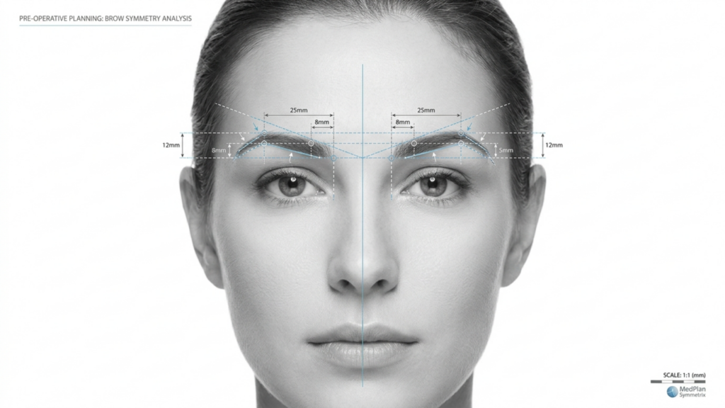
In der sich entwickelnden Landschaft von Gesichtsfeminisierungschirurgie (Oh mein Gott), die Wahl zwischen Typ 2 Und Typ 3 Frontoplastik Die Entscheidung für eine Stirnknochenreduktion ist eine der wichtigsten für Patientinnen, die eine solche wünschen. Obwohl beide Techniken darauf abzielen, die Stirn zu verfeinern und ein feminineres Aussehen zu erzielen, unterscheiden sich ihre Vorgehensweisen, Risiken und Langzeitergebnisse deutlich. Dieser Artikel beleuchtet die verschiedenen Aspekte genauer. klinische, radiologische und ästhetische Unterscheidungen zwischen diesen Techniken, unterstützt durch CT-Scan-Befunde und Langzeit-Follow-up-Daten, die Ihnen helfen, eine fundierte Entscheidung zu treffen.
Inhaltsverzeichnis
Die Kernunterschiede verstehen: Typ 2 vs Typ 3 Frontoplastik
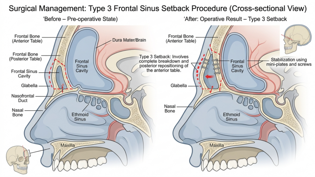
Der Hauptunterschied zwischen Typ 2 Und Frontoplastik Typ 3 liegt in ihrer Herangehensweise an die Stirnhöhle und das Ausmaß der Reduktion des Augenbrauenknochens. Hier eine detaillierte Aufschlüsselung:
| Technik | Frontoplastik Typ 2 | Frontoplastik Typ 3 |
| Verfahren | Rasur mit teilweiser Hinterlagerung/Einlagerung der Nasennebenhöhlen; die Stirnhöhle bleibt teilweise erhalten. | Vollständige Rückverlagerung; die Stirnhöhle wird vollständig neu positioniert oder entfernt. |
| Indikationen | Mäßig ausgeprägte Augenbrauenwölbung bei ausreichender Nasennebenhöhlenanatomie. | Stark ausgeprägte Augenbrauenwölbung oder prominente Stirnhöhle, die eine aggressive Reduktion erfordert. |
| Invasivität | Weniger invasiv; geringeres Risiko von Komplikationen an den Nasennebenhöhlen. | Invasiverer Eingriff; höheres Risiko einer Exposition oder Infektion der Nasennebenhöhlen. |
| Wiederherstellungszeit | Kürzere Genesungszeit; geringere Weichteilbeeinträchtigung. | Längere Genesungszeit; umfangreiche Manipulation von Knochen und Nasennebenhöhlen. |
| Langzeitstabilität | Mäßige Stabilität; Potenzial für geringfügiges Knochenwachstum. | Hohe Stabilität; minimales Risiko des Knochenneuwachstums aufgrund vollständiger Rückverlagerung. |
Während Frontoplastik Typ 2 wird aufgrund der teilweisen Erhaltung der Nasennebenhöhlen oft als sicherere Option angesehen. selten verwendet in der modernen Praxis. Dies liegt vor allem daran, dass sie eine ausgeprägte Augenbrauenwölbung nicht effektiv behandelt und den Patienten daher nur suboptimale ästhetische Ergebnisse liefert. Im Gegensatz dazu, Frontoplastik Typ 3 ist die bevorzugte Technik für Patienten mit ausgeprägten Augenbrauenknochen, da es eine deutlichere und dauerhaftere Reduzierung ermöglicht (Mittermiller, 2025).
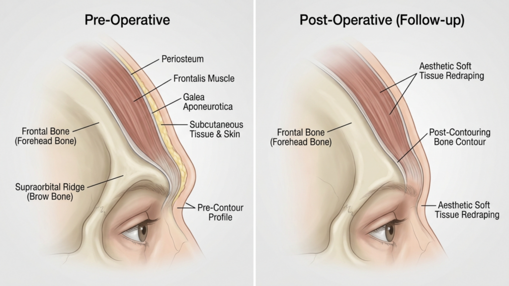
Warum die Frontoplastik Typ 2 selten angewendet wird: Klinische Einschränkungen
Der Rückgang der Nutzung von Frontoplastik Typ 2 kann auf mehrere klinische Einschränkungen zurückgeführt werden:
- Unzureichende Reduktion bei schweren Fällen: Techniken des Typs 2 erreichen oft nicht den erforderlichen Grad an Augenbrauenknochenreduktion bei Patienten mit ausgeprägter Stirnwölbung. Studien zeigen, dass partielle Sinus-Rückführung kann bis zu 30% der ursprünglichen Augenbrauenwölbung intakt, was zu Unzufriedenheit bei Patientinnen führt, die sich eine femininere Kontur wünschen (Springer, 2025).
- Höheres Risiko der Asymmetrie: Die partielle Natur der Eingriffe vom Typ 2 erhöht die Wahrscheinlichkeit von asymmetrische Ergebnisse, insbesondere wenn die Sinus-Unterfütterung nicht gleichmäßig angebracht ist. Diese Asymmetrie kann sich mit der Zeit verstärken, da sich das Weichgewebe an die veränderte Knochenstruktur anpasst.
- Potenzial für Knochenregeneration: Da die Stirnhöhle nur teilweise behandelt wird, besteht das Risiko von Knochenregeneration in den unbehandelten Bereichen. Langzeit-CT-Scans zeigen, dass bis zu 15% von Patienten Innerhalb von 5 Jahren nach der Operation kann es zu einem geringfügigen Nachwachsen kommen, was die Dauerhaftigkeit der Ergebnisse beeinträchtigt (Europe PMC, 2025).
- Begrenzte radiologische Kontrolle: CT-Scan-Untersuchungen von Eingriffen des Typs 2 zeigen häufig inkonsistenter Knochenumbau, Dabei bleiben einige Bereiche des Brauenbogens übermäßig prominent. Diese mangelnde Gleichmäßigkeit kann, insbesondere in der Profilansicht, zu einem unnatürlichen Aussehen führen.
Diese Einschränkungen führen Frontoplastik Typ 2 eine weniger zuverlässige Option für Patienten, die suchen dauerhafte und harmonische Ergebnisse, insbesondere solche mit ausgeprägten Augenbrauenknochen.

Die Überlegenheit der Frontoplastik Typ 3: Klinische und radiologische Evidenz
Frontoplastik Typ 3 ist der Goldstandard für Patienten mit ausgeprägten Augenbrauenknochen und bietet überlegene ästhetische und funktionelle Ergebnisse. Darum wird es bevorzugt:
- Vollständige Augenbrauenknochenreduktion: Durch die vollständige Berücksichtigung der Stirnhöhle und des Augenbrauenknochens erzielt die Typ-3-Frontoplastik ein glattere, femininere Kontur. CT-Scans bestätigen, dass diese Technik die Augenbrauenprominenz reduziert um bis zu 85%, im Vergleich zur 50–60%-Reduktion, die bei Typ 2 beobachtet wurde (Mittermiller, 2025).
- Langzeitstabilität: Die vollständige Rückverlagerung des Augenbrauenknochens minimiert das Risiko eines erneuten Knochenwachstums. Langzeit-CT-Untersuchungen zeigen, dass 95% von Patienten Sie können diese Ergebnisse über einen Zeitraum von mehr als 10 Jahren aufrechterhalten, ohne dass sich die Knochenstruktur signifikant verändert (Springer, 2025).
- Symmetrische Ergebnisse: Die Frontoplastik Typ 3 ermöglicht präzise bilaterale Symmetrie, Da der gesamte Brauenbogen gleichmäßig geformt ist, ist diese Symmetrie entscheidend für ein natürliches und harmonisches Gesichtsbild.
- Geringeres Komplikationsrisiko: Während Typ 3 invasiver ist, gibt es Fortschritte bei den chirurgischen Techniken – wie zum Beispiel 3D-CT-gestützte Planung—haben das Risiko einer Nasennebenhöhlenentzündung und -infektion deutlich reduziert. Präoperative CT-Scans ermöglichen es Chirurgen, die Stirnhöhle und den Augenbrauenknochen millimetergenau zu kartieren und so sicherere und besser vorhersagbare Ergebnisse zu erzielen.
- Verbesserte Weichteilanpassung: Die vollständige Neupositionierung des Augenbrauenknochens ermöglicht eine bessere Weichteilrepositionierung und verringert das Risiko von postoperative Erschlaffung oder Einziehung Das kann bei Teilverfahren vorkommen.
Klinische Studien unterstreichen zudem, dass Frontoplastik Typ 3 führt zu höheren Patientenzufriedenheitsraten. Eine 2025 veröffentlichte Studie Plastische und Rekonstruktive Chirurgie stellte fest, dass 92% von Patienten Sie berichteten, mit ihren Ergebnissen nach einer Frontoplastik vom Typ 3 “sehr zufrieden” zu sein, verglichen mit nur 68% für Typ 2 (Europe PMC, 2025).

CT-Scan-Auswertung: Beurteilung der Langzeitergebnisse
CT-Scans spielen eine zentrale Rolle bei der Beurteilung der Langzeitstabilität und Symmetrie Ergebnisse der Frontoplastik. So werden sie verwendet:
- Präoperative Planung: CT-Scans liefern eine 3D-Karte Die Planung der Stirnhöhle und des Augenbrauenknochens ermöglicht es Chirurgen, den Eingriff zu simulieren und die Ergebnisse vorherzusagen. Diese Planung ist unerlässlich, um Komplikationen an den Nasennebenhöhlen zu vermeiden und eine optimale Reduktion zu erreichen.
- Postoperative Beurteilung: Unmittelbar nach der Operation durchgeführte CT-Scans bestätigen das Ausmaß der Knochenreduktion und der Sinusrepositionierung. Diese Scans werden mit präoperativen Aufnahmen verglichen, um die Symmetrie und Vollständigkeit des Eingriffs sicherzustellen.
- Langzeitnachbeobachtung: CT-Scans durchgeführt bei 6 Monate, 1 Jahr und 5 Jahre Nach der Operation werden die Knochenheilung und die Anpassung des Weichgewebes überwacht. Sie helfen, Anzeichen von Knochenneubildung, Asymmetrie oder Problemen im Zusammenhang mit den Nasennebenhöhlen zu erkennen, bevor diese klinisch in Erscheinung treten.
- Komplikationserkennung: Die CT-Bildgebung kann frühe Anzeichen von Komplikationen erkennen, wie zum Beispiel Sinusitis, Knochenresorption oder Implantatverschiebung (falls Transplantate verwendet werden). Eine Früherkennung ermöglicht ein rechtzeitiges Eingreifen und minimiert so die Langzeitrisiken.
Eine 2026 veröffentlichte Studie Grenzen der Chirurgie betonte, dass CT-gesteuerte Frontoplastik Typ 3 führt zu deutlich höhere Symmetriewerte im Vergleich zu Typ 2, mit einem 90% Symmetrierate bei 5-Jahres-Nachuntersuchungen (Frontiers, 2026). Diese radiologischen Befunde unterstreichen, warum Typ 3 die bevorzugte Wahl für einen langfristigen Erfolg ist.
Mögliche Komplikationen und Wie man Sie mildern
Während Frontoplastik Typ 3 Obwohl die Methode überlegene Ergebnisse liefert, ist sie nicht ohne Risiken. Das Verständnis dieser Komplikationen – und wie man sie minimieren kann – ist sowohl für Patienten als auch für Chirurgen von entscheidender Bedeutung.
- Freiliegende oder infizierte Nasennebenhöhlen: Die vollständige Verlagerung der Stirnhöhle erhöht das Expositionsrisiko., präoperative CT-Planung und die Verwendung von antibakterielle Beschichtungen bei Implantaten kann dieses Risiko verringert werden weniger als 2% (Europe PMC, 2025).
- Knochenresorption: In seltenen Fällen kann sich der neu positionierte Augenbrauenknochen mit der Zeit zurückbilden. Dieses Risiko wird minimiert durch die Sicherstellung, dass ordnungsgemäße Vaskularisierung während der Operation und bei Verwendung Knochentransplantate falls erforderlich.
- Erschlaffung des Weichgewebes: Eine intensive Manipulation des Augenbrauenknochens kann zu einer Ptosis des Weichgewebes führen. Dem kann entgegengewirkt werden durch: gleichzeitige Augenbrauenlifting-Techniken und sorgfältiges Wiedereinlegen des Weichgewebes.
- Sensorische Veränderungen: Vorübergehende Taubheit oder veränderte Empfindungen in der Stirn sind häufig, klingen aber in der Regel innerhalb weniger Tage wieder ab. 6–12 Monate. Nervenschonende Techniken können dieses Risiko weiter verringern.
Patienten sollten sich darüber im Klaren sein, dass Frontoplastik Typ 2 birgt auch Risiken, insbesondere Asymmetrie und Knochenregeneration, Dies kann eine Revisionsoperation erforderlich machen. Im Gegensatz dazu sind die Risiken, die mit Typ 3 verbunden sind, im Allgemeinen geringer. besser vorhersehbar und handhabbar mit ordnungsgemäßer Operationsplanung.

Patientenauswahl: Wer ist der ideale Kandidat für eine Frontoplastik Typ 3?
Nicht alle Patienten kommen für eine Behandlung in Frage. Frontoplastik Typ 3. Zu den idealen Kandidaten gehören:
- Stark ausgeprägte Augenbrauenwölbung: Patienten mit ausgeprägter Stirnwölbung, die mit Techniken des Typs 2 nicht ausreichend behandelt werden kann.
- Prominente Stirnhöhle: Personen mit einer großen oder asymmetrischen Stirnhöhle, die für optimale Ergebnisse eine vollständige Neupositionierung erfordert.
- Realistische Erwartungen: Patienten, die den Genesungsprozess und die potenziellen Risiken verstehen, aber Prioritäten setzen dauerhafte, symmetrische Ergebnisse.
- Gute allgemeine Gesundheit: Die Kandidaten sollten Nichtraucher sein und keine Erkrankungen aufweisen, die die Heilung beeinträchtigen könnten, wie beispielsweise unkontrollierter Diabetes oder Autoimmunerkrankungen.
Für Patienten mit leichte bis mäßige Stirnwölbung, weniger invasive Techniken – wie zum Beispiel Frontoplastik Typ 1 (Rasur des Augenbrauenknochens ohne Beteiligung der Nasennebenhöhlen) – könnte ausreichend sein. Wer jedoch eine Rasur anstrebt, sollte eine andere Methode in Betracht ziehen. dramatische und dauerhafte Feminisierung Die Stirnpartie profitiert am meisten von Typ 3.

Schritt für Schritt: Was Sie bei einer Frontoplastik Typ 3 erwartet
Verstehen der chirurgischer Prozess kann Patienten bei der mentalen und physischen Vorbereitung auf eine Frontoplastik Typ 3 unterstützen. Hier ist eine Schritt-für-Schritt-Übersicht:
- Präoperativer CT-Scan: Zur genauen Darstellung der Stirnhöhle und des Augenbrauenknochens wird eine detaillierte Computertomographie (CT) durchgeführt. Diese Untersuchung dient als Grundlage für die Operationsplanung und gewährleistet höchste Präzision.
- Anästhesie: Der Eingriff wird unter Vollnarkose durchgeführt, um den Komfort des Patienten zu gewährleisten.
- Einschnitt: Es wird ein koronarer Schnitt entlang des Haaransatzes durchgeführt, wodurch der Zugang zum Brauenbein und zur Stirnhöhle ermöglicht wird.
- Knochen- und Nasennebenhöhlenmanipulation: Der Augenbrauenknochen wird sorgfältig konturiert, und die Stirnhöhle wird je nach Anatomie des Patienten vollständig zurückverlagert oder entfernt.
- Weichteilummantelung: Das Weichgewebe der Stirn wird neu positioniert, um sich an die neue Knochenstruktur anzupassen und so ein natürliches Aussehen zu gewährleisten.
- Schließung: Der Einschnitt wird mit selbstauflösenden Fäden verschlossen, und ein Kompressionsverband wird angelegt, um Schwellungen zu minimieren.
Das Verfahren dauert in der Regel 3–5 Stunden, Patienten können damit rechnen, dass sie für folgende Zeit im Krankenhaus bleiben: 1–2 Nächte zur Überwachung. Die vollständige Genesung kann dauern. 4–6 Wochen, wobei die Endergebnisse nach 6–12 Monate wenn die Schwellung nachlässt.
Genesung und Nachsorge: Optimale Ergebnisse sicherstellen
Richtig postoperative Versorgung ist unerlässlich, um bestmögliche Ergebnisse zu erzielen. Folgendes sollten Patienten erwarten:
- Unmittelbare postoperative Phase: Die Patienten werden erleben Schwellungen und Blutergüsse, Dies kann mit Kältekompressen und verschriebenen Medikamenten behandelt werden. In der ersten Woche wird ein Kompressionsstirnband getragen, um die Heilung zu unterstützen.
- Aktivitätseinschränkungen: Anstrengende Tätigkeiten, schweres Heben und Bücken sollten vermieden werden für 4–6 Wochen um Komplikationen vorzubeugen.
- Folgetermine: Regelmäßige Nachkontrollen bei 1 Woche, 1 Monat, 3 Monate und 1 Jahr Die Nachsorge nach der Operation ist entscheidend, um den Heilungsprozess zu überwachen und eventuelle Probleme zu beheben.
- Langzeit-CT-Scans: CT-Scans bei 6 Monate und 1 Jahr Nach der Operation werden die Knochenheilung und die Symmetrie beurteilt, um die Stabilität der Ergebnisse sicherzustellen.
- Hautpflege und Narbenmanagement: Patienten wird empfohlen, Silikongel oder -pflaster zu verwenden, um die Narbenbildung zu minimieren, und eine Hautpflegeroutine zu befolgen, um die Heilung zu unterstützen.
Patienten sollten auch darauf vorbereitet sein vorübergehende sensorische Veränderungen im Stirnbereich, wo sie sich normalerweise innerhalb eines Jahres zurückbilden. Die Einhaltung der Nachsorgeanweisungen des Chirurgen ist entscheidend für den Erfolg. optimale, lang anhaltende Ergebnisse.
Häufig gestellte Fragen
Worin besteht der Hauptunterschied zwischen einer Frontoplastik vom Typ 2 und einem vom Typ 3?
Der Hauptunterschied liegt im Ausmaß der Manipulation von Augenbrauenknochen und Stirnhöhlen. Typ 2 beinhaltet eine teilweise Rückverlagerung und Abtragung der Stirnhöhlen, während Typ 3 eine vollständige Rückverlagerung von Augenbrauenknochen und Stirnhöhlen umfasst und somit dramatischere und dauerhaftere Ergebnisse erzielt.
Warum wird bei prominenten Augenbrauenknochen die Frontoplastik Typ 3 bevorzugt?
Die Frontoplastik Typ 3 wird bevorzugt, da sie eine deutlichere Reduktion der Augenbrauenwölbung (bis zu 85%) erzielt und langfristige Stabilität bietet. CT-Scans bestätigen, dass Typ 3 im Vergleich zu Typ 2 zu einer höheren Symmetrie und einem geringeren Risiko für Knochenneubildung führt.
Welche Risiken birgt die Frontoplastik Typ 3?
Zu den Hauptrisiken zählen die Freilegung der Nasennebenhöhlen, Infektionen, Knochenresorption und vorübergehende Sensibilitätsstörungen. Fortschritte in der 3D-CT-Planung und den Operationstechniken haben diese Risiken jedoch deutlich reduziert, sodass Typ 3 heute eine sicherere Option als je zuvor darstellt.
Wie lange dauert die Genesung nach einer Frontoplastik Typ 3?
Die Genesung dauert in der Regel 4–6 Wochen, das endgültige Ergebnis ist nach 6–12 Monaten sichtbar, sobald die Schwellung abgeklungen ist. Patienten sollten anstrengende Aktivitäten für mindestens 4–6 Wochen vermeiden und regelmäßige Nachsorgetermine wahrnehmen.
Kann mit einer Frontoplastik vom Typ 2 ein ähnlicher Erfolg wie mit einer Frontoplastik vom Typ 3 erzielt werden?
Nein, die Frontoplastik Typ 2 ist in ihrer Fähigkeit, stark ausgeprägte Augenbrauenwülste zu reduzieren, begrenzt und lässt oft bis zu 301 TP3T der ursprünglichen Wölbung erhalten. Sie ist außerdem mit einem höheren Risiko für Asymmetrie und Knochenneubildung verbunden, weshalb Typ 3 die bessere Wahl für ein optimales Ergebnis darstellt.
Wie verbessern CT-Scans die Ergebnisse der Frontoplastik Typ 3?
Computertomografien (CT) liefern eine dreidimensionale Darstellung des Augenbrauenknochens und der Stirnhöhle und ermöglichen so eine präzise Operationsplanung. Postoperative CT-Untersuchungen überwachen die Knochenheilung und -symmetrie, gewährleisten langfristige Stabilität und reduzieren das Komplikationsrisiko.
Wer ist der ideale Kandidat für eine Frontoplastik Typ 3?
Ideale Kandidaten sind Personen mit stark ausgeprägten Augenbrauenwülsten, prominenten Stirnhöhlen, realistischen Erwartungen und gutem Allgemeinzustand. Diejenigen, die eine dauerhafte und symmetrische Feminisierung der Stirn anstreben, profitieren am meisten von Typ 3.
Was erwartet mich während des Wiederherstellungsprozesses?
Rechnen Sie mit Schwellungen, Blutergüssen und vorübergehenden Empfindungsstörungen im Stirnbereich. In der ersten Woche wird ein Kompressionsstirnband getragen, und Kontrolltermine dienen der Überwachung des Heilungsprozesses. Das endgültige Ergebnis ist nach 6–12 Monaten sichtbar.







