

Reconstruction du front est un élément essentiel de Soin du visage Chirurgie de féminisation (FFS) et les interventions craniofaciales, visant à obtenir un contour du visage harmonieux et esthétique. Parmi les techniques les plus controversées figurent Type 1 et cranioplastie de type 3, chacune proposant des approches distinctes pour remodeler le front. Type 1 implique fraisage ou rasage osseux, Tapez 3 nécessite une solution plus complexe ostéotomie et recul du sinus frontal. Comprendre les implications anatomiques, fonctionnelles et esthétiques de ces techniques est essentiel pour que les chirurgiens et les patients puissent prendre des décisions éclairées. Apprenez-en davantage sur le recul sinusal et la réduction de la bosse sourcilière dans notre guide complet. modelage du front.
Ce guide explore les différences structurelles entre la cranioplastie de type 1 et de type 3, leur impact sur épaisseur osseuse, anatomie du sinus frontal, et comment les chirurgiens déterminent l'approche la plus adaptée à chaque patient. À la fin de cet ouvrage, vous saurez clairement quelle technique correspond le mieux à vos besoins anatomiques et à vos objectifs esthétiques.
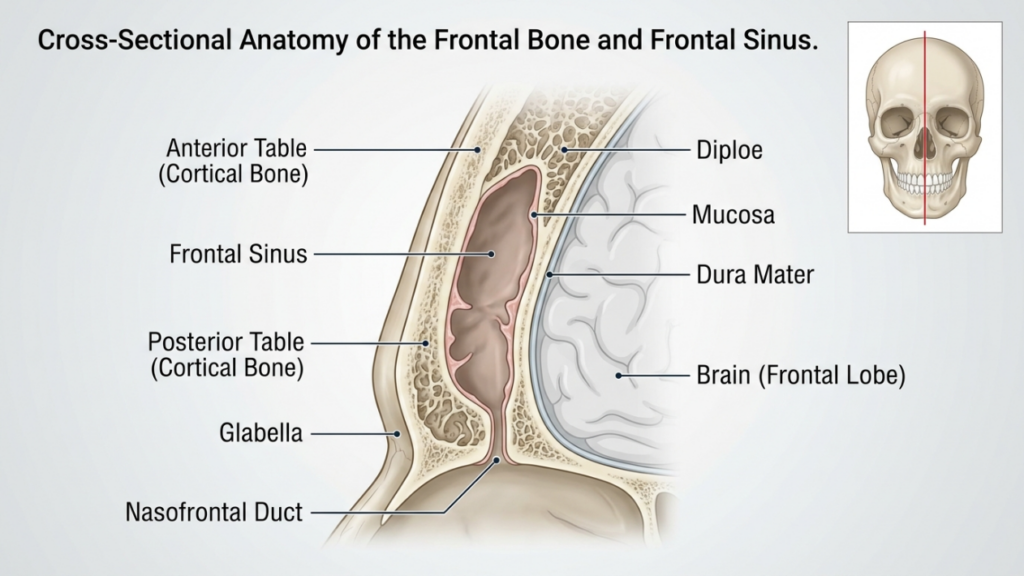
Table des matières
Bases anatomiques : os frontal et sinus
Le os frontal et sinus frontal jouent un rôle essentiel dans la reconstruction du front. L'os frontal forme la partie supérieure du visage et abrite le sinus frontal, une cavité creuse dont la taille et la forme varient d'un individu à l'autre. table antérieure Le sinus frontal est la couche osseuse externe qui contribue au contour du front, tandis que table postérieure Elle sépare les sinus du cerveau. L'épaisseur de ces plaques et le degré de pneumatisation des sinus (expansion remplie d'air) influencent le choix entre une cranioplastie de type 1 et de type 3.
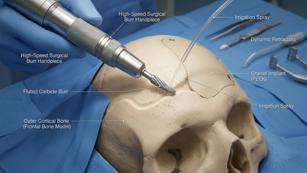
Dans cranioplastie de type 1, le chirurgien Cette technique utilise une fraise à grande vitesse pour amincir la corticale osseuse externe, réduisant ainsi la saillie sans léser le sinus frontal. Elle est idéale pour les patients présentant os frontal mince ou un léger surfaçage, car il préserve l'intégrité du sinus. Cependant, il peut s'avérer insuffisant chez les patients présentant un surfaçage important des sourcils ou une table antérieure épaisse, car un fraisage excessif peut compromettre la stabilité osseuse ou ne pas permettre d'obtenir le contour souhaité (Ousterhout, 2024).
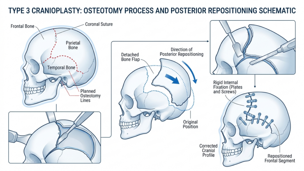
En revanche, cranioplastie de type 3 implique un ostéotomie— une incision contrôlée de la table antérieure du sinus frontal. Le segment osseux est ensuite repositionné vers l'arrière (recul) afin de réduire sa projection et d'obtenir un front plus lisse et plus féminin. Cette technique est réservée aux patientes présentant bosse de sourcil modérée à sévère ou une table antérieure épaisse, où le fraisage seul serait insuffisant. L'ostéotomie permet un remodelage précis tout en préservant la fonction protectrice du sinus (Féminisation du front : revue exploratoire de la littérature, 2024).
Cranioplastie de type 1 : rabotage et ébavurage
Aperçu technique
La cranioplastie de type 1 est l'option la moins invasive pour la reconstruction du front. Elle consiste à utiliser un fraise à grande vitesse réduire progressivement la proéminence de l'os frontal. chirurgien Elle consiste à raser méticuleusement la couche corticale externe, en évitant toute pénétration dans le sinus frontal. Cette technique est particulièrement efficace chez les patients atteints de :
- Sourcils légèrement à modérés
- Os frontal fin (moins de 5 mm)
- Absence de pneumatisation significative du sinus frontal
La procédure est réalisée par l'intermédiaire d'un incision coronale, Cette technique permet d'accéder au front tout en minimisant les cicatrices visibles. Le chirurgien utilise le toucher et des repères visuels pour garantir une réduction uniforme sans trop amincir l'os, ce qui pourrait entraîner une instabilité ou des irrégularités de contour.
Avantages de la cranioplastie de type 1
La cranioplastie de type 1 offre plusieurs avantages :
- Invasivité minimale : L'absence d'ostéotomie ou d'ablation osseuse réduit le traumatisme chirurgical et le temps de récupération.
- Risque de complications réduit : La préservation du sinus frontal minimise le risque de sinusite, de fuites de liquide céphalo-rachidien ou de formation de mucocèle.
- Temps opératoire plus court : L'intervention dure généralement 1 à 2 heures, ce qui en fait une option plus rapide que le type 3.
- Résultats prévisibles : Idéal pour les patientes présentant une légère bosse, pour lesquelles un contouring subtil suffit à obtenir une apparence féminine.
Limites de la cranioplastie de type 1
Bien que la cranioplastie de type 1 soit plus sûre et moins invasive, elle présente des limitations notables :
- Réduction limitée : Insuffisant pour les patients présentant une forte proéminence des sourcils ou des os frontaux épais.
- Risque de sur-éclaircissage : Un fraisage agressif peut fragiliser l'os, entraînant des irrégularités de contour ou des fractures.
- Féminisation incomplète : Peut ne pas permettre d'obtenir le résultat esthétique souhaité chez les patients présentant des traits masculins marqués.
Cranioplastie de type 3 : ostéotomie et recul
Aperçu technique
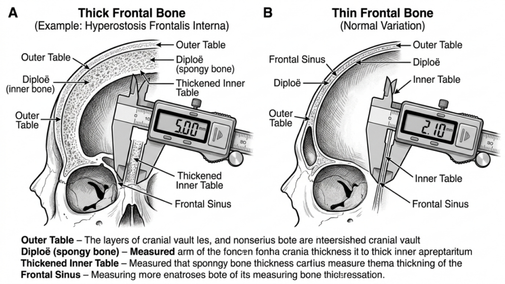
La cranioplastie de type 3 est une intervention plus complexe, conçue pour les patients atteints de bosse de sourcil modérée à sévère ou des os frontaux épais. Cela implique un ostéotomie— une incision précise de la paroi antérieure du sinus frontal — suivie d'un repositionnement du segment osseux vers l'arrière (recul). Cette technique permet une réduction significative de la projection du front et un contour plus harmonieux et féminin.
L'intervention est généralement réalisée par l'intermédiaire d'un incision coronale, permettant l'accès à l'os frontal et au sinus. Le chirurgien utilise un scie sagittale ou appareil piézoélectrique pour créer l'ostéotomie, en veillant à ce que la coupe suive la courbure naturelle du front. Le segment osseux est ensuite repositionné et fixé avec plaques et vis en titane ou des sutures résorbables. Cette technique est idéale pour les patients présentant :
- Sourcils très bombés
- Os frontal épais (supérieur à 5 mm)
- Pneumatisation significative du sinus frontal
La cranioplastie de type 3 nécessite une planification méticuleuse, impliquant souvent Planification chirurgicale virtuelle 3D (VSP) pour simuler l'ostéotomie et le recul. Ceci garantit la précision et minimise le risque de complications telles que la violation des sinus ou les fuites de liquide céphalo-rachidien (Planification chirurgicale virtuelle). Féminisation faciale de la Face Supérieure, 2025).
Avantages de la cranioplastie de type 3
La cranioplastie de type 3 offre plusieurs avantages aux patients présentant des traits masculins prononcés :
- Contourage significatif : Permet de réduire considérablement l'aspect bosselé des sourcils, pour un front plus lisse et plus féminin.
- Polyvalence: Convient aux patients présentant des os frontaux épais ou une pneumatisation sinusale étendue.
- Stabilité à long terme : Le segment osseux repositionné s'intègre bien, réduisant ainsi le risque d'irrégularités de contour au fil du temps.
- Personnalisation : La planification chirurgicale virtuelle permet des ajustements précis et adaptés à chaque patient afin d'obtenir des résultats optimaux.
Limites de la cranioplastie de type 3
Malgré son efficacité, la cranioplastie de type 3 comporte des risques et des complexités plus élevés :
- Durée opératoire accrue : L'intervention dure généralement de 3 à 5 heures et requiert une plus grande précision et une expertise accrue.
- Risque accru de complications : Les risques potentiels comprennent la sinusite, les fuites de liquide céphalo-rachidien ou la formation d'un mucocèle en cas de lésion du sinus.
- Récupération plus longue : Les patients peuvent ressentir un gonflement et une gêne prolongés par rapport à la cranioplastie de type 1.
- Coût: Le recours à l'imagerie avancée et aux outils chirurgicaux de pointe augmente le coût global de l'intervention.

Principales différences entre la cranioplastie de type 1 et la cranioplastie de type 3
| Fonctionnalité | Cranioplastie de type 1 | Cranioplastie de type 3 |
|---|---|---|
| Technique | fraisage/rasage osseux | Ostéotomie et recul |
| caractère invasif | Minimal | Modéré à élevé |
| Candidats idéaux | Sourcils légèrement à modérément bombés, os frontal fin | Sourcils fortement proéminents, os frontal épais |
| Temps chirurgical | 1 à 2 heures | 3 à 5 heures |
| Le temps de récupération | 1 à 2 semaines | 3 à 6 semaines |
| Risque de complications | Faible | Modéré à élevé |
| Coût | Inférieur | Plus élevé |
| Résultat esthétique | contouring subtil | Féminisation spectaculaire |
Comment les chirurgiens décident : Type 1 ou Type 3
Le choix entre la cranioplastie de type 1 et de type 3 dépend de plusieurs facteurs, notamment du patient. caractéristiques anatomiques, objectifs esthétiques, et risques chirurgicaux. Les chirurgiens s'appuient sur une combinaison de examen clinique, Imagerie 3D, et consultation du patient pour déterminer la technique la plus appropriée.
1. Évaluation anatomique
La première étape consiste à évaluer le patient épaisseur de l'os frontal et anatomie du sinus frontal. UN scanner ou Reconstruction 3D fournit des informations détaillées sur :
- Épaisseur osseuse : Les patients ayant des os frontaux fins (moins de 5 mm) sont mieux adaptés à une cranioplastie de type 1, tandis que ceux ayant des os plus épais peuvent nécessiter une cranioplastie de type 3.
- Pneumatisation des sinus : Une pneumatisation sinusale étendue peut nécessiter une cranioplastie de type 3 pour éviter de violer le sinus lors du fraisage.
- Degré d'autoritarisme : Les sourcils très proéminents nécessitent souvent une réduction drastique, possible uniquement grâce aux techniques de type 3.
2. Objectifs esthétiques
Les attentes esthétiques des patients jouent un rôle crucial dans le choix de la technique. Ceux qui recherchent féminisation subtile peuvent opter pour une cranioplastie de type 1, tandis que les personnes présentant des traits masculins marqués nécessitent souvent une cranioplastie de type 2. résultats transformateurs de type 3. Les chirurgiens discutent des résultats réalistes en fonction de l'anatomie du patient et des changements souhaités.
3. Risques chirurgicaux et convalescence
La cranioplastie de type 3 comporte des risques plus élevés, notamment complications des sinus, fuites de liquide céphalo-rachidien, et récupération prolongée. Les chirurgiens évaluent l'état de santé général du patient, sa tolérance à l'intervention et sa volonté de suivre les soins postopératoires. Il peut être conseillé aux patients présentant des affections médicales augmentant les risques chirurgicaux d'envisager une intervention de type 1 ou d'autres procédures.
4. Planification chirurgicale virtuelle (PCV)
Progrès dans planification chirurgicale virtuelle 3D L’impression 3D et la planification chirurgicale virtuelle ont révolutionné la cranioplastie. Les chirurgiens utilisent ces techniques pour simuler les ostéotomies, les reculs et les résultats, garantissant ainsi la précision et minimisant les risques. Cette technologie est particulièrement précieuse pour la cranioplastie de type 3, où un repositionnement osseux précis est essentiel (Impression 3D et planification chirurgicale virtuelle en chirurgie craniofaciale et orthognathique, 2025).
Soins postopératoires et récupération
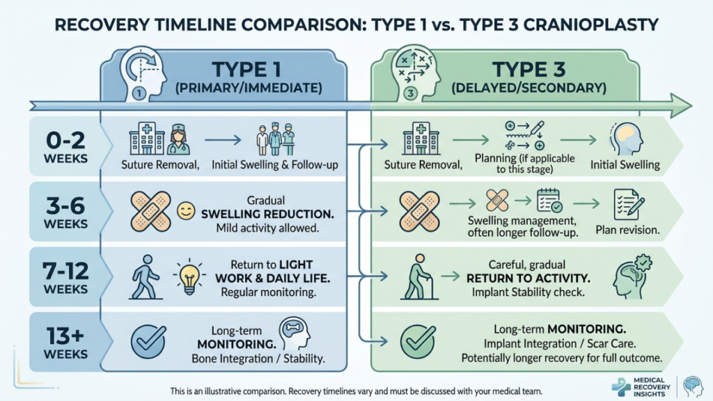
La durée de la convalescence varie considérablement entre la cranioplastie de type 1 et celle de type 3. Comprendre le processus postopératoire permet aux patients de mieux se préparer à une guérison optimale.
Récupération après une cranioplastie de type 1
Les patients subissant une cranioplastie de type 1 présentent généralement les symptômes suivants :
- Gonflement léger à modéré : Se résout en 1 à 2 semaines.
- Inconfort minimal : Géré avec des analgésiques en vente libre.
- Retour rapide aux activités : La plupart des patients reprennent leurs activités normales en deux semaines.
Récupération après une cranioplastie de type 3
La convalescence après une cranioplastie de type 3 est plus complexe en raison de la complexité de l'intervention :
- Gonflements et ecchymoses importants : Peut persister pendant 3 à 4 semaines.
- Douleur modérée : Des analgésiques sur ordonnance peuvent être nécessaires pendant la première semaine.
- Retour progressif aux activités : Les activités physiques intenses sont déconseillées pendant 4 à 6 semaines.
- Visites de suivi : Un suivi régulier est nécessaire pour assurer une bonne cicatrisation et traiter toute complication.
Les deux techniques exigent que les patients évitent soulever des charges lourdes, exercice intense, et pression directe sur le front Durant la phase initiale de convalescence, les chirurgiens fournissent des instructions postopératoires détaillées, notamment concernant les soins de la plaie, les restrictions d'activité et les signes de complications à surveiller.
Complications potentielles et comment les éviter
Bien que la cranioplastie soit généralement une intervention sûre, des complications peuvent survenir. La connaissance de ces risques et des mesures préventives est essentielle tant pour les chirurgiens que pour les patients.
Complications courantes
- Sinusite: Inflammation ou infection du sinus frontal, en particulier lors d'une cranioplastie de type 3 si le sinus est lésé.
- Fuite de liquide céphalorachidien (LCR) : Complication rare mais grave en cas de perforation de la paroi postérieure du sinus.
- Irrégularités de contour : Un amincissement osseux excessif dans le type 1 ou un recul incorrect dans le type 3 peuvent entraîner une asymétrie ou des crêtes visibles.
- Formation de mucocèle : L'obstruction des voies de drainage des sinus peut entraîner la formation de kystes remplis de mucus.
- Infection: Le risque est plus élevé dans le type 3 en raison de la durée opératoire plus longue et de l'utilisation d'implants.
Mesures préventives
Les chirurgiens utilisent plusieurs stratégies pour minimiser les complications :
- Planification chirurgicale précise : L'imagerie 3D et la planification chirurgicale virtuelle garantissent des ostéotomies et des reculs précis.
- Préservation des sinus : Éviter toute lésion du sinus frontal lors du fraisage ou de l'ostéotomie.
- Prophylaxie antibiotique : Administré en pré- et postopératoire pour réduire les risques d'infection.
- Surveillance postopératoire : Un suivi régulier est nécessaire pour détecter les signes précoces de complications.
- Éducation des patients : Informer les patients sur les soins appropriés à apporter aux plaies, les restrictions d'activité et les signes avant-coureurs de complications.
Alternatives aux cranioplasties de type 1 et de type 3
Pour les patients qui ne sont pas des candidats idéaux pour une cranioplastie de type 1 ou de type 3, des techniques alternatives peuvent être envisagées :
- Greffe de graisse: Le transfert de graisse autologue permet d'adoucir les contours du front sans modifier la structure osseuse. Cette technique est idéale pour les patients présentant de légères irrégularités ou ceux qui recherchent des solutions non chirurgicales.
- Implants personnalisés : Les implants préfabriqués, tels que PEEK (polyétheréthercétone) ou titane, Ces implants peuvent être utilisés pour augmenter ou remodeler le front sans ostéotomie. Ils sont particulièrement utiles pour les patients ayant une ossature fine ou des problèmes de sinus.
- Chirurgie orthognatique : Dans les cas où modelage du front fait partie d'un ensemble plus vaste féminisation faciale Dans le cadre de ce plan, des interventions orthognathiques (par exemple, une ostéotomie de Le Fort I) peuvent être combinées pour corriger l'alignement du tiers moyen du visage et de la mâchoire.
- Techniques endoscopiques : Les approches endoscopiques mini-invasives permettent de réduire la proéminence des sourcils grâce à des incisions plus petites et des temps de récupération plus rapides.
Témoignages de patients et résultats concrets
Les résultats concrets apportent un éclairage précieux sur l'efficacité et le taux de satisfaction des cranioplasties de type 1 et de type 3. Les témoignages de patients soulignent l'impact transformateur de ces interventions :
Étude de cas 1 : Cranioplastie de type 1
Une femme transgenre de 28 ans souhaitait une féminisation subtile de son front. Présentant un os frontal fin et une bosse peu prononcée, elle a opté pour une cranioplastie de type 1. L'intervention a permis d'obtenir un contour plus harmonieux avec un temps de convalescence minimal. Elle s'est déclarée très satisfaite, soulignant que le résultat correspondait à ses attentes d'une apparence naturelle et féminine.
Étude de cas 2 : Cranioplastie de type 3
Une femme transgenre de 35 ans présentait une forte proéminence des sourcils et un front épais. Une cranioplastie de type 3 a été réalisée, impliquant une ostéotomie et un recul du front. La réduction importante de la projection du front a considérablement féminisé ses traits. Bien que la convalescence ait été plus longue que prévu, elle s'est dite extrêmement satisfaite du résultat, affirmant que l'intervention avait “ changé sa vie ”.”
Étude de cas 3 : Chirurgie de révision
Une patiente de 40 ans avait initialement subi une cranioplastie de type 1, mais était insatisfaite du résultat discret. Elle a ensuite opté pour une cranioplastie de type 3 afin d'obtenir une féminisation plus marquée. L'intervention de révision a permis de répondre pleinement à ses attentes, démontrant ainsi l'importance de choisir la technique appropriée en fonction des besoins anatomiques et des objectifs esthétiques.
Le rôle de la planification chirurgicale virtuelle en cranioplastie
La planification chirurgicale virtuelle (PCV) a révolutionné la cranioplastie, notamment pour les interventions de type 3. La PCV permet aux chirurgiens de :
- Simuler des ostéotomies : Planifier avec précision les coupes osseuses et les repositionnements pour obtenir un contour optimal.
- Prédire les résultats : Visualiser les résultats postopératoires et adapter le plan chirurgical en conséquence.
- Minimiser les risques : Évitez les structures critiques telles que le sinus frontal et les nerfs supra-orbitaires.
- Améliorer la communication : Partager des modèles 3D avec les patients pour définir des attentes réalistes et améliorer le consentement éclairé.
Des études ont démontré que la planification chirurgicale virtuelle (PCV) réduit la durée opératoire, améliore la précision et accroît la satisfaction des patients (Impression 3D et planification chirurgicale virtuelle en chirurgie craniofaciale et orthognathique, 2025). Elle est désormais considérée comme la référence pour les cranioplasties complexes.

Questions fréquemment posées
Quelle est la principale différence entre la cranioplastie de type 1 et la cranioplastie de type 3 ?
La cranioplastie de type 1 consiste à limer ou à fraiser la couche externe de l'os frontal pour en réduire la proéminence, tandis que la cranioplastie de type 3 nécessite une ostéotomie (section osseuse) et un recul du segment frontal pour obtenir un contour plus marqué. La cranioplastie de type 1 est moins invasive et convient aux bosses frontales légères à modérées, tandis que la cranioplastie de type 3 est réservée aux bosses frontales importantes ou aux os frontaux épais.
Comment les chirurgiens choisissent-ils entre une cranioplastie de type 1 et une cranioplastie de type 3 ?
Les chirurgiens évaluent l'épaisseur de l'os frontal, le degré de proéminence des sourcils et l'anatomie des sinus frontaux du patient à l'aide de scanners et d'imagerie 3D. Le type 1 est privilégié en cas d'os plus fins et de proéminence légère, tandis que le type 3 est préféré en cas d'os plus épais, de proéminence importante ou de pneumatisation sinusale étendue. Les objectifs du patient et sa tolérance aux risques chirurgicaux influencent également la décision.
Quels sont les risques associés à la cranioplastie de type 3 ?
La cranioplastie de type 3 présente des risques plus élevés en raison de sa complexité, notamment la sinusite, les fuites de liquide céphalo-rachidien, les irrégularités de contour, la formation d'un mucocèle et l'infection. Ces risques sont minimisés grâce à une planification chirurgicale précise, la préservation des sinus et une surveillance postopératoire. Les patients sont informés des signes d'alerte et des soins de suivi afin de garantir une convalescence optimale.
Quelle est la durée de la période de convalescence pour une cranioplastie de type 1 par rapport à une cranioplastie de type 3 ?
La convalescence après une cranioplastie de type 1 dure généralement de 1 à 2 semaines et s'accompagne d'un léger gonflement et d'une gêne passagère. La cranioplastie de type 3 nécessite une convalescence plus longue, de 3 à 6 semaines, en raison de l'intervention plus importante et des risques de complications. Il est conseillé aux patients d'éviter les efforts physiques intenses et de suivre scrupuleusement les instructions postopératoires.
La cranioplastie de type 1 peut-elle donner les mêmes résultats que la cranioplastie de type 3 ?
Non, la cranioplastie de type 1 se limite à un remodelage subtil et est idéale pour les bosses frontales légères à modérées. La cranioplastie de type 3 permet une féminisation plus marquée en repositionnant le segment frontal, ce qui la rend adaptée aux bosses frontales importantes ou aux os frontaux épais. Le choix dépend des besoins anatomiques et des objectifs esthétiques du patient.
Quel rôle joue la planification chirurgicale virtuelle en cranioplastie ?
La planification chirurgicale virtuelle (PCV) permet aux chirurgiens de simuler les ostéotomies, d'en prédire les résultats et de minimiser les risques en évitant les structures critiques comme le sinus frontal. Elle améliore la précision, réduit la durée de l'intervention et accroît la satisfaction du patient en offrant une visualisation claire du plan chirurgical et des résultats attendus.
Existe-t-il des alternatives non chirurgicales à la cranioplastie pour la féminisation du front ?
Oui, les alternatives non chirurgicales comprennent greffe de graisse Pour adoucir les contours et remodeler le front sans ostéotomie, on peut recourir à des implants sur mesure (en PEEK ou en titane, par exemple). Ces options sont idéales pour les patientes présentant de légères irrégularités ou celles qui préfèrent éviter la chirurgie. Cependant, elles n'offrent pas nécessairement le même degré de féminisation que les techniques chirurgicales.
À quoi dois-je m'attendre lors de la consultation pour une reconstruction frontale ?
Lors de la consultation, votre chirurgien évaluera l'épaisseur de votre os frontal, l'anatomie de vos sinus et le degré de proéminence de votre crâne à l'aide d'un examen clinique et d'imagerie 3D. Il discutera de vos objectifs esthétiques, vous expliquera les différences entre les cranioplasties de type 1 et de type 3 et vous recommandera la technique la plus adaptée à votre anatomie et à vos attentes.







