

Феминизация лица Трансгендерная хирургия (FFS) превратилась в революционную область, предоставляя трансгендерным женщинам возможность привести черты лица в соответствие со своей гендерной идентичностью. К числу наиболее сложных и эффективных процедур относятся... Тип 3 Уменьшение лба, Это хирургическая методика, разработанная для коррекции выступающих надбровных дуг и структур лобной кости. Эта процедура – не просто эстетическая процедура, а тщательная реконструкция передней стенки лобной пазухи с изменением ее формы для достижения более мягкого, женственного контура. Однако сложность этой операции требует глубокого понимания ее особенностей. анатомические тонкости, этапы процедуры и потенциальные риски. для обеспечения как безопасности, так и оптимальных результатов.
В этом руководстве мы подробно рассмотрим... хирургические методы, анатомические особенности и риски связанный с типом 3 Уменьшение лба. Независимо от того, являетесь ли вы пациентом, изучающим свои возможности, или медицинским работником, стремящимся получить информацию, эта статья предоставит вам необходимые сведения. Технический, но доступный анализ Именно это делает данную процедуру краеугольным камнем FFS (феминизации лица и шеи).
Оглавление
Понимание методики уменьшения объема лба 3-го типа: чем она отличается от других?
Тип 3 Уменьшение лба, также известен как смещение лобной пазухи или краниопластика 3-го типа, отличается тем, что фокусируется на передняя стенка лобной пазухи. В отличие от процедур 1-го или 2-го типа, которые включают в себя шлифовку кости или частичное изменение ее формы, процедура 3-го типа требует... полное удаление, изменение формы и перемещение передней стенки. Этот метод особенно подходит для людей с Выраженные надбровные дуги и глубокие лобные пазухи, где более простые методы были бы недостаточны или могли бы привести к обнажению пазухи (Миттермиллер, 2025).
The ключевое отличие Особенность типа 3 заключается в его способности достигать драматическая феминизация коррекция лба достигается за счет изменения положения передней стенки лба. Такой подход не только уменьшает надбровную дугу, но и создает более гладкий и гармоничный контур лба, соответствующий женской эстетике лица.
Пошаговая хирургическая процедура: удаление, изменение формы и замена передней стенки
Процедура уменьшения лба 3-го типа — это многоэтапный процесс Для этого требуются точность, знание анатомии и передовые хирургические инструменты. Ниже приведено подробное описание каждого этапа:
1. Предоперационное планирование и визуализация
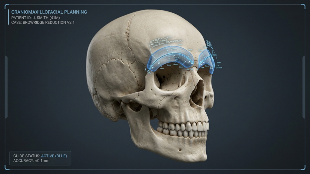
Перед операцией комплексная оценка проводится с использованием 3D КТ-сканирование и программное обеспечение для виртуального планирования хирургических операций. Этот шаг имеет решающее значение для:
- Измерение размеров лобной пазухи определить объем необходимого удаления и изменения формы кости.
- Визуализация носолобного соединения чтобы избежать таких осложнений, как утечка спинномозговой жидкости (СМЖ).
- Разработка индивидуальных направляющих для разрезов для каждого пациента. для обеспечения точности во время остеотомии (Обзорная статья о хирургии лица с учетом гендерных особенностей, 2025).
2. Разрез и обнажение
Хирург делает коронарный разрез (вдоль линии роста волос или внутри кожи головы) для доступа к лобной кости. Этот разрез делается стратегически для:
- Свести к минимуму видимые рубцы спрятав его в линию роста волос.
- Сохраните надкостницу. (мембрана, покрывающая кость) для поддержания кровоснабжения и снижения риска инфекции.
3. Остеотомия: удаление передней стенки
С использованием прецизионные хирургические инструменты, Хирург выполняет остеотомию для удалить переднюю стенку лобной пазухи. Этот шаг включает в себя:
- Разрезание кости по заранее спланированному маршруту для создания “костного лоскута”.”
- Осторожно приподнимите сегмент кости. чтобы избежать повреждения лобной пазухи или окружающих структур.
- Удаление любых костных перегородок внутри пазухи для обеспечения гладкого внутреннего контура (Феминизация лба, 2024).
4. Изменение формы костного лоскута
После удаления костный лоскут измененная форма на стерильном поле. Это включает в себя:
- Заусенец внутренней поверхности чтобы уменьшить его выступание и создать более плавный контур.
- Корректировка формы для соответствия желаемой женской эстетике, часто с использованием титановые пластины или винты для стабилизации.
- Обеспечение симметрии с остальной структурой лица (многофрагментная накладная реконструкция в Контурирование лба Хирургия, 2025).
5. Изменение положения и фиксация
Измененный костный лоскут перепозиционированный в более заднем и феминизированном положении. Этот этап включает в себя:
- Закрепление кости с титановые пластины и винты для обеспечения стабильности.
- Закрытие разреза с использованием рассасывающихся швов для минимизации образования рубцов.
- Наложение компрессионной повязки для уменьшения отека и поддержания нового контура.
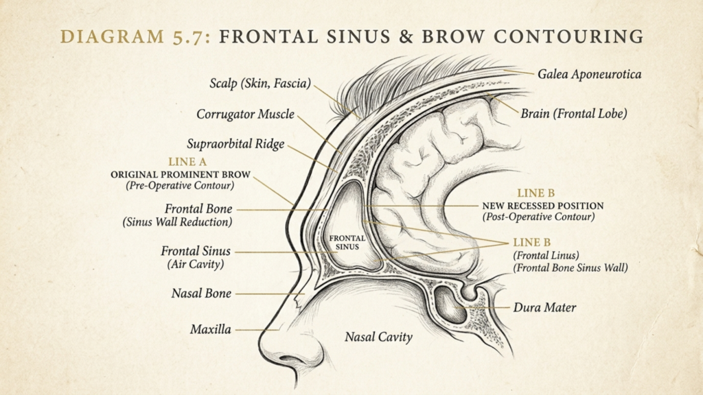
Анатомические особенности: лобная пазуха и окружающие структуры.
The лобная пазуха является критически важной анатомической структурой при редукции лба 3-го типа. размер, глубина и взаимосвязь Взаимодействия с окружающими костями и тканями напрямую влияют на хирургический подход и потенциальные риски. Ключевые анатомические аспекты включают:
1. Анатомия лобной пазухи
Лобная пазуха — это полая, заполненная воздухом полость Расположена в лобной кости. Ее передняя стенка образует надбровный дугу, а задняя стенка граничит с передней черепной ямкой. Ключевые особенности включают:
- Изменчивость размеров и формРазмеры синуса могут варьироваться от поверхностных до глубоко расположенных, что влияет на сложность процедуры.
- Толщина передней стенкиБолее толстая кость может потребовать более агрессивной коррекции формы, в то время как более тонкая кость увеличивает риск обнажения пазух.
- Наличие костных перегородокВнутренние разветвления в пазухе могут осложнить коррекцию формы и требуют тщательного удаления.

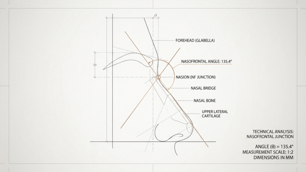
2. Связь с носолобной областью
The носолобное соединение Это место соединения лобной пазухи с носовыми костями. Эта область имеет решающее значение, потому что:
- Неправильное изменение формы может нарушить проходимость носовых дыхательных путей или изменить симметрию лица.
- Избыточная резекция может привести к “вогнутому” виду или нарушению функции пазух.
- Точные измерения необходимы для поддержания естественного перехода между лбом и переносицей (смещение лобной пазухи назад). Феминизация лица Хирургия, 2025).
3. Окружающие мягкие ткани и нервы
Лобная пазуха окружена мягкие ткани, мышцы и нервы которые необходимо сохранить во время операции. Ключевые структуры включают:
- Надблоковый и надглазничный нервыОтвечает за чувствительность в области лба и кожи головы. Повреждение может привести к онемению или хронической боли.
- лобная мышцаПриподнимает брови и способствует выразительности лица. Чрезмерное рассечение может привести к птозу (опущению) бровей.
- НадчерепСосудистая мембрана, питающая костный лоскут. Ее сохранение имеет решающее значение для заживления и снижения риска инфекции.
Возможные риски и осложнения: что должны знать пациенты.
Хотя операция по уменьшению объема лба 3-го типа очень эффективна, она не лишена рисков. Понимание этих потенциальных осложнений имеет важное значение для информированное принятие решений и послеоперационный уход. Ниже перечислены наиболее распространенные риски:
1. Утечка спинномозговой жидкости (СМЖ).
Утечка спинномозговой жидкости происходит, если задняя стенка лобной пазухи Случайное повреждение происходит во время остеотомии. Это серьезное осложнение, потому что:
- Это может привести к инфекциям. например, менингит, если его своевременно не вылечить.
- Симптомы включают в себя: Из носа или места разреза выделяется прозрачная жидкость, наблюдаются головные боли и тошнота.
- Лечение включает в себя хирургическое восстановление с использованием дурального трансплантата и, возможно, установкой поясничного дренажа (реконструкция лба типа 1, 2 или 3, 2025).
2. Инфекция и резорбция костной ткани
В месте хирургического вмешательства могут возникнуть инфекции, особенно если... надчереп поврежден или используются посторонние материалы (например, титановые пластины). К рискам относятся:
- Резорбция костной тканиОрганизм может рассосать измененный костный лоскут, что приведет к асимметрии или неровностям контура.
- Хронический синуситЕсли слизистая оболочка пазух повреждена, это может привести к длительному воспалению.
- Аппаратные сложностиПластины или винты могут стать ощутимыми на ощупь или инфицироваться, что потребует их удаления (многофрагментная накладная реконструкция). Контурирование лба Хирургия, 2025).
3. Сенсорные изменения и повреждение нервов
Повреждение надблоковый или надглазничный нерв Это может привести к временным или постоянным изменениям чувствительности. Пациенты могут испытывать:
- Онемение или покалывание на лбу или коже головы.
- Хроническая боль из-за раздражения нервов или образования рубцов.
- Изменённое выражение лица если поражена лобная мышца.
4. Неровности контура и асимметрия
Даже при тщательном планировании, асимметрия или неровные контуры может возникнуть по следующим причинам:
- Неравномерная коррекция формы костей во время процедуры.
- Послеоперационный отек Это приводит к неравномерному разрешению.
- Видимость оборудования если пластины или винты расположены не вровень с костью.
5. Долгосрочные функциональные и эстетические результаты
Хотя операция по уменьшению лба 3-го типа направлена на усиление феминизации, долгосрочные результаты зависят от:
- Правильное заживление костей: Обеспечение плавной интеграции измененной кости с окружающими структурами.
- Лечение рубцов: Минимизация видимых рубцов за счет тщательного выполнения разрезов и послеоперационного ухода.
- Удовлетворенность пациентовСогласование ожиданий с реалистичными результатами, поскольку анатомические особенности каждого человека различны.

Послеоперационный уход и восстановление: обеспечение оптимального заживления.
Восстановление после уменьшения объема лба 3-го типа – это... постепенный процесс Для этого требуется терпение и соблюдение послеоперационных инструкций. Ниже приведены ключевые шаги, способствующие заживлению и минимизации осложнений:
1. Непосредственный послеоперационный уход
В первые 48 часов сосредоточьтесь на следующем:
- Лечение отеков с холодными компрессами и держа голову в приподнятом положении.
- Приём назначенных врачом обезболивающих препаратов чтобы избежать дискомфорта.
- Избегайте перенапряжения и подъема тяжестей. для предотвращения повышения внутричерепного давления.
2. Первые две недели: мониторинг и гигиена.
В течение этого этапа:
- Следите за тем, чтобы место разреза было чистым и сухим. для предотвращения инфекции.
- Посещайте повторные приемы. следить за процессом заживления и решать любые возникающие проблемы.
- Избегайте курения и употребления алкоголя., поскольку они могут замедлить заживление и увеличить риск инфекций.
3. Недели 3–6: Постепенное возвращение к нормальной активности.
По мере спада отека:
- Возобновите легкие занятия. например, ходьба или работа за столом.
- Избегайте контактных видов спорта и интенсивных физических нагрузок. до получения разрешения от вашего хирурга.
- Используйте силиконовый гель или пластыри. чтобы свести к минимуму образование рубцов.
4. Долгосрочный уход: поддержание достигнутых результатов
Для обеспечения долговременных результатов:
- Защитите лоб от травм чтобы избежать смещения костного лоскута.
- Проконсультируйтесь со своим хирургом. Ежегодно для контроля стабильности костей и выявления любых поздних осложнений.
- Рассмотрите нехирургические методы улучшения внешности. например, филлеры или ботокс для дальнейшего уточнения контуров.

Альтернативы уменьшению лба 3-го типа: изучение менее инвазивных вариантов.
Хотя процедура уменьшения объема лба 3-го типа очень эффективна, она может подойти не всем. Альтернативные варианты включают:
1. Формирование контуров лба 1-го и 2-го типов
Для лиц с более мягкое набухание бровей, В некоторых случаях могут быть достаточны процедуры типа 1 или типа 2:
- Тип 1Включает в себя костная стружка Без удаления передней стенки. Идеально подходит для небольших уменьшений.
- Тип 2Комбайны частичная остеотомия С коррекцией формы, подходит для умеренно выступающих бровей.
2. Нехирургические варианты лечения
Для тех, кто ищет минимальное время простоя, К нехирургическим альтернативам относятся:
- Дермальные наполнители: Временное увеличение объема для смягчения надбровных дуг.
- Инъекции ботоксаРасслабьте лобную мышцу, чтобы уменьшить выступание бровей.
- Пересадка жираИспользуется собственный жир пациента для придания формы лбу.
3. Комбинированные процедуры
Для комплексной феминизации уменьшение лба 3-го типа может сочетаться со следующими процедурами:
- Продвижение линии роста волос уменьшить высоту лба.
- Ринопластика для уточнения контуров носа.
- Подтяжка бровей Приподнять брови для более молодого вида.
Заключение: Баланс между эстетикой, безопасностью и целями пациента.
Уменьшение объема лба 3-го типа — это мощный инструмент В хирургии феминизации лица предлагаются преобразующие результаты для трансгендерных женщин, стремящихся к более женственному контуру лба. Однако ее сложность требует экспертные знания, точность и глубокое понимание анатомии. для снижения таких рисков, как утечка спинномозговой жидкости, инфекции и неровности контуров.
Решение о проведении операции по уменьшению объема лба 3-го типа должно приниматься пациентами после консультации со специалистом. сертифицированный хирург Специалист по феминизации лица. Взвешивая преимущества и риски, а также изучая альтернативы, люди могут достичь желаемого результата. гармоничный баланс между эстетикой и безопасностью, в конечном итоге это повысит их уверенность в себе и укрепит соответствие их гендерной идентичности.
Тем, кто рассматривает возможность проведения этой процедуры, Клиника доктора МФО Компания предлагает специализированные услуги по уменьшению объема лба 3-го типа, гарантируя индивидуальный подход и оптимальные результаты. Свяжитесь с нами сегодня Запишитесь на консультацию и начните свой путь к более женственной внешности.
Часто задаваемые вопросы
В чём разница между уменьшением объёма лба 1-го, 2-го и 3-го типов?
Тип 1 включает в себя удаление костной ткани для незначительного уменьшения объема, Тип 2 сочетает частичную остеотомию с изменением формы для умеренного выступа надбровных дуг, а Тип 3 требует полного удаления, изменения формы и перемещения передней стенки лобной пазухи для значительной феминизации. Тип 3 является наиболее сложным и применяется у лиц с выступающими надбровными дугами и глубокими лобными пазухами.
Сколько времени занимает восстановление после уменьшения объема лба 3-го типа?
Период восстановления может варьироваться, но обычно соответствует следующим срокам: 1–2 недели для первоначального отека и дискомфорта, 3–6 недель для постепенного возвращения к нормальной активности и до 6 месяцев для достижения окончательных результатов по мере полного спада отека. Пациентам следует избегать интенсивных физических нагрузок в течение как минимум 6 недель для обеспечения правильного заживления.
Какие признаки указывают на утечку спинномозговой жидкости после уменьшения объема лба 3-го типа?
Симптомы утечки спинномозговой жидкости включают выделение прозрачной жидкости из носа или места разреза, постоянные головные боли, тошноту и соленый привкус во рту. При появлении любого из этих симптомов следует немедленно обратиться за медицинской помощью, поскольку нелеченная утечка спинномозговой жидкости может привести к серьезным инфекциям, таким как менингит.
Можно ли сочетать уменьшение лба 3-го типа с другими процедурами по коррекции формы лба?
Да, уменьшение объема лба 3-го типа часто сочетается с такими процедурами, как коррекция линии роста волос., ринопластика, а также подтяжка бровей для достижения комплексной феминизации лица. Сочетание процедур может улучшить общую гармонию и уменьшить необходимость в нескольких операциях.
Какие риски связаны с использованием титановых пластин при уменьшении объема лба 3-го типа?
Хотя титановые пластины обеспечивают стабильность, к рискам относятся пальпируемость имплантатов (ощущение пластин под кожей), инфекция и резорбция костной ткани вокруг пластин. В редких случаях пластины могут потребовать удаления, если они вызывают дискомфорт или осложнения.
Как минимизировать образование рубцов после операции по уменьшению объема лба 3-го типа?
Чтобы свести к минимуму образование рубцов, следуйте инструкциям хирурга по послеоперационному уходу, которые могут включать использование силиконового геля или пластырей, избегание пребывания на солнце и поддержание чистоты и увлажненности места разреза. Большинство рубцов со временем значительно бледнеют, особенно если они скрыты в линии роста волос.
Подходит ли процедура уменьшения объема лба 3-го типа всем?
Уменьшение лба 3-го типа идеально подходит для людей с выступающими надбровными дугами и глубокими лобными пазухами. Пациенты с менее выраженными надбровными дугами могут достичь удовлетворительных результатов с помощью процедур 1-го или 2-го типа. Необходима консультация со специалистом. хирург ФФС Крайне важно определить оптимальный подход, исходя из вашей анатомии и целей.
Чего следует ожидать во время консультации по поводу уменьшения объема лба 3-го типа?
В ходе консультации хирург изучит вашу историю болезни, проведет физический осмотр и с помощью 3D-визуализации оценит анатомию лобных пазух. Он обсудит этапы процедуры, риски и ожидаемые результаты, а также альтернативные варианты, такие как нехирургические методы или менее инвазивные хирургические техники.







