

Féminisation faciale La chirurgie de féminisation faciale (FFS) est devenue un domaine transformateur, offrant aux femmes transgenres la possibilité d'harmoniser leurs traits faciaux avec leur identité de genre. Parmi les interventions les plus complexes et les plus marquantes, on trouve : Tapez 3 Réduction du front, Cette technique chirurgicale vise à corriger les arcades sourcilières proéminentes et la structure osseuse frontale. Il ne s'agit pas d'une simple intervention esthétique : c'est une reconstruction méticuleuse de la paroi antérieure du sinus frontal, remodelée pour obtenir un contour plus doux et plus féminin. Cependant, la complexité de cette chirurgie exige une connaissance approfondie de ses mécanismes. subtilités anatomiques, étapes de la procédure et risques potentiels pour garantir à la fois la sécurité et des résultats optimaux.
Dans ce guide, nous explorons en profondeur… techniques chirurgicales, considérations anatomiques et risques associé au type 3 Réduction du front. Que vous soyez un patient explorant vos options ou un professionnel de la santé en quête d'informations, cet article vous apportera un éclairage précieux. explication technique mais accessible ce qui fait de cette procédure une pierre angulaire de la FFS.
Table des matières
Comprendre la réduction du front de type 3 : qu’est-ce qui la distingue ?
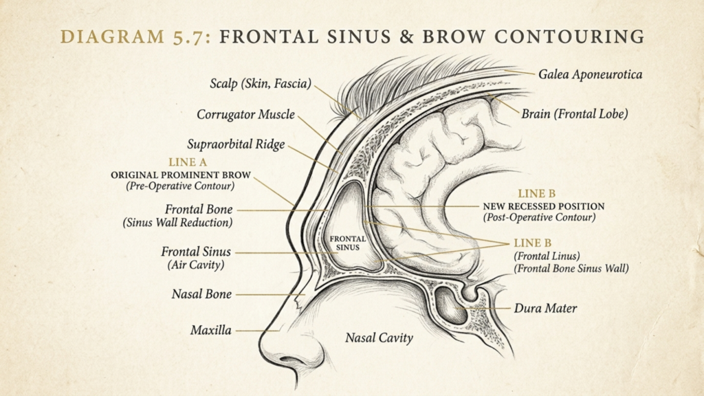
Tapez 3 Réduction du front, aussi connu sous le nom recul du sinus frontal ou cranioplastie de type 3, se distingue par son orientation vers le paroi antérieure du sinus frontal. Contrairement aux interventions de type 1 ou 2, qui consistent en un limage osseux ou un remodelage partiel, l'intervention de type 3 nécessite… retrait complet, remodelage et repositionnement de la paroi antérieure. Cette technique est particulièrement adaptée aux personnes présentant arcades sourcilières proéminentes et sinus frontaux profonds, là où des méthodes plus simples seraient insuffisantes ou risqueraient d'exposer le sinus (Mittermiller, 2025).
Le distinction clé Le type 3 réside dans sa capacité à atteindre un féminisation spectaculaire Le repositionnement de la paroi antérieure du front permet d'affiner le visage. Cette technique réduit non seulement l'arcade sourcilière, mais crée également un contour du front plus lisse et harmonieux, en accord avec les canons de beauté féminins.
Procédure chirurgicale étape par étape : ablation, remodelage et remplacement de la paroi antérieure
La procédure de réduction du front de type 3 est une processus en plusieurs étapes Cela exige précision, expertise anatomique et instruments chirurgicaux de pointe. Vous trouverez ci-dessous le détail de chaque étape :
1. Planification et imagerie préopératoires
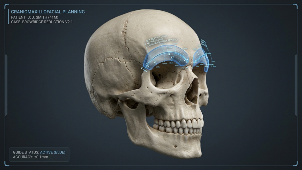
Avant l'opération, un évaluation complète est menée à l'aide de Scanners CT 3D et un logiciel de planification chirurgicale virtuelle. Cette étape est cruciale pour :
- Mesure des dimensions du sinus frontal pour déterminer l'étendue du retrait et du remodelage osseux nécessaires.
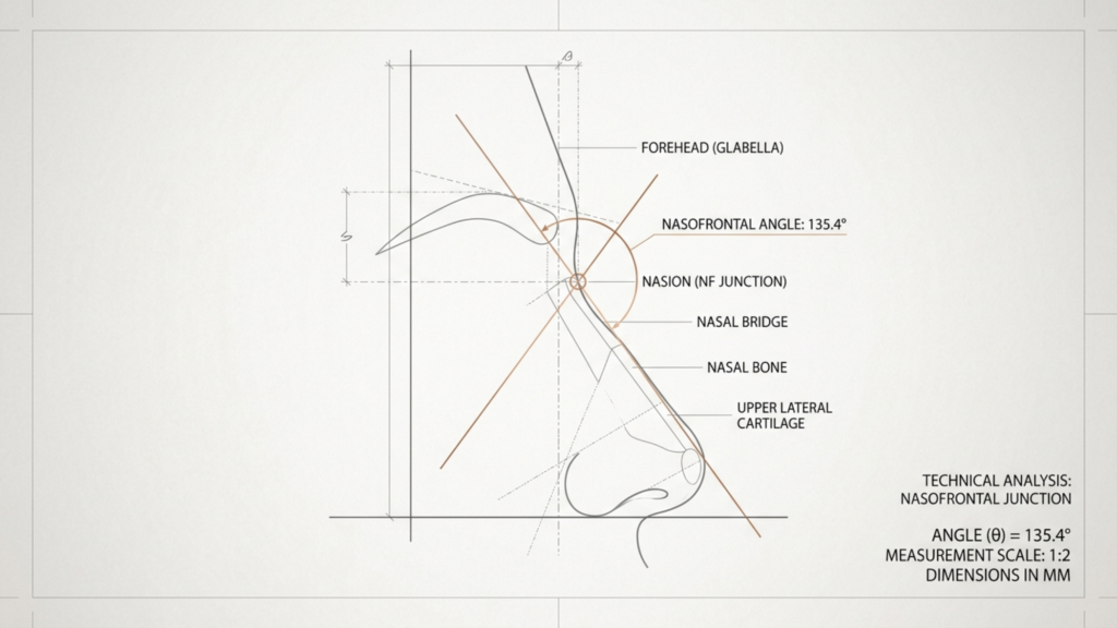
- Visualisation de la jonction nasofrontale pour éviter les complications telles que les fuites de liquide céphalo-rachidien (LCR).
- Conception de guides de coupe spécifiques au patient pour assurer la précision lors de l'ostéotomie (Revue narrative de la chirurgie de genre faciale, 2025).
2. Incision et exposition
Le chirurgien fait un incision coronale (le long de la ligne des cheveux ou dans le cuir chevelu) pour accéder à l'os frontal. Cette incision est stratégiquement placée pour :
- Minimiser les cicatrices visibles en le dissimulant dans la ligne des cheveux.
- Préserver le péricrâne (la membrane recouvrant l'os) pour maintenir l'apport sanguin et réduire le risque d'infection.
3. Ostéotomie : ablation de la paroi antérieure
En utilisant instruments chirurgicaux de précision, le chirurgien pratique une ostéotomie pour retirer la paroi antérieure du sinus frontal. Cette étape implique :
- Couper l'os le long de lignes préétablies pour créer un “ volet osseux ”.”
- Soulever délicatement le segment osseux afin d'éviter d'endommager le sinus frontal ou les structures environnantes.
- Éliminer les cloisons osseuses à l'intérieur du sinus pour assurer un contour intérieur lisse (Féminisation du front, 2024).
4. Remodelage du lambeau osseux
Une fois retirée, la membrane osseuse est remodelé sur un champ stérile. Cela implique :
- Ébavurage de la surface intérieure pour réduire sa projection et créer un contour plus lisse.
- Ajuster la forme pour correspondre à l'esthétique féminine recherchée, souvent en utilisant plaques ou vis en titane pour la stabilisation.
- Assurer la symétrie avec le reste de la structure faciale (Reconstruction par superposition multifragmentaire dans Remodelage du front Chirurgie, 2025).
5. Repositionnement et fixation
Le volet osseux remodelé est repositionné dans une zone plus postérieure et féminisée. Cette étape comprend :
- Fixer l'os avec plaques et vis en titane pour assurer la stabilité et une bonne cicatrisation.
- Fermeture de l'incision avec des sutures résorbables pour minimiser les cicatrices.
- Application d'un pansement compressif pour réduire l'enflure et soutenir le nouveau contour.
Considérations anatomiques : Le sinus frontal et les structures environnantes
Le sinus frontal est une structure anatomique essentielle dans la réduction du front de type 3. taille, profondeur et relation Les caractéristiques anatomiques des os et des tissus environnants influencent directement l'approche chirurgicale et les risques potentiels. Les principaux éléments à prendre en compte sont les suivants :
1. Anatomie du sinus frontal
Le sinus frontal est un cavité creuse remplie d'air Située dans l'os frontal, sa paroi antérieure forme l'arcade sourcilière, tandis que sa paroi postérieure borde la fosse crânienne antérieure. Ses principales caractéristiques sont :
- Variabilité de taille et de formeLa profondeur du sinus peut varier, allant de superficielle à profonde, ce qui influe sur la complexité de l'intervention.
- Épaisseur de la paroi antérieureUn os plus épais peut nécessiter un remodelage plus agressif, tandis qu'un os plus fin augmente le risque d'exposition des sinus.
- Présence de cloisons osseusesLes divisions internes au sein du sinus peuvent compliquer le remodelage et nécessitent une ablation minutieuse.

2. Relation avec la jonction nasofrontale
Le jonction nasofrontale C'est à la jonction du sinus frontal et des os nasaux. Cette zone est cruciale car :
- Remodelage inadéquat peuvent perturber les voies nasales ou altérer la symétrie faciale.
- Résection excessive peut donner un aspect “ creusé ” ou compromettre la fonction des sinus.
- Mesures précises sont essentiels pour maintenir une transition naturelle entre le front et l'arête nasale (Recul du sinus frontal dans Féminisation faciale Chirurgie, 2025).
3. Tissus mous et nerfs environnants
Le sinus frontal est entouré par tissus mous, muscles et nerfs qui doivent être préservées pendant l'intervention chirurgicale. Les structures clés comprennent :
- Nerfs supratrochléaire et supra-orbitaireResponsable de la sensibilité du front et du cuir chevelu. Des lésions peuvent entraîner un engourdissement ou des douleurs chroniques.
- Muscle frontalElle rehausse les sourcils et contribue aux expressions faciales. Une dissection excessive peut entraîner une ptose (affaissement) des sourcils.
- PéricrâneUne membrane vascularisée qui nourrit le volet osseux. Sa préservation est essentielle à la cicatrisation et à la réduction des risques d'infection.
Risques et complications potentiels : ce que les patients doivent savoir
Bien que la réduction du front de type 3 soit très efficace, elle n'est pas sans risques. Il est essentiel de comprendre ces complications potentielles. prise de décision éclairée et les soins postopératoires. Voici les risques les plus courants :
1. Fuite de liquide céphalo-rachidien (LCR)
Une fuite de LCR se produit si le paroi postérieure du sinus frontal une brèche accidentelle se produit lors de l'ostéotomie. Cette complication est grave car :
- Cela peut entraîner des infections comme la méningite si elle n'est pas traitée rapidement.
- Les symptômes incluent Écoulement de liquide clair par le nez ou le site d'incision, maux de tête et nausées.
- Le traitement implique réparation chirurgicale avec greffe durale et mise en place possible d'un drain lombaire (reconstruction du front de type 1, 2 ou 3, 2025).
2. Infection et résorption osseuse
Des infections peuvent survenir au niveau du site chirurgical, en particulier si le Le péricrâne est endommagé ou des matériaux étrangers (par exemple, des plaques de titane) sont utilisés. Les risques comprennent :
- Résorption osseuseLe corps peut absorber le lambeau osseux remodelé, ce qui peut entraîner une asymétrie ou des irrégularités de contour.
- sinusite chroniqueSi la muqueuse des sinus est endommagée, cela peut entraîner une inflammation chronique.
- Complications liées au matérielLes plaques ou les vis peuvent devenir palpables ou s'infecter, nécessitant leur retrait (Reconstruction par onlay multifragmentaire dans Remodelage du front Chirurgie, 2025).
3. Altérations sensorielles et lésions nerveuses
Dommages à nerfs supratrochléaires ou supra-orbitaires peut entraîner des modifications sensorielles temporaires ou permanentes. Les patients peuvent ressentir :
- Engourdissement ou picotements sur le front ou le cuir chevelu.
- La douleur chronique due à une irritation nerveuse ou à des cicatrices.
- Expressions faciales modifiées si le muscle frontal est touché.
4. Irrégularités et asymétrie des contours
Même avec une planification méticuleuse, asymétrie ou contours irréguliers peut survenir en raison de :
- remodelage osseux inégal pendant la procédure.
- œdème postopératoire qui se résout de manière inégale.
- visibilité matérielle si les plaques ou les vis ne sont pas positionnées au ras de l'os.
5. Résultats fonctionnels et esthétiques à long terme
Bien que la réduction du front de type 3 soit conçue pour accentuer la féminisation, les résultats à long terme dépendent de :
- bonne cicatrisation osseuse: S'assurer que l'os remodelé s'intègre harmonieusement aux structures environnantes.
- Gestion des cicatricesMinimiser les cicatrices visibles grâce à un placement précis des incisions et à des soins postopératoires appropriés.
- Satisfaction des patients: Adapter les attentes aux résultats réalistes, car l'anatomie individuelle varie.

Soins postopératoires et convalescence : assurer une cicatrisation optimale
La récupération après une réduction du front de type 3 est une processus graduel Cela exige de la patience et le respect des consignes postopératoires. Voici les étapes clés pour favoriser la guérison et minimiser les complications :
1. Soins postopératoires immédiats
Au cours des 48 premières heures, concentrez-vous sur :
- Gestion de l'enflure avec des compresses froides et en gardant la tête surélevée.
- Prendre des médicaments contre la douleur prescrits pour prévenir tout inconfort.
- Éviter les efforts excessifs ou le port de charges lourdes pour prévenir l'augmentation de la pression intracrânienne.
2. Les deux premières semaines : surveillance et hygiène
Durant cette phase :
- Gardez la zone d'incision propre et sèche. pour prévenir l'infection.
- Assistez aux rendez-vous de suivi pour surveiller la guérison et répondre à toute préoccupation.
- Évitez de fumer et de consommer de l'alcool., car elles peuvent nuire à la cicatrisation et augmenter les risques d'infection.
3. Semaines 3 à 6 : Reprise progressive des activités normales
Lorsque l'enflure diminue :
- Reprise d'activités légères comme la marche ou le travail de bureau.
- Évitez les sports de contact ou les exercices physiques intenses. jusqu'à ce que votre chirurgien vous y autorise.
- Utilisez du gel ou des feuilles de silicone pour minimiser les cicatrices.
4. Soins de longue durée : Maintenir les résultats
Pour garantir des résultats durables :
- Protégez votre front des traumatismes. pour éviter de déplacer le volet osseux.
- Prenez contact avec votre chirurgien. annuellement pour surveiller la stabilité osseuse et traiter toute complication tardive.
- Envisagez des améliorations non chirurgicales comme les injections de comblement ou de Botox pour affiner davantage les contours.

Alternatives à la réduction du front de type 3 : explorer des options moins invasives
Bien que la réduction du front de type 3 soit très efficace, elle peut ne pas convenir à tout le monde. Voici quelques alternatives :
1. Contourage du front de type 1 et de type 2
Pour les personnes atteintes de sourcils plus légèrement froncés, Les procédures de type 1 ou de type 2 peuvent suffire :
- Type 1: Implique rasage des os sans retirer la paroi antérieure. Idéal pour les réductions mineures.
- Type 2: Moissonneuses-batteuses ostéotomie partielle Avec remodelage, convient pour une élévation modérée des sourcils.
2. Options non chirurgicales
Pour ceux qui recherchent temps d'arrêt minimal, les alternatives non chirurgicales comprennent :
- Produits de comblement cutané: Ajout temporaire de volume pour adoucir les arcades sourcilières.
- Injections de BotoxDétendez le muscle frontal pour réduire la proéminence des sourcils.
- Greffe de graisse: Utilise la propre graisse du patient pour redessiner le contour du front.
3. Procédures combinées
Pour une féminisation complète, la réduction du front de type 3 peut être associée à :
- Avancement de la racine des cheveux pour réduire la hauteur du front.
- Rhinoplastie pour affiner les contours du nez.
- Lifting des sourcils pour rehausser les sourcils et obtenir un aspect plus jeune.
Conclusion : Concilier esthétique, sécurité et objectifs du patient
La réduction du front de type 3 est une outil puissant La chirurgie de féminisation faciale offre des résultats transformateurs aux femmes transgenres qui souhaitent un contour du front plus féminin. Cependant, sa complexité exige expertise, précision et une connaissance approfondie de l'anatomie afin d'atténuer les risques tels que les fuites de LCR, les infections et les irrégularités de contour.
Pour les patients, la décision de subir une réduction du front de type 3 doit être prise en consultation avec un médecin. chirurgien certifié qui se spécialise dans la chirurgie esthétique du visage. En évaluant les avantages et les risques et en explorant les alternatives, les personnes peuvent parvenir à un résultat optimal. équilibre harmonieux entre esthétique et sécurité, renforçant ainsi leur confiance en eux et leur adéquation avec leur identité de genre.
Pour ceux qui envisagent cette procédure, Clinique Dr MFO offre une expertise spécialisée en réduction du front de type 3, garantissant des soins personnalisés et des résultats optimaux. Contactez-nous aujourd'hui pour programmer une consultation et entamer votre parcours vers une apparence plus féminine.
Questions fréquemment posées
Quelle est la différence entre la réduction du front de type 1, de type 2 et de type 3 ?
Le type 1 consiste en un limage osseux pour des réductions mineures ; le type 2 associe une ostéotomie partielle à un remodelage pour une correction modérée des arcades sourcilières proéminentes ; et le type 3 requiert l’ablation complète, le remodelage et le repositionnement de la paroi antérieure du sinus frontal pour une féminisation importante. Le type 3 est le plus complexe et est réservé aux personnes présentant des arcades sourcilières saillantes et des sinus frontaux profonds.
Combien de temps dure la convalescence après une réduction du front de type 3 ?
La durée de la convalescence est variable, mais elle suit généralement le calendrier suivant : 1 à 2 semaines pour l’œdème et l’inconfort initiaux, 3 à 6 semaines pour une reprise progressive des activités normales, et jusqu’à 6 mois pour le résultat final une fois l’œdème complètement résorbé. Il est conseillé aux patients d’éviter les efforts physiques intenses pendant au moins 6 semaines afin de favoriser une bonne cicatrisation.
Quels sont les signes d'une fuite de LCR après une réduction frontale de type 3 ?
Les symptômes d'une fuite de liquide céphalo-rachidien comprennent un écoulement de liquide clair par le nez ou la cicatrice, des maux de tête persistants, des nausées et un goût salé dans la bouche. Si l'un de ces symptômes apparaît, consultez immédiatement un médecin, car une fuite de liquide céphalo-rachidien non traitée peut entraîner des infections graves comme la méningite.
La réduction du front de type 3 peut-elle être combinée à d'autres interventions de chirurgie esthétique du front ?
Oui, la réduction du front de type 3 est souvent associée à des interventions telles que l'avancement de la ligne capillaire., rhinoplastie, et un lifting des sourcils pour une féminisation faciale complète. Combiner ces interventions permet d'harmoniser le visage et de limiter le nombre d'opérations nécessaires.
Quels sont les risques liés à l'utilisation de plaques en titane dans la réduction du front de type 3 ?
Bien que les plaques en titane assurent la stabilité, les risques comprennent la palpation du matériel (sensation de présence des plaques sous la peau), l'infection et la résorption osseuse autour des plaques. Dans de rares cas, il peut être nécessaire de retirer les plaques si elles provoquent une gêne ou des complications.
Comment minimiser les cicatrices après une réduction du front de type 3 ?
Pour minimiser les cicatrices, suivez les instructions postopératoires de votre chirurgien, qui peuvent inclure l'utilisation de gel ou de pansements en silicone, l'évitement de l'exposition au soleil et le maintien d'une bonne hydratation de la zone d'incision. La plupart des cicatrices s'atténuent considérablement avec le temps, surtout si elles sont dissimulées dans la ligne des cheveux.
La réduction du front de type 3 convient-elle à tout le monde ?
La réduction du front de type 3 est idéale pour les personnes présentant des arcades sourcilières proéminentes et des sinus frontaux profonds. Celles dont les sourcils sont moins marqués peuvent obtenir des résultats satisfaisants avec les interventions de type 1 ou 2. Une consultation avec un spécialiste est recommandée. Chirurgien FFS Il est essentiel de déterminer la meilleure approche en fonction de votre anatomie et de vos objectifs.
À quoi dois-je m'attendre lors de la consultation pour une réduction du front de type 3 ?
Lors de la consultation, votre chirurgien examinera vos antécédents médicaux, procédera à un examen physique et utilisera l'imagerie 3D pour évaluer l'anatomie de vos sinus frontaux. Il vous expliquera les étapes de l'intervention, les risques et les résultats attendus, ainsi que les alternatives possibles comme les options non chirurgicales ou les techniques chirurgicales moins invasives.







