

Rekonstruktion der Stirn ist ein wichtiger Bestandteil von Gesichts Feminisierungschirurgie (Oh mein Gott) und kraniofaziale Eingriffe mit dem Ziel, eine harmonische und ästhetisch ansprechende Gesichtskontur zu erzielen. Zu den am meisten diskutierten Techniken gehören Typ 1 Und Kranioplastik Typ 3, Jede dieser Methoden bietet einen anderen Ansatz zur Umformung der Stirn. Typ 1 beinhaltet Knochenabfräsen oder -schaben, Typ 3 erfordert eine komplexere Osteotomie und Rückschlag der Stirnhöhle. Das Verständnis der anatomischen, funktionellen und ästhetischen Auswirkungen dieser Techniken ist sowohl für Chirurgen als auch für Patienten unerlässlich, um fundierte Entscheidungen treffen zu können. Erfahren Sie mehr über die Rückverlagerung der Stirnhöhle und die Reduzierung der Augenbrauenwölbung in unserem vollständigen Leitfaden. Konturierung der Stirn.
Dieser Leitfaden untersucht die strukturellen Unterschiede zwischen Kranioplastik Typ 1 und Typ 3 sowie deren Auswirkungen auf Knochendicke, Anatomie der Stirnhöhle, und wie Chirurgen die jeweils geeignetste Vorgehensweise für jeden Patienten bestimmen. Am Ende werden Sie Klarheit darüber gewinnen, welche Technik Ihren anatomischen Gegebenheiten und ästhetischen Zielen am besten entspricht.
Inhaltsverzeichnis
Die anatomische Grundlage: Stirnbein und Nasennebenhöhlen
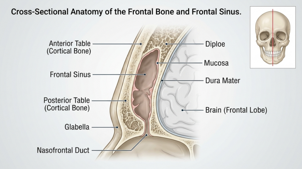
Der Stirnbein Und Stirnhöhle spielen eine entscheidende Rolle bei der Stirnrekonstruktion. Das Stirnbein bildet den oberen Teil des Gesichts und beherbergt die Stirnhöhle, einen Hohlraum, dessen Größe und Form von Mensch zu Mensch variiert. vorderer Tisch Die Stirnhöhle ist die äußere Knochenschicht, die zur Kontur der Stirn beiträgt, während die hintere Tafel Sie trennt die Nasennebenhöhlen vom Gehirn. Die Dicke dieser Platten und der Grad der Pneumatisierung (luftgefüllte Ausdehnung) der Nasennebenhöhlen beeinflussen die Wahl zwischen einer Kranioplastik vom Typ 1 und vom Typ 3.
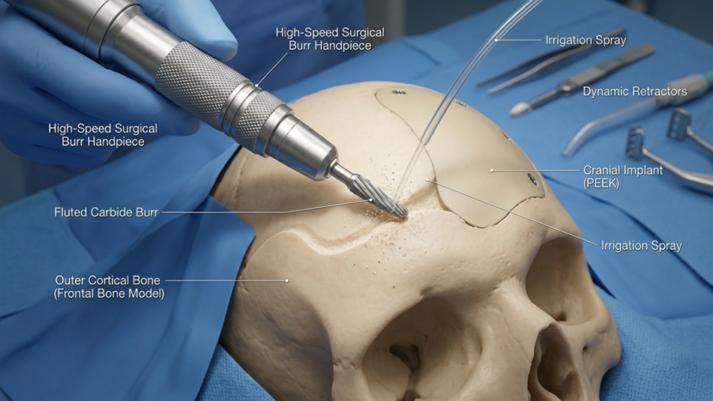
In Kranioplastik Typ 1, Die der Chirurg Dabei wird ein Hochgeschwindigkeitsbohrer verwendet, um die äußere Kortikalis abzutragen und so die Vorwölbung zu reduzieren, ohne die Stirnhöhle zu verletzen. Diese Technik ist ideal für Patienten mit dünnes Stirnbein oder minimales Abtragen, da dadurch die Integrität der Nasennebenhöhlen erhalten bleibt. Bei Patienten mit ausgeprägtem Augenbrauenwulst oder einer dicken vorderen Kieferhöhlenwand ist dies jedoch möglicherweise nicht ausreichend, da übermäßiges Abtragen die Knochenstabilität beeinträchtigen oder die gewünschte Kontur nicht erreichen kann (Ousterhout, 2024).
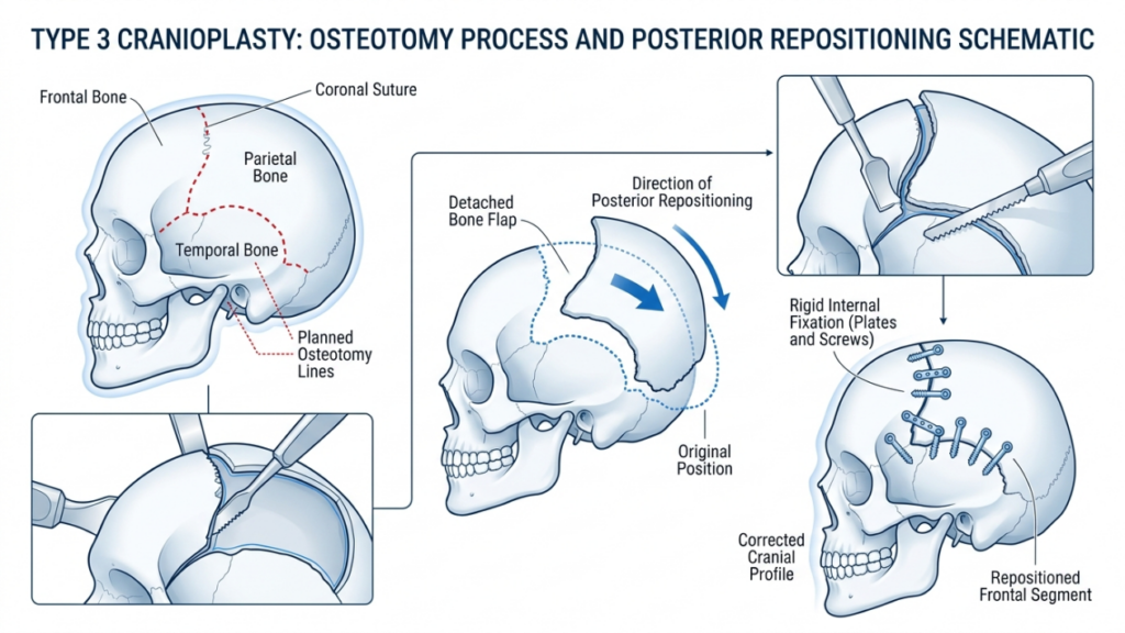
Im Gegensatz, Kranioplastik Typ 3 beinhaltet ein Osteotomie—ein kontrollierter Schnitt durch die vordere Wand der Stirnhöhle. Das Knochensegment wird anschließend nach hinten verlagert (Setback), um die Stirnprojektion zu reduzieren und eine glattere, femininere Stirnform zu erzielen. Diese Technik ist Patientinnen mit … vorbehalten. mäßige bis starke Stirnwölbung oder bei einer dicken vorderen Kieferhöhlenwand, wo ein alleiniges Abfräsen nicht ausreichen würde. Die Osteotomie ermöglicht eine präzise Umformung unter Erhalt der Schutzfunktion der Kieferhöhle (Feminisierung der Stirn: Eine Übersichtsarbeit, 2024).
Kranioplastik Typ 1: Abtragen und Fräsen
Technikübersicht
Die Kranioplastik Typ 1 ist die minimalinvasivste Methode zur Stirnrekonstruktion. Dabei wird eine Hochgeschwindigkeits-Grind um die Vorwölbung des Stirnbeins allmählich zu verringern. der Chirurg Die äußere Kortexschicht wird sorgfältig abgetragen, wobei ein Eindringen in die Stirnhöhle vermieden wird. Diese Technik ist besonders wirksam bei Patienten mit:
- Leichte bis mäßige Stirnwölbung
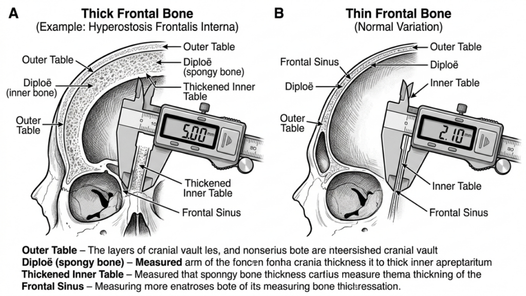
- Dünnes Stirnbein (weniger als 5 mm)
- Fehlen einer signifikanten Pneumatisierung der Stirnhöhlen
Der Eingriff wird mittels eines/einer Koronarschnitt, Diese Technik ermöglicht den Zugang zur Stirn und minimiert gleichzeitig die sichtbare Narbenbildung. Der Chirurg nutzt taktiles Feedback und visuelle Hinweise, um eine gleichmäßige Reduktion zu gewährleisten, ohne den Knochen zu stark auszudünnen, was zu Instabilität oder Konturunregelmäßigkeiten führen könnte.
Vorteile der Kranioplastik Typ 1
Die Kranioplastik Typ 1 bietet mehrere Vorteile:
- Minimale Invasivität: Der Verzicht auf eine Osteotomie oder Knochenentfernung reduziert das Operationstrauma und die Genesungszeit.
- Geringeres Komplikationsrisiko: Durch den Erhalt der Stirnhöhle wird das Risiko von Sinusitis, Liquorverlust oder Mukozelenbildung minimiert.
- Kürzere Operationszeit: Der Eingriff dauert in der Regel 1–2 Stunden und ist damit eine schnellere Option im Vergleich zu Typ 3.
- Vorhersehbare Ergebnisse: Ideal für Patientinnen mit leichter Vorwölbung, bei denen eine dezente Konturierung ausreicht, um ein feminines Erscheinungsbild zu erzielen.
Einschränkungen der Kranioplastik Typ 1
Obwohl die Kranioplastik Typ 1 sicherer und weniger invasiv ist, weist sie bemerkenswerte Einschränkungen auf:
- Begrenzte Reduzierung: Unzureichend bei Patienten mit stark ausgeprägten Augenbrauenwülsten oder dicken Stirnbeinen.
- Risiko der Überausdünnung: Aggressives Fräsen kann den Knochen schwächen und zu Konturunregelmäßigkeiten oder Frakturen führen.
- Unvollständige Feminisierung: Bei Patienten mit ausgeprägten maskulinen Gesichtszügen wird möglicherweise nicht das gewünschte ästhetische Ergebnis erzielt.
Kranioplastik Typ 3: Osteotomie und Rückverlagerung
Technikübersicht
Die Kranioplastik Typ 3 ist ein komplexeres Verfahren, das für Patienten mit … entwickelt wurde. mäßige bis starke Stirnwölbung oder verdickte Stirnbeine. Es beinhaltet ein OsteotomieEin präziser Schnitt durch die vordere Wand der Stirnhöhle, gefolgt von der Rückverlagerung des Knochensegments, ermöglicht eine deutliche Reduzierung der Stirnprojektion und eine harmonischere, femininere Kontur.
Der Eingriff wird typischerweise durch ein/eine Koronarschnitt, Dadurch wird der Zugang zum Stirnbein und zur Nasennebenhöhle ermöglicht. Der Chirurg verwendet ein Sagittalsäge oder piezoelektrisches Gerät Um die Osteotomie durchzuführen, muss darauf geachtet werden, dass der Schnitt der natürlichen Krümmung der Stirn folgt. Das Knochensegment wird dann repositioniert und fixiert mit Titanplatten und Schrauben oder resorbierbarem Nahtmaterial. Diese Technik eignet sich ideal für Patienten mit:
- Stark ausgeprägte Augenbrauenwölbung
- Dickes Stirnbein (größer als 5 mm)
- Signifikante Pneumatisierung der Stirnhöhlen
Die Kranioplastik Typ 3 erfordert eine sorgfältige Planung, die oft Folgendes umfasst: Virtuelle 3D-Operationsplanung (VSP) um die Osteotomie und den Rückverlagerungsvorgang zu simulieren. Dies gewährleistet Präzision und minimiert das Risiko von Komplikationen wie Nasennebenhöhlenverletzungen oder Liquorverlust (Virtuelle Operationsplanung in der virtuellen Operationsplanung). Gesichtsfeminisierung des oberen Gesichts, 2025).
Vorteile der Kranioplastik Typ 3
Die Kranioplastik Typ 3 bietet mehrere Vorteile für Patienten mit ausgeprägten männlichen Gesichtszügen:
- Signifikante Konturierung: Erzielt eine deutliche Reduzierung der Stirnwölbung und sorgt so für eine glattere, femininere Stirn.
- Vielseitigkeit: Geeignet für Patienten mit dicken Stirnbeinen oder ausgeprägter Pneumatisierung der Nasennebenhöhlen.
- Langzeitstabilität: Das neu positionierte Knochensegment integriert sich gut, wodurch das Risiko von Konturunregelmäßigkeiten im Laufe der Zeit verringert wird.
- Anpassung: Die virtuelle Operationsplanung ermöglicht präzise, patientenspezifische Anpassungen, um optimale Ergebnisse zu erzielen.
Einschränkungen der Kranioplastik Typ 3
Trotz ihrer Effektivität birgt die Kranioplastik Typ 3 höhere Risiken und Komplexitäten:
- Verlängerte Operationszeit: Der Eingriff dauert in der Regel 3–5 Stunden und erfordert ein hohes Maß an Präzision und Fachkenntnis.
- Höheres Komplikationsrisiko: Zu den potenziellen Risiken gehören Sinusitis, Liquorverlust oder die Bildung einer Mukozelenbildung bei Verletzung der Nasennebenhöhlen.
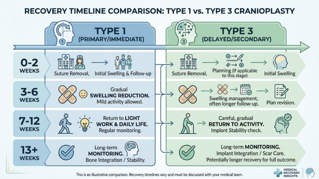
- Längere Erholung: Im Vergleich zur Kranioplastik Typ 1 können bei den Patienten länger anhaltende Schwellungen und Beschwerden auftreten.
- Kosten: Der Einsatz moderner Bildgebungs- und chirurgischer Instrumente erhöht die Gesamtkosten des Eingriffs.

Wesentliche Unterschiede zwischen Kranioplastik Typ 1 und Typ 3
| Besonderheit | Kranioplastik Typ 1 | Kranioplastik Typ 3 |
|---|---|---|
| Technik | Knochenentgraten/-schaben | Osteotomie und Rückverlagerung |
| Invasivität | Minimal | Mäßig bis hoch |
| Ideale Kandidaten | Leichte bis mäßige Augenbrauenwölbung, schmales Stirnbein | Stark ausgeprägte Augenbrauenwölbung, kräftiger Stirnknochen |
| Operationszeit | 1–2 Stunden | 3–5 Stunden |
| Wiederherstellungszeit | 1–2 Wochen | 3–6 Wochen |
| Komplikationsrisiko | Niedrig | Mäßig bis hoch |
| Kosten | Untere | Höher |
| Ästhetisches Ergebnis | Subtile Konturierung | Dramatische Feminisierung |
Wie Chirurgen entscheiden: Typ 1 vs. Typ 3
Die Wahl zwischen einer Kranioplastik vom Typ 1 und vom Typ 3 hängt von mehreren Faktoren ab, unter anderem vom Patienten. anatomische Merkmale, ästhetische Ziele, Und chirurgische Risiken. Chirurgen verlassen sich auf eine Kombination aus klinische Untersuchung, 3D-Bildgebung, Und Patientenberatung um die am besten geeignete Technik zu ermitteln.
1. Anatomische Beurteilung
Der erste Schritt besteht in der Beurteilung des Patienten. Dicke des Stirnbeins Und Anatomie der Stirnhöhle. A CT-Scan oder 3D-Rekonstruktion bietet detaillierte Einblicke in:
- Knochendicke: Patienten mit dünnen Stirnbeinen (weniger als 5 mm) eignen sich besser für eine Kranioplastik vom Typ 1, während bei Patienten mit dickeren Stirnbeinen möglicherweise eine Kranioplastik vom Typ 3 erforderlich ist.
- Sinuspneumatisierung: Eine ausgedehnte Pneumatisierung der Nasennebenhöhlen kann eine Kranioplastik vom Typ 3 erforderlich machen, um eine Verletzung der Nasennebenhöhlen während des Bohrens zu vermeiden.
- Grad der Vorherrschaft: Bei stark ausgeprägten Augenbrauenwülsten ist oft eine drastische Reduzierung erforderlich, die nur mit Techniken des Typs 3 erreicht werden kann.
2. Ästhetische Ziele
Die ästhetischen Erwartungen der Patienten spielen eine entscheidende Rolle bei der Wahl der Technik. Diejenigen, die suchen subtile Feminisierung Manche entscheiden sich für eine Kranioplastik Typ 1, während Personen mit ausgeprägten männlichen Gesichtszügen oft eine andere benötigen. transformative Ergebnisse Typ 3. Chirurgen besprechen realistische Ergebnisse auf der Grundlage der Anatomie des Patienten und der gewünschten Veränderungen.
3. Chirurgische Risiken und Genesung
Die Kranioplastik vom Typ 3 birgt höhere Risiken, darunter Komplikationen der Nebenhöhlen, Liquorverlust, Und verlängerte Genesung. Chirurgen beurteilen den allgemeinen Gesundheitszustand des Patienten, seine Operationstoleranz und seine Bereitschaft zur Einhaltung der postoperativen Nachsorge. Patienten mit Erkrankungen, die das Operationsrisiko erhöhen, wird gegebenenfalls empfohlen, eine Operation vom Typ 1 oder alternative Verfahren in Betracht zu ziehen.
4. Virtuelle Operationsplanung (VSP)
Fortschritte in Virtuelle 3D-Operationsplanung Die virtuelle Operationsplanung (VSP) hat die Kranioplastik revolutioniert. Chirurgen nutzen sie, um Osteotomien, Rückverlagerungen und Ergebnisse zu simulieren und so Präzision zu gewährleisten und Risiken zu minimieren. Diese Technologie ist besonders wertvoll für die Kranioplastik Typ 3, bei der eine genaue Knochenrepositionierung entscheidend ist (3D-Druck und virtuelle Operationsplanung in der kraniofazialen und orthognathen Chirurgie, 2025).
Postoperative Pflege und Genesung
Die Genesung verläuft nach einer Kranioplastik vom Typ 1 und Typ 3 deutlich unterschiedlich. Ein Verständnis des postoperativen Ablaufs hilft Patienten, sich auf einen reibungslosen Heilungsprozess vorzubereiten.
Genesung nach Kranioplastik Typ 1
Patienten, die sich einer Kranioplastik Typ 1 unterziehen, erleben typischerweise Folgendes:
- Leichte bis mittelschwere Schwellung: Heilt innerhalb von 1–2 Wochen ab.
- Minimale Beschwerden: Die Beschwerden wurden mit rezeptfreien Schmerzmitteln behandelt.
- Schnelle Rückkehr zu Aktivitäten: Die meisten Patienten können innerhalb von 2 Wochen ihre normalen Aktivitäten wieder aufnehmen.
Genesung nach Kranioplastik Typ 3
Die Genesung nach einer Kranioplastik Typ 3 ist aufgrund der Komplexität des Eingriffs aufwendiger:
- Erhebliche Schwellungen und Blutergüsse: Kann 3–4 Wochen anhalten.
- Mäßige Schmerzen: Für die erste Woche können verschreibungspflichtige Schmerzmittel erforderlich sein.
- Allmähliche Rückkehr zu Aktivitäten: Anstrengende Aktivitäten sind für 4–6 Wochen eingeschränkt.
- Folgebesuche: Regelmäßige Kontrollen, um eine ordnungsgemäße Heilung zu gewährleisten und eventuelle Komplikationen zu behandeln.
Beide Techniken erfordern von den Patienten, dass sie Folgendes vermeiden: schweres Heben, anstrengende Übungen, Und direkter Druck auf die Stirn Während der ersten Erholungsphase geben Chirurgen detaillierte postoperative Anweisungen, einschließlich Wundversorgung, Aktivitätseinschränkungen und Anzeichen von Komplikationen, auf die zu achten ist.
Mögliche Komplikationen und wie man sie vermeidet
Obwohl eine Kranioplastik im Allgemeinen sicher ist, können Komplikationen auftreten. Das Bewusstsein für diese Risiken und vorbeugenden Maßnahmen ist sowohl für Chirurgen als auch für Patienten von entscheidender Bedeutung.
Häufige Komplikationen
- Sinusitis: Entzündung oder Infektion der Stirnhöhle, insbesondere bei Kranioplastik Typ 3, wenn die Stirnhöhle verletzt wird.
- Liquorleck (CSF): Eine seltene, aber schwerwiegende Komplikation tritt ein, wenn die hintere Wand der Nasennebenhöhlen verletzt wird.
- Konturunregelmäßigkeiten: Eine zu starke Ausdünnung des Knochens bei Typ 1 oder eine unsachgemäße Rückverlagerung bei Typ 3 können zu Asymmetrie oder sichtbaren Graten führen.
- Mukozelenbildung: Eine Verstopfung der Abflusswege der Nasennebenhöhlen kann zur Bildung von mit Schleim gefüllten Zysten führen.
- Infektion: Das Risiko ist bei Typ 3 aufgrund der längeren Operationszeit und der Verwendung von Implantaten höher.
Präventive Maßnahmen
Chirurgen wenden verschiedene Strategien an, um Komplikationen zu minimieren:
- Präzise Operationsplanung: 3D-Bildgebung und virtuelle Operationsplanung gewährleisten präzise Osteotomien und Rückverlagerungen.
- Erhalt der Nasennebenhöhlen: Eine Verletzung der Stirnhöhle während des Bohrens oder der Osteotomie vermeiden.
- Antibiotikaprophylaxe: Wird vor und nach der Operation verabreicht, um das Infektionsrisiko zu verringern.
- Postoperative Überwachung: Regelmäßige Nachuntersuchungen zur Früherkennung von Komplikationen.
- Patientenaufklärung: Patienten über die richtige Wundversorgung, Aktivitätseinschränkungen und Warnzeichen von Komplikationen aufklären.
Alternativen zur Kranioplastik Typ 1 und Typ 3
Für Patienten, die nicht die idealen Kandidaten für eine Kranioplastik vom Typ 1 oder Typ 3 sind, können alternative Techniken in Betracht gezogen werden:
- Fetttransplantation: Die Eigenfetttransplantation kann Stirnkonturen weicher gestalten, ohne die Knochenstruktur zu verändern. Dies ist ideal für Patienten mit leichten Unregelmäßigkeiten oder solche, die eine nicht-operative Behandlungsmöglichkeit suchen.
- Maßgefertigte Implantate: Vorgefertigte Implantate, wie zum Beispiel PEEK (Polyetheretherketon) oder Titan, Sie können zur Vergrößerung oder Umformung der Stirn ohne Osteotomie eingesetzt werden. Sie eignen sich besonders für Patienten mit dünnen Knochen oder Nasennebenhöhlenproblemen.
- Kieferorthopädische Chirurgie: In Fällen, in denen Konturierung der Stirn ist Teil eines umfassenderen Gesichtsfeminisierung Bei der Planung können orthognathe Eingriffe (z. B. eine Le-Fort-I-Osteotomie) kombiniert werden, um die Ausrichtung des Mittelgesichts und des Kiefers zu verbessern.
- Endoskopische Techniken: Minimalinvasive endoskopische Verfahren können die Augenbrauenwölbung durch kleinere Einschnitte und schnellere Genesungszeiten reduzieren.
Patientenberichte und Ergebnisse aus der Praxis
Ergebnisse aus der Praxis liefern wertvolle Erkenntnisse über die Wirksamkeit und die Zufriedenheitsraten der Kranioplastik Typ 1 und Typ 3. Erfahrungsberichte von Patienten unterstreichen die transformative Wirkung dieser Eingriffe:
Fallbeispiel 1: Kranioplastik Typ 1
Eine 28-jährige Transfrau wünschte sich eine dezente Feminisierung ihrer Stirn. Aufgrund ihres schmalen Stirnbeins und der geringen Stirnwölbung entschied sie sich für eine Kranioplastik Typ 1. Der Eingriff führte zu einer harmonischeren Kontur bei minimaler Ausfallzeit. Sie zeigte sich sehr zufrieden und betonte, dass das Ergebnis ihren Erwartungen an ein natürliches, feminines Aussehen entsprach.
Fallbeispiel 2: Kranioplastik Typ 3
Eine 35-jährige Transfrau stellte sich mit stark ausgeprägten Augenbrauen und einem verdickten Stirnbein vor. Es wurde eine Kranioplastik Typ 3 durchgeführt, die eine Osteotomie und eine Rückverlagerung des Stirnbeins umfasste. Die deutliche Reduzierung der Stirnprojektion führte zu einer signifikanten Feminisierung ihrer Gesichtszüge. Obwohl die Genesung länger dauerte, zeigte sie sich äußerst zufrieden mit dem Ergebnis und erklärte, der Eingriff habe ihr Leben verändert.“
Fallbeispiel 3: Revisionsoperation
Eine 40-jährige Patientin unterzog sich zunächst einer Kranioplastik Typ 1, war jedoch mit dem dezenten Ergebnis unzufrieden. Sie entschied sich später für eine Kranioplastik Typ 3, um eine deutlichere Feminisierung zu erzielen. Die Revisionsoperation konnte ihre Bedenken erfolgreich ausräumen und verdeutlichte die Wichtigkeit der Wahl der richtigen Technik basierend auf den anatomischen Gegebenheiten und ästhetischen Zielen.
Die Rolle der virtuellen Operationsplanung in der Kranioplastik
Die virtuelle Operationsplanung (VSP) hat sich in der Kranioplastik, insbesondere bei Eingriffen des Typs 3, als bahnbrechend erwiesen. VSP ermöglicht es Chirurgen:
- Osteotomien simulieren: Die Knochenschnitte und -repositionierungen müssen präzise geplant werden, um eine optimale Konturierung zu erreichen.
- Ergebnisse vorhersagen: Visualisieren Sie die postoperativen Ergebnisse und passen Sie den Operationsplan entsprechend an.
- Risiken minimieren: Vermeiden Sie kritische Strukturen wie die Stirnhöhle und die Supraorbitalnerven.
- Kommunikation verbessern: Teilen Sie 3D-Modelle mit Patienten, um realistische Erwartungen zu wecken und die informierte Einwilligung zu verbessern.
Studien haben gezeigt, dass die virtuelle Operationsplanung (VSP) die Operationszeit verkürzt, die Genauigkeit verbessert und die Patientenzufriedenheit erhöht (3D-Druck und virtuelle Operationsplanung in der kraniofazialen und orthognathen Chirurgie, 2025). Sie gilt heute als Goldstandard für komplexe Kranioplastiken.

Häufig gestellte Fragen
Worin besteht der Hauptunterschied zwischen einer Kranioplastik vom Typ 1 und einer vom Typ 3?
Bei der Kranioplastik Typ 1 wird die äußere Schicht des Stirnbeins abgetragen oder gefräst, um dessen Vorwölbung zu reduzieren. Die Kranioplastik Typ 3 hingegen erfordert eine Osteotomie (Knochenschnitt) und eine Rückverlagerung des Stirnbeinsegments, um eine stärkere Konturierung zu erzielen. Typ 1 ist weniger invasiv und eignet sich für leichte bis mittelschwere Vorwölbungen, während Typ 3 schweren Vorwölbungen oder dicken Stirnbeinen vorbehalten ist.
Wie entscheiden Chirurgen zwischen einer Kranioplastik vom Typ 1 und vom Typ 3?
Chirurgen beurteilen mithilfe von CT-Scans und 3D-Bildgebung die Dicke des Stirnbeins, den Grad der Augenbrauenwölbung und die Anatomie der Stirnhöhle. Typ 1 wird bei dünneren Knochen und leichter Wölbung gewählt, während Typ 3 bei dickeren Knochen, starker Wölbung oder ausgeprägter Pneumatisierung der Stirnhöhle bevorzugt wird. Die Ziele des Patienten und seine Risikotoleranz beeinflussen die Entscheidung ebenfalls.
Welche Risiken sind mit einer Kranioplastik vom Typ 3 verbunden?
Die Kranioplastik Typ 3 birgt aufgrund ihrer Komplexität höhere Risiken, darunter Sinusitis, Liquorverlust, Konturunregelmäßigkeiten, Mukozelenbildung und Infektionen. Diese Risiken werden durch präzise Operationsplanung, Erhalt der Nasennebenhöhlen und postoperative Überwachung minimiert. Patienten werden über Warnzeichen und Nachsorge aufgeklärt, um eine reibungslose Genesung zu gewährleisten.
Wie lange dauert die Genesung nach einer Kranioplastik Typ 1 im Vergleich zu Typ 3?
Die Genesung nach einer Kranioplastik Typ 1 dauert in der Regel 1–2 Wochen und ist mit leichten Schwellungen und Beschwerden verbunden. Eine Kranioplastik Typ 3 erfordert aufgrund des umfangreicheren Eingriffs und möglicher Komplikationen eine längere Genesungszeit von 3–6 Wochen. Patienten wird empfohlen, anstrengende Aktivitäten zu vermeiden und die postoperativen Anweisungen genau zu befolgen.
Kann eine Kranioplastik vom Typ 1 die gleichen Ergebnisse erzielen wie eine vom Typ 3?
Nein, die Kranioplastik Typ 1 beschränkt sich auf subtile Konturierungen und eignet sich ideal bei leichter bis mittelschwerer Vorwölbung des Stirnbeins. Die Kranioplastik Typ 3 erzielt eine deutlichere Feminisierung durch die Repositionierung des Stirnbeinsegments und ist daher für stark ausgeprägte Vorwölbungen oder dicke Stirnbeine geeignet. Die Wahl der Methode hängt von den anatomischen Gegebenheiten und ästhetischen Zielen des Patienten ab.
Welche Rolle spielt die virtuelle Operationsplanung bei der Kranioplastik?
Die virtuelle Operationsplanung (VSP) ermöglicht es Chirurgen, Osteotomien zu simulieren, Ergebnisse vorherzusagen und Risiken zu minimieren, indem kritische Strukturen wie die Stirnhöhle geschont werden. Sie erhöht die Präzision, verkürzt die Operationszeit und steigert die Patientenzufriedenheit durch eine klare Visualisierung des Operationsplans und der zu erwartenden Ergebnisse.
Gibt es nicht-operative Alternativen zur Kranioplastik zur Feminisierung der Stirn?
Ja, zu den nicht-chirurgischen Alternativen gehören Fetttransplantation Konturglättung und individuell angefertigte Implantate (z. B. aus PEEK oder Titan) ermöglichen die Umformung der Stirn ohne Osteotomie. Diese Optionen eignen sich ideal für Patientinnen mit leichten Unregelmäßigkeiten oder solche, die eine Operation vermeiden möchten. Allerdings erreichen sie möglicherweise nicht denselben Grad an Feminisierung wie operative Verfahren.
Was kann ich während des Beratungsgesprächs zur Stirnrekonstruktion erwarten?
Im Rahmen des Beratungsgesprächs beurteilt Ihr Chirurg anhand einer klinischen Untersuchung und 3D-Bildgebung die Dicke Ihres Stirnbeins, die Anatomie der Nasennebenhöhlen und den Grad der Stirnwölbung. Er bespricht mit Ihnen Ihre ästhetischen Ziele, erläutert die Unterschiede zwischen Kranioplastik Typ 1 und Typ 3 und empfiehlt Ihnen die für Ihre Anatomie und Ihre Erwartungen am besten geeignete Technik.







