

Reconstrucción de frente es un componente crítico de Facial Cirugía de Feminización (FFS) y procedimientos craneofaciales, con el objetivo de lograr un contorno facial armonioso y estéticamente agradable. Entre las técnicas más debatidas se encuentran: Tipo 1 y Craneoplastia tipo 3, cada una ofreciendo enfoques distintos para remodelar la frente. Tipo 1 implica fresado o afeitado del hueso, Tipo 3 requiere un más complejo osteotomía y retroceso del seno frontal. Comprender las implicaciones anatómicas, funcionales y estéticas de estas técnicas es esencial tanto para los cirujanos como para los pacientes a fin de tomar decisiones informadas. Obtenga más información sobre el retroceso del seno y la reducción de la protuberancia de la ceja en nuestra guía completa sobre contorno de la frente.
Esta guía explora las diferencias estructurales entre la craneoplastia tipo 1 y tipo 3, su impacto en espesor óseo, anatomía del seno frontal, y cómo los cirujanos determinan el enfoque más adecuado para cada paciente. Al finalizar, comprenderá con claridad qué técnica se ajusta mejor a sus necesidades anatómicas y objetivos estéticos.
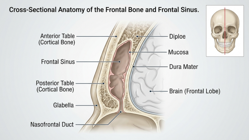
Tabla de contenido
Fundamentos anatómicos: Hueso frontal y seno maxilar
El hueso frontal y seno frontal desempeñan un papel fundamental en la reconstrucción de la frente. El hueso frontal forma la parte superior de la cara y alberga el seno frontal, una cavidad hueca que varía en tamaño y forma entre individuos. tabla anterior del seno frontal es la capa externa del hueso que contribuye al contorno de la frente, mientras que el tabla posterior separa el seno del cerebro. El grosor de estas tablas y el grado de neumatización del seno (expansión llena de aire) influyen en la elección entre craneoplastia de tipo 1 y tipo 3.
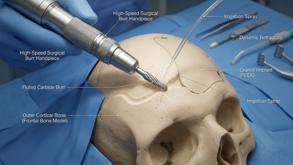
En Craneoplastia tipo 1, el cirujano Utiliza una fresa de alta velocidad para rebajar el hueso cortical externo, reduciendo la prominencia sin dañar el seno frontal. Esta técnica es ideal para pacientes con hueso frontal delgado o un mínimo abombamiento, ya que preserva la integridad del seno. Sin embargo, puede no ser suficiente para pacientes con un abombamiento significativo de la ceja o una tabla anterior gruesa, ya que un fresado excesivo puede comprometer la estabilidad ósea o no lograr el contorno deseado (Ousterhout, 2024).
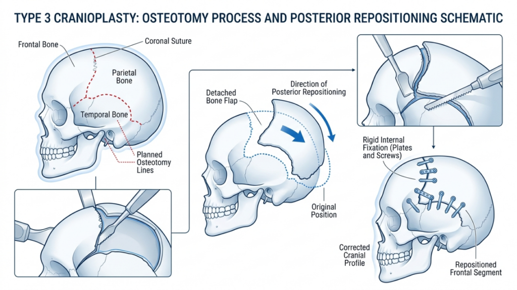
Por el contrario, Craneoplastia tipo 3 implica un osteotomía—un corte controlado a través de la tabla anterior del seno frontal. El segmento óseo se reposiciona hacia atrás (retroceso) para reducir la proyección y crear una frente más suave y femenina. Esta técnica está reservada para pacientes con protuberancia de la frente de moderada a severa o una tabla anterior gruesa, donde el fresado por sí solo sería insuficiente. La osteotomía permite una remodelación precisa manteniendo la función protectora del seno (Feminización de la frente: una revisión bibliográfica exploratoria, 2024).
Craneoplastia tipo 1: Raspado y fresado
Descripción general de la técnica
La craneoplastia tipo 1 es la opción menos invasiva para la reconstrucción de la frente. Implica el uso de un fresa de alta velocidad para reducir gradualmente la prominencia del hueso frontal. cirujano Se realiza un afeitado meticuloso de la capa cortical externa, evitando la penetración en el seno frontal. Esta técnica es particularmente eficaz para pacientes con:
- Protuberancias leves a moderadas en las cejas
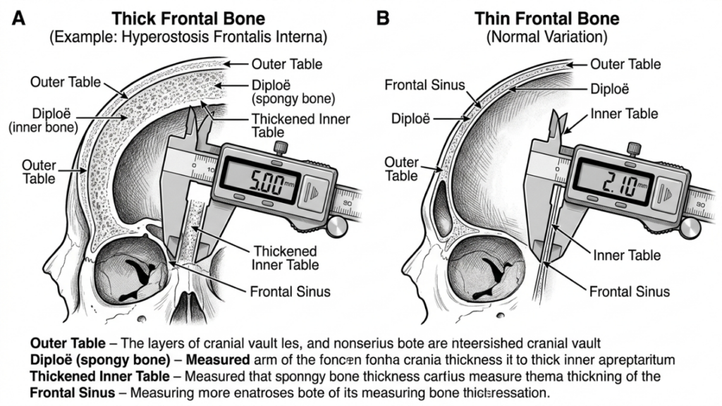
- Hueso frontal delgado (menos de 5 mm)
- Ausencia de neumatización significativa del seno frontal
El procedimiento se realiza a través de un incisión coronal, Esto permite acceder a la frente minimizando las cicatrices visibles. El cirujano utiliza la retroalimentación táctil y las señales visuales para asegurar una reducción uniforme sin adelgazar demasiado el hueso, lo que podría provocar inestabilidad o irregularidades en el contorno.
Ventajas de la craneoplastia tipo 1
La craneoplastia de tipo 1 ofrece varios beneficios:
- Mínima invasividad: La ausencia de osteotomía o extracción ósea reduce el trauma quirúrgico y el tiempo de recuperación.
- Menor riesgo de complicaciones: Preservar el seno frontal minimiza el riesgo de sinusitis, fugas de líquido cefalorraquídeo o formación de mucocele.
- Tiempo operatorio más corto: El procedimiento suele durar entre 1 y 2 horas, lo que lo convierte en una opción más rápida en comparación con el Tipo 3.
- Resultados predecibles: Ideal para pacientes con protuberancias leves en la piel, donde un contorno sutil es suficiente para lograr una apariencia femenina.
Limitaciones de la craneoplastia tipo 1
Si bien la craneoplastia de tipo 1 es más segura y menos invasiva, presenta limitaciones importantes:
- Reducción limitada: Insuficiente para pacientes con cejas prominentes o huesos frontales gruesos.
- Riesgo de un adelgazamiento excesivo: Un fresado agresivo puede debilitar el hueso, provocando irregularidades en el contorno o fracturas.
- Feminización incompleta: Es posible que no se logre el resultado estético deseado en pacientes con rasgos masculinos pronunciados.
Craneoplastia tipo 3: Osteotomía y retroceso
Descripción general de la técnica
La craneoplastia tipo 3 es un procedimiento más complejo diseñado para pacientes con protuberancia de la frente de moderada a severa o huesos frontales gruesos. Implica un osteotomía—un corte preciso a través de la tabla anterior del seno frontal— seguido del reposicionamiento del segmento óseo hacia atrás (retroceso). Esta técnica permite una reducción significativa de la proyección de la frente y un contorno más suave y femenino.
El procedimiento se realiza normalmente a través de un incisión coronal, proporcionando acceso al hueso frontal y al seno. El cirujano utiliza un sierra sagital o dispositivo piezoeléctrico para crear la osteotomía, asegurándose de que el corte siga la curvatura natural de la frente. Luego, el segmento óseo se reposiciona y se fija con placas y tornillos de titanio o suturas reabsorbibles. Esta técnica es ideal para pacientes con:
- Protuberancia severa en las cejas
- Hueso frontal grueso (mayor de 5 mm)
- Neumatización significativa del seno frontal
La craneoplastia tipo 3 requiere una planificación meticulosa, que a menudo implica Planificación quirúrgica virtual en 3D (VSP) para simular la osteotomía y el retroceso. Esto garantiza precisión y minimiza el riesgo de complicaciones como la violación del seno o fugas de líquido cefalorraquídeo (Planificación quirúrgica virtual en Feminización Facial de la cara superior, 2025).
Ventajas de la craneoplastia tipo 3
La craneoplastia de tipo 3 ofrece varias ventajas para pacientes con rasgos masculinos pronunciados:
- Contorneado significativo: Consigue una reducción drástica de las cejas prominentes, creando una frente más suave y femenina.
- Versatilidad: Indicado para pacientes con huesos frontales gruesos o neumatización sinusal extensa.
- Estabilidad a largo plazo: El segmento óseo reposicionado se integra bien, reduciendo el riesgo de irregularidades en el contorno con el paso del tiempo.
- Personalización: La planificación quirúrgica virtual permite realizar ajustes precisos y personalizados para cada paciente, con el fin de lograr resultados óptimos.
Limitaciones de la craneoplastia tipo 3
A pesar de su eficacia, la craneoplastia de tipo 3 conlleva mayores riesgos y complejidades:
- Mayor tiempo quirúrgico: El procedimiento suele durar entre 3 y 5 horas, y requiere mayor precisión y experiencia.
- Mayor riesgo de complicaciones: Entre los riesgos potenciales se incluyen la sinusitis, las fugas de líquido cefalorraquídeo o la formación de mucocele si se produce una lesión en el seno paranasal.
- Recuperación más prolongada: Los pacientes pueden experimentar hinchazón y molestias prolongadas en comparación con la craneoplastia de tipo 1.
- Costo: El uso de técnicas avanzadas de imagen y herramientas quirúrgicas incrementa el coste total del procedimiento.

Principales diferencias entre la craneoplastia tipo 1 y tipo 3
| Característica | Craneoplastia tipo 1 | Craneoplastia tipo 3 |
|---|---|---|
| Técnica | fresado/desbaste óseo | Osteotomía y retroceso |
| Invasividad | Mínimo | Moderado a alto |
| Candidatos ideales | Cejas ligeramente a moderadamente prominentes, hueso frontal delgado. | Protuberancia cefálica severa, hueso frontal grueso |
| Tiempo quirúrgico | 1–2 horas | 3–5 horas |
| Tiempo de recuperación | 1-2 semanas | 3–6 semanas |
| Riesgo de complicaciones | Bajo | Moderado a alto |
| Costo | Más bajo | Más alto |
| Resultado estético | Contorneado sutil | Feminización dramática |
Cómo deciden los cirujanos: Tipo 1 vs. Tipo 3
La elección entre craneoplastia tipo 1 y tipo 3 depende de varios factores, incluido el paciente. características anatómicas, objetivos estéticos, y riesgos quirúrgicos. Los cirujanos se basan en una combinación de examen clínico, Imágenes 3D, y consulta del paciente para determinar la técnica más apropiada.
1. Evaluación anatómica
El primer paso es evaluar al paciente espesor del hueso frontal y anatomía del seno frontal. A tomografía computarizada o Reconstrucción 3D Proporciona información detallada sobre:
- Grosor del hueso: Los pacientes con huesos frontales delgados (menos de 5 mm) son más aptos para la craneoplastia de tipo 1, mientras que aquellos con huesos más gruesos pueden requerir la de tipo 3.
- Neumatización de los senos paranasales: La neumatización extensa del seno maxilar puede requerir una craneoplastia de tipo 3 para evitar dañar el seno durante el fresado.
- Grado de autoridad: La protuberancia severa en las cejas a menudo requiere una reducción drástica que solo se puede lograr con las técnicas de tipo 3.
2. Objetivos estéticos
Las expectativas estéticas de los pacientes juegan un papel crucial en la selección de la técnica. Aquellos que buscan feminización sutil pueden optar por la craneoplastia tipo 1, mientras que las personas con rasgos masculinos pronunciados a menudo requieren la resultados transformadores del tipo 3. Los cirujanos analizan los resultados realistas en función de la anatomía del paciente y los cambios deseados.
3. Riesgos quirúrgicos y recuperación
La craneoplastia de tipo 3 conlleva mayores riesgos, entre ellos: complicaciones sinusales, fugas de líquido cefalorraquídeo, y recuperación prolongada. Los cirujanos evalúan el estado de salud general del paciente, su tolerancia a la cirugía y su disposición a seguir las indicaciones postoperatorias. A los pacientes con afecciones médicas que aumentan los riesgos quirúrgicos se les puede recomendar que consideren procedimientos de tipo 1 o alternativos.
4. Planificación Quirúrgica Virtual (PQV)
Avances en Planificación quirúrgica virtual en 3D Han revolucionado la craneoplastia. Los cirujanos utilizan la planificación quirúrgica virtual (VSP) para simular osteotomías, retrocesos y resultados, garantizando precisión y minimizando riesgos. Esta tecnología es especialmente valiosa para la craneoplastia de tipo 3, donde el reposicionamiento óseo preciso es fundamental (Impresión 3D y planificación quirúrgica virtual en cirugía craneofacial y ortognática, 2025).
Cuidados postoperatorios y recuperación
La recuperación varía significativamente entre la craneoplastia de tipo 1 y la de tipo 3. Comprender el proceso postoperatorio ayuda a los pacientes a prepararse para una recuperación sin complicaciones.
Recuperación tras craneoplastia tipo 1
Los pacientes sometidos a craneoplastia de tipo 1 suelen experimentar:
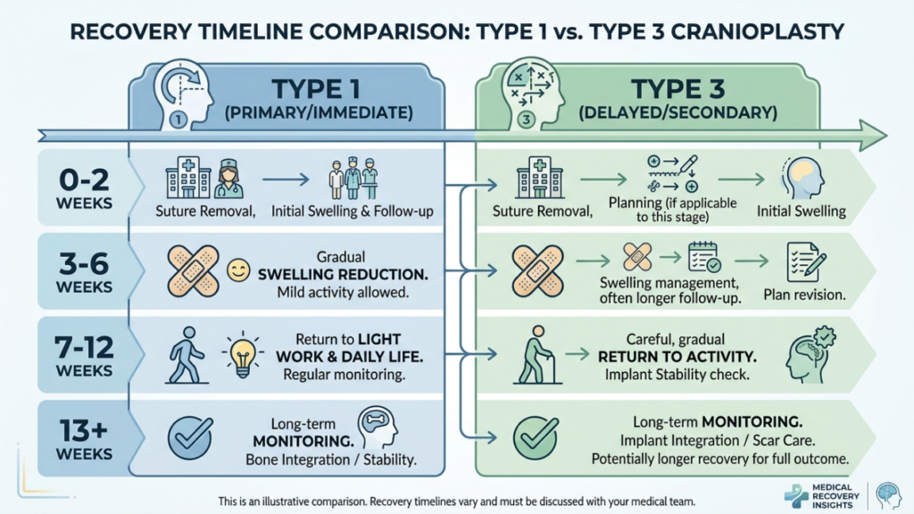
- Hinchazón leve a moderada: Se resuelve en 1-2 semanas.
- Mínimas molestias: Se controló con analgésicos de venta libre.
- Regreso rápido a las actividades: La mayoría de los pacientes retoman sus actividades normales en un plazo de dos semanas.
Recuperación tras craneoplastia tipo 3
La recuperación tras una craneoplastia de tipo 3 es más compleja debido a la dificultad del procedimiento:
- Hinchazón y hematomas significativos: Puede persistir durante 3-4 semanas.
- Dolor moderado: Es posible que se requieran analgésicos recetados durante la primera semana.
- Regreso gradual a las actividades: Las actividades extenuantes están restringidas durante 4 a 6 semanas.
- Visitas de seguimiento: Seguimiento periódico para asegurar una correcta cicatrización y abordar cualquier complicación.
Ambas técnicas requieren que los pacientes eviten levantamiento de objetos pesados, ejercicio extenuante, y presión directa sobre la frente Durante la fase inicial de recuperación, los cirujanos proporcionan instrucciones postoperatorias detalladas, que incluyen el cuidado de la herida, las restricciones de actividad y los signos de posibles complicaciones a los que hay que prestar atención.
Posibles complicaciones y cómo evitarlas
Si bien la craneoplastia es generalmente segura, pueden surgir complicaciones. Es fundamental que tanto los cirujanos como los pacientes conozcan estos riesgos y las medidas preventivas.
Complicaciones comunes
- Sinusitis: Inflamación o infección del seno frontal, especialmente en la craneoplastia de tipo 3 si el seno está dañado.
- Fuga de líquido cefalorraquídeo (LCR): Complicación poco frecuente pero grave si se perfora la tabla posterior del seno maxilar.
- Irregularidades del contorno: Un adelgazamiento excesivo del hueso en el tipo 1 o un retroceso inadecuado en el tipo 3 pueden provocar asimetría o crestas visibles.
- Formación de mucocele: La obstrucción de las vías de drenaje sinusal puede provocar la formación de quistes llenos de mucosidad.
- Infección: El riesgo es mayor en el tipo 3 debido al mayor tiempo quirúrgico y al uso de implantes.
Medidas preventivas
Los cirujanos emplean diversas estrategias para minimizar las complicaciones:
- Planificación quirúrgica precisa: Las imágenes 3D y la planificación quirúrgica virtual garantizan osteotomías y retrocesos precisos.
- Preservación de los senos paranasales: Evitar la lesión del seno frontal durante el fresado o la osteotomía.
- Profilaxis antibiótica: Se administra antes y después de la cirugía para reducir los riesgos de infección.
- Monitoreo postoperatorio: Controles periódicos para detectar signos tempranos de complicaciones.
- Educación del paciente: Instruir a los pacientes sobre el cuidado adecuado de las heridas, las restricciones de actividad y las señales de alerta de posibles complicaciones.
Alternativas a la craneoplastia tipo 1 y tipo 3
Para los pacientes que no son candidatos ideales para la craneoplastia de tipo 1 o tipo 3, se pueden considerar técnicas alternativas:
- Injerto de grasa: La transferencia de grasa autóloga puede suavizar los contornos de la frente sin alterar la estructura ósea. Esto es ideal para pacientes con irregularidades leves o para quienes buscan opciones no quirúrgicas.
- Implantes personalizados: Implantes prefabricados, como PEEK (poliéter éter cetona) o titanio, Pueden utilizarse para aumentar o remodelar la frente sin necesidad de osteotomía. Son especialmente útiles para pacientes con huesos delgados o complicaciones sinusales.
- Cirugía Ortognática: En los casos en que contorno de la frente forma parte de un proyecto más amplio feminización facial En el plan de tratamiento, se pueden combinar procedimientos ortognáticos (por ejemplo, osteotomía Le Fort I) para corregir la alineación del tercio medio facial y la mandíbula.
- Técnicas endoscópicas: Los abordajes endoscópicos mínimamente invasivos pueden reducir la protuberancia de la ceja gracias a incisiones más pequeñas y tiempos de recuperación más rápidos.
Testimonios de pacientes y resultados en la vida real
Los resultados en la práctica clínica ofrecen información valiosa sobre la eficacia y los índices de satisfacción de la craneoplastia de tipo 1 y tipo 3. Los testimonios de los pacientes destacan el impacto transformador de estos procedimientos:
Caso práctico 1: Craneoplastia tipo 1
Una mujer transgénero de 28 años buscaba una feminización sutil de su frente. Con un hueso frontal delgado y una protuberancia mínima, optó por una craneoplastia tipo 1. El procedimiento logró un contorno más suave con un tiempo de recuperación mínimo. Manifestó una gran satisfacción, señalando que los resultados se ajustaban a sus expectativas de una apariencia natural y femenina.
Caso práctico 2: Craneoplastia tipo 3
Una mujer transgénero de 35 años presentaba una marcada prominencia de las cejas y un hueso frontal grueso. Se le realizó una craneoplastia tipo 3, que incluyó osteotomía y retroceso. La drástica reducción de la proyección de la frente feminizó significativamente sus rasgos faciales. Si bien la recuperación fue más prolongada, expresó una inmensa satisfacción con los resultados, afirmando que el procedimiento le había cambiado la vida.“
Caso práctico 3: Cirugía de revisión
Una paciente de 40 años se sometió inicialmente a una craneoplastia tipo 1, pero no quedó satisfecha con los resultados poco convincentes. Posteriormente, optó por una craneoplastia tipo 3 para lograr una feminización más marcada. La cirugía de revisión resolvió satisfactoriamente sus inquietudes, demostrando la importancia de seleccionar la técnica adecuada según las necesidades anatómicas y los objetivos estéticos.
El papel de la planificación quirúrgica virtual en la craneoplastia
La planificación quirúrgica virtual (VSP) se ha convertido en un factor revolucionario en la craneoplastia, especialmente para los procedimientos de tipo 3. La VSP permite a los cirujanos:
- Simular osteotomías: Planifique con precisión los cortes y el reposicionamiento óseo para lograr un contorno óptimo.
- Predecir resultados: Visualice los resultados postoperatorios y ajuste el plan quirúrgico en consecuencia.
- Minimizar riesgos: Evite dañar estructuras críticas como el seno frontal y los nervios supraorbitarios.
- Mejorar la comunicación: Comparta modelos 3D con los pacientes para establecer expectativas realistas y mejorar el consentimiento informado.
Los estudios han demostrado que la planificación quirúrgica virtual (VSP) reduce el tiempo operatorio, mejora la precisión y aumenta la satisfacción del paciente (Impresión 3D y planificación quirúrgica virtual en cirugía craneofacial y ortognática, 2025). Actualmente, se considera el método de referencia para procedimientos complejos de craneoplastia.

Preguntas frecuentes
¿Cuál es la principal diferencia entre la craneoplastia de tipo 1 y la de tipo 3?
La craneoplastia tipo 1 consiste en limar o fresar la capa externa del hueso frontal para reducir su prominencia, mientras que la craneoplastia tipo 3 requiere una osteotomía (corte óseo) y un retroceso del segmento del hueso frontal para lograr un contorno más definido. La craneoplastia tipo 1 es menos invasiva y adecuada para protuberancias leves a moderadas, mientras que la tipo 3 se reserva para protuberancias severas o huesos frontales gruesos.
¿Cómo deciden los cirujanos entre una craneoplastia de tipo 1 y una de tipo 3?
Los cirujanos evalúan el grosor del hueso frontal del paciente, el grado de abombamiento de la ceja y la anatomía del seno frontal mediante tomografía computarizada e imágenes 3D. Se elige el tipo 1 para huesos más delgados y abombamiento leve, mientras que el tipo 3 se prefiere para huesos más gruesos, abombamiento severo o neumatización extensa del seno. Los objetivos del paciente y su tolerancia a los riesgos quirúrgicos también influyen en la decisión.
¿Cuáles son los riesgos asociados a la craneoplastia de tipo 3?
La craneoplastia tipo 3 conlleva mayores riesgos debido a su complejidad, incluyendo sinusitis, fugas de líquido cefalorraquídeo, irregularidades en el contorno, formación de mucocele e infección. Estos riesgos se minimizan mediante una planificación quirúrgica precisa, la preservación de los senos paranasales y el seguimiento postoperatorio. Se informa a los pacientes sobre los signos de alerta y los cuidados posteriores para garantizar una recuperación sin complicaciones.
¿Cuánto dura el período de recuperación entre una craneoplastia de tipo 1 y una de tipo 3?
La recuperación tras una craneoplastia tipo 1 suele durar entre 1 y 2 semanas, con leve hinchazón y molestias. La craneoplastia tipo 3 requiere una recuperación más prolongada, de 3 a 6 semanas, debido a la mayor complejidad de la cirugía y las posibles complicaciones. Se recomienda a los pacientes evitar actividades extenuantes y seguir al pie de la letra las instrucciones postoperatorias.
¿Puede la craneoplastia de tipo 1 lograr los mismos resultados que la de tipo 3?
No, la craneoplastia tipo 1 se limita a un contorno sutil y es ideal para protuberancias leves a moderadas. La craneoplastia tipo 3 logra una feminización más marcada mediante el reposicionamiento del segmento del hueso frontal, por lo que resulta adecuada para protuberancias severas o huesos frontales gruesos. La elección depende de las necesidades anatómicas y los objetivos estéticos de la paciente.
¿Qué papel desempeña la planificación quirúrgica virtual en la craneoplastia?
La planificación quirúrgica virtual (PQV) permite a los cirujanos simular osteotomías, predecir resultados y minimizar riesgos evitando estructuras críticas como el seno frontal. Mejora la precisión, reduce el tiempo operatorio y aumenta la satisfacción del paciente al proporcionar una visualización clara del plan quirúrgico y los resultados esperados.
¿Existen alternativas no quirúrgicas a la craneoplastia para la feminización de la frente?
Sí, las alternativas no quirúrgicas incluyen: injerto de grasa Para suavizar los contornos y remodelar la frente con implantes personalizados (por ejemplo, de PEEK o titanio) sin necesidad de osteotomía, estas opciones son ideales para pacientes con irregularidades leves o que prefieren evitar la cirugía. Sin embargo, es posible que no logren el mismo grado de feminización que las técnicas quirúrgicas.
¿Qué debo esperar durante la consulta para la reconstrucción de la frente?
Durante la consulta, su cirujano evaluará el grosor de su hueso frontal, la anatomía de los senos paranasales y el grado de abombamiento mediante examen clínico e imágenes 3D. Analizará sus objetivos estéticos, le explicará las diferencias entre la craneoplastia tipo 1 y tipo 3, y le recomendará la técnica más adecuada según su anatomía y expectativas.







