
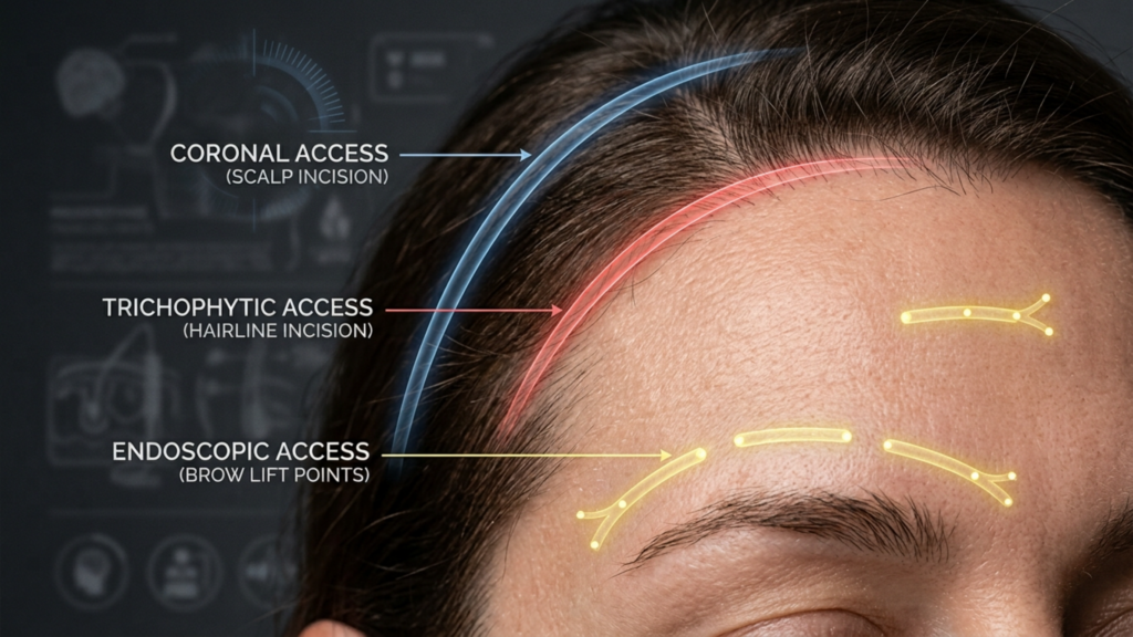
Wat als de allerbelangrijkste beslissing bij uw voorhoofdvervrouwelijkingsprocedure niets te maken heeft met het hervormen van de botten, maar alles met de locatie van de ingreep? chirurg De incisie die gekozen wordt om bij het wenkbrauwbot te komen, bepaalt echter de zichtbaarheid van het litteken, de haarlijn, het herstel van het gevoel en zelfs of uw chirurg kan de laterale orbitale randen voldoende bereiken. coronale versus trichofytische versus endoscopische wenkbrauwtoegang De keuze voor de botstructuur vormt het architectonische fundament waarop elke andere chirurgische ingreep rust. Een verkeerde keuze kan er zelfs toe leiden dat perfect uitgevoerde botreconstructies een zichtbaar litteken, een terugtrekkende haargrens of een permanent gevoelloze hoofdhuid achterlaten.
Na honderden optredens gezichtsfeminisering procedures bij Dokter MFO Kliniek in Antalya, Ik heb zelf gezien hoe de keuze van de incisie bepalend kan zijn voor de tevredenheid op de lange termijn. Patiënten komen binnen met de gedachte aan millimeters wenkbrauwbotreductie. Ze vertrekken teleurgesteld door een litteken dat ze nooit hadden verwacht. Deze gids biedt iets wat geen enkele andere bron biedt: een zij-aan-zij vergelijking in de vorm van een beslissingsboom van alle drie de incisies voor toegang tot het voorhoofd, zodat u uw consultatie ingaat met precieze, bruikbare kennis. U leert precies hoe elke aanpak presteert op zeven cruciale aspecten: kwaliteit van de blootstelling, behoud van de haargrens, Denk hierbij aan factoren zoals de zichtbaarheid van het litteken, sensorische verstoring, risico op haaruitval, herstelsnelheid en de toegang van de chirurg tot de oogkasrand. Met behulp van het bijgevoegde zelfbeoordelingsschema kunt u de ideale incisie bepalen voordat u überhaupt met een chirurg spreekt.
Inhoudsopgave
De anatomie van de toegang tot het voorhoofd: waarom de keuze van de incisie bepalend is voor het resultaat van uw FFS-behandeling.
Voor een feminiserende operatie aan het voorhoofd is directe fysieke toegang tot het voorhoofdsbeen, de supraorbitale randen en de voorhoofdsholten nodig. De chirurg moet deze structuren kunnen zien, aanraken en hervormen. Elk type incisie vertegenwoordigt een andere afweging tussen hoeveel van het voorhoofd de chirurg kan zien en manipuleren en hoeveel schade de hoofdhuid en haarlijn oplopen. Geen enkele methode biedt onbeperkte toegang zonder esthetische kosten. Coronale incisie FFFS De endoscopische benadering biedt de breedste toegang, maar resulteert wel in een lang litteken en aanzienlijke verstoring van het gevoel. De endoscopische benadering verbergt littekens prachtig, maar beperkt het bereik van de chirurg. De trichofytische incisie volgt een middenweg waardoor het litteken direct naar de haargrens verschuift.
Houd rekening met een feit dat de meeste patiënten verrast: de supraorbitale en supratrochleaire zenuwen – verantwoordelijk voor het gevoel in uw voorhoofd en de voorkant van uw hoofdhuid – lopen dwars door het operatiegebied. Een coronale incisie snijdt onvermijdelijk de achterste takken van deze zenuwen over de gehele breedte van de hoofdhuid door. Endoscopische incisies behouden de meeste zenuwbanen omdat de dissectie onder de hoofdhuid doorgaat in plaats van er dwars doorheen te snijden. Deze ene anatomische realiteit verklaart waarom patiënten die een coronale incisie ondergaan, twaalf tot achttien maanden last hebben van gevoelloosheid, terwijl patiënten die een endoscopische incisie ondergaan vaak binnen zes weken hun volledige gevoel terugkrijgen. De incisie is niet zomaar een toegangspunt. Het is de belangrijkste factor die uw herstelproces bepaalt.
Coronale incisie FFS: maximale blootstelling, maximale compromissen
De coronale incisie, ook wel bicoronale of coronale flaptechniek genoemd, bestaat uit een enkele, doorlopende incisie van oor tot oor over de bovenkant van de hoofdhuid, meestal vier tot zes centimeter achter de haarlijn. De chirurg tilt vervolgens de gehele voorhoofdflap naar beneden om het voorhoofdsbeen bloot te leggen, van de haarlijn tot de oogkassen. Deze techniek wordt al decennialang gebruikt in de craniofaciale chirurgie omdat ze een ongeëvenaard zicht op het operatiegebied biedt.
Belichtingskwaliteit en toegang tot de orbitale rand
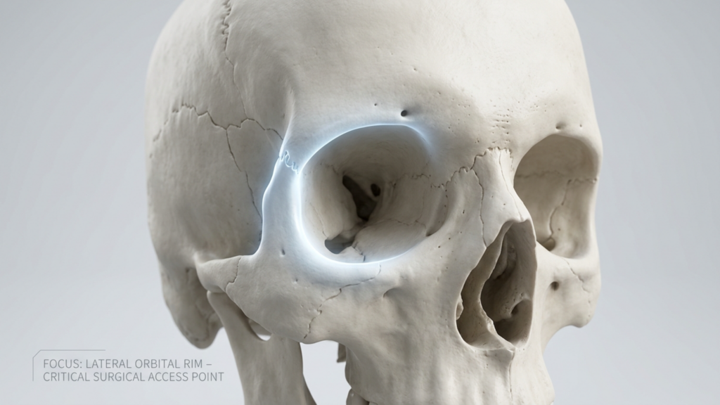
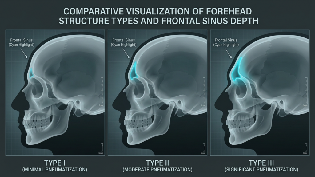
De coronaire benadering is de gouden standaard voor toegang tot het wenkbrauwbot. Wanneer de gehele voorhoofdsflap naar beneden wordt geklapt, krijgt de chirurg direct en ononderbroken zicht op beide laterale orbitale randen, de glabella, de frontale sinus en de temporale fossa. Voor voorhoofden van type III, waarbij een agressieve terugplaatsing van de sinuswand en een aanzienlijke botreductie nodig zijn, is dit niveau van zichtbaarheid onvervangbaar. De chirurg kan instrumenten onder elke hoek plaatsen, de symmetrie in realtime visueel controleren en onverwachte anatomische bevindingen aanpakken zonder te hoeven worstelen met beperkt zicht.
Behoud van de haargrens en risico op haarverlies
Hier komt de scherpste rand van de coronale incisie tevoorschijn. Omdat de incisie achter de haarlijn zit, blijft de positie van de haarlijn zelf anatomisch onveranderd. De incisie creëert echter een zone met permanent haarverlies langs de hechting. Spanningsluiting, elektrocoagulatie en postoperatieve littekenverbreding kunnen een zichtbare kale strook van vier tot zes centimeter achter de haarlijn veroorzaken. Bij patiënten met dun haar of een van nature hoge haarlijn is deze kale zone opvallend zichtbaar. Risico op beschadiging van de hoofdhuidzenuwen Dit is een aanzienlijke ingreep, omdat de coronale incisie de achterste takken van het supraorbitale zenuwcomplex aan beide zijden doorsnijdt, wat leidt tot gevoelloosheid over de gehele bovenste hoofdhuid die achttien maanden of langer kan aanhouden.
Zichtbaarheid van littekens en herstel
Het litteken bij de coronale benadering bevindt zich in de behaarde hoofdhuid, waardoor het onzichtbaar is bij lang en dicht haar. Patiënten met dik haar kunnen het litteken binnen enkele weken gemakkelijk verbergen. Patiënten met fijn, dun of teruglopend haar hebben hier meer moeite mee. De coronale benadering vereist de langste herstelperiode omdat de gehele voorhoofdflap moet worden opgetild en opnieuw gedrapeerd. De uitgebreide dissectie veroorzaakt aanzienlijke zwelling en blauwe plekken die doorgaans drie tot vier weken nodig hebben om zichtbaar te verdwijnen. Volledig herstel van het gevoel duurt aanzienlijk langer, waarbij sommige patiënten melding maken van blijvende plekken met verminderd gevoel langs de incisielijn.
Trichofytische incisie-haarlijn: het compromis in het grensgebied
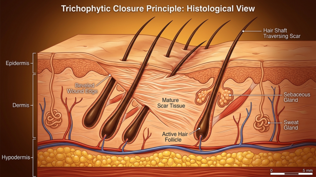
Bij de trichofytische incisie wordt de snede precies op de haarlijn geplaatst – de anatomische grens waar de huid van het voorhoofd overgaat in de behaarde hoofdhuid. De chirurg maakt de incisie schuin, waarbij de haarzakjes worden doorgesneden, zodat het haar door en voor het resulterende litteken heen groeit. Wanneer deze techniek vakkundig wordt uitgevoerd, ontstaat een litteken dat zo goed gecamoufleerd is dat het vrijwel onzichtbaar wordt, zelfs wanneer het haar naar achteren is getrokken.
Belichtingskwaliteit en toegang tot de orbitale rand
De trichofytische benadering biedt een vrijwel identieke visualisatie als de coronale techniek. Omdat de incisie zich op de haarlijn bevindt in plaats van erachter, klapt de chirurg de voorhoofdflap op een vergelijkbare manier naar beneden, waardoor een breed zicht op het voorhoofdsbeen en beide oogkassen wordt verkregen. Het belangrijkste verschil zit in de positionering: de trichofytische incisie verplaatst het litteken van een verborgen locatie in de hoofdhuid naar de rand van de haarlijn. Voor patiënten die tegelijkertijd een andere ingreep nodig hebben... voorhoofd contouren En voorhoofdverkleining—wat betekent dat de haarlijn naar voren moet worden gebracht—is de trichofytische incisie niet zomaar een redelijke keuze. Het is de enige logische optie, omdat de chirurg hiermee overtollige huid van het voorhoofd kan verwijderen en de haarlijn naar voren kan verplaatsen, terwijl hij tegelijkertijd toegang krijgt tot het onderliggende bot.
Behoud van de haargrens en risico op haarverlies
Behoud van de haargrens, verdorie! De trichofytische methode werkt uitstekend wanneer de techniek wordt uitgevoerd door een ervaren chirurg. De schuine incisie zorgt ervoor dat de haarzakjes aan de bovenrand haar blijven laten groeien door de littekenlijn heen. Trichofytische incisies brengen echter een duidelijk en onderschat risico met zich mee: als de wond onder spanning wordt gesloten – wat vaak voorkomt bij een aanzienlijke haarlijnverplaatsing – kan er follikelsterfte optreden langs beide randen van de incisie. Dit resulteert in een smalle strook haarverlies precies bij de haarlijn, wat ironisch genoeg de meest zichtbare plek is. Ik heb meerdere patiënten van andere klinieken behandeld waar agressieve haarlijnverplaatsing littekenalopecia langs de incisielijn veroorzaakte, waardoor later een haartransplantatie nodig was om de haarlijn te herstellen.
Zichtbaarheid van littekens en sensorische verstoring
Het trichofytische litteken bevindt zich op de overgang tussen de haargrens en de hoofdhuid. Wanneer er haar door het litteken groeit zoals bedoeld, wordt het uitzonderlijk goed verborgen. Het litteken blijft echter zichtbaar gedurende de eerste drie tot vier maanden van de genezing, voordat het haar door het littekenweefsel heen groeit. Patiënten moeten dit tijdsverloop begrijpen en zich voorbereiden op een periode waarin het verbergen van de incisie aanpassingen in de haarstijl vereist. Sensorische verstoring is vergelijkbaar met de coronale benadering in het centrale deel van het voorhoofd, omdat dezelfde zenuwtakken worden doorgesneden tijdens het losmaken van de huidflap. De achterste hoofdhuid behoudt echter gevoel, omdat de incisie zich niet tot dat gebied uitstrekt. Patiënten beschrijven doorgaans gevoelloosheid die beperkt is tot het voorhoofd zelf, terwijl de hoofdhuid achter de incisie gevoelig blijft.
Endoscopische toegang tot de wenkbrauw: de minimalistische aanpak met maximale beperkingen
Bij een endoscopische benadering van de wenkbrauw worden twee tot vijf kleine incisies van elk één tot twee centimeter gemaakt in de behaarde hoofdhuid. De chirurg brengt via deze openingen een endoscoop en instrumenten in en maakt een dissectie onder het periost van het voorhoofd om toegang te krijgen tot het voorhoofdsbeen en de supraorbitale randen. Deze methode is populair geworden omdat er geen lang litteken ontstaat en de zenuwbeschadiging aanzienlijk wordt verminderd.
Belichtingskwaliteit: de cruciale beperking
Endoscopische toegang tot de wenkbrauw Deze methode biedt een visualisatie die fundamenteel verschilt van open benaderingen. De chirurg bekijkt het operatiegebied via een camera en werkt in een subperiostale tunnel die de bewegingsvrijheid van instrumenten beperkt en de dissectieboog verkleint. Hoewel de endoscoop een uitstekend zicht biedt op het centrale deel van het voorhoofd en de glabella, is de toegang tot de laterale orbitaranden – de benige bogen die de buitenste ooghoeken omlijsten – beperkt. Voor voorhoofden van type I en type II, waarbij contourcorrectie nodig is zonder grote sinusoperaties, is endoscopische toegang vaak voldoende. Voor voorhoofden van type III, waarbij de wand van de frontale sinus moet worden teruggeplaatst met aanzienlijke botverwijdering, dwingt de endoscopische benadering de chirurg om te werken via kleine openingen, wat de operatietijd verlengt en de precisie van het modelleren van het bot bij de orbitaranden beperkt.
Een belangrijk detail: endoscopische toegang werkt het beste in combinatie met andere endoscopische procedures. Patiënten die gelijktijdig een endoscopische ingreep ondergaan, kunnen hier het beste gebruik van maken. endoscopische temporale lift Bij ingrepen aan het middengezicht profiteren mensen van gedeelde toegangspoorten. De incisiestrategie wordt efficiënter in plaats van beperkend wanneer meerdere anatomische regio's via hetzelfde operatieveld behandeld moeten worden.
Behoud van de haargrens, risico op haarverlies en sensorische impact
De endoscopische methode blinkt uit in het behoud van de haarlijn. Kleine incisies in de hoofdhuid veroorzaken een verwaarloosbaar haarverlies – doorgaans minder dan twintig haarzakjes per poort. De positie en vorm van de haarlijn blijven onaangetast. Risico op beschadiging van de hoofdhuidzenuwen De impact is aanzienlijk kleiner omdat de dissectie onder het periost plaatsvindt zonder belangrijke zenuwbanen te doorsnijden. De meeste endoscopische patiënten melden lichte gevoelloosheid in het voorhoofd die binnen vier tot zes weken verdwijnt. Dit snelle herstel van het gevoel is het grootste functionele voordeel van de benadering en maakt endoscopische toegang de voorkeursmethode voor patiënten die er vooral op uit zijn hun bestaande anatomie zo min mogelijk te verstoren.
Zichtbaarheid van littekens en herstelsnelheid
Endoscopische littekens zijn volledig verborgen in de behaarde hoofdhuid. Elke incisie van één centimeter geneest als een klein littekentje dat zelfs onder kort haar onzichtbaar is. Het herstel verloopt sneller dan bij zowel de coronale als de trichofytische benadering, omdat de dissectie beperkt is. De meeste patiënten kunnen binnen zeven tot tien dagen hun lichte activiteiten hervatten. De zwelling neemt sneller af omdat de uitgebreide subcutane verstoring die bij open benaderingen optreedt, ontbreekt. De afweging blijft: u profiteert van een snel herstel en onzichtbare littekens, maar accepteert beperkte chirurgische toegang die de grondigheid van de botcontouring, met name bij de laterale wenkbrauwstructuur, in gevaar kan brengen.
Vergelijking van feminiseringsincisies voor het voorhoofd: de zevendimensionale matrix
Er bestaat geen enkele bron die alle drie incisietypen naast elkaar plaatst op basis van alle cruciale variabelen. Hieronder volgt het vergelijkende kader dat ik in mijn eigen praktijk gebruik bij het bespreken van de incisiestrategie met patiënten. Elke dimensie wordt beoordeeld op een schaal van vijf punten, waarbij vijf staat voor de meest gunstige uitkomst en één voor de minst gunstige uitkomst voor de patiënt.
Vergelijkingstabel: Zeven klinische dimensies
| Klinische dimensie | Coronale incisie | Trichofytische incisie | Endoscopische incisie |
|---|---|---|---|
| Blootstellingskwaliteit | ★★★★★ (Volledig panoramisch uitzicht) | ★★★★☆ (Vrijwel identiek aan coronaal) | ★★★☆☆ (Tunnel, camera-afhankelijk) |
| Behoud van de haargrens | ★★★★☆ (Stabiele positie; risico op kale strook) | ★★★☆☆ (Verhoogt de haargrens; risico op spanningsalopecia) | ★★★★★ (Haarlijn volledig intact) |
| Zichtbaarheid van littekens | ★★★★☆ (Verborgen bij dicht haar) | ★★★★☆ (Er groeit haar door het litteken heen) | ★★★★★ (Kleine incisies, volledig verborgen) |
| Zintuiglijke stoornis | ★★☆☆☆ (12-18+ maanden gevoelloosheid) | ★★★☆☆ (6-12 maanden gevoelloosheid in het voorhoofd) | ★★★★☆ (4-6 weken lichte gevoelloosheid) |
| Risico op haaruitval | ★★★☆☆ (Kale strook langs de incisie) | ★★★☆☆ (Verlies van haarzakjes aan de rand is mogelijk) | ★★★★★ (Minimale follikelbeschadiging) |
| Herstelsnelheid | ★★☆☆☆ (Zichtbare zwelling na 3-4 weken) | ★★★☆☆ (Zichtbare zwelling gedurende 2-3 weken) | ★★★★☆ (Zichtbare zwelling gedurende 1-2 weken) |
| Toegang tot de orbitale rand | ★★★★★ (Direct, onbeperkt) | ★★★★☆ (Directe, lichte velgbeperking) | ★★☆☆☆ (Beperkt bereik van de zijrand) |
Deze vergelijking laat zien waarom geen enkele incisie op alle vlakken superieur is. De coronale en trichofytische benaderingen blinken uit in zichtbaarheid en toegang tot de orbitarand – prioriteiten voor de operatie. De endoscopische benadering blinkt uit in het behoud van de haargrens, sensorische bescherming en herstelsnelheid – prioriteiten voor de patiëntervaring. De incisie jij kiest Je moet aansluiten bij welke van deze dimensies het zwaarst weegt voor jouw specifieke anatomie en persoonlijke prioriteiten.
Toegang tot het wenkbrauwbot: wat uw chirurg precies moet kunnen bereiken
Patiënten begrijpen zelden waarom toegang tot de orbitarand zo belangrijk is. De laterale orbitaranden – de benige bogen in de buitenste ooghoeken – vormen het breedste punt van het mannelijke voorhoofd. Tijdens een feminisering van het voorhoofd is het verkleinen of contouren van deze randen essentieel voor het verkrijgen van een zachtere, rondere wenkbrauwcontour. Onvoldoende toegang tot dit gebied leidt tot een veelvoorkomend resultaat: een centraal gefeminiseerd voorhoofd met aanhoudend prominente laterale orbitaranden die het mannelijke skelet verraden.

Via een coronale of trichofytische incisie kan de chirurg de laterale orbitaranden bereiken met directe visualisatie en instrumentmanipulatie in elk vlak. Bij een endoscopische benadering bevinden de laterale randen zich aan de uiterste rand van het operatiegebied. De endoscoop en instrumenten moeten door smalle tunnels worden geleid en de benaderingshoek wordt tangentieel in plaats van loodrecht. Voor kleine reducties – het afronden van de laterale rand in plaats van grote botreconstructies – is deze beperkte toegang voldoende. Voor substantiële contourcorrecties van de laterale orbitarand dwingt de endoscopische benadering tot compromissen in precisie, waardoor het buitenste wenkbrauwframe mogelijk onvoldoende gefeminiseerd blijft. Vraag uw chirurg direct: hoeveel werk aan de laterale orbitarand is nodig en kunt u dit endoscopisch uitvoeren? Hun eerlijke antwoord bepaalt of endoscopische toegang voor u geschikt is.
Risico op zenuwschade aan de hoofdhuid: een nadere blik op de sensorische gevolgen
De nervus supraorbitalis verlaat de schedel via het foramen supraorbitale (de inkeping boven de oogkas) en vertakt zich vervolgens over het voorhoofd en de voorste hoofdhuid. De nervus supratrochlearis loopt mediaal en innerveert het centrale deel van het voorhoofd en de glabella (de voorhoofdsrand). Deze zenuwen vormen de belangrijkste sensorische innervatielijn van het voorhoofd. Een coronale incisie op de kruin van de hoofdhuid doorsnijdt de opstijgende takken van deze zenuwen over de volledige breedte. Anatomieboeken beschrijven dit als een verwachte, tijdelijke zenuwbeschadiging. In de klinische praktijk is herstel echter noch gegarandeerd, noch volledig.
Ongeveer vijftien procent van de patiënten die een coronale incisie ondergaan, ervaart blijvende sensorische veranderingen – zoals aanhoudende gevoelloosheid, verhoogde gevoeligheid of dysesthesie (een onaangenaam tintelend gevoel) in de frontopariëtale hoofdhuid. De trichofytische incisie deelt deze centrale zenuwbeschadiging, omdat het proces van het optillen van de flap op vergelijkbare wijze takken doorsnijdt ter hoogte van de haarlijn. Endoscopische toegang verandert dit fundamenteel. Subperiostale dissectie spaart de supraorbitale en supratrochleaire zenuwstammen, omdat deze zenuwen oppervlakkig ten opzichte van het dissectievlak lopen. De kleine incisies voor de poorten veroorzaken slechts lichte, gelokaliseerde zenuwirritatie in plaats van een wijdverspreide beschadiging. Voor patiënten die sensorische integriteit belangrijk vinden – en velen beseffen de waarde ervan pas als ze die kwijt zijn – biedt de endoscopische benadering een aanzienlijk beter veiligheidsprofiel.
Haarlijnbehoud FFS: Het dilemma van dun haar
Patiënten met dik, dicht haar hebben veel vrijheid bij de keuze van de incisie, omdat elk litteken effectief gecamoufleerd kan worden. Patiënten met dun, fijn of teruglopend haar staan voor een veel beperktere keuze. Voor deze personen is de kale strook van de coronale incisie een ramp, omdat er onvoldoende haardichtheid is om deze te verbergen. Het mechanisme waarbij haar door het litteken heen groeit bij de trichofytische incisie is afhankelijk van de bestaande follikeldichtheid – als de haarlijn al dun is, zijn er minder follikels die door het litteken heen kunnen groeien. De endoscopische benadering blinkt juist hier uit: kleine incisies met poorten offeren vrijwel geen follikels op en de haarlijn blijft volledig intact.

Neem deze klinische realiteit in overweging: een patiënt met Norwood Klasse II haaruitval ondergaat een trichofytische incisie met een haarlijnverplaatsing van drie centimeter. De spanning bij het sluiten van de incisie rekt de toch al dunne haarlijn verder uit, wat leidt tot extra haaruitval. Zes maanden na de operatie is de haarlijn weliswaar verplaatst, maar is nu doorschijnend ter hoogte van de incisielijn. De patiënt heeft vervolgens een haartransplantatie nodig om de dichtheid te herstellen – een tweede ingreep die met een endoscopische benadering voorkomen had kunnen worden. Ik benadruk dit scenario niet om de trichofytische techniek af te raden, maar om te benadrukken dat Behoud van de haargrens, verdorie! Het vereist niet alleen een beoordeling van de locatie van de incisie, maar ook van de manier waarop de krachten die op de wondsluiting inwerken, de haardichtheid in uw specifieke geval beïnvloeden.
FFS-incisiebeslissingsboom: een klinisch stroomschema voor zelfevaluatie door de patiënt
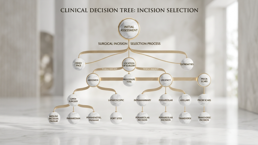
Hieronder staat de beslissingsboom die ik voor mijn praktijk heb ontwikkeld. Dokter MFO Kliniek. Het vertaalt de complexe wisselwerking van anatomische variabelen naar een stapsgewijze zelfbeoordeling die patiënten begeleidt naar het meest geschikte type incisie vóór het formele consult. Beantwoord de vragen eerlijk, en de weg zal zich openbaren.
Stapsgewijs zelfevaluatiestroomschema
Vraag 1: Heeft u een huidverkleining op het voorhoofd of een haarlijncorrectie nodig?
- JA → Ga verder naar vraag 2A.
- NEE → Ga verder naar vraag 2B.
Vraag 2A: U heeft een haarlijncorrectie nodig. Is uw haardichtheid voldoende om de spanning bij de haargrens te weerstaan?
- JA → Trichofytische incisie Dit is uw belangrijkste optie. De chirurg zal de haarlijn verplaatsen en tegelijkertijd via dezelfde incisie toegang krijgen tot het bot.
- NEE → Overweeg een coronale insnijding met een aparte haartransplantatie een behandeling om de haarlijn te verlagen en zo haaruitval door spanning aan de rand van de haarlijn te voorkomen.
Vraag 2B: U hoeft uw haarlijn niet naar voren te plaatsen. Wordt uw voorhoofd geclassificeerd als type III met aanzienlijke betrokkenheid van de voorhoofdsholte, waardoor een grote terugplaatsing van de haarlijn nodig is?
- JA → Is uw haardichtheid voldoende om een litteken op uw achterste hoofdhuid te camoufleren?
- NEE (Uw voorhoofd is type I of type II, waardoor contourcorrectie nodig is zonder grote sinusproblemen) → Ga verder naar vraag 3.
Vraag 3: U heeft een voorhoofd van type I of type II. Is een aanzienlijke contourcorrectie van de laterale oogkasrand nodig?
- JA → Trichofytische of coronale incisie Geeft de chirurg directe toegang tot de laterale rand. Endoscopische toegang is mogelijk onvoldoende voor uitgebreider werk aan de laterale zijde.
- NEE → Behoren snel herstel en minimale sensorische verstoring tot uw top drie prioriteiten?
Deze beslissingsboom filtert de marketingpraatjes rondom elk type incisie. Elke aanpak heeft echte sterke en zwakke punten. Het juiste antwoord hangt af van uw anatomie, uw voorhoofdtype, uw haardichtheid en uw persoonlijke prioriteiten – niet van de standaardvoorkeur van een chirurg.
Herstelsnelheid en -tijdlijn: wat u van elke aanpak kunt verwachten.
De herstelperiode heeft direct invloed op uw vermogen om weer aan het werk te gaan, sociale contacten te onderhouden en uw normale leven te hervatten. Inzicht in de hersteltijd die elk type incisie vereist, helpt u realistisch te plannen.

Endoscopisch herstel: De meeste patiënten hervatten hun bureauwerk binnen zeven tot tien dagen. De zwelling bereikt een piek na achtenveertig uur en neemt merkbaar af na tien dagen. Het sensorisch herstel is binnen vier tot zes weken voltooid. Volledige, onbeperkte activiteit, inclusief sporten, is na drie weken mogelijk.
Trichofytisch herstel: Bureauwerk kan doorgaans na tien tot veertien dagen worden hervat. De zwelling bereikt een piek rond dag drie en neemt na twee tot drie weken aanzienlijk af. Het litteken bij de haarlijn is het meest zichtbaar tussen week twee en zes, voordat er weer haar begint te groeien. Het herstel van de sensorische functie in het midden van het voorhoofd duurt zes tot twaalf maanden. Volledige activiteit is na vier weken weer mogelijk.
Coronaal herstel: Het hervatten van bureauwerk duurt twee tot drie weken vanwege de uitgebreide zwelling van het voorhoofd en de gevolgen van de wijdverspreide sensorische verstoring. De zwelling bereikt een piek tussen dag drie en vijf en kan vier tot zes weken aanhouden voordat deze volledig is verdwenen. Het sensorisch herstel duurt twaalf tot achttien maanden, waarbij mogelijk blijvende veranderingen optreden. Normaal gesproken kan men na zes weken, met goedkeuring van het chirurgisch team, de normale activiteiten hervatten.
Gecombineerde procedures: wanneer één incisie meerdere doelen dient
Efficiëntie tijdens een operatie is belangrijk. Wanneer een patiënt een operatie nodig heeft... voorhoofd contouren Naast een temporale lift, wenkbrauwlift of verjonging van het bovenste deel van het gezicht, moet de incisiestrategie meerdere doelen tegelijk dienen. De coronale incisie is bij uitstek geschikt voor een wenkbrauwlift, omdat de voorhoofdsflap al is opgetild. De trichofytische incisie sluit naadloos aan op een haarlijncorrectie, omdat beide doelen – de cosmetische verbetering en de chirurgische toegang – dezelfde incisielijn delen. Endoscopische toegang kan elegant worden gecombineerd met endoscopische procedures aan de slapen en het middengezicht via gedeelde poortincisies, waardoor de chirurg het voorhoofd, de slapen en het middengezicht kan behandelen zonder extra littekens te creëren.
Bij Dr. MFO Kliniek, Ik evalueer routinematig of het combineren van procedures via één enkele incisie betere algehele resultaten oplevert dan het afzonderlijk behandelen van elk gebied. Een patiënt die voorhoofdcontouren, een lichte wenkbrauwverzakking en een holte in de slapen nodig heeft, kan het meest gebaat zijn bij een endoscopische benadering waarbij alle drie de gebieden via dezelfde kleine poorten worden aangepakt. Een patiënt met een bult op het voorhoofd, een hoge haarlijn en ernstige wenkbrauwptosis behaalt het beste resultaat met een trichofytische incisie die de haarlijn naar voren brengt, het bot bereikt en de wenkbrauw lift in één gecoördineerde manoeuvre.
Littekenrijping: de twaalf maanden durende realiteit waar niemand over praat
Elk litteken doorloopt een voorspelbaar rijpingsproces. De ontstekingsfase duurt één tot twee weken, waarin het litteken rood, verhoogd en stevig aanvoelt. De proliferatiefase loopt van week twee tot en met week twaalf, wanneer de collageenproductie piekt en het litteken dikker en prominenter kan lijken dan tijdens de eerste genezingsfase. De remodelleringsfase duurt van drie tot en met twaalf maanden, waarin het litteken geleidelijk zachter, vlakker en minder opvallend wordt. Patiënten die hun litteken na zes weken beoordelen – terwijl het zich nog in de proliferatiefase bevindt – raken vaak onnodig in paniek. Het ware uiterlijk van een litteken op het voorhoofd kan pas na twaalf maanden worden beoordeeld.
Deze realiteit heeft praktische implicaties voor de keuze van de incisie. Als u kiest voor een trichofytische incisie, zal het litteken bij de haarlijn er na acht weken zorgwekkend uitzien. Dit is normaal. Het haar is nog niet door het litteken heen gegroeid en het weefsel is nog steeds in actieve remodellering. Door twaalf maanden te wachten voordat u een oordeel velt, voorkomt u onnodige angst en voortijdige hersteloperaties. Een patiënt die deze tijdlijn begrijpt, gaat de operatie in met realistische verwachtingen in plaats van de valse hoop op een nauwelijks zichtbaar litteken na één maand.
Praktische toepassing: Uw vijfstappenplan voor het kiezen van een incisie
Zet bovenstaande kennis om in concrete actie. Volg deze vijf stappen om jouw ideale incisietype te bepalen en ervoor te pleiten:
- Bepaal je voorhoofdstype. Maak een laterale cefalogram of een 3D CT-scan. Een radioloog of chirurg moet bepalen of uw voorhoofd type I (minimale bultvorming, intacte sinuswand), type II (bultvorming met ondiepe sinusbetrokkenheid) of type III (diepe frontale sinus die terugplaatsing vereist) vertoont. Type III vereist bijna altijd een open toegang – coronaal of trichofytisch. Type I en II komen mogelijk in aanmerking voor een endoscopische of open benadering, afhankelijk van andere variabelen.
- Meet de hoogte en dichtheid van uw haargrens. Gebruik een liniaal om de afstand van uw voorhoofd tot uw haarlijn te meten. Metingen van meer dan zes centimeter duiden meestal op een hoog voorhoofd dat baat heeft bij een haarlijncorrectie – wat wijst op trichofytische haargroei. Beoordeel uw haardichtheid door te kijken of uw hoofdhuid zichtbaar is door uw haar op de kruin en bij de slapen. Zichtbare hoofdhuid wijst op een lage haardichtheid, wat de mogelijkheden voor littekencamouflage beperkt en endoscopische toegang bevordert.
- Rangschik je persoonlijke prioriteiten. Noteer de zeven dimensies uit de bovenstaande vergelijkingstabel: blootstelling, behoud van de haarlijn, zichtbaarheid van het litteken, sensorische verstoring, risico op haaruitval, herstelsnelheid en toegang tot de oogkasrand. Nummer ze van meest belangrijk naar minst belangrijk. Uw twee of drie belangrijkste prioriteiten onthullen welk type incisie het beste aansluit bij uw waarden. Patiënten die sensorisch behoud en snel herstel het belangrijkst vinden, kiezen vaak voor een endoscopische ingreep. Patiënten die blootstelling en toegang tot de oogkasrand het belangrijkst vinden, kiezen vaak voor een open benadering.
- Doorloop de bovenstaande beslissingsboom. Beantwoord elke vraag in het stroomschema eerlijk op basis van uw klinische bevindingen. Noteer de uitkomst. Vergelijk deze vervolgens met uw prioriteitsrangschikking uit stap drie. Als beide in dezelfde richting wijzen, heeft u uw antwoord. Als ze tegenstrijdig zijn, vormt de discrepantie zelf het gespreksonderwerp voor uw chirurgisch consult.
- Presenteer uw analyse tijdens het overleg. Neem uw voorhoofdclassificatie, haarlijnmetingen, prioriteitsrangschikking en de uitkomst van de beslissingsboom mee naar uw afspraak met de chirurg. Deze voorbereiding transformeert het gesprek van passief informatie ontvangen naar actieve, gezamenlijke besluitvorming. Een chirurg die de autonomie van de patiënt respecteert, zal deze mate van betrokkenheid waarderen en zijn of haar aanbeveling daarop aanpassen.
Uw keuze maken coronale versus trichofytische versus endoscopische wenkbrauwtoegang De keuze voor een incisie is geen beslissing die je volledig kunt delegeren. Je anatomie bepaalt de parameters. Je prioriteiten geven de richting aan. De expertise van je chirurg zorgt voor de uitvoering van het plan. Neem de beslissing in eigen handen, en je bent verantwoordelijk voor het resultaat. Klaar om te bepalen welke incisie het beste bij jouw unieke anatomie past? Vraag nu een persoonlijk adviesgesprek aan. Kom naar de Dr. MFO-kliniek en ontvang een uitgebreide beoordeling van uw voorhoofd met een op maat gemaakt incisieadvies.
Veelgestelde vragen
Waarom is het type incisie belangrijker dan de techniek voor het hervormen van het bot bij voorhoofdvervrouwing?
De incisie bepaalt de kwaliteit van de toegang die uw chirurg biedt, de zichtbaarheid van uw litteken, het risico op beschadiging van de zenuwen, de kans op haaruitval en de hersteltijd. Zelfs een perfecte botcontouring kan een slecht gekozen incisie, die een zichtbaar litteken of blijvende gevoelloosheid achterlaat, niet compenseren. De incisie vormt de structurele basis van het uiteindelijke resultaat.
Hoe weet ik of ik een coronale, trichofytische of endoscopische incisie nodig heb voor FFS?
Uw voorhoofdtype, de hoogte van uw haarlijn, de haardichtheid en uw persoonlijke prioriteiten bepalen de beste aanpak. Voorhoofden van type III met diepe sinusbetrokkenheid vereisen doorgaans een coronale of trichofytische toegang. Patiënten die een haarlijnverplaatsing nodig hebben, hebben baat bij een trichofytische incisie. Voorhoofden van type I of II met een goede haardichtheid komen mogelijk in aanmerking voor endoscopische toegang.
Zal ik haar verliezen op de incisieplek na een feminiseringsoperatie aan mijn voorhoofd?
Haarverlies varieert afhankelijk van het type incisie. Bij coronale incisies bestaat het risico op een permanente kale strook langs de hechting. Bij trichofytische incisies kunnen haarfollikels aan de rand verloren gaan als de sluiting te strak wordt aangetrokken. Bij endoscopische incisies worden minder dan twintig follikels per poort verwijderd, waardoor haarverlies voor de meeste patiënten vrijwel verwaarloosbaar is.
Hoe lang houdt de gevoelloosheid van de hoofdhuid aan na elk type incisie?
Patiënten die een endoscopische behandeling ondergaan, krijgen doorgaans binnen vier tot zes weken hun gevoel terug. Patiënten met trichofytische tumoren ervaren zes tot twaalf maanden lang gevoelloosheid in het voorhoofd. Patiënten met coronale tumoren hebben twaalf tot achttien maanden last van gevoelloosheid op de hoofdhuid, waarbij ongeveer vijftien procent een blijvende verandering in het gevoel ervaart.
Kan ik een haarlijncorrectie ondergaan via een endoscopische incisie?
Nee. Endoscopische incisies kunnen de haarlijn niet naar voren brengen, omdat er geen huid van het voorhoofd wordt verwijderd. Om de haarlijn naar voren te brengen, moet overtollig voorhoofdsweefsel worden verwijderd. Dit vereist een trichofytische of coronale incisie, waarmee de chirurg huid kan verwijderen en de haarlijn naar voren kan verplaatsen.
Is het trichofytische litteken zichtbaar wanneer het haar naar achteren wordt getrokken?
Gedurende de eerste drie tot vier maanden blijft het litteken zichtbaar bij de haargrens. Nadat er na ongeveer vier tot zes maanden haar door het litteken begint te groeien, neemt de zichtbaarheid aanzienlijk af. Na twaalf maanden zijn de meeste goed uitgevoerde trichofytische littekens zelfs met opgetrokken haar nauwelijks nog te zien.
Wat gebeurt er als mijn chirurg de laterale orbitale randen niet endoscopisch kan bereiken?
Onvoldoende contourcorrectie van de laterale orbitarand zorgt ervoor dat de buitenste wenkbrauwbotten prominent aanwezig blijven, waardoor een mannelijk kader behouden blijft ondanks de feminisering van het centrale voorhoofd. Als er aanzienlijke correctie van de laterale orbitarand nodig is, dient uw chirurg een open benadering aan te bevelen – coronaal of trichofytisch – in plaats van het contourresultaat te compromitteren door beperkte endoscopische toegang.







