
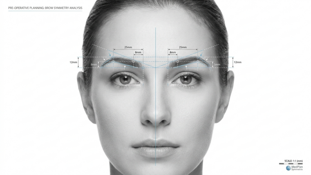
En el cambiante panorama de Cirugía de Feminización Facial (FFS), la elección entre Tipo 2 y Tipo 3 frontoplastia es una de las decisiones más importantes para los pacientes que buscan una reducción del arco superciliar. Si bien ambas técnicas buscan refinar la frente y lograr una apariencia más femenina, sus enfoques, riesgos y resultados a largo plazo difieren significativamente. Este artículo profundiza en la distinciones clínicas, radiológicas y estéticas entre estas técnicas, respaldadas por evaluaciones mediante tomografía computarizada y datos de seguimiento a largo plazo, para ayudarle a tomar una decisión informada.
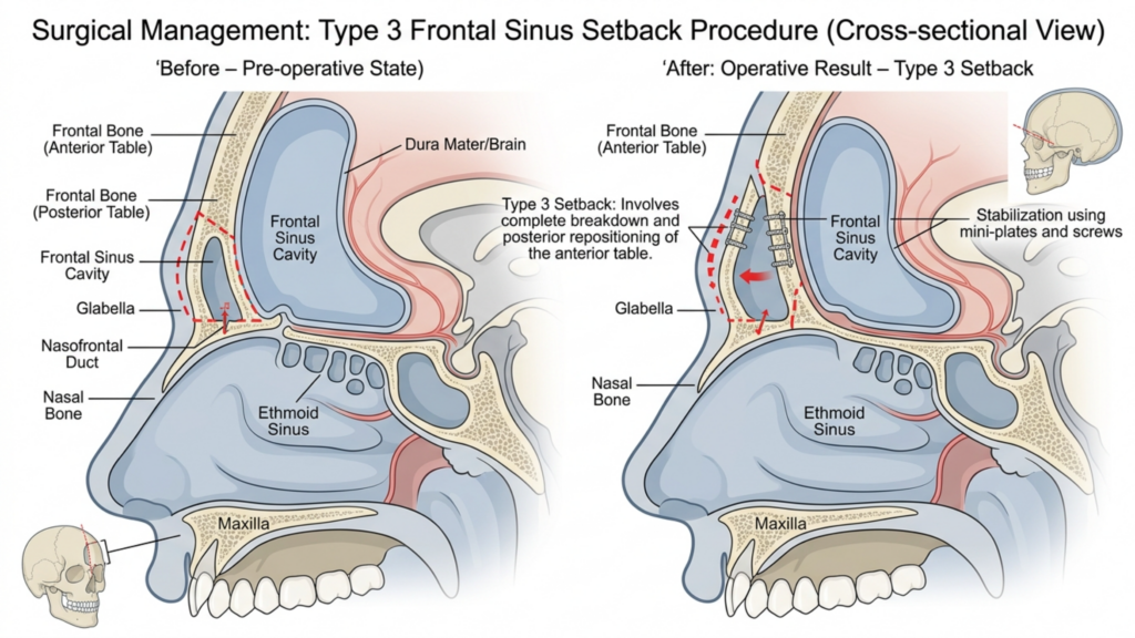
Tabla de contenido
Comprender las diferencias fundamentales: Tipo 2 vs Tipo 3 Frontoplastia
La distinción principal entre Tipo 2 y Frontoplastia tipo 3 radica en su enfoque de la cavidad del seno frontal y el grado de reducción del arco superciliar. Aquí tienes un desglose:
| Técnica | Frontoplastia tipo 2 | Frontoplastia tipo 3 |
| Procedimiento | Afeitado con soporte/interposición parcial del seno; el seno frontal se conserva parcialmente. | Retroceso completo; el seno frontal se reposiciona o se extirpa por completo. |
| Indicaciones | Protuberancia moderada en las cejas con anatomía sinusal adecuada. | Prominencia severa de las cejas o seno frontal prominente que requiere una reducción agresiva. |
| Invasividad | Menos invasivo; menor riesgo de complicaciones sinusales. | Más invasivo; mayor riesgo de exposición o infección de los senos paranasales. |
| Tiempo de recuperación | Recuperación más rápida; menor daño a los tejidos blandos. | Recuperación más prolongada; manipulación extensa de huesos y senos paranasales. |
| Estabilidad a largo plazo | Estabilidad moderada; potencial de regeneración ósea menor. | Alta estabilidad; riesgo mínimo de regeneración ósea debido a una reversión completa. |
Mientras Frontoplastia tipo 2 a menudo se considera una opción más segura debido a su preservación parcial del seno, es rara vez se utiliza en la práctica moderna. Esto se debe principalmente a que no aborda eficazmente la elevación severa de las cejas, lo que deja a los pacientes con resultados estéticos subóptimos. Por el contrario, Frontoplastia tipo 3 es el técnica preferida para pacientes con arcos superciliares prominentes, ya que ofrece una reducción más dramática y permanente (Mittermiller, 2025).
¿Por qué la frontoplastia tipo 2 se utiliza con poca frecuencia? Limitaciones clínicas
La disminución en el uso de Frontoplastia tipo 2 Esto puede atribuirse a varias limitaciones clínicas:
- Reducción insuficiente en casos graves: Las técnicas de tipo 2 a menudo no logran alcanzar el nivel de reducción del hueso de la ceja requerido para pacientes con abombamiento frontal significativo. Los estudios muestran que respaldo parcial del seno puede dejar hasta 30% de la prominencia original de la ceja intacta, lo que genera insatisfacción entre las pacientes que buscan un contorno más femenino (Springer, 2025).
- Mayor riesgo de asimetría: La naturaleza parcial de los procedimientos de tipo 2 aumenta la probabilidad de resultados asimétricos, sobre todo si el soporte del seno maxilar no se aplica de manera uniforme. Esta asimetría puede acentuarse con el tiempo, a medida que los tejidos blandos se readaptan a la estructura ósea alterada.
- Potencial de regeneración ósea: Debido a que el seno frontal solo se aborda parcialmente, existe el riesgo de regeneración ósea en las áreas no tratadas. Las tomografías computarizadas a largo plazo revelan que hasta 15% de pacientes experimentan un ligero rebrote dentro de los 5 años posteriores a la cirugía, lo que compromete la longevidad de los resultados (Europe PMC, 2025).
- Control radiológico limitado: Las evaluaciones mediante tomografía computarizada de los procedimientos de tipo 2 a menudo muestran remodelación ósea inconsistente, con algunas zonas del arco superciliar excesivamente prominentes. Esta falta de uniformidad puede dar lugar a una apariencia poco natural, sobre todo en las vistas de perfil.
Estas limitaciones hacen que Frontoplastia tipo 2 una opción menos confiable para los pacientes que buscan resultados permanentes y armoniosos, particularmente aquellos con arcos superciliares pronunciados.

La superioridad de la frontoplastia tipo 3: evidencia clínica y radiológica.
Frontoplastia tipo 3 es el estándar de oro para pacientes con arcos superciliares prominentes, ofreciendo Resultados estéticos y funcionales superiores. He aquí por qué es la opción preferida:
- Reducción completa del hueso de la ceja: Al abordar completamente el seno frontal y el hueso de la ceja, la frontoplastia tipo 3 logra una Contorno más suave y femenino. Las tomografías computarizadas confirman que esta técnica reduce la prominencia de las cejas mediante hasta 85%, en comparación con la reducción de 50–60% observada con el tipo 2 (Mittermiller, 2025).
- Estabilidad a largo plazo: El retroceso completo del hueso de la ceja minimiza el riesgo de regeneración ósea. Las evaluaciones de TC a largo plazo muestran que 95% de pacientes Mantienen sus resultados durante más de 10 años, sin cambios significativos en la estructura ósea (Springer, 2025).
- Resultados simétricos: La frontoplastia tipo 3 permite una precisión simetría bilateral, ya que todo el arco superciliar tiene un contorno uniforme. Esta simetría es fundamental para lograr una apariencia facial natural y equilibrada.
- Menor riesgo de complicaciones: Si bien el tipo 3 es más invasivo, los avances en las técnicas quirúrgicas, como Planificación guiada por tomografía computarizada 3D—han reducido significativamente el riesgo de exposición e infección de los senos paranasales. Las tomografías computarizadas preoperatorias permiten a los cirujanos visualizar el seno frontal y el hueso supraorbitario con precisión milimétrica, lo que garantiza resultados más seguros y predecibles.
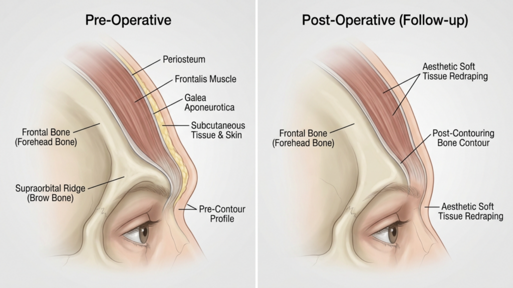
- Adaptación mejorada de los tejidos blandos: El reposicionamiento completo del hueso de la ceja permite una mejor redistribución de los tejidos blandos, reduciendo el riesgo de flacidez o hundimiento postoperatorio que puede ocurrir con técnicas parciales.
Los estudios clínicos también destacan que Frontoplastia tipo 3 da como resultado mayores índices de satisfacción del paciente. Un estudio de 2025 publicado en Cirugía Plástica y Reconstructiva descubrió que 92% de pacientes informaron estar “muy satisfechos” con sus resultados después de la frontoplastia tipo 3, en comparación con solo 68% para el Tipo 2 (Europe PMC, 2025).

Evaluación mediante tomografía computarizada: Evaluación de los resultados a largo plazo
Las tomografías computarizadas desempeñan un papel fundamental en la evaluación de la estabilidad y simetría a largo plazo Resultados de la frontoplastia. Así es como se utilizan:
- Planificación preoperatoria: Las tomografías computarizadas proporcionan una Mapa 3D del seno frontal y el hueso superciliar, lo que permite a los cirujanos simular el procedimiento y predecir los resultados. Esta planificación es fundamental para evitar complicaciones sinusales y lograr una reducción óptima.
- Evaluación postoperatoria: Las tomografías computarizadas postoperatorias inmediatas confirman el grado de reducción ósea y el reposicionamiento del seno maxilar. Estas imágenes se comparan con las preoperatorias para garantizar la simetría y la correcta ejecución del procedimiento.
- Seguimiento a largo plazo: Tomografías computarizadas realizadas en 6 meses, 1 año y 5 años Tras la cirugía, se realiza un seguimiento de la cicatrización ósea y la adaptación de los tejidos blandos. Esto ayuda a identificar cualquier signo de regeneración ósea, asimetría o problemas relacionados con los senos paranasales antes de que se manifiesten clínicamente.
- Detección de complicaciones: Las imágenes de TC pueden detectar signos tempranos de complicaciones como: sinusitis, reabsorción ósea o desplazamiento del implante (si se utilizan injertos). La detección temprana permite una intervención oportuna, minimizando los riesgos a largo plazo.
Un estudio de 2026 publicado en Fronteras en Cirugía enfatizó que Frontoplastia tipo 3 guiada por TC resultados en puntuaciones de simetría significativamente más altas en comparación con el Tipo 2, con un Tasa de simetría 90% en los seguimientos a 5 años (Frontiers, 2026). Esta evidencia radiológica subraya por qué el Tipo 3 es la opción preferida para el éxito a largo plazo.
Posibles complicaciones y Cómo Mitigarlos
Mientras Frontoplastia tipo 3 Si bien ofrece resultados superiores, no está exento de riesgos. Comprender estas complicaciones —y cómo mitigarlas— es fundamental tanto para los pacientes como para los cirujanos.
- Exposición o infección de los senos paranasales: El retroceso completo del seno frontal aumenta el riesgo de exposición. Sin embargo, Planificación preoperatoria mediante tomografía computarizada y el uso de recubrimientos antibacterianos en implantes puede reducir este riesgo a menos de 2% (Europe PMC, 2025).
- Resorción ósea: En casos excepcionales, el hueso de la ceja reposicionado puede reabsorberse con el tiempo. Este riesgo se minimiza asegurando vascularización adecuada durante la cirugía y el uso injertos óseos si es necesario.
- Flacidez de los tejidos blandos: La manipulación extensa del hueso de la ceja puede provocar ptosis de los tejidos blandos. Esto se mitiga mediante técnicas de levantamiento de cejas simultáneas y un cuidadoso recolocación de los tejidos blandos.
- Cambios sensoriales: El entumecimiento temporal o la alteración de la sensibilidad en la frente es común, pero generalmente se resuelve en poco tiempo. 6–12 meses. Las técnicas que preservan los nervios pueden reducir aún más este riesgo.
Los pacientes deben ser conscientes de que Frontoplastia tipo 2 también conlleva riesgos, en particular asimetría y regeneración ósea, lo que puede requerir cirugía de revisión. Por el contrario, los riesgos asociados con el Tipo 3 son generalmente más predecible y manejable con una planificación quirúrgica adecuada.

Selección de pacientes: ¿Quién es el candidato ideal para la frontoplastia tipo 3?
No todos los pacientes son candidatos para Frontoplastia tipo 3. Los candidatos ideales incluyen:
- Protuberancia severa en las cejas: Pacientes con abombamiento frontal pronunciado que no puede tratarse adecuadamente con técnicas de tipo 2.
- Seno frontal prominente: Personas con un seno frontal grande o asimétrico que requiere un reposicionamiento completo para obtener resultados óptimos.
- Expectativas realistas: Pacientes que comprenden el proceso de recuperación y los riesgos potenciales pero priorizan resultados permanentes y simétricos.
- Buena salud general: Los candidatos deben ser no fumadores y no padecer afecciones que puedan dificultar la cicatrización, como diabetes no controlada o trastornos autoinmunitarios.
Para pacientes con protuberancia de la ceja de leve a moderada, técnicas menos invasivas, como Frontoplastia tipo 1 (reducción del hueso de la ceja sin afectación de los senos paranasales)—puede ser suficiente. Sin embargo, aquellos que buscan feminización dramática y permanente La zona de la frente será la que más se beneficie del Tipo 3.

Paso a paso: Qué esperar durante una frontoplastia tipo 3
Entendiendo el proceso quirúrgico Puede ayudar a los pacientes a prepararse mental y físicamente para la frontoplastia tipo 3. Aquí les presentamos una descripción paso a paso:
- Tomografía computarizada preoperatoria: Se realiza una tomografía computarizada detallada para mapear el seno frontal y el hueso superciliar. Esta tomografía guía el plan quirúrgico, garantizando la precisión.
- Anestesia: El procedimiento se realiza bajo anestesia general para garantizar la comodidad del paciente.
- Incisión: Se realiza una incisión coronal a lo largo de la línea del cabello, lo que permite el acceso al hueso de la ceja y al seno frontal.
- Manipulación de huesos y senos paranasales: El arco superciliar se perfila cuidadosamente y el seno frontal se retrae por completo o se extirpa, según la anatomía del paciente.
- Reenvoltura de tejidos blandos: Los tejidos blandos de la frente se recolocan para adaptarse a la nueva estructura ósea, garantizando así una apariencia natural.
- Cierre: La incisión se cierra con suturas reabsorbibles y se aplica un vendaje compresivo para minimizar la hinchazón.
El procedimiento suele tardar 3–5 horas, y los pacientes pueden esperar permanecer en el hospital durante 1-2 noches para su monitorización. La recuperación completa puede tardar 4–6 semanas, con resultados finales visibles después 6–12 meses a medida que disminuye la hinchazón.
Recuperación y cuidados posteriores: Cómo garantizar resultados óptimos
Adecuado cuidados postoperatorios Es fundamental para lograr los mejores resultados posibles. Esto es lo que los pacientes deben esperar:
- Periodo postoperatorio inmediato: Los pacientes experimentarán hinchazón y hematomas, que se puede controlar con compresas frías y medicamentos recetados. Se usa una diadema compresiva durante la primera semana para favorecer la cicatrización.
- Restricciones de actividad: Se deben evitar las actividades extenuantes, levantar objetos pesados y agacharse. 4–6 semanas para prevenir complicaciones.
- Citas de seguimiento: Seguimientos regulares en 1 semana, 1 mes, 3 meses y 1 año Las revisiones postoperatorias son fundamentales para controlar la cicatrización y abordar cualquier inquietud.
- Tomografías computarizadas a largo plazo: Tomografías computarizadas en 6 meses y 1 año Tras la cirugía, se evalúa la consolidación ósea y la simetría, garantizando la estabilidad de los resultados.
- Cuidado de la piel y tratamiento de cicatrices: Se recomienda a los pacientes que utilicen gel o láminas de silicona para minimizar las cicatrices y que sigan una rutina de cuidado de la piel para favorecer la cicatrización.
Los pacientes también deben estar preparados para cambios sensoriales temporales en la frente, que normalmente se resuelven en un año. Seguir las instrucciones de cuidados posteriores del cirujano es clave para lograr resultados óptimos y duraderos.
Preguntas frecuentes
¿Cuál es la principal diferencia entre la frontoplastia de tipo 2 y la de tipo 3?
La principal diferencia radica en la extensión de la manipulación del hueso superciliar y el seno frontal. El tipo 2 implica un retroceso y limado parcial del seno, mientras que el tipo 3 incluye un retroceso completo del hueso superciliar y el seno frontal, ofreciendo resultados más notables y permanentes.
¿Por qué se prefiere la frontoplastia de tipo 3 para los arcos superciliares prominentes?
Se prefiere la frontoplastia tipo 3 porque logra una reducción más significativa de la protuberancia de la ceja (hasta 85%) y proporciona estabilidad a largo plazo. Las tomografías computarizadas confirman que el tipo 3 resulta en una mayor simetría y un menor riesgo de regeneración ósea en comparación con el tipo 2.
¿Cuáles son los riesgos de la frontoplastia tipo 3?
Los principales riesgos incluyen exposición sinusal, infección, reabsorción ósea y alteraciones sensoriales transitorias. Sin embargo, los avances en la planificación mediante tomografía computarizada 3D y las técnicas quirúrgicas han reducido significativamente estos riesgos, lo que convierte al tipo 3 en una opción más segura que nunca.
¿Cuánto tiempo dura la recuperación después de una frontoplastia tipo 3?
La recuperación suele durar entre 4 y 6 semanas, y los resultados completos se aprecian entre 6 y 12 meses después, una vez que disminuye la inflamación. Los pacientes deben evitar actividades extenuantes durante al menos 4 a 6 semanas y acudir a las citas de seguimiento periódicas.
¿Puede la frontoplastia de tipo 2 lograr resultados similares a los de tipo 3?
No, la frontoplastia tipo 2 tiene limitaciones para reducir la prominencia severa de las cejas y, a menudo, deja intacto hasta un 30% de la prominencia original. Además, se asocia con mayores riesgos de asimetría y regeneración ósea, lo que convierte a la frontoplastia tipo 3 en la mejor opción para obtener resultados espectaculares.
¿Cómo mejoran las tomografías computarizadas los resultados de la frontoplastia tipo 3?
Las tomografías computarizadas proporcionan un mapa tridimensional del hueso superciliar y el seno frontal, lo que permite una planificación quirúrgica precisa. Las tomografías computarizadas postoperatorias monitorizan la consolidación y la simetría ósea, garantizando la estabilidad a largo plazo y reduciendo el riesgo de complicaciones.
¿Quién es el candidato ideal para una frontoplastia de tipo 3?
Las candidatas ideales son personas con cejas muy prominentes, seno frontal prominente, expectativas realistas y buena salud en general. Quienes busquen una feminización permanente y simétrica de la frente se beneficiarán más del Tipo 3.
¿Qué debo esperar durante el proceso de recuperación?
Es normal que se presenten hinchazón, hematomas y cambios sensoriales temporales en la frente. Durante la primera semana, deberá usar una diadema compresiva y se programarán citas de seguimiento para controlar la recuperación. Los resultados completos se aprecian después de 6 a 12 meses.







