

Femminilizzazione facciale La chirurgia (FFS) si è evoluta in un campo trasformativo, offrendo alle donne transgender l'opportunità di allineare i loro tratti del viso alla loro identità di genere. Tra le procedure più complesse e di maggiore impatto c'è Digitare 3 Riduzione della fronte, una tecnica chirurgica progettata per affrontare le arcate sopracciliari prominenti e le strutture ossee frontali. Questa procedura non riguarda solo l'estetica: è una meticolosa ricostruzione della parete anteriore del seno frontale, rimodellandola per ottenere un contorno più morbido e femminile. Tuttavia, la complessità di questo intervento chirurgico richiede una profonda comprensione della sua Complessità anatomiche, fasi procedurali e potenziali rischi per garantire sia la sicurezza che risultati ottimali.
In questa guida, approfondiremo il tecniche chirurgiche, considerazioni anatomiche e rischi associato al tipo 3 Riduzione della fronte. Che tu sia un paziente che sta esplorando le sue opzioni o un professionista medico in cerca di approfondimenti, questo articolo fornisce un spiegazione tecnica ma accessibile di ciò che rende questa procedura un pilastro della FFS.
Sommario
Comprendere la riduzione della fronte di tipo 3: cosa la distingue dalle altre?
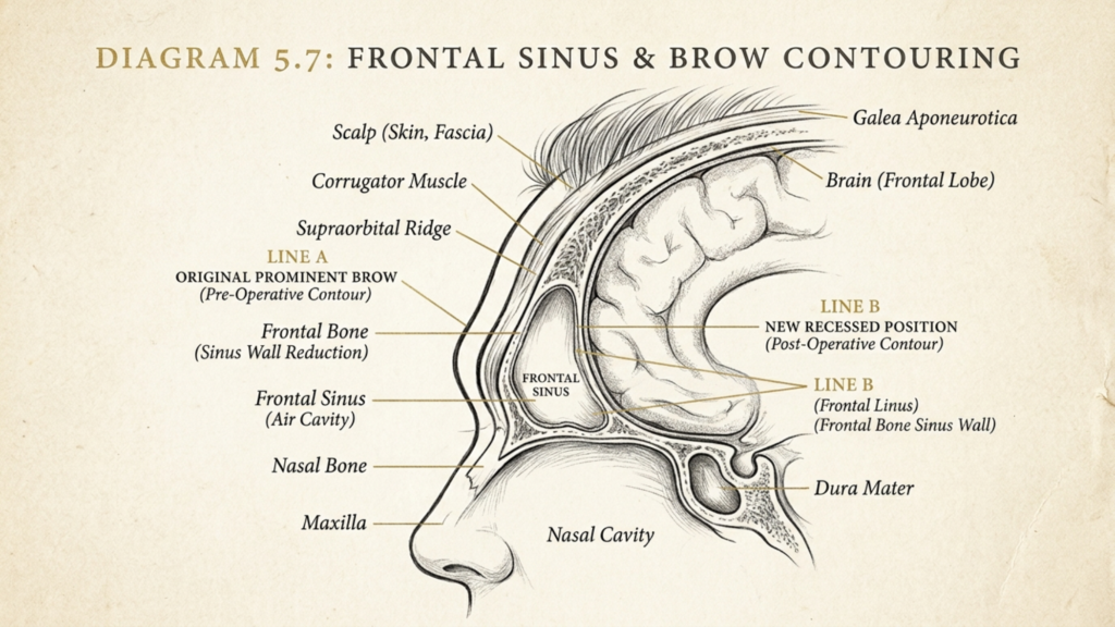
Digitare 3 Riduzione della fronte, conosciuto anche come arretramento del seno frontale O cranioplastica di tipo 3, si distingue per la sua attenzione al parete anteriore del seno frontale. A differenza delle procedure di tipo 1 o di tipo 2, che prevedono la rasatura o il rimodellamento parziale dell'osso, il tipo 3 richiede la rimozione completa, rimodellamento e riposizionamento della parete anteriore. Questa tecnica è particolarmente adatta a individui con arcate sopracciliari prominenti e seni frontali profondi, dove metodi più semplici sarebbero insufficienti o rischierebbero di esporre il seno (Mittermiller, 2025).
IL distinzione fondamentale della tipologia 3 risiede nella sua capacità di raggiungere un femminilizzazione drammatica della fronte sfruttando il riposizionamento della parete anteriore. Questo approccio non solo riduce l'arcata sopracciliare, ma crea anche un contorno della fronte più liscio e armonioso, in linea con l'estetica del viso femminile.
Procedura chirurgica passo passo: rimozione, rimodellamento e riposizionamento della parete anteriore
La procedura di riduzione della fronte di tipo 3 è una processo in più fasi che richiede precisione, competenza anatomica e strumenti chirurgici avanzati. Di seguito è riportata una descrizione dettagliata di ciascuna fase:
1. Pianificazione preoperatoria e diagnostica per immagini
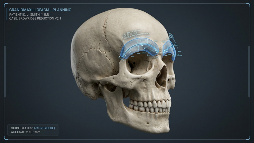
Prima dell'intervento chirurgico, un valutazione completa viene condotto utilizzando scansioni TC 3D e software di pianificazione chirurgica virtuale. Questo passaggio è fondamentale per:
- Misurazione delle dimensioni del seno frontale per determinare l'entità della rimozione ossea e del rimodellamento necessari.
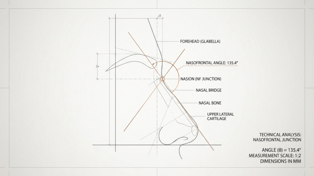
- Visualizzazione della giunzione nasofrontale per evitare complicazioni come perdite di liquido cerebrospinale (CSF).
- Progettazione di guide di taglio specifiche per il paziente per garantire la precisione durante l'osteotomia (Narrative Review of Facial Gender Surgery, 2025).
2. Incisione ed esposizione
Il chirurgo esegue un incisione coronale (lungo l'attaccatura dei capelli o all'interno del cuoio capelluto) per accedere all'osso frontale. Questa incisione è posizionata strategicamente per:
- Ridurre al minimo le cicatrici visibili nascondendolo tra i capelli.
- Preservare il pericranio (la membrana che ricopre l'osso) per mantenere l'afflusso di sangue e ridurre il rischio di infezione.
3. Osteotomia: rimozione della parete anteriore
Utilizzando strumenti chirurgici di precisione, il chirurgo esegue un'osteotomia per rimuovere la parete anteriore del seno frontale. Questo passaggio prevede:
- Tagliare l'osso seguendo linee prestabilite per creare un "lembo osseo".“
- Sollevare con cautela il segmento osseo per evitare di danneggiare il seno frontale o le strutture circostanti.
- Rimozione di eventuali setti ossei all'interno del seno per garantire un contorno interno liscio (Femminilizzazione della fronte, 2024).
4. Rimodelamento del lembo osseo
Una volta rimosso, il lembo osseo è rimodellato su un campo sterile. Ciò implica:
- Fresatura della superficie interna per ridurne la sporgenza e creare un contorno più armonioso.
- Regolare la forma per adattarsi all'estetica femminile desiderata, spesso utilizzando piastre o viti in titanio per la stabilizzazione.
- Garantire la simmetria con il resto della struttura facciale (Ricostruzione Onlay Multi-fragment in Contorno della fronte Chirurgia, 2025).
5. Riposizionamento e fissaggio
Il lembo osseo rimodellato è riposizionato in una posizione più posteriore e femminilizzata. Questo passaggio include:
- Fissare l'osso con piastre e viti in titanio per garantire stabilità e una corretta guarigione.
- Chiusura dell'incisione con suture riassorbibili per ridurre al minimo le cicatrici.
- Applicazione di una medicazione compressiva per ridurre il gonfiore e sostenere il nuovo contorno.
Considerazioni anatomiche: il seno frontale e le strutture circostanti
IL seno frontale è una struttura anatomica critica nella riduzione della fronte di tipo 3. La sua dimensione, profondità e relazione Le condizioni delle ossa e dei tessuti circostanti influenzano direttamente l'approccio chirurgico e i potenziali rischi. Le principali considerazioni anatomiche includono:
1. Anatomia del seno frontale
Il seno frontale è un cavità vuota piena d'aria Situato all'interno dell'osso frontale. La sua parete anteriore forma l'arcata sopracciliare, mentre la parete posteriore confina con la fossa cranica anteriore. Le caratteristiche principali includono:
- Variabilità di dimensioni e formeLa posizione del seno mascellare può variare da superficiale a profondamente incavata, influenzando la complessità della procedura.
- Spessore della parete anterioreUn osso più spesso può richiedere un rimodellamento più aggressivo, mentre un osso più sottile aumenta il rischio di esposizione dei seni paranasali.
- Presenza di setti osseiLe divisioni interne all'interno del seno mascellare possono complicare il rimodellamento e richiedono un'attenta rimozione.

2. Relazione con la giunzione nasofrontale
IL giunzione nasofrontale È il punto in cui il seno frontale incontra le ossa nasali. Questa zona è fondamentale perché:
- Rimodelamento improprio possono ostruire le vie aeree nasali o alterare la simmetria facciale.
- Resezione eccessiva può portare a un aspetto "incavato" o compromettere la funzionalità dei seni paranasali.
- Misurazioni precise sono essenziali per mantenere una transizione naturale tra la fronte e il ponte nasale (arretramento del seno frontale in Femminilizzazione facciale Chirurgia, 2025).
3. Tessuti molli e nervi circostanti
Il seno frontale è circondato da tessuti molli, muscoli e nervi che devono essere preservate durante l'intervento chirurgico. Le strutture chiave includono:
- Nervi sovratrocleare e sovraorbitaleResponsabile della sensibilità nella fronte e nel cuoio capelluto. Un danno può causare intorpidimento o dolore cronico.
- muscolo frontaleSolleva le sopracciglia e contribuisce all'espressività del viso. Un'eccessiva dissezione può portare alla ptosi (abbassamento) delle sopracciglia.
- Pericranio: Una membrana vascolare che nutre il lembo osseo. Preservarla è fondamentale per la guarigione e per ridurre i rischi di infezione.
Rischi e complicazioni potenziali: cosa devono sapere i pazienti
Sebbene la riduzione della fronte di tipo 3 sia altamente efficace, non è priva di rischi. Comprendere queste potenziali complicazioni è essenziale per processo decisionale informato e cure post-operatorie. Di seguito sono elencati i rischi più comuni:
1. Perdita di liquido cerebrospinale (CSF)
Si verifica una perdita di liquido cerebrospinale se parete posteriore del seno frontale si verifica una lesione accidentale durante l'osteotomia. Questa complicanza è grave perché:
- Può portare a infezioni come la meningite se non curata tempestivamente.
- I sintomi includono Fuoriuscita di liquido trasparente dal naso o dalla sede dell'incisione, mal di testa e nausea.
- Il trattamento comporta Riparazione chirurgica con innesto durale ed eventuale posizionamento di drenaggio lombare (Ricostruzione della fronte di tipo 1, 2 o 3, 2025).
2. Infezione e riassorbimento osseo
Le infezioni possono verificarsi nel sito chirurgico, in particolare se il pericranio è danneggiato oppure vengono utilizzati materiali estranei (ad esempio, placche di titanio). I rischi includono:
- riassorbimento osseoIl corpo potrebbe riassorbire il lembo osseo rimodellato, causando asimmetria o irregolarità del contorno.
- sinusite cronicaSe la mucosa dei seni paranasali viene danneggiata, può verificarsi un'infiammazione cronica.
- Complicazioni hardware: Le placche o le viti possono diventare palpabili o infette, richiedendo la rimozione (Ricostruzione Onlay Multi-fragment in Contorno della fronte Chirurgia, 2025).
3. Alterazioni sensoriali e danni ai nervi
Danni al nervi sovratrocleare o sovraorbitale può provocare alterazioni sensoriali temporanee o permanenti. I pazienti possono manifestare:
- Intorpidimento o formicolio sulla fronte o sul cuoio capelluto.
- Dolore cronico a causa di irritazione o cicatrizzazione dei nervi.
- Espressioni facciali alterate se il muscolo frontale è interessato.
4. Irregolarità e asimmetria del contorno
Anche con una pianificazione meticolosa, asimmetria o contorni irregolari può verificarsi a causa di:
- Rimodellamento osseo irregolare durante la procedura.
- gonfiore post-operatorio che si risolve in modo non uniforme.
- visibilità dell'hardware se le placche o le viti non sono posizionate a filo con l'osso.
5. Risultati funzionali ed estetici a lungo termine
Sebbene la riduzione della fronte di tipo 3 sia progettata per favorire la femminilizzazione, i risultati a lungo termine dipendono da:
- Corretta guarigione ossea: Garantire che l'osso rimodellato si integri armoniosamente con le strutture circostanti.
- Gestione delle cicatriciRiduzione al minimo delle cicatrici visibili grazie a un'attenta pianificazione dell'incisione e alla cura post-operatoria.
- Soddisfazione del pazienteAllineare le aspettative a risultati realistici, poiché l'anatomia individuale varia.

Assistenza post-operatoria e recupero: garantire una guarigione ottimale
Il recupero dalla riduzione della fronte di tipo 3 è un processo graduale Ciò richiede pazienza e il rispetto delle istruzioni post-operatorie. Di seguito sono riportati i passaggi chiave per favorire la guarigione e ridurre al minimo le complicazioni:
1. Cure postoperatorie immediate
Nelle prime 48 ore, concentrati su:
- Gestione del gonfiore con impacchi freddi e tenendo la testa sollevata.
- Assunzione di farmaci antidolorifici prescritti per prevenire il disagio.
- Evitare sforzi o sollevamenti pesanti per prevenire l'aumento della pressione intracranica.
2. Prime due settimane: monitoraggio e igiene
Durante questa fase:
- Mantenere la zona dell'incisione pulita e asciutta. per prevenire l'infezione.
- Partecipa agli appuntamenti di controllo. per monitorare la guarigione e affrontare eventuali problemi.
- Evitare di fumare e di consumare alcolici., poiché possono compromettere la guarigione e aumentare il rischio di infezioni.
3. Settimane 3-6: Ritorno graduale alle normali attività
Man mano che il gonfiore si riduce:
- Riprendere le attività leggere come camminare o lavorare alla scrivania.
- Evita gli sport di contatto o l'esercizio fisico intenso. fino a quando non riceverai l'autorizzazione dal chirurgo.
- Utilizzare gel o fogli di silicone per ridurre al minimo le cicatrici.
4. Assistenza a lungo termine: mantenimento dei risultati
Per garantire risultati duraturi:
- Proteggi la fronte dai traumi per evitare lo spostamento del lembo osseo.
- Effettua un controllo con il tuo chirurgo. annualmente per monitorare la stabilità ossea e affrontare eventuali complicazioni tardive.
- Valutare i miglioramenti non chirurgici come ad esempio filler o Botox per ridefinire ulteriormente i contorni.

Alternative alla riduzione della fronte di tipo 3: alla scoperta di opzioni meno invasive
Sebbene la riduzione della fronte di tipo 3 sia molto efficace, potrebbe non essere adatta a tutti. Le alternative includono:
1. Contouring della fronte di tipo 1 e tipo 2
Per le persone con una leggera sporgenza delle sopracciglia, Le procedure di tipo 1 o di tipo 2 possono essere sufficienti:
- Tipo 1: Coinvolge rasatura delle ossa senza rimuovere la parete anteriore. Ideale per riduzioni minori.
- Tipo 2: Combina osteotomia parziale con rimodellamento, adatto per sopracciglia moderatamente sporgenti.
2. Opzioni non chirurgiche
Per chi cerca tempi di inattività minimi, Le alternative non chirurgiche includono:
- Filler dermiciAumento temporaneo di volume per ammorbidire le arcate sopracciliari.
- Iniezioni di botulinoRilassare il muscolo frontale per ridurre la prominenza delle sopracciglia.
- Innesto di grassoUtilizza il tessuto adiposo del paziente stesso per rimodellare la fronte.
3. Procedure combinate
Per una femminilizzazione completa, la riduzione della fronte di tipo 3 può essere combinata con:
- Avanzamento dell'attaccatura dei capelli per ridurre l'altezza della fronte.
- Rinoplastica per affinare i contorni del naso.
- Sollevamento della fronte per sollevare le sopracciglia e ottenere un aspetto più giovane.
Conclusione: trovare un equilibrio tra estetica, sicurezza e obiettivi del paziente.
La riduzione della fronte di tipo 3 è una strumento potente nella chirurgia di femminilizzazione facciale, offrendo risultati trasformativi per le donne transgender che cercano un contorno della fronte più femminile. Tuttavia, la sua complessità richiede competenza, precisione e una conoscenza approfondita dell'anatomia per mitigare rischi quali perdite di liquido cerebrospinale, infezioni e irregolarità del profilo.
Per i pazienti, la decisione di sottoporsi a una riduzione della fronte di tipo 3 dovrebbe essere presa in consultazione con un chirurgo abilitato che è specializzato in FFS. Valutando i benefici rispetto ai rischi ed esplorando le alternative, gli individui possono raggiungere un equilibrio armonioso tra estetica e sicurezza, rafforzando in definitiva la loro fiducia e la coerenza con la propria identità di genere.
Per coloro che stanno valutando questa procedura, Clinica del dottor MFO Offre competenze specialistiche nella riduzione della fronte di tipo 3, garantendo cure personalizzate e risultati ottimali. Contattaci oggi per fissare un appuntamento e iniziare il tuo percorso verso un aspetto più femminile.
Domande frequenti
Qual è la differenza tra la riduzione della fronte di tipo 1, di tipo 2 e di tipo 3?
Il tipo 1 prevede la limatura ossea per riduzioni minori, il tipo 2 combina l'osteotomia parziale con il rimodellamento per un moderato sviluppo delle arcate sopracciliari, e il tipo 3 richiede la rimozione completa, il rimodellamento e il riposizionamento della parete anteriore del seno frontale per una significativa femminilizzazione. Il tipo 3 è il più complesso ed è riservato a individui con arcate sopracciliari prominenti e seni frontali profondi.
Quanto tempo ci vuole per riprendersi dopo una riduzione della fronte di tipo 3?
Il tempo di recupero varia da persona a persona, ma generalmente segue questa tempistica: 1-2 settimane per il gonfiore e il fastidio iniziali, 3-6 settimane per un graduale ritorno alle normali attività e fino a 6 mesi per i risultati definitivi, quando il gonfiore si riduce completamente. I pazienti dovrebbero evitare attività faticose per almeno 6 settimane per garantire una corretta guarigione.
Quali sono i segni di una perdita di liquido cerebrospinale dopo una riduzione della fronte di tipo 3?
I sintomi di una perdita di liquido cerebrospinale includono la fuoriuscita di liquido trasparente dal naso o dalla sede dell'incisione, mal di testa persistente, nausea e un sapore salato in bocca. Se si manifesta uno qualsiasi di questi sintomi, consultare immediatamente un medico, poiché le perdite di liquido cerebrospinale non trattate possono portare a gravi infezioni come la meningite.
È possibile combinare la riduzione della fronte di tipo 3 con altre procedure di chirurgia di femminilizzazione del viso?
Sì, la riduzione della fronte di tipo 3 viene spesso combinata con procedure come l'avanzamento dell'attaccatura dei capelli, rinoplastica, e lifting delle sopracciglia per ottenere una femminilizzazione completa del viso. La combinazione di diverse procedure può migliorare l'armonia generale e ridurre la necessità di interventi chirurgici multipli.
Quali sono i rischi dell'utilizzo di placche in titanio nella riduzione della fronte di tipo 3?
Sebbene le placche in titanio offrano stabilità, i rischi includono la palpabilità del materiale (la presenza delle placche sotto la pelle), l'infezione e il riassorbimento osseo intorno alle placche. In rari casi, potrebbe essere necessario rimuovere le placche se causano fastidio o complicazioni.
Come posso ridurre al minimo le cicatrici dopo una riduzione della fronte di tipo 3?
Per ridurre al minimo le cicatrici, segui le istruzioni del chirurgo per la cura post-operatoria, che possono includere l'uso di gel o fogli di silicone, evitare l'esposizione al sole e mantenere la zona dell'incisione pulita e idratata. La maggior parte delle cicatrici si attenua notevolmente nel tempo, soprattutto se nascoste tra i capelli.
L'intervento di riduzione della fronte di tipo 3 è adatto a tutti?
La riduzione della fronte di tipo 3 è ideale per le persone con arcate sopracciliari prominenti e seni frontali profondi. Chi presenta un'arcata sopracciliare più lieve può ottenere risultati soddisfacenti con le procedure di tipo 1 o di tipo 2. È consigliabile consultare uno specialista. Chirurgo FFS è essenziale determinare l'approccio migliore in base alla tua anatomia e ai tuoi obiettivi.
Cosa devo aspettarmi durante la visita per la riduzione della fronte di tipo 3?
Durante la visita, il chirurgo esaminerà la tua anamnesi, eseguirà un esame obiettivo e utilizzerà la tecnologia di imaging 3D per valutare l'anatomia del tuo seno frontale. Discuterà con te le fasi della procedura, i rischi e i risultati attesi, nonché le alternative, come le opzioni non chirurgiche o le tecniche chirurgiche meno invasive.







