
Forehead reconstruction is a critical component of Gezichtsfeminisering Chirurgie (FFS) and craniofacial procedures, aiming to achieve a harmonious and aesthetically pleasing facial contour. Among the most debated techniques are Type 1 En Type 3 cranioplastie, each offering distinct approaches to reshaping the forehead. While Type 1 involves bone burring or shaving, Soort 3 requires a more complex osteotomie en tegenslag of the frontal sinus. Understanding the anatomical, functional, and aesthetic implications of these techniques is essential for both surgeons and patients to make informed decisions.
This guide explores the structural differences between Type 1 and Type 3 cranioplasty, their impact on bone thickness, frontal sinus anatomy, and how surgeons determine the most suitable approach for each patient. By the end, you will gain clarity on which technique aligns best with your anatomical needs and aesthetic goals.
Inhoudsopgave
De anatomische basis: het voorhoofdsbeen en de sinussen
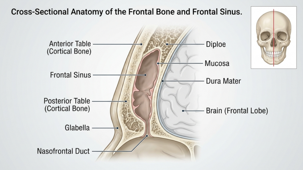
De voorhoofdsbeen En frontale sinus play a pivotal role in forehead reconstruction. The frontal bone forms the upper part of the face and houses the frontal sinus, a hollow cavity that varies in size and shape among individuals. The anterior table of the frontal sinus is the outer layer of bone that contributes to the forehead’s contour, while the posterior table separates the sinus from the brain. The thickness of these tables and the degree of sinus pneumatization (air-filled expansion) influence the choice between Type 1 and Type 3 cranioplasty.
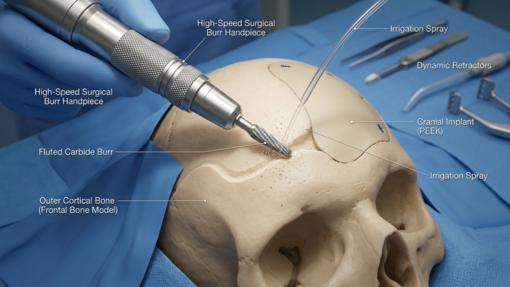
In Type 1 cranioplasty, de chirurg uses a high-speed burr to shave down the outer cortical bone, reducing prominence without violating the frontal sinus. This technique is ideal for patients with thin frontal bone or minimal bossing, as it preserves the sinus’s integrity. However, it may not be sufficient for patients with significant brow bossing or a thick anterior table, as excessive burring can compromise bone stability or fail to achieve the desired contour (Ousterhout, 2024).
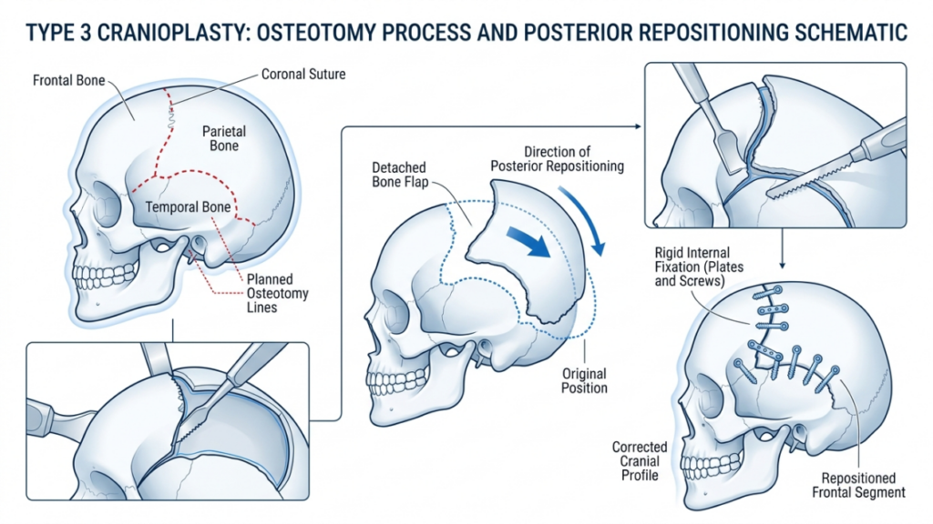
Daarentegen, Type 3 cranioplastie involves an osteotomie—a controlled cut through the anterior table of the frontal sinus. The bone segment is then repositioned backward (setback) to reduce projection and create a smoother, more feminine forehead. This technique is reserved for patients with moderate to severe brow bossing or a thick anterior table, where burring alone would be inadequate. The osteotomy allows for precise reshaping while maintaining the sinus’s protective function (Feminization of the Forehead: A Scoping Literature Review, 2024).
Cranioplastiek type 1: afschaven en frezen
Techniekoverzicht
Type 1 cranioplasty is the least invasive option for forehead reconstruction. It involves using a high-speed burr to gradually reduce the prominence of the frontal bone. The chirurg meticulously shaves the outer cortical layer, avoiding penetration into the frontal sinus. This technique is particularly effective for patients with:
- Mild to moderate brow bossing
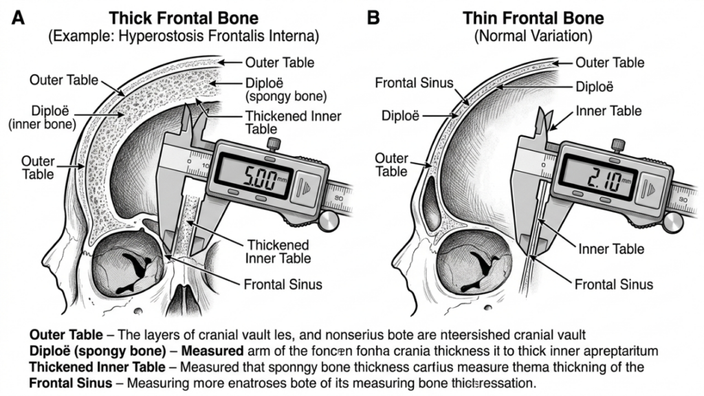
- Thin frontal bone (less than 5 mm)
- Absence of significant frontal sinus pneumatization
The procedure is performed through a coronale insnijding, which allows access to the forehead while minimizing visible scarring. The surgeon uses tactile feedback and visual cues to ensure uniform reduction without over-thinning the bone, which could lead to instability or contour irregularities.
Voordelen van cranioplastie type 1
Type 1 cranioplasty offers several benefits:
- Minimal Invasiveness: No osteotomy or bone removal reduces surgical trauma and recovery time.
- Lager risico op complicaties: Preserving the frontal sinus minimizes the risk of sinusitis, cerebrospinal fluid leaks, or mucocele formation.
- Kortere operatietijd: The procedure typically takes 1–2 hours, making it a quicker option compared to Type 3.
- Predictable Results: Ideal for patients with mild bossing, where subtle contouring is sufficient to achieve a feminine appearance.
Beperkingen van cranioplastie type 1
While Type 1 cranioplasty is safer and less invasive, it has notable limitations:
- Limited Reduction: Insufficient for patients with severe brow bossing or thick frontal bones.
- Risk of Over-Thinning: Aggressive burring can weaken the bone, leading to contour irregularities or fractures.
- Onvolledige feminisering: May not achieve the desired aesthetic outcome for patients with pronounced masculine features.
Cranioplastiek type 3: Osteotomie en terugplaatsing
Techniekoverzicht
Type 3 cranioplasty is a more complex procedure designed for patients with moderate to severe brow bossing or thick frontal bones. It involves an osteotomie—a precise cut through the anterior table of the frontal sinus—followed by repositioning the bone segment backward (setback). This technique allows for significant reduction in forehead projection and a smoother, more feminine contour.
The procedure is typically performed through a coronale insnijding, providing access to the frontal bone and sinus. The surgeon uses a sagittal saw or piezoelectric device to create the osteotomy, ensuring the cut follows the natural curvature of the forehead. The bone segment is then repositioned and secured with titanium platen en schroeven or resorbable sutures. This technique is ideal for patients with:
- Severe brow bossing
- Thick frontal bone (greater than 5 mm)
- Significant frontal sinus pneumatization
Type 3 cranioplasty requires meticulous planning, often involving 3D virtuele chirurgische planning (VSP) to simulate the osteotomy and setback. This ensures precision and minimizes the risk of complications such as sinus violation or cerebrospinal fluid leaks (Virtual Surgical Planning in Gezichtsfeminisering of the Upper Face, 2025).
Voordelen van cranioplastie type 3
Type 3 cranioplasty offers several advantages for patients with pronounced masculine features:
- Significant Contouring: Achieves dramatic reduction in brow bossing, creating a smoother, more feminine forehead.
- Veelzijdigheid: Suitable for patients with thick frontal bones or extensive sinus pneumatization.
- Stabiliteit op lange termijn: The repositioned bone segment integrates well, reducing the risk of contour irregularities over time.
- Aanpassing: Virtual surgical planning allows for precise, patient-specific adjustments to achieve optimal results.
Beperkingen van cranioplastie type 3
Despite its effectiveness, Type 3 cranioplasty carries higher risks and complexities:
- Increased Surgical Time: The procedure typically takes 3–5 hours, requiring greater precision and expertise.
- Hoger risico op complicaties: Potential risks include sinusitis, cerebrospinal fluid leaks, or mucocele formation if the sinus is violated.
- Langer herstel: Patients may experience prolonged swelling and discomfort compared to Type 1 cranioplasty.
- Kosten: The use of advanced imaging and surgical tools increases the overall cost of the procedure.

Belangrijkste verschillen tussen cranioplastiek type 1 en type 3
| Functie | Cranioplastie type 1 | Type 3 Cranioplastie |
|---|---|---|
| Techniek | Bone burring/shaving | Osteotomy and setback |
| Invasiviteit | Minimaal | Matig tot hoog |
| Ideale kandidaten | Mild to moderate brow bossing, thin frontal bone | Severe brow bossing, thick frontal bone |
| Operatietijd | 1–2 hours | 3-5 uur |
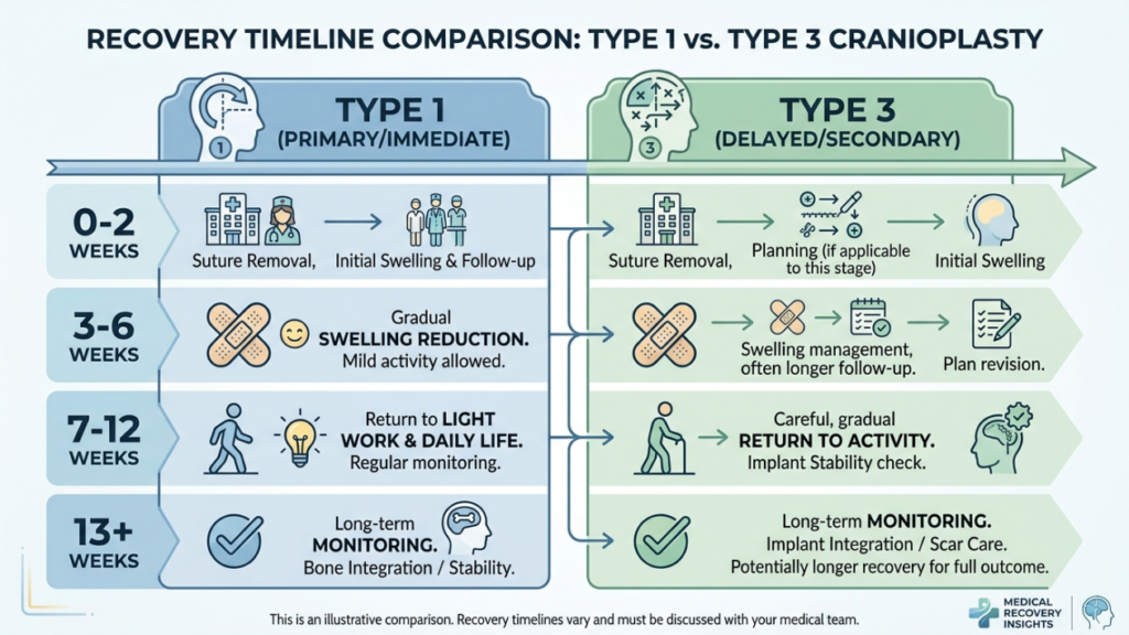
| Hersteltijd | 1–2 weken | 3-6 weken |
| Risk of Complications | Laag | Matig tot hoog |
| Kosten | Onder | Hoger |
| Esthetisch resultaat | Subtle contouring | Dramatic feminization |
Hoe chirurgen beslissen: Type 1 versus Type 3
The choice between Type 1 and Type 3 cranioplasty depends on several factors, including the patient’s anatomical features, aesthetic goals, En surgical risks. Surgeons rely on a combination of clinical examination, 3D-beeldvorming, En patient consultation to determine the most appropriate technique.
1. Anatomische beoordeling
The first step is evaluating the patient’s frontal bone thickness En frontal sinus anatomy. A CT scan of 3D-reconstructie provides detailed insights into:
- Botdikte: Patients with thin frontal bones (less than 5 mm) are better suited for Type 1 cranioplasty, while those with thicker bones may require Type 3.
- Sinus Pneumatization: Extensive sinus pneumatization may necessitate Type 3 cranioplasty to avoid violating the sinus during burring.
- Degree of Bossing: Severe brow bossing often requires the dramatic reduction achievable only with Type 3 techniques.
2. Esthetische doelen
Patients’ aesthetic expectations play a crucial role in technique selection. Those seeking subtle feminization may opt for Type 1 cranioplasty, while individuals with pronounced masculine features often require the transformative results of Type 3. Surgeons discuss realistic outcomes based on the patient’s anatomy and desired changes.
3. Chirurgische risico's en herstel
Type 3 cranioplasty carries higher risks, including sinus complications, cerebrospinal fluid leaks, En prolonged recovery. Surgeons assess the patient’s overall health, tolerance for surgery, and willingness to adhere to postoperative care. Patients with medical conditions that increase surgical risks may be advised to consider Type 1 or alternative procedures.
4. Virtuele chirurgische planning (VSP)
Advancements in 3D virtual surgical planning have revolutionized cranioplasty. Surgeons use VSP to simulate osteotomies, setbacks, and outcomes, ensuring precision and minimizing risks. This technology is particularly valuable for Type 3 cranioplasty, where accurate bone repositioning is critical (3D Printing and Virtual Surgical Planning in Craniofacial and Orthognathic Surgery, 2025).
Postoperatieve zorg en herstel
Recovery varies significantly between Type 1 and Type 3 cranioplasty. Understanding the postoperative process helps patients prepare for a smooth healing journey.
Herstel na cranioplastie type 1
Patients undergoing Type 1 cranioplasty typically experience:
- Mild to Moderate Swelling: Resolves within 1–2 weeks.
- Minimal Discomfort: Managed with over-the-counter pain medications.
- Snelle terugkeer naar activiteiten: Most patients resume normal activities within 2 weeks.
Herstel na cranioplastie type 3
Recovery from Type 3 cranioplasty is more involved due to the complexity of the procedure:
- Significante zwelling en kneuzing: May persist for 3–4 weeks.
- Moderate Pain: Prescription pain medications may be required for the first week.
- Geleidelijke terugkeer naar activiteiten: Strenuous activities are restricted for 4–6 weeks.
- Vervolgbezoeken: Regular monitoring to ensure proper healing and address any complications.
Both techniques require patients to avoid heavy lifting, zware inspanning, En direct pressure on the forehead during the initial recovery phase. Surgeons provide detailed postoperative instructions, including wound care, activity restrictions, and signs of complications to watch for.
Mogelijke complicaties en hoe u ze kunt vermijden
While cranioplasty is generally safe, complications can arise. Awareness of these risks and preventive measures is crucial for both surgeons and patients.
Veelvoorkomende complicaties
- Sinusitis: Inflammation or infection of the frontal sinus, particularly in Type 3 cranioplasty if the sinus is violated.
- Lekkage van hersenvocht (CSF): Rare but serious complication if the posterior table of the sinus is breached.
- Onregelmatigheden in contouren: Over-thinning of bone in Type 1 or improper setback in Type 3 can lead to asymmetry or visible ridges.
- Mucocele-vorming: Blockage of sinus drainage pathways can result in mucus-filled cysts.
- Infectie: Risk is higher in Type 3 due to the longer operative time and use of implants.
Preventieve maatregelen
Surgeons employ several strategies to minimize complications:
- Nauwkeurige chirurgische planning: 3D imaging and virtual surgical planning ensure accurate osteotomies and setbacks.
- Sinus Preservation: Avoiding violation of the frontal sinus during burring or osteotomy.
- Antibiotic Prophylaxis: Administered pre- and postoperatively to reduce infection risks.
- Postoperatieve monitoring: Regular follow-ups to detect early signs of complications.
- Patiënteneducatie: Instructing patients on proper wound care, activity restrictions, and warning signs of complications.
Alternatieven voor cranioplastiek type 1 en type 3
For patients who are not ideal candidates for Type 1 or Type 3 cranioplasty, alternative techniques may be considered:
- Vettransplantatie: Autologous fat transfer can soften forehead contours without altering bone structure. This is ideal for patients with mild irregularities or those seeking non-surgical options.
- Custom Implants: Pre-fabricated implants, such as PEEK (polyether ether ketone) of titanium, can be used to augment or reshape the forehead without osteotomy. These are particularly useful for patients with thin bones or sinus complications.
- Orthognatische chirurgie: In gevallen waarin voorhoofd contouren is part of a broader facial feminization plan, orthognathic procedures (e.g., Le Fort I osteotomy) may be combined to address midface and jaw alignment.
- Endoscopic Techniques: Minimally invasive endoscopic approaches can reduce brow bossing with smaller incisions and faster recovery times.
Patiëntgetuigenissen en resultaten uit de praktijk
Real-world outcomes provide valuable insights into the effectiveness and satisfaction rates of Type 1 and Type 3 cranioplasty. Patient testimonials highlight the transformative impact of these procedures:
Case Study 1: Type 1 Cranioplasty
A 28-year-old transgender woman sought subtle feminization of her forehead. With a thin frontal bone and minimal bossing, she opted for Type 1 cranioplasty. The procedure achieved a smoother contour with minimal downtime. She reported high satisfaction, noting that the results aligned with her expectations for a natural, feminine appearance.
Case Study 2: Type 3 Cranioplasty
A 35-year-old transgender woman presented with severe brow bossing and a thick frontal bone. Type 3 cranioplasty was performed, involving osteotomy and setback. The dramatic reduction in forehead projection significantly feminized her facial features. While recovery took longer, she expressed immense satisfaction with the results, stating that the procedure “changed her life.”
Case Study 3: Revision Surgery
A 40-year-old patient initially underwent Type 1 cranioplasty but was dissatisfied with the subtle results. She later opted for Type 3 cranioplasty to achieve more dramatic feminization. The revision surgery successfully addressed her concerns, demonstrating the importance of selecting the right technique based on anatomical needs and aesthetic goals.
De rol van virtuele chirurgische planning bij cranioplastiek
Virtual surgical planning (VSP) has become a game-changer in cranioplasty, particularly for Type 3 procedures. VSP allows surgeons to:
- Simulate Osteotomies: Precisely plan bone cuts and repositioning to achieve optimal contouring.
- Predict Outcomes: Visualize postoperative results and adjust the surgical plan accordingly.
- Minimaliseer risico's: Avoid critical structures such as the frontal sinus and supraorbital nerves.
- Enhance Communication: Share 3D models with patients to set realistic expectations and improve informed consent.
Studies have shown that VSP reduces operative time, improves accuracy, and enhances patient satisfaction (3D Printing and Virtual Surgical Planning in Craniofacial and Orthognathic Surgery, 2025). It is now considered the gold standard for complex cranioplasty procedures.

Veelgestelde vragen
Wat is het belangrijkste verschil tussen een cranioplastie van type 1 en type 3?
Bij een cranioplastie type 1 wordt de buitenste laag van het voorhoofdsbeen afgeschaafd of afgevlakt om de prominentie te verminderen, terwijl bij een cranioplastie type 3 een osteotomie (botdoorsnijding) en terugplaatsing van het voorhoofdsbeensegment nodig zijn om een meer uitgesproken contour te bereiken. Type 1 is minder ingrijpend en geschikt voor milde tot matige bulten, terwijl type 3 is voorbehouden aan ernstige bulten of dikke voorhoofdsbeenderen.
Hoe maken chirurgen de keuze tussen een cranioplastie van type 1 en type 3?
Chirurgen beoordelen de dikte van het voorhoofdsbeen, de mate van wenkbrauwuitstulping en de anatomie van de voorhoofdsholten van de patiënt met behulp van CT-scans en 3D-beeldvorming. Type 1 wordt gekozen bij dunnere botten en een lichte uitstulping, terwijl Type 3 de voorkeur heeft bij dikkere botten, een ernstige uitstulping of uitgebreide pneumatisatie van de voorhoofdsholten. De doelen van de patiënt en de bereidheid tot het nemen van chirurgische risico's spelen ook een rol bij de beslissing.
Welke risico's zijn verbonden aan een cranioplastie type 3?
Een cranioplastie type 3 brengt vanwege de complexiteit hogere risico's met zich mee, waaronder sinusitis, lekkage van hersenvocht, contourafwijkingen, mucocelevorming en infectie. Deze risico's worden geminimaliseerd door nauwkeurige chirurgische planning, behoud van de sinussen en postoperatieve monitoring. Patiënten worden geïnformeerd over waarschuwingssignalen en nazorg om een voorspoedig herstel te garanderen.
Hoe lang duurt het herstel na een cranioplastie van type 1 vergeleken met type 3?
Het herstel na een cranioplastie van type 1 duurt doorgaans 1-2 weken, met lichte zwelling en ongemak. Een cranioplastie van type 3 vereist een langer herstel van 3-6 weken vanwege de uitgebreidere ingreep en mogelijke complicaties. Patiënten wordt geadviseerd zware inspanningen te vermijden en de postoperatieve instructies nauwgezet op te volgen.
Kan een cranioplastie van type 1 dezelfde resultaten opleveren als een cranioplastie van type 3?
Nee, een cranioplastie type 1 beperkt zich tot subtiele contourcorrecties en is ideaal bij een lichte tot matige bult op het voorhoofd. Cranioplastie type 3 bereikt een meer dramatische feminisering door het herpositioneren van het voorhoofdsbeensegment, waardoor het geschikt is voor een ernstige bult op het voorhoofd of een dik voorhoofdsbeen. De keuze hangt af van de anatomische behoeften en esthetische doelen van de patiënt.
Welke rol speelt virtuele chirurgische planning bij cranioplastiek?
Virtuele chirurgische planning (VSP) stelt chirurgen in staat osteotomieën te simuleren, de uitkomst te voorspellen en risico's te minimaliseren door kritieke structuren zoals de voorhoofdsholte te vermijden. Het verbetert de precisie, verkort de operatietijd en verhoogt de patiënttevredenheid door een duidelijke visualisatie van het chirurgische plan en de verwachte resultaten te bieden.
Zijn er niet-chirurgische alternatieven voor cranioplastiek om het voorhoofd te feminiseren?
Yes, non-surgical alternatives include vet enten to soften contours and custom implants (e.g., PEEK or titanium) to reshape the forehead without osteotomy. These options are ideal for patients with mild irregularities or those who prefer to avoid surgery. However, they may not achieve the same level of feminization as surgical techniques.
Wat kan ik verwachten tijdens het consult voor een voorhoofdreconstructie?
Tijdens het consult beoordeelt uw chirurg de dikte van uw voorhoofdsbeen, de anatomie van uw sinussen en de mate van botuitstulping aan de hand van een klinisch onderzoek en 3D-beeldvorming. Hij of zij bespreekt uw esthetische wensen, legt de verschillen tussen cranioplastiek type 1 en type 3 uit en adviseert de meest geschikte techniek op basis van uw anatomie en verwachtingen.