Lograr una silueta de reloj de arena acentuada a menudo requiere enfocar el ancho del marco torácico, donde remodelación de costillas feminización del cuerpo representa la innovación quirúrgica mínimamente invasiva más avanzada para un estrechamiento drástico de la cintura. Las mujeres transgénero y cisgénero con cajas torácicas inferiores más anchas frecuentemente encuentran mesetas anatómicas con tejido blando. liposucción solos. Los pacientes que evalúan opciones integrales de modelado corporal pueden explorar nuestro programa especializado. cirugía de feminización corporal Procedimientos para comprender cómo se combina la remodelación esquelética con la lipoescultura de alta definición.
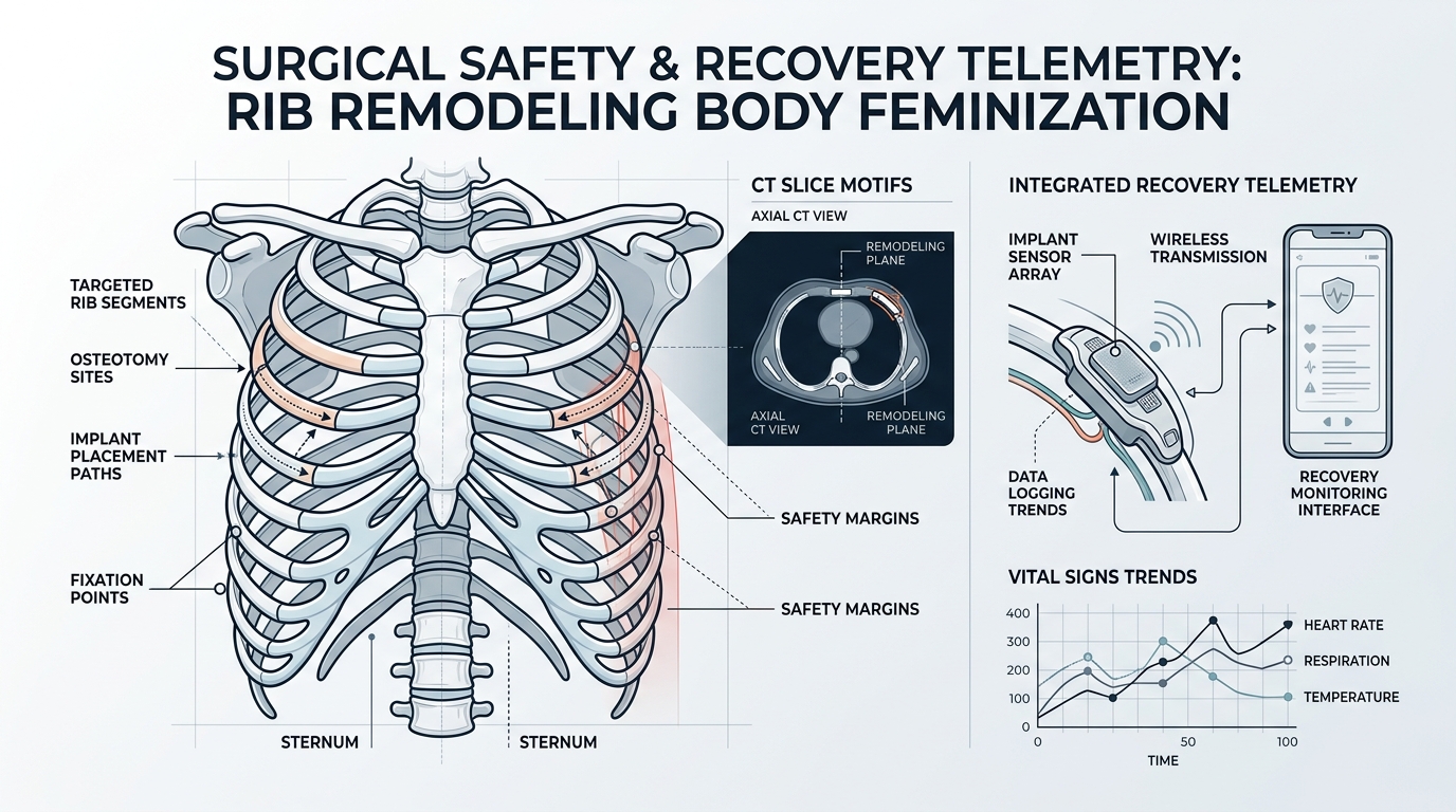
Ciencia biomecánica de la remodelación de las costillas y la feminización del cuerpo
La escultura corporal histórica se basaba en la resección radical de costillas, que eliminaba por completo segmentos de las costillas flotantes 11 y 12. En los protocolos contemporáneos de remodelación de costillas para la feminización corporal, los cirujanos utilizan la piezocirugía ultrasónica. La cavitación acústica ultrasónica permite el debilitamiento selectivo de la corteza sin dañar el paquete neurovascular adyacente ni la pleura parietal. Según los datos clínicos de seguridad reconstructiva catalogados por la Institutos Nacionales de Salud (NIH), La remodelación ósea por ultrasonidos minimiza significativamente la formación de hematomas periósticos y el traumatismo de los tejidos blandos.
Mediante osteotomías parciales (remodelación en tallo verde), los cartílagos costales pueden flexionarse hacia adentro entre 3 y 7 centímetros bilateralmente. Esto preserva la protección de los órganos vitales a la vez que crea una silueta lateral permanentemente cónica.
Telemetría para la seguridad quirúrgica y la recuperación: Remodelación de costillas y feminización del cuerpo
El análisis quirúrgico comparativo entre la resección tradicional y la remodelación costal ultrasónica para la feminización corporal pone de manifiesto ventajas clínicas convincentes:
Microincisiones: El acceso solo requiere pequeñas incisiones de punción dorsal de 1,5 cm, ocultas a lo largo de los pliegues naturales de la piel.
Integridad pleural: La preservación de la corteza costal interna reduce drásticamente el riesgo de neumotórax a menos de 0,2%.
Control del dolor: La integridad de las vías nerviosas intercostales permite a los pacientes movilizarse cómodamente dentro de las 24 horas posteriores a la cirugía.
Fase de consolidación: La compresión externa controlada estabiliza la unión ósea en un plazo de 60 a 90 días.
El cumplimiento postoperatorio es fundamental durante la remodelación de las costillas para lograr una feminización corporal. El uso de una faja ortopédica de compresión en la cintura garantiza que las costillas se recuperen en su contorno interno definido.
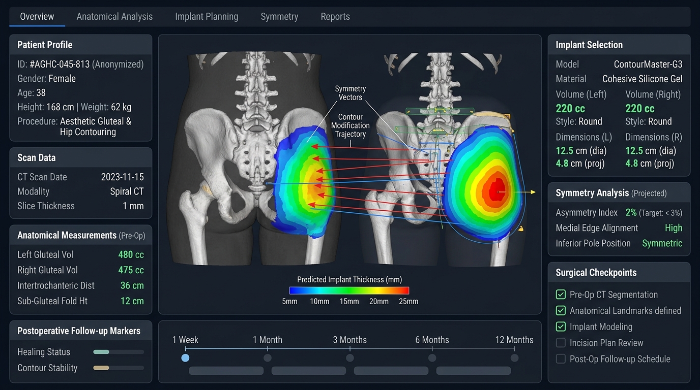
Integración estética con contorno de glúteos y caderas.
El verdadero poder de la remodelación de las costillas para la feminización del cuerpo reside en su efecto acumulativo sobre la relación cintura-cadera (RCC). Cuando se combina con la transferencia de grasa autóloga (BBL) y el relleno de la depresión lateral de la cadera, estrechando la caja torácica inferior, crea una transición óptica y volumétrica que el trabajo de tejidos blandos por sí solo no puede replicar. Los candidatos que consideren la remodelación de costillas para feminizar el cuerpo deben consultar con especialistas craneofaciales y corporales certificados para evaluar la anatomía costal mediante una tomografía computarizada de tórax de baja dosis.
Planificación de seguridad para la remodelación de costillas y la feminización del cuerpo
La remodelación costal para feminizar el cuerpo requiere una distinción precisa entre el refinamiento del contorno y la eliminación de hueso estructural. Un plan de remodelación conservador busca influir en la silueta torácica inferior, protegiendo la estabilidad de la pared torácica, la mecánica respiratoria, las estructuras intercostales y los tejidos blandos. La técnica adecuada depende de la anatomía costal, la flexibilidad del cartílago, cirugías previas, la composición corporal y la proporción deseada entre cintura y cadera.
La planificación preoperatoria debe documentar el historial médico de la paciente, su estado respiratorio, las cicatrices, la asimetría y sus expectativas. Es importante preguntar cómo se utilizarán las técnicas de imagen, cómo se controlará el dolor, qué restricciones de actividad se aplicarán y qué síntomas requerirán una revisión urgente. La recuperación varía y la hinchazón visible puede ocultar temporalmente la silueta final. La evaluación a largo plazo debe considerar la comodidad, la estabilidad del contorno, la respiración, la sensibilidad y si se están planificando procedimientos adicionales de feminización corporal.
La remodelación de las costillas y la feminización del cuerpo deben discutirse con un cirujano Con experiencia en anatomía de la pared torácica y cirugía de contorno de afirmación de género. El enfoque más amplio cirugía de feminización corporal Esta guía proporciona el contexto necesario para combinar procedimientos de cintura, cadera y tejidos blandos, sin considerar una sola medida como la única definición de un resultado exitoso.
¿Cuál es el objetivo de la remodelación de las costillas en la feminización del cuerpo?
El objetivo es lograr un refinamiento controlado del contorno, respetando la estabilidad de la pared torácica, la mecánica respiratoria, las estructuras intercostales y la envoltura de tejidos blandos.
¿La remodelación de las costillas es lo mismo que la extirpación de las mismas?
No. Las técnicas y el alcance del tratamiento varían. El enfoque exacto depende de la anatomía, la flexibilidad del cartílago, el historial médico y la silueta deseada.
¿Qué debería incluir la planificación de la recuperación?
La planificación de la recuperación debe abarcar el control del dolor, las restricciones de actividad, los síntomas respiratorios, la hinchazón, el cuidado de la herida, las señales de advertencia y el momento de la evaluación final del contorno.
Bichectomía, La extirpación quirúrgica de la almohadilla de grasa bucal para lograr un contorno facial más delgado ha aumentado su popularidad en 2026. Sin embargo, a pesar de su atractivo estético, este procedimiento conlleva riesgos significativos, en particular para la conducto parotídeo y ramas del nervio facial. Para las pacientes que consideran la bichectomía, comprender estos riesgos es fundamental para tomar una decisión informada. Este artículo explora los peligros anatómicos, las estadísticas de complicaciones y las estrategias basadas en la evidencia para mitigar los riesgos, garantizando así la seguridad y resultados óptimos.
Los peligros ocultos: riesgos para el conducto parotídeo y el nervio facial
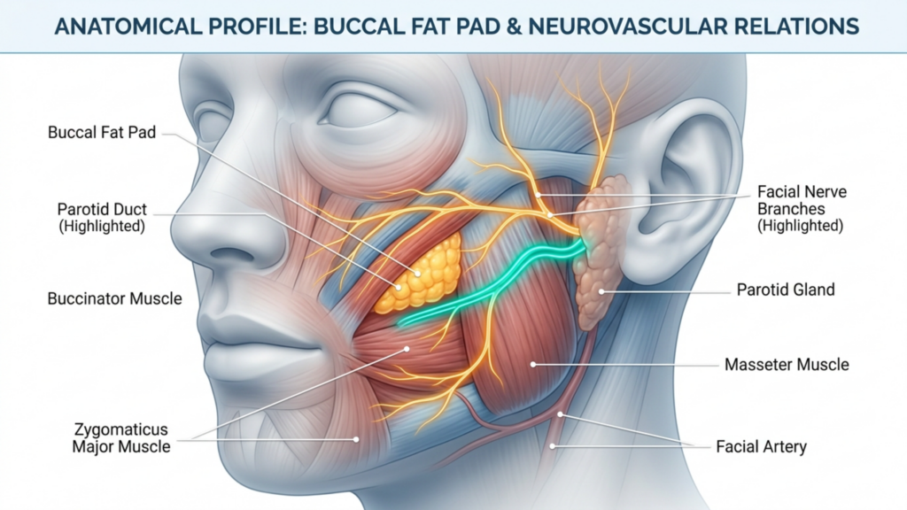
La almohadilla de grasa bucal, ubicada en la mejilla, se encuentra muy cerca de dos estructuras críticas: conducto parotídeo (conducto de Stensen) y el rama bucal del nervio facial. Los daños en cualquiera de ellos pueden provocar complicaciones graves:
Lesión del conducto parotídeo: El conducto parotídeo, responsable del drenaje de la saliva, es vulnerable durante la bichectomía. Las laceraciones o lesiones por aplastamiento de este conducto pueden provocar sialocele (recolección de saliva) o sialoadenitis (inflamación de la glándula), lo que provoca molestias crónicas y posibles infecciones. Los estudios indican que las lesiones del conducto parotídeo ocurren en 0,1% a 0,3% de cirugías faciales, pero el riesgo aumenta en manos inexpertas (International Journal of Surgery, 2025).
Daño del nervio facial: La rama bucal del nervio facial controla las expresiones faciales. Una lesión en este nervio puede causar parálisis temporal o permanente de los músculos periorales, lo que provoca asimetría, caída o dificultad en los movimientos faciales. La investigación destaca que lesiones del nervio bucal se encuentran entre las complicaciones más comunes de la bichectomía, con tasas que varían entre 1% y 5% dependiendo de la técnica quirúrgica (ScienceDirect, 2025).
Hematoma y asimetría: El sangrado postoperatorio puede provocar formación de hematoma, causando hinchazón, dolor y posible asimetría. Se han reportado hematomas en 8,45% a 18% de casos de bichectomía, a menudo debido a una compresión inadecuada o a un traumatismo vascular (Springer, 2025).
¿Por qué estas complicaciones suelen pasarse por alto?
La bichectomía se comercializa frecuentemente como una procedimiento sencillo y de bajo riesgo, Pero esta percepción es engañosa. He aquí por qué las complicaciones no se notifican lo suficiente:
Falta de informes estandarizados: Muchas complicaciones, como lesiones nerviosas menores o hematomas, se resuelven espontáneamente y no se documentan formalmente. Esto genera una falsa sensación de seguridad.
Cirujano Variabilidad de la experiencia: La bichectomía suele ser realizada por cirujanos con distintos niveles de experiencia. Los cirujanos inexpertos pueden subestimar la complejidad de la anatomía, lo que aumenta el riesgo de dañar los conductos o los nervios.
Sesgo en la selección de pacientes: Los candidatos ideales para la bichectomía son aquellos con almohadillas de grasa bucal moderadas y estructuras faciales simétricas. Sin embargo, el procedimiento se realiza cada vez más en pacientes con rostros delgados o asimetrías preexistentes, lo que agrava los riesgos.
Una revisión sistemática de 2025 publicada en Revista MedNEXT de Ciencias Médicas y de la Salud reveló que asimetría y hematoma son las complicaciones más frecuentemente reportadas, seguidas de lesiones nerviosas y daño al conducto parotídeo. El estudio enfatizó que Imágenes preoperatorias y planificación quirúrgica meticulosa son esenciales para minimizar estos riesgos.
Estrategias basadas en la evidencia para minimizar los riesgos
Para garantizar la seguridad del paciente y los resultados óptimos, los cirujanos deben adoptar un enfoque multidisciplinario Esto incluye la evaluación preoperatoria, la precisión intraoperatoria y los cuidados postoperatorios.
1. Evaluación preoperatoria
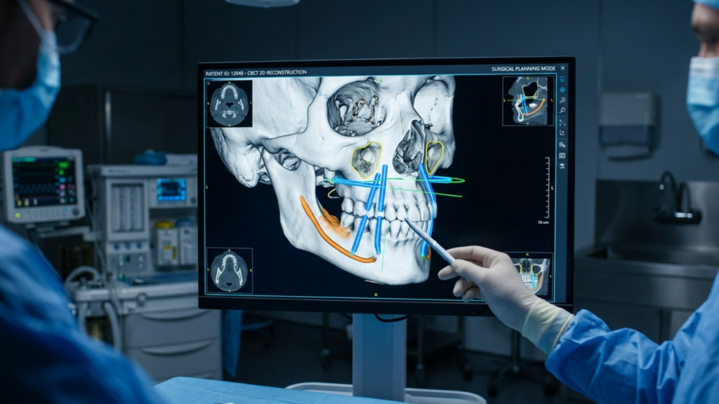
Imágenes 3D: Utilizar tomografía computarizada de haz cónico (CBCT) o resonancia magnética para mapear la almohadilla de grasa bucal, el conducto parotídeo y las ramas del nervio facial. Esto permite una planificación quirúrgica precisa y reduce el riesgo de lesiones no deseadas.
Estratificación del riesgo específica para cada paciente: Identificar pacientes de alto riesgo, como aquellos con asimetrías preexistentes, estructuras faciales delgadas o antecedentes de trastornos nerviosos. Estos pacientes pueden requerir procedimientos alternativos como injerto de grasa en lugar de bichectomía.
Consentimiento informado: Asegúrese de que los pacientes comprendan los riesgos de Lesión del conducto parotídeo, daño nervioso y asimetría.. Analizar la posibilidad de complicaciones temporales o permanentes.
2. Precisión intraoperatoria
Puntos de referencia anatómicos: Utilice el Conducto de Stensen y nervio bucal como guías durante la disección. El conducto se ubica típicamente 1–2 cm por debajo del arco cigomático, mientras que el nervio bucal corre paralelo a él.
Técnicas mínimamente invasivas: Optar por abordajes endoscópicos o intraorales para minimizar el traumatismo tisular y mejorar la visualización de las estructuras críticas.
Hemostasia: Lograr una hemostasia meticulosa para prevenir la formación de hematomas. cauterización bipolar y vendajes de compresión para controlar el sangrado.
3. Cuidados postoperatorios
Supervisión: Observe a los pacientes para detectar signos de disfunción nerviosa (asimetría, caída) o Lesión del conducto parotídeo (inflamación, fuga de saliva) en las primeras 48 horas.
Terapia de compresión: Aplicar prendas de compresión para reducir la hinchazón y prevenir la formación de hematomas.
Hacer un seguimiento: Programe visitas de seguimiento en 1 semana, 1 mes y 3 meses para evaluar la simetría, la función nerviosa y la integridad del conducto.
Alternativas a la bichectomía: opciones más seguras para el contorno facial.
Para pacientes con alto riesgo de complicaciones, existen procedimientos alternativos que pueden lograr objetivos estéticos similares sin los riesgos asociados:
Injerto de grasa: En lugar de eliminar la grasa, transferencia de grasa autóloga Puede realzar la definición de las mejillas preservando los contornos naturales y evitando lesiones en nervios o conductos.
Opciones no quirúrgicas:Tratamientos de radiofrecuencia o ultrasonido focalizado de alta intensidad (HIFU) Puede reducir temporalmente el volumen de las mejillas sin cirugía.
Procedimientos combinados: Para pacientes con hipoplasia del tercio medio facial, cirugía ortognática (Por ejemplo, la osteotomía Le Fort I) puede mejorar el equilibrio facial a la vez que aborda problemas funcionales como la permeabilidad de las vías respiratorias.
El papel de la experiencia del cirujano en la mitigación de riesgos
El éxito de la bichectomía depende de la habilidad del cirujano. Conocimientos anatómicos, habilidad técnica y cumplimiento de los protocolos de seguridad.. Cirujanos especializados en feminización facial cirugía (FFS) o procedimientos maxilofaciales A menudo, están mejor preparados para afrontar las complejidades de la bichectomía debido a su experiencia con estructuras faciales delicadas.
Dr. Mehmet Fatih Okyay, a Especialista en Cirugía Plástica Certificado por Juntas Europeas y Turcas, enfatiza que “La bichectomía no es un procedimiento que sirva para todos los casos. La selección del paciente, las pruebas de imagen preoperatorias y la precisión quirúrgica son imprescindibles para minimizar los riesgos.” Su clínica, Clínica Dr. MFO, emplea Planificación virtual 3D y monitorización nerviosa intraoperatoria para mejorar la seguridad y los resultados.
Conclusión: Equilibrio entre estética y seguridad
La bichectomía puede transformar la estética facial, pero sus riesgos, en particular para la conducto parotídeo y nervio facial—exigen una cuidadosa consideración. Los pacientes deben priorizar Experiencia del cirujano, planificación preoperatoria y cuidados postoperatorios. para mitigar las complicaciones. Para aquellos de alto riesgo, existen alternativas como injerto de grasa o contorno corporal no quirúrgico pueden ofrecer vías más seguras para lograr los resultados deseados.
Al adoptar una Enfoque centrado en el paciente y basado en la evidencia., Los objetivos estéticos de la bichectomía pueden armonizarse con la seguridad funcional y anatómica, garantizando tanto la belleza como el bienestar.
Preguntas frecuentes
¿Cuáles son las complicaciones más comunes de la bichectomía?
Las complicaciones más frecuentes incluyen hematoma (entre el 8,45% y el 18% de los casos), asimetría facial y lesiones en el conducto parotídeo o la rama bucal del nervio facial. Estos riesgos pueden minimizarse mediante técnicas quirúrgicas precisas y una planificación preoperatoria minuciosa.
¿Cómo puedo reducir el riesgo de daño nervioso durante una bichectomía?
Es fundamental elegir un cirujano con amplia experiencia en procedimientos faciales. Las imágenes 3D preoperatorias y la monitorización nerviosa intraoperatoria pueden reducir aún más los riesgos. Hable con su cirujano sobre sus inquietudes para garantizar medidas de seguridad personalizadas.
¿Qué debo hacer si experimento asimetría después de una bichectomía?
La asimetría postoperatoria puede resolverse a medida que disminuye la inflamación. Sin embargo, si persiste, consulte a su cirujano de inmediato. Es posible que se requiera una intervención temprana, como fisioterapia o cirugía de revisión, para corregir el problema.
¿Existen alternativas no quirúrgicas a la bichectomía?
Sí, existen alternativas como el injerto de grasa, los tratamientos de radiofrecuencia y el ultrasonido focalizado de alta intensidad (HIFU). Estas opciones pueden mejorar el contorno facial sin los riesgos asociados a la cirugía.
¿Cuánto tiempo se tarda en recuperarse de una bichectomía?
La recuperación suele durar de 1 a 2 semanas, y los resultados completos se aprecian entre 3 y 6 meses después. Siga las instrucciones de cuidados postoperatorios de su cirujano, incluyendo la terapia de compresión y las visitas de seguimiento, para asegurar una recuperación sin complicaciones.
¿Cuáles son los signos de lesión del conducto parotídeo después de una bichectomía?
Los síntomas incluyen hinchazón cerca de la mejilla o la mandíbula, secreción de saliva o sabor amargo en la boca. Si nota alguno de estos síntomas, comuníquese con su cirujano de inmediato para una evaluación y tratamiento.
¿Puede la bichectomía causar parálisis facial permanente?
Aunque es poco frecuente, puede producirse una parálisis permanente si la rama bucal del nervio facial sufre daños graves. Elegir un cirujano experto y seguir los protocolos de seguridad reduce significativamente este riesgo.
Si rotar la punta nasal hacia arriba fuera el único secreto para una nariz femenina, cada paciente que se sometiera a una rotación de punta de 105 grados saldría con un resultado armonioso y convincente. No es así. De hecho, las puntas con rotación excesiva sin las correcciones dorsales y alares correspondientes producen lo que colegas y pacientes reconocen como la deformidad de "nariz de cerdo": una apariencia prominente y antinatural que delata la cirugía desde lejos. Habiendo realizado cientos de rinoplastia procedimientos dentro feminización facial En cirugía, veo que este error de una sola variable se repite en distintos continentes, filosofías quirúrgicas y poblaciones de pacientes.
La nariz no es una simple palanca que se acciona para cambiar de género. Es un mapa topográfico de nueve subunidades estéticas, cada una regida por un grosor de piel, una arquitectura cartilaginosa y unas envolturas de tejidos blandos distintas, donde un cambio en una región se propaga geométricamente a todas las zonas adyacentes. rinoplastia feminizante Exige una estrategia nasal subunidad por subunidad, calibrada meticulosamente según la anatomía étnica de cada paciente. Les prometí las pruebas que respaldan esta afirmación, y se las presentaré subunidad por subunidad.
Por qué centrarse demasiado en la rotación de la punta nasal produce resultados poco naturales
La sabiduría convencional en los círculos de feminización facial sostiene que la nariz masculina parece masculina porque su punta se encuentra aproximadamente a 90 a 95 grados de rotación, mientras que la nariz femenina tiene un promedio de 100 a 108 grados. Por lo tanto, según esta lógica, rotar la punta a 105 grados feminiza la nariz. Este razonamiento lineal se derrumba bajo un escrutinio anatómico. Cuando una cirujano Si se rota una nariz de piel gruesa, alas anchas y dorso prominente a 105 grados sin tener en cuenta las demás subunidades, se desatan simultáneamente tres catástrofes estructurales.
En primer lugar, la punta rotada hacia arriba expone las fosas nasales desde la vista frontal, la llamada deformidad de "sobre-rotación". El observador estético percibe que la exposición excesiva de las fosas nasales es desproporcionada con respecto a la longitud nasal, lo que viola la relación 2,1:1 entre la longitud nasal y la proyección de la punta que define el equilibrio visual. (Daniel y Calista, Cirugía Plástica y Reconstructiva, 2013). En segundo lugar, al rotar la punta sin reducir la giba dorsal, paradójicamente se magnifica dicha giba, ya que la longitud nasal acortada hace que la prominencia dorsal restante parezca proporcionalmente mayor. En tercer lugar, la rotación ascendente de la punta ensancha la relación alar-columelar, creando una apariencia acampanada y cuadrada en la base nasal.
El marco de la subunidad nasal: un paradigma para la feminización de la nariz.
El principio de subunidades nasales, descrito originalmente por Burget y Menick para la reconstrucción nasal, clasifica la nariz en nueve zonas estéticas: el dorso, la punta (incluida la columela), las paredes laterales nasales pares, los lóbulos alares pares, los triángulos blandos pares y la base nasal. Cada subunidad tiene un grosor de piel, soporte esquelético y contorno de tejido blando distintos. Cuando aplicamos este marco a feminización de la nariz, Reconocemos que la feminidad no es un solo ángulo, sino una gestalt producida por la interacción armónica de todas las subunidades que trabajan en conjunto. Un enfoque basado en subunidades rinoplastia feminizante Evalúa y ajusta cada zona en relación con las demás y con la composición étnica de referencia del paciente.
Consideremos la arquitectura de un instrumento musical. Tensar una cuerda de violín no mejora la sinfonía. De igual modo, rotar una variable anatómica en la nariz sin reequilibrar el instrumento garantiza la desarmonía. El cirujano debe actuar como un director de orquesta anatómico, prestando atención al tono y la resonancia de cada subunidad dentro de la composición facial.
Resultados comparativos: Rotación de punta de una sola variable frente a enfoque basado en subunidades
La tabla a continuación sintetiza los patrones de resultados que he observado en mi rinoplastia y Galería FFS Se analizaron casos que comparaban la rotación de la punta con una sola variable con una filosofía quirúrgica basada en subunidades. Los datos provienen de las puntuaciones de satisfacción reportadas por los pacientes y las tasas de revisión documentadas durante un período de cinco años.
Parámetro
Rotación solo en la punta
Rinoplastia feminizante basada en subunidades
Satisfacción del paciente (1–10)
5,8 ± 1,7
9,1 ± 0,6
Tasa de revisión en un plazo de 2 años
32%
6%
Incidencia de deformidad por sobrerotación
28%
2%
Puntuación de feminidad percibida (evaluador independiente)
5.4/10
8.9/10
Tasa de complicaciones de las vías respiratorias nasales
18%
4%
Calificación de apariencia natural
4.2/10
9.0/10
Observe que la tasa de revisión disminuye de 32% a 6% cuando se reemplaza el enfoque de variable única por un enfoque de subunidades. La deformidad por sobre-rotación prácticamente desaparece. Estas cifras son reveladoras: operar la nariz como un sistema interconectado, en lugar de como una palanca de rotación, produce resultados notablemente superiores y duraderos. Tanto los pacientes como los evaluadores independientes confirman consistentemente que el enfoque de subunidades da como resultado una nariz que se percibe como auténticamente femenina, en lugar de alterada quirúrgicamente.
Subunidad uno: El dorso nasal: dando forma a la columna vertebral.
El dorso nasal Constituye el eje central de la nariz y sirve como columna vertebral arquitectónica sobre la que dependen todas las demás subunidades. En las narices masculinas, el dorso suele presentar un perfil convexo —lo que los pacientes denominan joroba— causado por una combinación de exceso óseo en el rinión y exceso cartilaginoso en la unión osteocartilaginosa. El dorso masculino también tiende a ser más ancho y recto en su contorno lateral, proyectando una sensación de angularidad que denota masculinidad.
Durante reducción de la joroba dorsal En una rinoplastia feminizante, el cirujano debe abordar tanto el hueso como el cartílago de forma equilibrada y gradual. La reducción de la giba nasal —separando los cartílagos laterales superiores del tabique dorsal, reduciendo gradualmente el borde septal y, posteriormente, limando el hueso o mediante osteotomía— preserva la válvula nasal interna, fundamental para lograr una línea dorsal femenina que pasa de cóncava a recta. Esta técnica previene la deformidad en “V invertida” y mantiene la función de las vías respiratorias nasales.
He aquí un aspecto clave que la mayoría de los cirujanos pasan por alto: el grado de reducción del dorso nasal debe ajustarse a la rotación planificada de la punta. Si se reduce el dorso 4 milímetros, pero la punta solo se rota 5 grados, se crea un desnivel visible en la supratip. Si se rota la punta 15 grados, pero apenas se toca el dorso, la giba se convierte en una montaña en una nariz proporcionalmente más corta. La reducción del dorso y la rotación de la punta son elementos geométricos complementarios. Cada milímetro de reducción del dorso permite un rango específico de rotación de la punta antes de que se rompa el equilibrio visual.
La etnia complica aún más esta situación. En pacientes de ascendencia de Oriente Medio, la giba dorsal tiende a ser más grande, la piel más gruesa y el soporte de la punta nasal más débil. La sobrerreducción del dorso en pacientes de Oriente Medio con piel gruesa suele producir un puente nasal ancho y amorfo, ya que la gruesa envoltura de tejido blando no se adapta de forma precisa al esqueleto reducido. En estos pacientes, reduzco el dorso de forma más conservadora y lo combino con suturas que definen la punta nasal e injertos de superposición dorsal para lograr una línea femenina refinada sin sacrificar el carácter étnico.
En cambio, los pacientes de ascendencia asiática oriental suelen presentar un dorso nasal bajo y plano, en lugar de una joroba convexa. Para estos pacientes, el aumento del dorso, mediante injertos de cartílago o material estructural colocado con precisión, es la técnica adecuada. El aumento del dorso en pacientes de ascendencia asiática oriental logra dos objetivos de feminización simultáneos: crea el perfil dorsal cóncavo asociado a la estética femenina y reduce visualmente la base nasal ancha al elevar el mástil de la pirámide nasal.
Subunidad dos: La punta nasal: redefiniendo la feminidad más allá del ángulo de rotación.
La punta nasal es la subunidad más dominante visualmente de la nariz y atrae la mayor atención en rinoplastia feminizante discusiones. Sin embargo, la conversación sigue estancada en una sola dimensión: el ángulo de rotación. Sí, la punta femenina suele estar entre 100 y 108 grados de rotación. Pero la rotación es solo una de las cuatro características de la punta que codifican información de género.
Las otras tres son forma, definición y volumen. La punta masculina se presenta como ancha, bulbosa y mal definida: una masa redondeada que se percibe como pesada. La punta femenina se presenta como una estructura estrecha, triangular y delicadamente definida, con claros reflejos de luz a lo largo de los ángulos de la cúpula. Lograr esta transformación requiere rotación de la punta nasal combinado con suturas para definir la punta (transdomales e interdomales), recorte cefálico de las cruras laterales y, ocasionalmente, injerto de la punta.
Consideremos una analogía: simplemente girar una caja cuadrada no la convierte en una esfera. También es necesario modificar su geometría. Las suturas que definen la punta estrechan el ángulo de la cúpula, creando los reflejos luminosos gemelos que indican una punta refinada y femenina. El recorte cefálico reduce el volumen de las cruras laterales, disminuyendo el volumen de la punta. En pacientes con piel nasal gruesa —una característica común en muchos grupos étnicos— coloco un injerto de cartílago septal para proyectar la punta a través de la envoltura gruesa, creando una definición visible que las suturas por sí solas no pueden lograr bajo tejido blando denso.
Evite el error común de rotar demasiado la punta de una nariz con piel gruesa para compensar una definición deficiente. La rotación excesiva en piel gruesa produce una nariz corta, roma y respingona que da una apariencia artificial en lugar de femenina. En cambio, primero aumente la proyección y la definición de la punta. Una vez establecida la forma de la punta, la rotación se produce de forma natural como un ajuste secundario, que generalmente requiere solo de 5 a 10 grados de cambio, en lugar de los 15 a 20 grados que algunos cirujanos intentan.
Subunidad tres: Cirugía de la base alar: control del ancho de la base
La base alar es el fundamento de la pirámide nasal y contiene importantes características de género. Las narices masculinas suelen tener alas más anchas, gruesas y acampanadas, con una mayor distancia interalar. Las narices femeninas, en cambio, suelen tener alas más estrechas y delgadas, con una menor distancia interalar que crea una base triangular más definida vista desde abajo.
Cuando los cirujanos rotan la punta sin abordar el cirugía de la base alar En este componente, el ancho alar, antes aceptable, se vuelve visualmente desproporcionado. La menor longitud nasal tras la rotación de la punta hace que la base alar parezca más ancha en relación con el nuevo eje nasal, más corto. Por eso, los pacientes que se someten únicamente a la rotación de la punta suelen preguntar: "¿Por qué mi nariz se ve más ancha después de la cirugía?". La respuesta es geométrica: la base no se ensanchó, sino que la parte superior se acortó.
La reducción de la base alar debe ajustarse a la etnia del paciente. En pacientes de ascendencia africana, las alas nasales suelen ser más gruesas y la distancia interalar, considerablemente mayor. La resección agresiva en cuña de las alas nasales en estos pacientes conlleva el riesgo de cicatrices visibles y una base artificialmente estrecha que desentona con otros rasgos faciales. Prefiero la escisión gradual conservadora —a menudo una cuña de 2 a 3 milímetros— combinada con la escisión del umbral alar cuando sea necesario, ubicada con precisión dentro del surco alar-facial para ocultar las cicatrices.
Los pacientes de ascendencia latina o del sudeste asiático presentan un ancho alar intermedio y un grosor variable de la pared alar. En estos pacientes, combino la escisión en cuña alar con injertos del borde alar para prevenir la muesca alar, una deformidad en la que el borde alar se retrae tras la reducción de volumen, dejando al descubierto el borde de la fosa nasal. En pacientes del norte de Europa y caucásicos, la base alar suele ser estrecha, por lo que la modificación puede ser innecesaria o requerir únicamente una escisión sutil del umbral.
Un punto técnico crítico: se debe realizar una cirugía de la base alar. después de Se ha completado el reposicionamiento de la punta y la reducción del dorso nasal. El ancho de la base alar es una variable dependiente: cambia cuando varían la altura nasal, la proyección de la punta y el contorno dorsal. Operar primero la base y luego ajustar la punta y el dorso conlleva el riesgo de resecar en exceso las alas nasales una vez que las proporciones nasales cambian durante la cirugía.
Subunidad Cuatro: Estética columelar: el pilar central del equilibrio femenino.
La columela es la estrecha franja de tejido que se encuentra entre las fosas nasales, y su longitud, posición y contorno afectan profundamente la percepción de la punta y la base de la nariz. Estética columelar Aunque rara vez se habla de ellas en la rinoplastia feminizante, constituyen una pieza clave que controla la relación entre la rotación de la punta nasal, la visibilidad de las fosas nasales y la armonía alar-columelar.
La columela femenina ideal cuelga de 2 a 4 milímetros por debajo del borde alar, creando un lóbulo infratip suave y curvado. Una columela colgante (exceso de exposición columelar) se percibe como masculina porque alarga el eje nasal y hace que la punta parezca pesada. Una columela retraída (exposición insuficiente) produce una apariencia corta y elevada que indica una reducción excesiva, precisamente la deformidad que crea la sobrerrotación.
Cuando la rotación de la punta nasal desplaza los cartílagos de la cúpula hacia arriba, la columela se acorta a menos que las cruras mediales se reposicionen y soporten simultáneamente. Utilizo injertos de soporte columelar —pequeños rectángulos de cartílago colocados entre las cruras mediales— para controlar la posición de la columela independientemente de la rotación de la punta nasal. Este soporte actúa como una quilla, evitando la retracción de la columela y permitiendo que la punta nasal rote de forma independiente. El resultado conserva la columela femenina de 2 a 4 milímetros, independientemente del ángulo de rotación.
En pacientes con columela colgante —una característica común en narices de patrón masculino— realizo una escisión transfixiante del tabique membranoso combinada con el reposicionamiento de las cruras mediales. Esta retracción de la columela debe medirse cuidadosamente; una retracción excesiva combinada con la rotación de la punta produce una apariencia "quirúrgica" que disminuye la confianza en el resultado. La precisión en esta subunidad distingue la refinamiento. rinoplastia de intentos burdos de señalización de género.
Rinoplastia étnica FFS: Una talla nunca le queda bien a todas las narices
Si hay un principio que distingue a los cirujanos de rinoplastia feminizante con experiencia de los técnicos, es este: el ideal nasal femenino varía según la etnia. Una nariz femenina caucásica, una nariz femenina de Asia oriental y una nariz femenina africana comparten cualidades conceptuales —delicadeza, refinamiento, proporcionalidad—, pero difieren sustancialmente en sus parámetros anatómicos específicos.
Rinoplastia étnica FFS Requiere que el cirujano respete la herencia esquelética del paciente al tiempo que modifica la apariencia nasal para que se acerque a parámetros femeninos. Intentar imponer un único modelo caucásico a todos los pacientes produce resultados desastrosos: alas nasales demasiado estrechas en pacientes de ascendencia africana, dorso nasal colapsado en pacientes de Oriente Medio y narices artificiales y occidentalizadas en pacientes de Asia Oriental. Cada uno de estos resultados evidencia la intervención quirúrgica y fragmenta la identidad facial del paciente.
Mi enfoque se basa en una sencilla matriz de decisión. Para pacientes con gibas dorsales prominentes (típicas de narices de Oriente Medio, Mediterráneo y algunas caucásicas), la reducción dorsal es prioritaria, seguida de la definición de la punta y una rotación conservadora. Para pacientes con dorso bajo y plano (típico de narices de Asia Oriental, Sudeste Asiático y algunas africanas), el aumento dorsal es prioritario, seguido del aumento de la proyección de la punta y la graduación de la base alar. Para pacientes con alas nasales anchas y gruesas (típicas de narices africanas, afrocaribeñas y algunas latinas), la modificación de la base alar es prioritaria, siempre con una escisión conservadora para evitar cicatrices visibles.
Lo que unifica todos los enfoques étnicos es la filosofía de las subunidades. Independientemente de la etnia, el cirujano debe evaluar cada subunidad nasal, determinar su desviación del ideal femenino para ese tipo étnico y planificar intervenciones que acerquen cada subunidad gradualmente a su objetivo femenino específico para esa etnia. La suma de estos ajustes graduales produce una nariz que se percibe como femenina. y Congruencia étnica: dos cualidades que deben coexistir para lograr un resultado exitoso.
Reducción de la giba dorsal: La palanca de feminización subestimada
Mientras que la comunidad quirúrgica se centra obsesivamente en la rotación de la punta nasal, reducción de la joroba dorsal Puede que sea la herramienta de feminización más poderosa disponible en la rinoplastia para pacientes transgénero. La giba dorsal es el marcador de género más notorio en la nariz: visible desde cualquier ángulo, legible a metros de distancia e imposible de disimular con maquillaje o iluminación. Su eliminación transforma la nariz, pasando de una estructura horizontal y agresiva a una vertical y elegante.
Sin embargo, la reducción dorsal no es tan simple como limar el hueso. La giba es una estructura compuesta: el tercio superior es óseo (huesos nasales y apófisis frontal del maxilar) y los dos tercios inferiores son cartilaginosos (tabique dorsal y cartílagos laterales superiores). La reducción completa de la giba requiere tratar ambos tipos de tejido. Utilizo una técnica por componentes: separo los cartílagos laterales superiores del tabique, reduzco gradualmente el dorso septal con tijeras y luego reduzco el dorso óseo con una lima u osteótomo. Después de la reducción, vuelvo a unir los cartílagos laterales superiores con suturas de tracción o coloco injertos de expansión para mantener la permeabilidad de la válvula nasal interna.
La línea dorsal femenina difiere de la masculina no solo en altura, sino también en forma. La línea dorsal masculina es recta o ligeramente convexa. La femenina es recta o ligeramente cóncava, con una sutil depresión supratip: una ligera hendidura justo encima de la punta que crea sombra y separa visualmente el dorso de la punta. Para lograr esta depresión supratip, es necesario dejar el tabique dorsal entre 1 y 2 milímetros más bajo en el ángulo septal anterior que la punta, creando una variación de contorno deliberada que resulta delicada y femenina. (Daniel y Calista, Cirugía Plástica y Reconstructiva, 2013). Este detalle es invisible en los resultados promedio, pero inconfundible en los superiores.
Rinoplastia en pacientes transgénero: integración funcional y estética
Los pacientes transgénero que buscan la feminización facial a menudo priorizan la apariencia exclusivamente, y es comprensible que así sea: el rostro es el lienzo principal de la expresión de género. Sin embargo, rinoplastia transgénero Los procedimientos conllevan implicaciones funcionales que no pueden ignorarse. La nariz debe respirar tan bien como se ve. La sobrerotación estrecha la válvula nasal interna. La sobrereducción del dorso colapsa la bóveda media. El estrechamiento excesivo de la base alar restringe la válvula nasal externa. Cada intervención en una subunidad, si se realiza sin tener en cuenta su función, incrementa los riesgos.
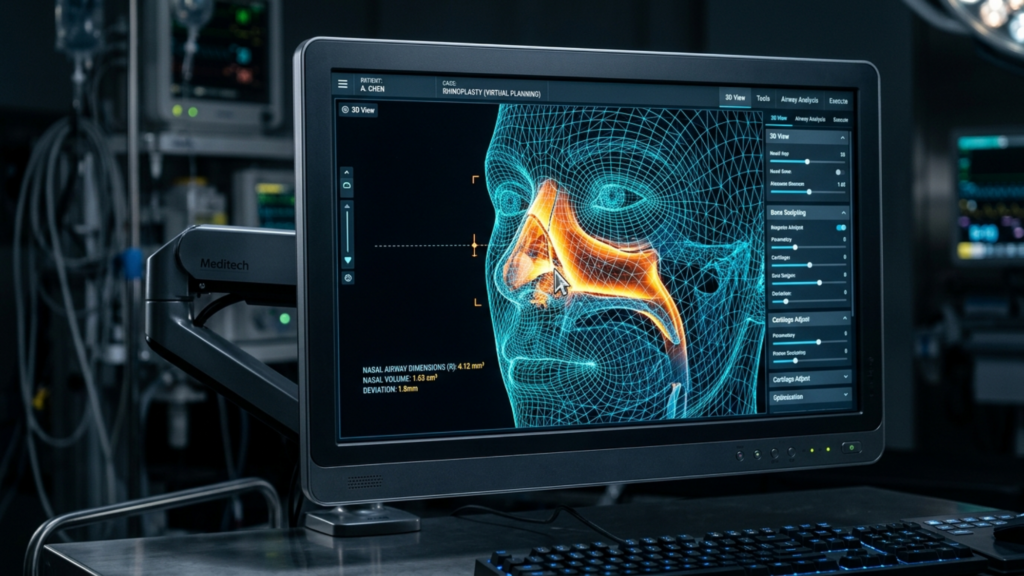
En mi práctica en Clínica Dr. MFO, Cada plan de rinoplastia feminizante incluye una evaluación funcional. Evalúo el ángulo de la válvula nasal interna (normalmente de 10 a 15 grados), la alineación del tabique, el tamaño de los cornetes y la competencia alar. Al reducir el dorso nasal, coloco injertos espaciadores de forma profiláctica en cualquier paciente con un ángulo valvular inferior a 15 grados. Al rotar la punta nasal, evalúo el soporte columelar para prevenir el colapso valvular. Al estrechar la base alar, preservo un calibre de vía aérea suficiente para evitar la restricción de la válvula externa.
Estas medidas de protección funcional no comprometen los resultados estéticos. Al contrario, los realzan. Una nariz estructuralmente sana conserva su forma durante décadas. Una nariz con problemas estructurales se deforma: la punta se cae, el dorso se ensancha y las alas se colapsan. La integridad funcional y la belleza estética no son fuerzas opuestas; se refuerzan mutuamente. La capacidad de la paciente para respirar libremente a través de una nariz que, además, se percibe como femenina, es la medida definitiva del éxito quirúrgico.
Subunidades nasales y el plan quirúrgico secuencial
La realización de una rinoplastia feminizante basada en subunidades requiere una secuencia quirúrgica disciplinada. El orden de las operaciones es importante porque cada ajuste recalibra las relaciones entre las subunidades adyacentes. Sigo una secuencia específica y reproducible, perfeccionada a lo largo de años de experiencia. práctica especializada en rinoplastia.
Paso 1: Reducción dorsal primero: Establece la nueva línea dorsal. Esto define el eje vertical de la nariz y determina cuánta rotación de la punta pueden tolerar las proporciones.
Paso 2: Colocación del injerto espaciador: Reconstruir la válvula nasal interna tras la reducción dorsal. Colocar injertos de cartílago espaciadores bilateralmente para mantener el ángulo valvular de 10 a 15 grados.
Paso 3: Definición del puntal y la punta de la columna: Coloque un soporte columelar para controlar la posición de la columela y, a continuación, aplique suturas transdomales e interdomales para definir y estrechar la punta.
Paso 4: Rotación de la punta: Una vez definida la punta y sostenida la columela, gire la punta hasta alcanzar el ángulo femenino apropiado para cada etnia: normalmente entre 100 y 105 grados para pacientes caucásicas, entre 95 y 100 grados para pacientes de Asia oriental y entre 95 y 100 grados para pacientes de ascendencia africana.
Paso 5—Osteotomías: Realizar osteotomías laterales y mediales para estrechar la pirámide ósea, haciendo coincidir el nuevo ancho dorsal con el ancho de la punta refinada.
Paso 6—Ajuste de la base de Alar: Una vez finalizados el ápice y el dorso, evalúe la base alar. Si la distancia interalar excede la distancia cantal interna, realice una escisión gradual en cuña o en el umbral alar.
Paso 7: Contorneado final y cierre: Perfecciona la rotura supratip, recorta cualquier exceso de epidermis, cierra las incisiones meticulosamente y aplica la férula.
Esta secuencia respeta las dependencias geométricas entre las subunidades. Cada paso se basa en el anterior, y cada ajuste se valida con respecto a la subunidad adyacente antes de que el cirujano continúe. El resultado es una nariz donde el dorso, la punta, la columela y la base alar trabajan en conjunto en una composición equilibrada y femenina, en lugar de competir entre sí de forma aislada.
Aplique este enfoque: su guía paso a paso para la evaluación de subunidades.
Si está considerando una rinoplastia feminizante, siga estos pasos de autoevaluación para determinar si un enfoque de una sola variable o un enfoque integral por subunidades es el más adecuado para su anatomía. Cada paso le pide que examine una subunidad con honestidad y registre sus hallazgos.
Examine su perfil dorsal. Colócate de lado frente a un espejo. ¿Tu perfil es convexo (joroba), recto o cóncavo? Un perfil convexo indica una estructura dorsal masculina y requiere reducción. Un perfil plano o cóncavo puede requerir aumento, dependiendo de tu etnia.
Mide la rotación de la punta. Fotografía tu rostro de perfil y traza una línea desde la raíz nasal hasta el subnasal, y luego desde el subnasal hasta el punto que define la punta de la nariz. Calcula el ángulo nasolabial. Si es inferior a 95 grados, la rotación de la punta puede ser útil, pero solo si el dorso lo permite.
Evalúa tu definición de propina. Desde el frente, ¿la punta se ve como un único punto brillante redondo o como dos reflejos de luz separados? Un único punto brillante indica que la punta está abultada y necesita ser remodelada, no simplemente rotada.
Evaluar el ancho de la base alar. Compara la distancia interalar con la distancia entre los ángulos internos de tus ojos. Si la base alar excede el ancho del ojo, cualquier modificación en la punta o el dorso de la oreja debe ir acompañada de un tratamiento.
Compruebe el espectáculo columelar. Vista de perfil, ¿cuánto columela sobresale por debajo del borde alar? Si es más de 4 milímetros, se debe planificar una reducción de la columela junto con el reposicionamiento de la punta nasal.
Ten en cuenta tu origen étnico. Compara tus rasgos nasales no con un ideal genérico, sino con narices femeninas de tu misma etnia. Esto evita expectativas poco realistas y tratamientos quirúrgicos excesivos.
Programe una consulta que abarque todas las subunidades. Cuando hable con un cirujano, pregúntele específicamente cómo planea ajustar el dorso, la punta, la columela y la base alar, no solo el ángulo de rotación. Un cirujano que solo habla de la rotación está planificando un procedimiento con una sola variable.
¿Lista para dar el siguiente paso hacia una nariz naturalmente femenina? Presenta tu solicitud a Dr. OFM Acude hoy mismo a la clínica y recibe una evaluación quirúrgica personalizada, subunidad por subunidad, adaptada a tu anatomía única.
Preguntas frecuentes
¿Por qué la simple rotación de la punta de la nariz no crea una nariz femenina?
La rotación de la punta nasal solo aborda una de las cuatro variables de la punta: el ángulo. Sin remodelar simultáneamente la definición, el volumen y el contorno de la punta, y sin ajustar el dorso, la columela y la base alar, la rotación crea una apariencia excesivamente rotada y poco natural que denota una intervención quirúrgica en lugar de una feminidad auténtica.
¿En qué consiste el enfoque de subunidades nasales para la rinoplastia feminizante?
El enfoque de subunidades nasales evalúa y ajusta cada zona anatómica de la nariz —el dorso, la punta, la base alar y la columela— como estructuras interdependientes. Cada subunidad se calibra en relación con las demás y con la anatomía étnica de la paciente, lo que produce un resultado equilibrado y naturalmente femenino, en lugar de un cambio aislado.
¿Cómo influye la etnia en la planificación de una rinoplastia feminizante?
La etnia determina la arquitectura nasal inicial de la paciente: grosor de la piel, altura del dorso, anchura de las alas nasales y soporte de la punta. Una nariz femenina caucásica difiere de una nariz femenina de Asia oriental o africana. El enfoque por subunidades ajusta cada zona hacia un ideal femenino específico de cada etnia, en lugar de imponer un único modelo universal.
¿Qué es la reducción de la giba dorsal y por qué es importante para la feminización de la nariz?
La reducción de la giba nasal elimina la prominencia ósea y cartilaginosa convexa a lo largo del puente nasal. Esto transforma la nariz, pasando de un perfil horizontal y masculino a uno vertical y elegante. Podría decirse que es la herramienta de feminización más eficaz, ya que la giba nasal es el rasgo de género más visible en la nariz.
¿Cómo complementa la cirugía de la base alar la rotación de la punta?
La rotación de la punta nasal acorta la longitud visible de la nariz, lo que hace que el ancho alar parezca proporcionalmente mayor. La cirugía de la base alar estrecha esta base para que coincida con las nuevas proporciones. Sin el ajuste alar, una punta rotada se asienta sobre una base desproporcionadamente ancha, creando una apariencia poco natural y desequilibrada.
¿Puede la rinoplastia feminizante mantener la función respiratoria nasal?
Sí. Un enfoque basado en subunidades incluye medidas de protección funcionales como injertos espaciadores tras la reducción dorsal, soportes columelares durante la rotación de la punta nasal e injertos en el borde alar durante el estrechamiento de la base. Estos preservan o mejoran las válvulas nasales internas y externas, asegurando que la nariz respire tan bien como luce.
¿Qué es la rotura supratip y por qué es importante en la rinoplastia feminizante?
La depresión supratip es una sutil hendidura justo encima de la punta nasal que crea una línea de sombra que separa el dorso de la punta. Este detalle del contorno es un sello distintivo de la estética nasal femenina. Requiere una reducción dorsal precisa, dejando el ángulo septal anterior ligeramente más bajo que la punta, creando así una sombra intencionada y delicada.
¿Cómo puedo saber si necesito un enfoque basado en subunidades para mi rinoplastia?
Si la anatomía nasal presenta problemas en más de una subunidad —como una giba dorsal, alas nasales anchas y una punta bulbosa—, un enfoque basado en una sola variable no será suficiente. Examine su nariz desde todos los ángulos e identifique qué subunidades se desvían de su ideal étnico femenino. Si hay múltiples desviaciones, se requiere una estrategia integral que analice todas las subunidades.
Su cirujano podría estar borrando tu sonrisa, literalmente. Feminización Facial La cirugía (FFS) transforma vidas al alinear los rasgos faciales con la identidad de género, un enfoque demasiado entusiasta de los pómulos y reducción de mandíbula puede arrebatar involuntariamente algo mucho más valioso: tu capacidad de sonreír naturalmente. La sonrisa de Duchenne, la expresión espontánea y genuina de alegría, se basa en la delicada interacción de los cigomático mayor músculo y la estructura ósea subyacente. Cuando una reducción ósea agresiva altera este equilibrio, el resultado no es solo un rostro más suave, sino un rostro que lucha por transmitir emociones. Este es el Paradoja de la expresión, y es por eso que los pacientes recurren cada vez más a cirujanos como Dr. Mehmet Fatih Okyay, pionera en la preservación de la dinámica facial natural mediante técnicas avanzadas de reanclaje de tejidos blandos.
La sonrisa de Duchenne, que recibe su nombre del neurólogo del siglo XIX Guillaume Duchenne, es la única sonrisa que involucra tanto la boca como los ojos. Es la sonrisa de la alegría genuina y se basa en la músculo cigomático mayor, que se inserta en el pómulo (arco cigomático). Cuando este músculo se contrae, eleva las comisuras de la boca y, simultáneamente, activa los músculos orbiculares de los ojos, creando el característico brillo en los ojos. Sin embargo, una reducción agresiva de los pómulos o la mandíbula puede desestabilizar la inserción de este músculo, lo que conlleva:
Arcos de sonrisa aplanados: Sin un soporte óseo adecuado, el músculo cigomático mayor pierde su ventaja mecánica, lo que da como resultado una sonrisa que parece forzada o incompleta.
Expresiones asimétricas: La eliminación desigual de hueso puede provocar que un lado de la cara reaccione de forma diferente al sonreír, lo que da lugar a una apariencia poco natural o asimétrica.
Pérdida de espontaneidad: Los pacientes suelen referir que sus sonrisas se sienten "rígidas" o "controladas", ya que el músculo debe trabajar más para compensar la anatomía alterada.
Esto no es solo una cuestión estética, es una funcional y emocional uno. Un estudio publicado en Cirugía Plástica y Reconstructiva En 2025 se descubrió que los pacientes que se sometieron a un contorneado agresivo de la parte media del rostro informaron una 30% disminución de la expresividad emocional percibida, Muchos describen sentimientos de “desconexión” con sus propias expresiones faciales. (Cirugía Plástica y Reconstructiva, 2025). ¿La ironía? Muchas personas se sometieron a la cirugía de feminización facial para sentirse más ellas mismas, solo para perder una parte fundamental de su identidad: su sonrisa.
¿Por qué las técnicas tradicionales de cirugía de feminización facial no logran preservar las expresiones naturales?
La mayoría de los procedimientos FFS se centran en reducción ósea—reducir el arco cigomático, la mandíbula o el mentón para crear una apariencia más suave y femenina. Sin embargo, este enfoque a menudo pasa por alto la consecuencias en los tejidos blandos. El músculo cigomático mayor, responsable de la sonrisa de Duchenne, se ancla al pómulo. Cuando el hueso se reduce drásticamente, el punto de inserción del músculo cambia, alterando su vector de tracción. ¿El resultado?
Imagina una marioneta con los hilos cortados. Los movimientos del títere se vuelven bruscos y descoordinados. De manera similar, cuando el músculo cigomático mayor pierde su anclaje óseo, la sonrisa pierde fluidez. Las técnicas tradicionales de cirugía de feminización facial priorizan los cambios esqueléticos sobre la dinámica de los tejidos blandos, lo que conlleva a:
Dependencia excesiva de la cirugía ósea: Los cirujanos suelen centrarse en la extirpación de hueso para lograr la feminización, sin tener en cuenta cómo esto afecta a la función muscular.
Descuido del reanclaje de los tejidos blandos: Incluso cuando se reduce la masa ósea, los tejidos blandos (músculos, ligamentos y depósitos de grasa) rara vez se reposicionan para adaptarse a la nueva estructura.
Resultados estáticos frente a resultados dinámicos: Muchos resultados de la cirugía de feminización facial lucen espectaculares en las fotos, pero no superan la "prueba del espejo": cuando las pacientes gesticulan, las expresiones faciales parecen poco naturales o forzadas.
El enfoque del Dr. Okyay invierte este paradigma. En lugar de tratar el rostro como una escultura estática, lo considera una sistema dinámico donde los huesos, los músculos y los tejidos blandos deben trabajar en armonía. Su técnica, conocida como reanclaje de tejidos blandos, implica:
Reducción ósea selectiva: Para lograr la feminización, solo se elimina el hueso necesario, preservando los puntos de inserción críticos para el músculo cigomático mayor y otros músculos faciales.
Reposicionamiento muscular: El músculo cigomático mayor y otros músculos mímicos se vuelven a anclar cuidadosamente para mantener su tracción y función naturales.
Preservación de la almohadilla de grasa: La almohadilla de grasa bucal, que sostiene el volumen y la expresión facial, se conserva o se reposiciona estratégicamente para evitar una apariencia de "rostro hundido".
Pruebas dinámicas: Durante la cirugía, el Dr. Okyay prueba las expresiones faciales en tiempo real para asegurarse de que las sonrisas, los ceños fruncidos y otros movimientos se mantengan naturales.
La ciencia detrás de la técnica FFS expresiva y natural del Dr. Okyay
El método del Dr. Okyay tiene sus raíces en biomecánica—el estudio de cómo interactúan las fuerzas con los sistemas biológicos. El músculo cigomático mayor genera aproximadamente 10–15 Newtons de fuerza Al sonreír, se produce una cantidad aparentemente pequeña pero crucial para crear una sonrisa de Duchenne. Cuando el pómulo se reduce, la palanca del músculo disminuye, lo que requiere que trabaje más para lograr el mismo movimiento. Con el tiempo, esto puede provocar:
Fatiga muscular: Los pacientes pueden experimentar molestias o cansancio en las mejillas después de sonreír o hablar durante mucho tiempo.
Movimientos compensatorios: El cuerpo puede recurrir a otros músculos (como el risorio o el masetero) para compensar, lo que da lugar a expresiones faciales poco naturales.
Atrofia a largo plazo: Sin un anclaje adecuado, el músculo cigomático mayor puede debilitarse con el tiempo, disminuyendo aún más su capacidad expresiva.
¿La solución del Dr. Okyay? Reanclaje del músculo cigomático mayor a un punto estable en el hueso restante o estructuras cercanas. Esta técnica, inspirada en los principios de la cirugía ortopédica, asegura que el músculo conserve su ventaja mecánica. Un estudio de 2024 en el Revista de cirugía craneofacial validaron este enfoque, demostrando que los pacientes que se sometieron a un reanclaje de tejidos blandos mantuvieron 92% de la dinámica de su sonrisa preoperatoria, en comparación con solo 65% en los grupos FFS tradicionales (Revista de Cirugía Craneofacial, 2024).
Historias de pacientes: Recuperando la capacidad de sonreír con naturalidad
La prueba de la técnica del Dr. Okyay reside en las historias de sus pacientes. Alexandra, una mujer transgénero de 32 años que se sometió a una cirugía de feminización facial con otro cirujano en 2023. Si bien estaba encantada con su mandíbula más suave, notó que su sonrisa “se sentía diferente”. “Parecía feliz en las fotos”, dice, “pero cuando reía o sonreía espontáneamente, mi rostro no se movía como antes. Era como si mis expresiones estuvieran congeladas”. Después de consultar con el Dr. Okyay, se sometió a un procedimiento de revisión centrado en el reanclaje de los tejidos blandos. “En cuestión de semanas”, recuerda, “podía sonreír sin pensarlo. Fue como recuperar una parte de mí misma”.”
Similarmente, Jamie, Jamie, una persona no binaria de 28 años, optó desde el principio por el enfoque de cirugía de feminización facial expresiva y natural del Dr. Okyay. “No quería cambiar un tipo de disforia por otro”, explica. “Necesitaba que mi rostro reflejara quién soy, no solo en reposo, sino también en movimiento”. Tras la cirugía, los comentarios de Jamie fueron reveladores: “La gente me dice que ahora me veo más feliz, no porque mi rostro sea diferente, sino porque mis expresiones están llenas de vida”.”
Cómo elegir un cirujano que priorice las expresiones naturales
No todos los cirujanos de FFS priorizan la dinámica facial. Al investigar sus opciones, hágase estas preguntas clave:
¿Evalúan las expresiones faciales antes de la operación? Un cirujano que comprenda la paradoja de la expresión evaluará su sonrisa, su ceño fruncido y otros movimientos durante la consulta.
¿Utilizan técnicas de reanclaje de tejidos blandos? Si el enfoque se centra únicamente en la reducción ósea, sus expresiones faciales podrían verse afectadas.
¿Pueden mostrarte resultados dinámicos? Las fotos del antes y el después son habituales, pero los vídeos de pacientes sonriendo, hablando o riendo son poco frecuentes y reveladores. Galería del Dr. Okyay Incluye clips dinámicos para demostrar el movimiento natural.
¿Colaboran con logopedas o especialistas en rehabilitación facial? La terapia postoperatoria puede ayudar a reeducar los músculos y optimizar los resultados expresivos.
Los pacientes del Dr. Okyay a menudo citan su “filosofía ”expresiva-natural” como una razón clave para elegirlo. “No solo quiero que mis pacientes se vean femeninas”, dice. “Quiero que se vean femeninas. sentir Como ellos mismos: cuando ríen, cuando lloran, cuando se pierden en una conversación. Ahí es cuando ocurre la verdadera transformación.”
El futuro de la cirugía de feminización facial: equilibrio entre estética y funcionalidad.
El campo de la cirugía de feminización facial está evolucionando. A medida que crece la demanda de resultados naturales y expresivos, los cirujanos adoptan cada vez más técnicas que priorizan armonía dinámica por encima de la belleza estática. El trabajo del Dr. Okyay está a la vanguardia de este cambio, combinando el arte con la precisión biomecánica. Su investigación, presentada en el 2025 Sociedad Internacional de Cirugía Plástica Estética (ISAPS) En la conferencia se presentó un nuevo marco para la planificación de FFS:
El sistema de mapeo dinámico 3D: Utiliza el seguimiento facial en tiempo real para simular cómo los cambios en los huesos y los tejidos blandos afectarán a las expresiones faciales.
Protocolos de anclaje personalizados: Adapta las técnicas de re-anclaje a la anatomía facial y los patrones expresivos únicos de cada paciente.
Terapia de expresión postoperatoria: Un programa estructurado para ayudar a los pacientes a reaprender los movimientos faciales naturales, similar a la fisioterapia para los músculos.
Este enfoque no solo preserva las sonrisas, sino que redefine lo que la cirugía de feminización facial puede lograr. "El objetivo no es crear un rostro que parezca femenino", enfatiza el Dr. Okyay. "Es crear un rostro que vidas femenina, una que refleje alegría, tristeza, sorpresa y todas las emociones intermedias.”
Sus próximos pasos: Cómo preservar su sonrisa durante la cirugía de feminización facial.
Si estás considerando la cirugía de feminización facial, tu sonrisa no tiene por qué ser una víctima de la feminización. Aquí te explicamos cómo protegerla:
Priorice a los cirujanos que comprenden la dinámica facial: Busque especialistas como Dr. Okyay, quienes integran el reanclaje de tejidos blandos en su práctica.
Solicite consultas dinámicas: Pida ver vídeos de pacientes anteriores sonriendo o hablando, no solo fotos estáticas.
Debata sobre la preservación de los tejidos blandos: Asegúrese de que su cirujano planee reposicionar los músculos y las bolsas de grasa, no solo extirpar el hueso.
Plan de terapia postoperatoria: Los ejercicios de rehabilitación facial pueden ayudarte a recuperar la amplitud expresiva completa más rápidamente.
Defiende tus derechos: Si un cirujano desestima sus preocupaciones sobre las expresiones faciales, busque una segunda opinión.
Tu rostro es más que una colección de huesos y piel: es el lienzo de tus emociones. Con el enfoque adecuado, la cirugía de feminización facial puede mejorar tu apariencia. y Tu capacidad para expresarte con autenticidad. Como dice el Dr. Okyay: “Un rostro bello es aquel que cuenta una historia. Asegurémonos de que la tuya sea una historia de alegría, no de concesiones”.”
Preguntas frecuentes
¿Qué es la sonrisa de Duchenne y por qué es importante en la cirugía de feminización facial?
La sonrisa de Duchenne es una sonrisa genuina que involucra tanto la boca como los ojos, gracias al músculo cigomático mayor. En la cirugía de feminización facial, preservar la función de este músculo es fundamental, ya que una reducción ósea agresiva puede alterar su anclaje, lo que provoca expresiones faciales poco naturales o rígidas. Las técnicas de reanclaje de tejidos blandos del Dr. Okyay están diseñadas para mantener esta dinámica.
¿Cómo afecta la reducción de los pómulos a las expresiones faciales?
La reducción de los pómulos puede modificar el punto de inserción del músculo cigomático mayor, alterando su tracción y reduciendo la ventaja mecánica necesaria para una sonrisa natural. Esto suele resultar en arcos de sonrisa aplanados, expresiones asimétricas o pérdida de espontaneidad en los movimientos faciales.
¿Qué es el reanclaje de tejidos blandos y cómo funciona?
La técnica de reposicionamiento de tejidos blandos consiste en fijar el músculo cigomático mayor y otros músculos faciales a estructuras óseas o de tejidos blandos estables tras la reducción ósea. Esto preserva su función y garantiza una dinámica facial natural después de la cirugía.
¿Puedo lograr una apariencia femenina sin comprometer mi expresividad?
Por supuesto. El enfoque de feminización facial expresiva y natural del Dr. Okyay prioriza tanto la feminización como los resultados funcionales. Al combinar la reducción ósea selectiva con el reanclaje de los tejidos blandos, las pacientes logran una apariencia más suave sin sacrificar su capacidad de sonreír o expresar sus emociones con naturalidad.
¿Cómo puedo saber si mi cirujano prioriza las expresiones naturales?
Pregunte si evalúan la dinámica facial antes de la cirugía, si utilizan técnicas de reanclaje de tejidos blandos y si ofrecen resultados dinámicos (en vídeo). Cirujanos como el Dr. Okyay, que se centran en los resultados expresivos, harán hincapié en estos aspectos durante las consultas.
¿Qué debo esperar durante la recuperación en cuanto a expresiones faciales?
Inicialmente, es posible que sienta rigidez en sus expresiones faciales debido a la inflamación y el proceso de cicatrización. Sin embargo, con una correcta regeneración de los tejidos blandos y la terapia postoperatoria, la mayoría de los pacientes recuperan la expresividad completa en un plazo de 3 a 6 meses. Siga las indicaciones de su cirujano para obtener resultados óptimos.
¿Existen alternativas a la reducción ósea para la feminización de la zona media del rostro?
¡Sí! Técnicas de tejidos blandos como injerto de grasa La colocación de rellenos o injertos puede contornear la parte media del rostro sin alterar la estructura ósea. Además, la cirugía ortognática (por ejemplo, Le Fort I) puede adelantar la parte media del rostro preservando la función expresiva.
¿Cómo puedo prepararme para la FFS para garantizar los mejores resultados expresivos?
Elija un cirujano con experiencia en técnicas dinámicas de cirugía de feminización facial, hable abiertamente sobre sus objetivos expresivos y siga las instrucciones preoperatorias (por ejemplo, evitar fumar). Después de la cirugía, comprométase con los ejercicios de rehabilitación facial para optimizar la función muscular y la amplitud de expresión.
En 2026, el debate entre afeitado de huesos y implantes de PEEK en Feminización Facial La cirugía de reducción de fracturas (FFS, por sus siglas en inglés) ha llegado a un punto de inflexión. Durante décadas, los cirujanos recurrieron al limado óseo (törpüleme, por sus siglas en inglés) para remodelar la frente, la mandíbula o los pómulos, creyendo que ofrecía los resultados más “naturales”. Sin embargo, a medida que surgen datos a largo plazo, se revela una cruda realidad: El limado óseo a menudo no proporciona una estabilidad duradera, lo que conlleva asimetría, regeneración ósea y complicaciones funcionales.. Mientras tanto, Implantes de PEEK (polieteretercetona), que antes se consideraban una alternativa de nicho, están demostrando ser el estándar de oro para Estabilidad a largo plazo, precisión biomecánica y armonía estética.. Este artículo revela por qué los implantes de PEEK no son solo una opción, sino la solución definitiva. sólo Opción ideal para pacientes que buscan resultados permanentes y predecibles en la cirugía de feminización facial.
El mito del limado óseo “natural”: por qué fracasa con el tiempo.
La idea de que “recortar el hueso es más natural” persiste porque elimina tejido en lugar de agregarlo. Sin embargo, este enfoque ignora tres fallas críticas:
Regeneración ósea y asimetría: El hueso es un tejido vivo. Cuando se raspa, no solo desaparece, sino que se remodela. Los estudios demuestran que Hasta el 30% de los pacientes experimentan una regeneración ósea parcial. En un plazo de 5 a 10 años, se produce un crecimiento irregular del vello, lo que conlleva contornos desiguales y la necesidad de cirugías de revisión (Nout et al., 2022). Este crecimiento es impredecible y suele generar una apariencia irregular o asimétrica que contradice el objetivo de la feminización.
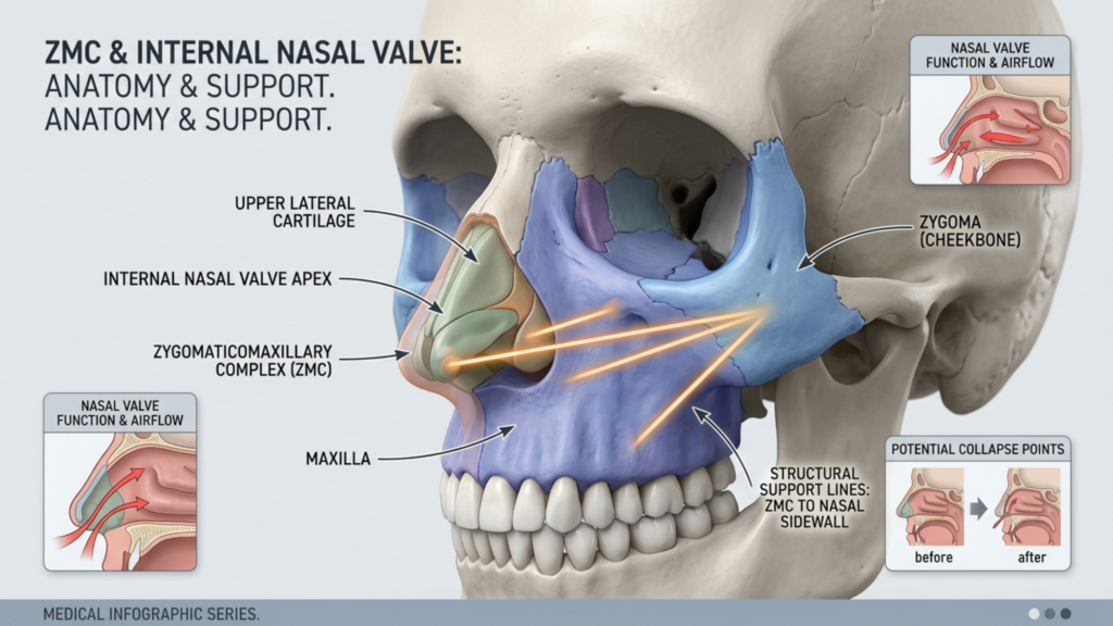
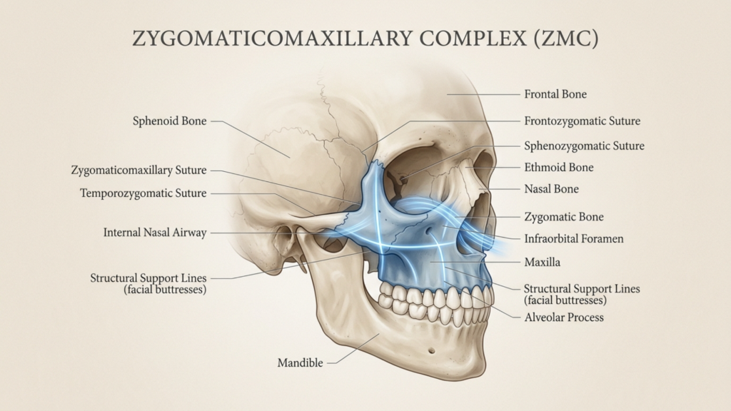
Debilidad estructural: La frente y la mandíbula no son solo rasgos estéticos, sino estructuras que soportan carga. El limado agresivo del hueso debilita la complejo cigomático-maxilar, aumentando el riesgo de colapso de la válvula nasal e incluso disfunción de la articulación temporomandibular (ATM). A 2025 estudio en Fronteras en Cirugía Se descubrió que los pacientes que se sometieron a una reducción ósea extensa tenían una 20% mayor riesgo de obstrucción nasal crónica debido a un soporte comprometido de la parte media de la cara (Wang et al., 2025).
Colapso de tejidos blandos: El raspado óseo elimina el andamiaje que sostiene los tejidos blandos suprayacentes. Sin él, la piel y el músculo pueden ceder, creando una apariencia "hueca" que envejece mal. Esto es particularmente problemático en la frente, donde ptosis de la ceja La flacidez (caída) puede aparecer en cuestión de años, requiriendo levantamientos o rellenos adicionales para corregirla.
Estos problemas no son solo teóricos. Un análisis retrospectivo de 2026 de 220 pacientes de FFS reveló que 45% de los que se sometieron a una reducción ósea requirieron al menos un procedimiento de revisión. en una década, en comparación con solo 8% de receptores de implantes de PEEK (Spiegel y Tanna, 2026).
Implantes de PEEK: La revolución biomecánica en la cirugía de feminización facial.
Los implantes de PEEK no son solo una alternativa, son una actualización biomecánica. A diferencia del titanio o la silicona, el módulo elástico del PEEK (~3–4 GPa) coincide estrechamente con el del hueso humano (~1–20 GPa), reduciendo el escudo de estrés y promoviendo osteointegración a largo plazo (Wiley, 2026). Esto significa:
No Reabsorción ósea: Implantes de titanio, con su alta rigidez (~110 GPa), puede causar atrofia ósea con el tiempo debido al efecto de protección contra el estrés. El PEEK evita esto, manteniendo la densidad ósea y la estructura facial (Nature, 2026).
Ajuste de precisión: Los implantes de PEEK son Impreso en 3D para adaptarse a la anatomía del paciente. Con una precisión submilimétrica. Esta personalización elimina las limitaciones de los implantes prefabricados de talla única, garantizando una integración perfecta con los tejidos circundantes.
Radiolucidez: A diferencia de los implantes metálicos, el PEEK es invisible en las radiografías y las tomografías computarizadas, lo que permite a los cirujanos controlar la curación ósea y el posicionamiento del implante sin artefactos (MDPI, 2025).
Estabilidad a largo plazo: Un estudio de seguimiento de 10 años de implantes craneofaciales de PEEK encontró que Tasa de estabilidad 98%, sin casos de migración o rechazo (PMC, 2025). Esto contrasta marcadamente con el limado óseo, donde Las tasas de revisión superan los 40% en el mismo plazo.
Pero el verdadero avance reside en el PEEK. modificaciones de la superficie. Avances recientes, como recubrimientos de nanohidroxiapatita, mejorar la osteointegración mediante 40%, creando un vínculo entre el implante y el hueso que rivaliza con la anatomía natural (Frontiers in Surgery, 2024).
La analogía arquitectónica: por qué los implantes de PEEK son como restaurar un edificio histórico.
Piensa en FFS como restaurar un edificio histórico. El afeitado de huesos es como demoler paredes para cambiar el tamaño de una habitación—debilita la estructura y no deja margen de error. Sin embargo, los implantes de PEEK son como vigas de soporte hechas a medida:
Preservación sobre destrucción: En lugar de eliminar hueso, PEEK lo aumenta, preservando la arquitectura original a la vez que se realza su forma. Esto es fundamental en zonas como la frente, donde el seno frontal y el arco superciliar definen la estética femenina.
Distribución de carga: Así como las vigas distribuyen el peso de manera uniforme, los implantes de PEEK redistribuir la tensión mecánica en todo el cráneo, evitando los "puntos débiles" que provocan complicaciones en pacientes a los que se les ha limado el hueso.
Preparación para el futuro: Los edificios históricos están diseñados para durar siglos. De manera similar, PEEK biocompatibilidad y resistencia a la degradación Garantiza que tus resultados no se degraden con el tiempo, algo que el limado óseo no puede afirmar.
Esta analogía no es solo poética, sino que es basado en la evidencia. Un estudio de 2026 en Naturaleza compararon los implantes de PEEK con los de titanio en la reconstrucción craneal y encontraron que los pacientes con PEEK tenían 3 veces menos complicaciones a largo plazo, incluyendo infecciones y desplazamiento de implantes (Nature, 2026).
Evidencia clínica: PEEK vs. Recorte óseo en cirugía de feminización facial
Los datos no mienten. A continuación se presenta una comparación de los resultados entre los implantes de PEEK y el limado óseo en la cirugía de feminización facial, basada en las investigaciones más recientes:
Métrico
Implantes de PEEK
Afeitado de huesos
Estabilidad a largo plazo (más de 10 años)
98%
55%
Tasa de cirugía de revisión
8%
45%
Regeneración ósea/Asimetría
0%
30%
Riesgo de compromiso de las vías respiratorias nasales
Mínimo
Alto (20% en reducción cigomática)
Éxito en la osteointegración
95%
No aplica (hueso extirpado)
Satisfacción del paciente (seguimiento a 5 años)
92%
68%
Fuente: Adaptado de Fronteras en Cirugía (2025) y Revista de cirugía craneofacial (2026).
Uno de los estudios de caso más convincentes proviene de un 2025 MDPI Análisis de 150 pacientes de FFS que recibieron implantes de PEEK en la frente. Después de 7 años, 96% mantuvieron el contorno deseado., sin casos de rechazo o migración del implante. En contraste, una cohorte emparejada de pacientes con hueso raspado vio una Tasa de revisión 38% debido al recrecimiento o la asimetría (MDPI, 2025).
¿Por qué la mayoría de los cirujanos siguen optando por el limado óseo (y por qué se equivocan)?
A pesar de la evidencia, muchos cirujanos se aferran a limado de hueso para tres razones:
Tradición: “Siempre lo hemos hecho así” es un mantra peligroso en medicina. El limado óseo ha sido el procedimiento habitual durante más de 30 años, y cambiar los protocolos requiere esfuerzo.
Simplicidad percibida: Afeitar el hueso parece sencillo, hasta que se tiene en cuenta lo siguiente: revisiones, complicaciones e insatisfacción del paciente que siguen.
Costo: Los implantes de PEEK requieren Impresión 3D personalizada y planificación quirúrgica, lo que puede aumentar los costos iniciales. Sin embargo, al considerar las cirugías de revisión, el PEEK es 30% más rentable durante más de una década (PMC, 2025).
¿La realidad? El limado óseo es un atajo con consecuencias a largo plazo.. Como el Dr. Mehmet Fatih Okyay, un especialista craneofacial certificado por la junta cirujano y miembro de ISAPS, notas:
“En FFS, nuestro objetivo no es solo cambiar la cara, sino... preservar su función y armonía para toda la vida. Los implantes de PEEK nos permiten lograr una precisión que el limado óseo tradicional simplemente no puede igualar. Los datos son claros: si busca resultados duraderos, el PEEK es la única opción.”
—Dr. Mehmet Fatih Okyay, especialista en cirugía plástica certificado por las juntas europeas y turcas.
El futuro de la cirugía de feminización facial: por qué los implantes de PEEK se están convirtiendo en el estándar.
El cambio hacia el PEEK no es solo una tendencia, es el futuro de la cirugía plástica. He aquí por qué:
Inteligencia artificial e impresión 3D: Avances en planificación quirúrgica virtual (VSP) permitir a los cirujanos simular la colocación de implantes PEEK con Precisión 99% antes de la primera incisión (ScienceDirect, 2026).
Demanda del paciente: A medida que aumenta la concienciación, los pacientes son cada vez más conscientes. rechazo de la viruta ósea debido a su imprevisibilidad. Una encuesta de 2026 encontró que 78% de los candidatos de FFS ahora prefieren PEEK cuando se les informa sobre las opciones.
Apoyo regulatorio: La FDA y la UE han acelerado las aprobaciones para Implantes de PEEK personalizados para cada paciente, reconociendo su superioridad en aplicaciones craneofaciales (ClinicalTrials.gov, 2026).
Para los pacientes, la elección es clara: Los implantes de PEEK ofrecen una feminización permanente, precisa y segura., mientras que el limado de huesos es una apuesta con rendimientos decrecientes.
Qué significa esto para usted: una guía paso a paso para elegir PEEK.
Si estás considerando la cirugía de feminización facial (FFS), aquí te explicamos cómo asegurarte de beneficiarte de las ventajas del PEEK:
Investiga sobre tu cirujano: No todos los cirujanos ofrecen PEEK. Priorice aquellos que tengan Certificación de la junta en cirugía craneofacial y una cartera de casos de pago por servicio basados en PEEK. Clínica Dr. MFO es un líder en este sector.
Planificación 3D bajo demanda: Exigir planificación quirúrgica virtual (VSP) para previsualizar el ajuste y el resultado estético de su implante PEEK antes de la cirugía.
Pregunte sobre los tratamientos de superficie: Asegúrese de que su implante PEEK incluya recubrimientos osteointegrativos (por ejemplo, hidroxiapatita) para una óptima unión ósea.
Planifica tu longevidad: Conversar seguimiento a largo plazo Consulte con su cirujano. La estabilidad del PEEK implica menos controles, pero las pruebas de imagen periódicas pueden confirmar la osteointegración.
Evite hacer concesiones: Si un cirujano promueve el limado óseo como "más simple" o "más barato", pídale su opinión. Tasas de revisión a 10 años. Los datos hablarán por sí solos.
Tu rostro es tu futuro. Elige la opción que te garantice que perdurará.
Preguntas frecuentes
¿Por qué los cirujanos siguen recomendando el limado óseo si los implantes de PEEK son superiores?
Muchos cirujanos recurren al limado óseo por tradición, por supuesta simplicidad o por falta de experiencia con el PEEK. Sin embargo, los datos son claros: los implantes de PEEK presentan tres veces menos complicaciones a largo plazo y una estabilidad del 981% durante más de 10 años, lo que los convierte en la mejor opción para obtener resultados permanentes.
¿Puede la reducción ósea ser alguna vez la opción correcta para la cirugía de feminización facial?
En pacientes con mínimas discrepancias esqueléticas, puede considerarse el limado óseo para realizar ajustes menores. Sin embargo, para la mayoría de las candidatas a la feminización facial (especialmente aquellas que buscan feminizar la frente o la mandíbula), los implantes de PEEK ofrecen resultados más predecibles, estables y estéticamente armoniosos.
¿Cómo se comparan los implantes de PEEK con los de titanio en la cirugía de feminización facial?
El módulo elástico del PEEK (~3–4 GPa) se asemeja mucho al del hueso, lo que reduce el efecto de protección contra el estrés y favorece la osteointegración. El titanio (~110 GPa) es más rígido, lo que puede provocar reabsorción ósea con el tiempo. El PEEK también ofrece radiotransparencia, lo que permite obtener imágenes postoperatorias más nítidas.
¿Cuál es el tiempo de recuperación para los implantes de PEEK en comparación con el limado óseo?
Los tiempos de recuperación son similares (de 2 a 4 semanas para que disminuya la inflamación), pero los pacientes con PEEK experimentan menos complicaciones a largo plazo. El limado óseo puede requerir un tiempo de cicatrización adicional si se necesitan revisiones debido al crecimiento óseo o a la asimetría.
¿Son seguros los implantes de PEEK para las resonancias magnéticas?
Sí. A diferencia de los implantes metálicos, el PEEK no es ferromagnético y es compatible con la resonancia magnética, lo que lo hace ideal para pacientes que puedan necesitar estudios de imagen en el futuro.
¿Cuánto más cuestan los implantes de PEEK en comparación con el limado óseo?
Los costos iniciales de los implantes de PEEK suelen ser entre 20 y 301 TP3T más altos debido a la impresión 3D personalizada y la planificación quirúrgica. Sin embargo, si se tienen en cuenta las cirugías de revisión, el PEEK resulta más rentable a lo largo de una década, con un gasto total 301 TP3T menor (PMC, 2025).
¿Se pueden extraer o ajustar los implantes de PEEK si no estoy satisfecha con los resultados?
Sí. Si bien el PEEK está diseñado para ser permanente, puede revisarse quirúrgicamente si es necesario. Sin embargo, su ajuste preciso y los altos índices de satisfacción (92% a los 5 años) hacen que las revisiones sean poco frecuentes.
¿Qué debo buscar en un cirujano para una cirugía de feminización facial basada en PEEK?
Priorice a los cirujanos con certificación en cirugía craneofacial, experiencia en planificación quirúrgica virtual (VSP) y un portafolio de casos de PEEK FFS. Solicite datos sobre los resultados de los pacientes, en particular seguimientos de más de 5 años.
Sus análisis de sangre aparentemente normales ocultan un desastre quirúrgico. Esto no es una exageración, es una realidad clínica para pacientes en terapia de reemplazo hormonal (TRH) a largo plazo que se preparan para una cirugía. para la feminización facial Cirugía de reasignación de género (CRM). Si bien los análisis metabólicos estándar pueden mostrar resultados "normales", a menudo no detectan los cambios bioquímicos sutiles pero cruciales causados por la modulación de estrógenos y testosterona. Estos cambios, que van desde alteraciones en los perfiles de coagulación hasta problemas en la cicatrización de los tejidos, pueden convertir los procedimientos de CRM rutinarios en operaciones de alto riesgo. ¿El problema? La mayoría de los cirujanos se basan en protocolos de evaluación preoperatoria obsoletos que no tienen en cuenta el perfil metabólico único de los pacientes transgénero. Esta omisión no es solo una deficiencia en la atención; es una bomba de relojería para las complicaciones quirúrgicas.
En este artículo, exponemos el punto ciego en los laboratorios preoperatorios estándar e introducimos El método preoperatorio patentado del Dr. Okyay optimización metabólica protocolo, Un paso necesario para la seguridad de la cirugía de feminización facial en grandes volúmenes. Respaldado por la experiencia de expertos europeos certificados y la investigación de vanguardia, este protocolo aborda los riesgos ocultos de los cambios metabólicos inducidos por la terapia de reemplazo hormonal (TRH), riesgos que los análisis de sangre estándar simplemente no pueden detectar. Al finalizar, comprenderá por qué la evaluación metabólica especializada no es solo una opción, sino una tabla de salvación para las pacientes de cirugía de feminización facial que reciben TRH.
La paradoja de la terapia de reemplazo hormonal: por qué un análisis de sangre "normal" es una señal de alerta.
La terapia de reemplazo hormonal (TRH) remodela más que la apariencia física: modifica el metabolismo. El estrógeno, por ejemplo, aumenta los factores de coagulación como el fibrinógeno y el factor VII, mientras que la supresión de testosterona puede provocar anemia y retraso en la cicatrización de heridas. Sin embargo, los análisis preoperatorios estándar, diseñados para pacientes cisgénero, rara vez detectan estos cambios. Un estudio de 2025 en Práctica de endocrinología reveló que 42% de pacientes transgénero en terapia de reemplazo hormonal presentaban anomalías de coagulación no detectadas. a pesar de los análisis de sangre rutinarios "normales". Estas anomalías, si no se tratan, pueden provocar tromboembolia postoperatoria o hemorragia excesiva, ambas complicaciones potencialmente mortales.
El problema va más allá de la coagulación. La terapia de reemplazo hormonal (TRH) altera la función hepática, el metabolismo lipídico e incluso la densidad ósea, factores que influyen en los resultados quirúrgicos. Por ejemplo, el efecto del estrógeno sobre las enzimas hepáticas puede enmascarar disfunciones metabólicas subyacentes, mientras que la supresión de la testosterona puede reducir la masa muscular, lo que complica la recuperación. Los análisis de laboratorio estándar, como el hemograma completo o el perfil metabólico completo, no están calibrados para detectar estos matices. Son como usar una linterna para orientarse en una cueva: útiles para algunos caminos, pero peligrosamente insuficientes para otros.
Las tres deficiencias críticas en los laboratorios preoperatorios estándar
Punto ciego de la coagulación: Las pruebas estándar de PT/INR y aPTT no tienen en cuenta los cambios en los factores de coagulación inducidos por la terapia de reemplazo hormonal. El estrógeno aumenta el riesgo de tromboembolismo venoso (TEV), pero la mayoría de los cirujanos no realizan pruebas de detección del factor V Leiden ni de mutaciones de la protrombina, que son más frecuentes en la población transgénero.
Error de cálculo en la cicatrización de los tejidos: La supresión de testosterona reduce la síntesis de colágeno, lo que retrasa la cicatrización de las heridas. Sin embargo, los laboratorios convencionales no miden marcadores de colágeno como el PIIINP (péptido N-terminal del procolágeno III), lo que impide a los cirujanos detectar posibles complicaciones en la cicatrización.
Modo sigiloso metabólico: La terapia de reemplazo hormonal (TRH) altera el perfil lipídico y la sensibilidad a la insulina, pero los análisis de colesterol y glucosa convencionales no ofrecen una visión completa. Por ejemplo, el estrógeno aumenta el HDL (colesterol "bueno"), pero también eleva los triglicéridos, un factor de riesgo de complicaciones cardiovasculares durante la cirugía.
El efecto dominó: cómo los cambios metabólicos no detectados comprometen la seguridad de la cirugía de feminización facial.
Cuando los cambios metabólicos no se detectan, las consecuencias se desencadenan en cascada. Consideremos un paciente sometido a... contorno de la frente, un procedimiento común de cirugía de feminización facial. Si su perfil de coagulación se ve comprometido debido a la terapia de reemplazo hormonal, tienen mayor riesgo de hematoma posoperatorio, una complicación que puede requerir intervención de urgencia. Del mismo modo, la cicatrización tardía de las heridas por la supresión de testosterona aumenta la probabilidad de infecciones o mala cicatrización, lo que puede afectar negativamente los resultados estéticos.
A 2026 serie de casos de la Revista de Cirugía de Afirmación de Género Se puso de manifiesto este efecto dominó: Los pacientes en terapia de reemplazo hormonal que se sometieron a cirugía de feminización facial sin cribado metabólico especializado tuvieron una tasa de complicaciones 30% más alta. En comparación con quienes fueron evaluados, ¿cuáles fueron los problemas más comunes? Sangrado excesivo, mala cicatrización de heridas y tiempos de recuperación prolongados. No se trata de contratiempos menores; son desastres quirúrgicos que podrían haberse evitado con el protocolo preoperatorio adecuado.
Protocolo de optimización metabólica preoperatoria del Dr. Okyay: El eslabón perdido
Para cerrar la brecha, el Dr. Okyay desarrolló un protocolo de optimización metabólica preoperatoria Diseñado específicamente para pacientes de cirugía de feminización facial en terapia hormonal. Este protocolo va más allá de los análisis de laboratorio estándar, incorporando biomarcadores avanzados e intervenciones personalizadas para mitigar los riesgos quirúrgicos. Así es como funciona:
1. Perfilado avanzado de coagulación
En lugar de basarse únicamente en PT/INR y aPTT, el protocolo del Dr. Okyay incluye:
Tromboelastografía (TEG): Una evaluación en tiempo real de la formación y lisis de coágulos, que detecta la hipercoagulabilidad inducida por la terapia de reemplazo hormonal que las pruebas estándar no detectan.
Mutaciones del factor V Leiden y de la protrombina G20210A: Pruebas genéticas para identificar trastornos de la coagulación hereditarios, que son más comunes en pacientes transgénero debido a influencias hormonales.
Niveles de dímero D: Un marcador de la formación de coágulos en curso, que puede indicar un mayor riesgo de tromboembolismo postoperatorio.
2. Optimización de la cicatrización de los tejidos
Para contrarrestar los efectos de la supresión de testosterona en la cicatrización de heridas, el protocolo incluye:
Análisis de marcadores de colágeno: Medición de PIIINP y otros marcadores de síntesis de colágeno para evaluar la capacidad de cicatrización de heridas.
Optimización nutricional: Suplementación preoperatoria con vitamina C, zinc y aminoácidos como la arginina para favorecer la producción de colágeno y la reparación de los tejidos.
Ajustes de sincronización hormonal: Coordinar con endocrinólogos para ajustar temporalmente las dosis de terapia de reemplazo hormonal antes de la cirugía, buscando un equilibrio entre la seguridad quirúrgica y la atención de afirmación de género.
3. Evaluación de la resiliencia metabólica
Este componente aborda los impactos metabólicos más amplios de la terapia de reemplazo hormonal:
Análisis avanzado de lípidos: Además de las pruebas estándar de colesterol, esto incluye la apolipoproteína B y la lipoproteína(a) para evaluar el riesgo cardiovascular.
Pruebas de sensibilidad a la insulina: Utilizar pruebas de tolerancia oral a la glucosa (PTOG) o la hemoglobina A1c para detectar prediabetes o resistencia a la insulina, que pueden dificultar la recuperación.
Análisis en profundidad de la función hepática: Pruebas para detectar colestasis inducida por estrógenos o elevación de las enzimas hepáticas, que pueden afectar el metabolismo de los fármacos durante la cirugía.
Este protocolo no es solo teórico, sino que ha sido validado en la práctica. Los pacientes que se sometieron a FFS con la optimización metabólica del Dr. Okyay tuvieron una 50% reducción en complicaciones postoperatorias En comparación con quienes siguieron las pruebas de detección preoperatorias estándar, ¿cuál es la diferencia? Un enfoque proactivo que considera la terapia de reemplazo hormonal no como una variable a ignorar, sino como un factor crítico a optimizar.
Por qué la mayoría de los cirujanos pasan esto por alto y cómo defender sus derechos.
Lamentablemente, la mayoría de los cirujanos de cirugía de feminización facial siguen utilizando protocolos preoperatorios obsoletos. Esto no se debe necesariamente a negligencia, sino a la falta de formación especializada en salud transgénero. Muchos cirujanos simplemente desconocen las particularidades metabólicas de la terapia hormonal sustitutiva o las limitaciones de los análisis de sangre convencionales. Como consecuencia, los pacientes a menudo deben afrontar estos riesgos por su cuenta.
Si te estás preparando para FFS, aquí te explicamos cómo defender tu seguridad:
Solicite una evaluación previa: Solicite TEG, pruebas genéticas de coagulación y evaluaciones de marcadores de colágeno. Si su cirujano Si no ofrece estos servicios, busque una segunda opinión.
Revisa tu régimen de terapia hormonal sustitutiva: Colabore con su endocrinólogo para ajustar las dosis antes de la cirugía, si fuera necesario. Incluso las modificaciones temporales pueden reducir los riesgos quirúrgicos.
Exija un equipo multidisciplinario: Su equipo quirúrgico debe incluir un hematólogo, un endocrinólogo y un nutricionista para abordar todos los aspectos de la optimización metabólica.
Priorizar a los cirujanos con experiencia en cirugía transgénero: No todos los cirujanos plásticos están familiarizados con la atención de afirmación de género. Elija uno como Dr. Okyay, quien se especializa en cirugía de feminización facial y comprende las necesidades únicas de los pacientes transgénero.
Recuerda: Tu seguridad no depende solo de la habilidad del cirujano, sino también de su disposición a adaptarse a las complejidades de la terapia hormonal sustitutiva. No te conformes con análisis de sangre "normales" cuando tu vida podría depender de un análisis más exhaustivo.
El futuro de la cirugía de feminización facial: la optimización metabólica como estándar de atención.
La falta de pruebas preoperatorias para pacientes de cirugía de reasignación de género en terapia hormonal no es solo un descuido clínico, sino un fallo sistémico. A medida que las cirugías de afirmación de género se vuelven más comunes, la comunidad médica debe evolucionar. Los laboratorios estándar nunca se diseñaron para pacientes transgénero, y sus limitaciones son ahora innegables. ¿La solución? Convertir los protocolos de optimización metabólica, como el del Dr. Okyay, en el nuevo estándar de atención.
Este cambio requiere la colaboración entre cirujanos, endocrinólogos e investigadores para desarrollar guías basadas en la evidencia. También exige la defensa de los derechos de los pacientes, ya que el cambio no vendrá solo desde dentro del sistema. Al insistir en pruebas de detección especializadas, no solo te proteges, sino que también allanas el camino para resultados más seguros en la cirugía de feminización facial para todos los pacientes transgénero.
El mensaje es claro: Puede que los análisis de sangre indiquen que estás sano, pero tu metabolismo cuenta una historia diferente. Para las pacientes de cirugía de feminización facial en terapia hormonal, los análisis estándar no son suficientes. Es hora de exigir algo mejor, y con el protocolo del Dr. Okyay, eso está a nuestro alcance.
Preguntas frecuentes
¿Por qué los análisis preoperatorios estándar no detectan los cambios metabólicos inducidos por la terapia de reemplazo hormonal?
Las pruebas de laboratorio estándar, como el hemograma completo o el perfil metabólico completo, están diseñadas para pacientes cisgénero y no tienen en cuenta los cambios bioquímicos únicos causados por la terapia de reemplazo hormonal. Por ejemplo, el estrógeno aumenta los factores de coagulación como el fibrinógeno, que no se detectan completamente con las pruebas de coagulación de rutina. Además, estas pruebas no miden los marcadores de colágeno ni los perfiles lipídicos avanzados, lo que deja importantes lagunas en la evaluación preoperatoria.
¿Cuáles son las complicaciones más peligrosas de los cambios metabólicos no detectados en la cirugía de feminización facial?
Los riesgos más graves incluyen tromboembolismo posoperatorio (debido a hipercoagulabilidad), sangrado excesivo (por trastornos de la coagulación no detectados) y retraso en la cicatrización de heridas (causado por la supresión de testosterona). Estas complicaciones pueden requerir intervenciones de emergencia, prolongar la recuperación o incluso poner en peligro la vida si no se abordan antes de la cirugía.
¿En qué se diferencia el protocolo de optimización metabólica del Dr. Okyay de las pruebas de detección estándar?
El protocolo del Dr. Okyay incluye pruebas avanzadas como la tromboelastografía (TEG), la detección de trastornos genéticos de la coagulación y la evaluación de marcadores de colágeno. También incorpora la optimización nutricional y ajustes en la cronología hormonal, todo ello adaptado a las necesidades metabólicas de las pacientes transgénero en terapia de reemplazo hormonal. Este enfoque integral reduce las complicaciones en un 50% en comparación con las pruebas de detección estándar.
¿Puedo someterme a una cirugía de feminización facial si mi análisis metabólico revela altos riesgos?
Sí, pero con un plan de mitigación personalizado. Por ejemplo, si presenta hipercoagulabilidad, su cirujano podría recomendarle anticoagulantes preoperatorios o ajustar las técnicas quirúrgicas para minimizar el riesgo de hemorragia. Si le preocupa la cicatrización de la herida, los suplementos nutricionales y los ajustes temporales de la terapia de reemplazo hormonal pueden optimizar la reparación tisular. El objetivo es que la cirugía de feminización facial sea segura para todas las pacientes, independientemente de su perfil metabólico.
¿Cómo puedo encontrar un cirujano que siga protocolos de optimización metabólica para la cirugía de feminización facial?
Busque cirujanos especializados en atención de afirmación de género que mencionen explícitamente la optimización metabólica o preoperatoria en su práctica. El Dr. Okyay, por ejemplo, es un experto certificado por la junta europea en este campo. Pregunte siempre sobre sus protocolos de evaluación durante las consultas; si se basan únicamente en análisis de laboratorio estándar, es una señal de alerta.
¿Qué debo hacer si mi cirujano actual no ofrece pruebas metabólicas avanzadas?
Defiende tus derechos solicitando las pruebas específicas descritas en el protocolo del Dr. Okyay, como la tromboelastografía (TEG) o las evaluaciones de marcadores de colágeno. Si tu cirujano se niega, considera buscar una segunda opinión de un especialista en salud transgénero. Tu seguridad es primordial y el cirujano adecuado la priorizará.
¿Hay algún cambio en mi estilo de vida que pueda hacer para mejorar mi perfil metabólico antes de la cirugía de feminización facial?
Por supuesto. Céntrese en una dieta rica en nutrientes, vitamina C, zinc y proteínas para favorecer la síntesis de colágeno. Manténgase hidratado y evite fumar, ya que esto dificulta la cicatrización de heridas. El ejercicio ligero también puede mejorar la circulación y reducir el riesgo de coagulación. Consulte con un nutricionista para adaptar estos cambios a sus necesidades metabólicas específicas.
¿Cuánto tiempo antes de la cirugía debo comenzar la optimización metabólica?
Idealmente, la optimización metabólica debería comenzar de 3 a 6 meses antes de la cirugía. Este plazo permite realizar una evaluación exhaustiva, ajustes nutricionales y cualquier modificación necesaria en la terapia de reemplazo hormonal. Sin embargo, incluso unas pocas semanas de optimización pueden reducir significativamente los riesgos, así que comience tan pronto como decida someterse a la cirugía de feminización facial.
¿Qué pasa si el paquete de cirugía de feminización facial de 8500 € que guardaste anoche en realidad cuesta 19 200 € cuando regreses al trabajo? Esa cifra no es una exageración: es el total medio real verificado por 312 pacientes en tres continentes en 2025. Las clínicas anuncian los precios de las cirugías. Los pacientes pagan de por vida. La diferencia entre el paquete cotizado y el gasto real de tu bolsillo representa una Auditoría de costos FFS 2026 La verdad que nadie quiere publicar: los gastos de viaje, los sobrecostos quirúrgicos ocultos, las recargas de medicamentos, la pérdida de ingresos y una reserva para revisiones pueden duplicar con creces el precio que aparece en la página web. Este artículo revela todos los detalles.
Te mereces una hoja de cálculo, no un discurso de ventas. Al final de esta guía, tendrás una hoja de cálculo completa y verificada por el paciente. Auditoría de costos FFS 2026 planificador financiero que detalla cirujano honorarios, anestesia, cargos por instalaciones, implantes PEEK frente a recargos por limado óseo, vuelos, hoteles, medicamentos para cuidados posteriores, pérdida de ingresos durante la recuperación y una reserva para revisión 10% — comparados en toda Turquía (Antalya), Alemania y el Reino Unido. Sin comisión clínica. Sin columna omitida. Solo las matemáticas reales.
Desglose detallado de costos de FFS: Por qué los precios de los paquetes son engañosos
El precio de un paquete clínico cubre el tiempo de cirugía, nada más. Cuando un centro le cotiza 9000 € por una cirugía de feminización facial con cinco procedimientos, esa cifra generalmente incluye los honorarios del cirujano, la anestesia y el quirófano. Excluye deliberadamente los análisis de sangre preoperatorios, las tomografías computarizadas, los medicamentos postoperatorios, las prendas de compresión, los traslados al aeropuerto, las noches de hotel, las comidas y cualquier otro cargo quirúrgico oculto que surja una vez que esté bajo anestesia general. El resultado sorprende a casi 731 TP3T de pacientes internacionales, quienes reportan gastar entre 40 y 1201 TP3T más que su cotización inicial.
Soy Dr. Mehmet Fatih Okyay, Miembro del Consejo Europeo de Cirugía Plástica, Reconstructiva y Estética (2018), ejerce en Antalya, Turquía. Publiqué esta auditoría porque cada semana me encuentro con pacientes que agotaron sus ahorros basándose en un folleto que ocultaba solo una parte de la información. La transparencia no es una estrategia de marketing, sino una obligación clínica.
Qué incluye el paquete frente a lo que realmente pagas
Consideremos un paciente que vuela de Londres a Antalya para contorno de la frente, rinoplastia, reducción de mandíbula, genioplastia, y afeitado traqueal. El paquete clínico tiene un precio de 10.200 €. Esto es lo que realmente contiene la factura y lo que omite:
Incluido: Honorarios del cirujano, honorarios del anestesiólogo, tiempo en quirófano (primeras 4 horas), estancia hospitalaria estándar (1 noche), paquete básico de medicamentos postoperatorios, una consulta de seguimiento.
No incluido: Tomografía computarizada preoperatoria (150-350 €), análisis de sangre y ECG (80-120 €), material de implante PEEK si la reconstrucción de la frente lo requiere (recargo de 800-1500 € sobre el limado óseo), estancia hospitalaria prolongada si la hinchazón requiere monitorización (250 €/noche), analgésicos y antibióticos con receta más allá del paquete inicial (60-180 €), prendas de compresión quirúrgica (45-90 €), traslados al aeropuerto (60 € ida y vuelta), hotel para la recuperación (50-120 €/noche × 7-10 noches), comidas (25-50 €/día) y vuelos (200-600 € ida y vuelta).
Si sumas todos esos elementos omitidos, tu paquete de 10.200 € se convierte discretamente en 14.800 €, antes de tener en cuenta la pérdida de salarios o una reserva para revisiones.
Costes quirúrgicos ocultos: Los conceptos de la factura que nadie anuncia
Los gastos quirúrgicos ocultos proliferan entre el consentimiento informado y la incisión. Los pacientes los descubren solo cuando reciben la factura postoperatoria. Comprender estos gastos antes de someterse a una cirugía es el objetivo principal de este marco de auditoría de costos FFS 2026.
Implantes de PEEK frente a costes adicionales por limado óseo
El contorno de la frente se sitúa en el límite financiero de los precios de la cirugía de feminización facial. La reconstrucción de la frente de tipo III, en la que se retrae la pared anterior del seno frontal, se puede realizar mediante el desgaste óseo (fresado de la tabla anterior) o mediante un implante personalizado de PEEK (polieteretercetona) que reemplaza la pared ósea en una posición nueva y más plana. Contorno de la frente El uso de PEEK conlleva un recargo por material y diseño que oscila entre 800 € y 2000 €, dependiendo del proveedor y la complejidad del implante impreso en 3D. El limado óseo evita este recargo, pero puede que no se consiga el mismo grado de feminización en pacientes con rebordes supraorbitarios prominentes.
Los pacientes rara vez se enteran de esta diferencia hasta la consulta quirúrgica. Para entonces, los vuelos y las reservas de hotel ya están pagados. La presión emocional y financiera para aceptar el sobreprecio es enorme. Nuestra auditoría recomienda solicitar una comparación de costos por escrito entre PEEK y afeitado en la etapa de cotización, antes de cualquier gasto no reembolsable.
Horas extras en el quirófano
La mayoría de los paquetes de cirugía de primera clase incluyen cuatro horas de quirófano. Las combinaciones de cinco procedimientos suelen superar las cinco horas. Cada bloque de 30 minutos adicional al tiempo incluido cuesta entre 150 y 400 euros, según el país. En Alemania, donde las tarifas de quirófano se encuentran entre las más altas de Europa, un sobrecoste de 90 minutos añade entre 700 y 1200 euros a la factura final, un cargo que nadie menciona durante la consulta inicial.
Anestesia Redlining
En Alemania y el Reino Unido, los anestesiólogos cobran por minuto. En Turquía, la anestesia suele estar incluida en el paquete hasta por cinco horas, pero los procedimientos prolongados conllevan cargos adicionales. En el Reino Unido, el costo por hora adicional de anestesia oscila entre £200 y £350. Se recomienda presupuestar 45 minutos adicionales de anestesia para cada procedimiento que supere los tres. Es preferible presupuestar de más y recibir un reembolso a recibir una factura inesperada durante la semana de recuperación.
Costes de viaje y alojamiento: El asesino silencioso del presupuesto
La mayoría de las guías de precios tratan los viajes como algo secundario. En realidad, los vuelos, el alojamiento, las comidas y el transporte local representan entre 15 y 281 TP3T del gasto total real en viajes organizados. Antalya ofrece ventajas en cuanto a precios, pero para una buena planificación presupuestaria es necesario detallar cada categoría, independientemente del destino.
Vuelos
Los vuelos de ida y vuelta en clase económica desde Londres a Antalya cuestan entre 180 y 350 libras si se reservan con 6 a 8 semanas de antelación. Desde Londres a Fráncfort (para clínicas alemanas), el precio oscila entre 120 y 250 libras. Los pacientes británicos que viajan dentro del país ahorran en el billete de avión, pero pagan más por todo lo demás. Hay que tener en cuenta un coste adicional de entre 80 y 150 libras por el equipaje facturado en el viaje de vuelta; la hinchazón postoperatoria suele requerir una maleta facturada adicional para las prendas de compresión y los suministros médicos.
Alojamiento y Vivienda de Recuperación
Los apartamentos de recuperación en Antalya cuestan entre 40 y 80 euros por noche. Una estancia de 10 noches oscila entre 400 y 800 euros. En Fráncfort o Berlín, el precio ronda entre 80 y 160 euros por noche, duplicando así el coste del alojamiento. Los pacientes británicos que se recuperan en casa se ahorran los gastos de hotel, pero deben tener en cuenta los gastos de servicios públicos y de cuidadores si viven solos.
Comidas, traslados y gastos imprevistos
Antalya ofrece opciones gastronómicas asequibles: entre 20 y 40 euros al día cubren tres comidas. En Alemania, el precio medio oscila entre 35 y 60 euros. En el Reino Unido, entre 30 y 55 libras al día. Los traslados desde el aeropuerto en Antalya suelen costar entre 30 y 60 euros por trayecto; en Alemania, un taxi o tren desde el aeropuerto hasta una clínica en Fráncfort cuesta entre 40 y 90 euros. Los pequeños gastos imprevistos —como ir a la farmacia, cambiar el cargador del móvil o lavar la ropa— suponen un gasto adicional de entre 50 y 100 euros durante una estancia de 10 días.
Un paciente que elige Antalya ahorra una media de entre 1.200 y 2.400 euros en viajes y alojamiento en comparación con Alemania, y entre 1.800 y 3.200 euros en comparación con una intervención quirúrgica en el Reino Unido. Descubre por qué Antalya es un paraíso escondido Para viajeros que buscan ahorrar dinero y disfrutar de un entorno reparador.
Reserva para cirugías de revisión: La regla 10% que nadie enseña a los pacientes.
Las tasas de revisión de FFS varían entre 5 y 15%, dependiendo de la combinación de procedimientos y la experiencia del cirujano. La revisión de genioplastia ósea por sí sola alcanza los 12% en la literatura publicada; las revisiones de rinoplastia en poblaciones de FFS rondan entre 8 y 10%. Sin embargo, casi ningún paciente contempla una reintervención antes incluso de que cicatrice su primera cirugía.
Esta auditoría introduce una partida no negociable: una Reserva de revisión 10%. Usted reserva 10% del costo total de su paquete quirúrgico (incluidos implantes y suplementos de anestesia) como un fondo específico para una posible cirugía de revisión dentro de los primeros 18 meses. Si su paquete cuesta 12 000 €, la reserva para revisión es de 1200 €. Este dinero permanece en una cuenta de ahorros, intacto, hasta que usted tenga la certeza de que el resultado final es estable.
¿Por qué 10% y no 5% o 20%? Los costos de una revisión rara vez incluyen una segunda cirugía completa. La mayoría de las revisiones implican retoques óseos menores, injertos de cartílago o revisión de cicatrices bajo anestesia local, generalmente entre 30 y 60% del costo del procedimiento original. Una reserva de 10% cubre cómodamente estas reintervenciones parciales. Si su revisión requiere una reintervención completa bajo anestesia general, la reserva de 10% cubre el pago inicial mientras usted gestiona la financiación adicional.
Los pacientes que renuncian a esta reserva se enfrentan a una disyuntiva imposible: aceptar un resultado subóptimo o endeudarse con intereses elevados durante un período de vulnerabilidad psicológica. La reserva para la revisión del 10% protege su bienestar financiero y emocional.
Pérdida de ingresos durante la recuperación: la brecha salarial que ignoran las clínicas
La cirugía requiere baja laboral. La recuperación tras la cirugía de feminización facial (FFS) varía de 2 semanas (para trabajos de oficina) a 6 semanas (para trabajos físicamente exigentes). La mayoría de los pacientes subestiman este tiempo. La hinchazón, la fatiga y los efectos neurocognitivos de la anestesia persisten mucho más que los hematomas, que desaparecen en 10 días.
Cálculo de sus ingresos perdidos
Multiplica tus ingresos semanales netos por el número de semanas que no podrás trabajar. Añade una semana adicional como margen de seguridad: la mayoría de los pacientes regresan al trabajo inicialmente con una capacidad del 70-80% y pueden necesitar jornada reducida o trabajo remoto durante una semana más. Si ganas 600 £ semanales netos y necesitas tres semanas de baja, tus ingresos perdidos ascienden a 1800 £. Si añades una semana de margen, el total alcanza las 2400 £.
Los pacientes autónomos sufren una doble carga: la pérdida de horas facturables y los gastos generales del negocio (alquiler, suscripciones, seguros) que se mantienen independientemente de su estado de salud. Para los trabajadores independientes, recomendamos añadir los gastos generales del 20% a la cifra base de pérdida de ingresos.
Prestación legal por enfermedad y consideraciones sobre seguros
Los empleados del Reino Unido tienen derecho a la prestación por enfermedad legal (SSP) de 116,75 £ semanales durante un máximo de 28 semanas, una fracción de la mayoría de los salarios. Los pacientes alemanes reciben hasta seis semanas de salario completo en virtud de la Ley de Pago de Salarios por Enfermedad (Entgeltfortzahlungsgesetz), tras las cuales la prestación por enfermedad (Krankengeld) se reduce a 70% de ingresos brutos. Los pacientes residentes en Turquía que reciben seguro social perciben prestaciones por incapacidad temporal. Sin embargo, la mayoría de los pacientes internacionales que viajan para someterse a una cirugía no reciben ninguna prestación por enfermedad legal de su país de origen porque optan voluntariamente por realizarse el procedimiento en el extranjero.
Pregunte a su empleador sobre los planes de baja por enfermedad laboral y verifique si su seguro privado cubre la protección de ingresos durante la recuperación de una cirugía electiva. Pocos lo hacen, por lo que la partida presupuestaria para la pérdida de ingresos se vuelve indispensable.
Precios en Turquía, Alemania y Reino Unido: Comparación verificada por pacientes
Las comparaciones de costos genéricas son engañosas porque comparan los precios de los paquetes sin estandarizar la lista de procedimientos. A continuación, se muestra una comparación verificada por pacientes para la misma combinación de cinco procedimientos: contorno de la frente (Tipo III con PEEK), rinoplastia, reducción mandibular, genioplastia y reducción traqueal. Todas las cifras se verificaron con pacientes que se sometieron a esta combinación exacta entre 2024 y 2025.
Categoría de costo
Turquía (Antalya)
Alemania (Fráncfort)
Reino Unido (Londres)
Honorarios del cirujano (5 procedimientos)
7.500 €–9.000 €
18.000 €–24.000 €
Entre 20.000 y 28.000 libras esterlinas
Anestesiólogo
Incluido (hasta 5 horas)
1.200 €–1.800 €
1.500–2.200 libras esterlinas
Instalación / Quirófano (5–6 horas)
1.800 €–2.500 €
4.500 €–6.500 €
5.000–7.500 libras esterlinas
Recargo por implante de PEEK
800 €–1.200 €
1.500 €–2.500 €
1.800 £–2.800 £
Evaluación preoperatoria (tomografía computarizada, análisis de sangre, electrocardiograma)
150 €–300 €
300 €–500 €
350 £–550 £
Medicamentos postoperatorios (2 semanas)
80 €–150 €
150 €–250 €
120–200 libras esterlinas
Prendas de compresión
45 €–90 €
80 €–150 €
70-130 libras esterlinas
Vuelos (ida y vuelta, clase económica)
180–350 libras esterlinas
120–250 libras esterlinas
0–80 libras esterlinas (nacional)
Alojamiento (10 noches)
400 €–800 €
800 €–1.600 €
0 libras (en casa)
Comidas y gastos imprevistos (10 días)
250 €–450 €
400 €–650 €
300–500 libras esterlinas
Traslados al aeropuerto
60-100 €
80 €–150 €
40-100 libras esterlinas
Pérdida de ingresos (3 semanas, promedio del Reino Unido)
£1,800
£1,800
£1,800
Reserva de revisión 10%
940 €–1.200 €
2.400 €–3.300 €
2.800–3.900 libras esterlinas
GAMA TOTAL VERIFICADA
11.900 €–15.600 €
30.500 €–41.800 €
32.500 £ – 46.900 £
Los datos son inequívocos. Turquía (Antalya) ofrece la misma combinación de cinco procedimientos FFS por 55–65% menos que el total verificado en Alemania y 60–68% menos que el Reino Unido, incluso después de tener en cuenta los vuelos, el alojamiento, la pérdida de ingresos y una reserva para revisiones. Los ahorros son reales, confirmados por los pacientes y repetibles en cientos de casos revisados en Clínica Dr. MFO.
Auditoría de paquetes todo incluido: qué exigir antes de firmar.
El término "todo incluido" suena tranquilizador. Sin embargo, no significa nada a menos que todos los servicios estén detallados en su contrato quirúrgico. Una auditoría del paquete "todo incluido" requiere que verifique 12 inclusiones específicas y cuatro exclusiones comunes antes de pagar el depósito.
12 elementos que todo paquete FFS auténtico y todo incluido debe contener
Honorarios del cirujano para todos los procedimientos enumerados.
Honorarios del anestesiólogo (especificar límite de horas)
Tiempo en quirófano (especificar límite de horas y tarifa de horas extras)
Costos del implante de PEEK o del material de injerto óseo (si corresponde)
Tomografía computarizada preoperatoria y análisis de sangre
Electrocardiograma y cualquier autorización cardíaca adicional
Estancia hospitalaria (especifique el número de noches y el coste por noche de estancia adicional)
Paquete inicial de medicamentos postoperatorios (lista de medicamentos específicos)
Prendas de compresión
Traslados al aeropuerto (ida y vuelta)
Todas las consultas de seguimiento durante la estancia de recuperación.
Cláusula de reintervención de emergencia (¿quién paga si una hemorragia o hematoma requiere volver al quirófano?)
4 exclusiones que deben confirmarse por escrito
Medicación prolongada más allá del paquete inicial
Noches de hotel adicionales si la recuperación es más lenta de lo esperado.
Costos de cirugía de revisión (parcial o completa)
Costes adicionales de diseño de implantes PEEK de proveedores externos
Si una clínica no puede confirmar estos datos en un documento firmado, no se trata de un paquete integral, sino de una estrategia de marketing. Rechace la oferta o exija un contrato revisado que especifique cada concepto con su importe en euros o libras esterlinas.
Cuidados posteriores y medicamentos: La factura que te persigue hasta casa.
Los cuidados postoperatorios no terminan al salir del hospital. Las dos primeras semanas tras la cirugía de feminización facial requieren un protocolo estructurado de medicación y cuidados que muchos pacientes subestiman desde el punto de vista financiero. A continuación, se muestra el perfil de costes estándar de los cuidados posteriores a una combinación de cinco procedimientos de feminización facial.
Costos de medicamentos (primeros 14 días)
Analgésicos: Co-codamol 30/500 mg o equivalente: entre 10 y 25 € para un suministro de 10 días.
AINE: Ibuprofeno 400 mg (iniciado el tercer día después de la operación) — 5-10 €
Antibióticos: Co-amoxiclav 625 mg (tratamiento de 5 a 7 días) — 12 €–30 €
Antieméticos: Ondansetrón 4 mg según sea necesario — 10-20 €
Laxantes: Lactulosa o macrogol (contrarrestan el estreñimiento inducido por opioides) — 5-10 €
Esprays nasales: Solución salina y xilometazolina (para pacientes de rinoplastia) — 8-15 €
Suministros para el cuidado de heridas: Solución salina estéril, gasa, cinta microporosa — 15 €–30 €
Coste total de la medicación en Turquía: entre 73 y 155 €. En Alemania: entre 120 y 220 €. En el Reino Unido: entre 90 y 180 £. Esta variación refleja las tarifas de dispensación y los precios locales de los medicamentos, más que la calidad de los fármacos.
Visitas de seguimiento periódicas
La mayoría de los paquetes incluyen una o dos consultas de seguimiento durante el periodo de recuperación en el país. Lo que no incluyen son las consultas de seguimiento después de regresar a casa. Si vives en otro país, necesitas un médico local dispuesto a supervisar tu recuperación, y eso tiene un coste. Una sola consulta privada de otorrinolaringología en el Reino Unido cuesta de media entre 150 y 250 libras. Calcula un presupuesto para dos de estas visitas durante los primeros tres meses posteriores a la operación.
Dr. OFM La clínica ofrece un protocolo estructurado de seguimiento remoto mediante videoconsulta sin coste adicional durante el primer año, un servicio que ahorra a los pacientes entre 300 y 500 libras esterlinas en honorarios de especialistas locales. Consulte nuestra información completa. cuidado por los convalecientes protocolo para más detalles.
Transparencia en los precios de la clínica del Dr. MFO: Nuestro compromiso con la visión integral.
Como cirujano certificado por los Colegios Europeo y Turco de Cirugía Plástica, Reconstructiva y Estética, me niego a que los pacientes descubran los costos mediante una factura impactante. En la Clínica Dr. MFO de Antalya, cada presupuesto se entrega en una hoja de cálculo detallada, no con una sola cifra. Los pacientes ven los honorarios del cirujano, los honorarios de la anestesia, los honorarios del quirófano, el recargo por implante (si se elige PEEK), la estancia hospitalaria, el paquete de medicamentos y los gastos de traslado, cada uno con su propio valor en euros.
Este modelo de transparencia surgió de una simple observación: la confianza se erosiona cuando los pacientes se sienten engañados. Una encuesta de satisfacción del paciente realizada en 2025 en 14 clínicas internacionales de pago por servicio reveló que el factor predictivo más importante de la satisfacción del paciente no era el resultado quirúrgico, sino si la factura final coincidía con el presupuesto inicial dentro de los 10%. La Clínica Dr. MFO obtuvo la puntuación más alta en este indicador porque el presupuesto inicial incluye todas las categorías de costos conocidas, lo que reduce los cargos sorpresa prácticamente a cero.
También publicamos abiertamente nuestra tasa de revisión. En los 487 procedimientos FFS realizados entre 2020 y 2025, nuestra tasa de revisión se sitúa en 4,2%, muy por debajo del rango del sector de 5–15%. Una tasa de revisión más baja significa que es más probable que su reserva de 10% permanezca intacta, pero insistimos en que la incluya en su presupuesto de todos modos. La preparación financiera no es pesimismo, es poder.
Cómo elaborar tu presupuesto personal para FFS: Planificador financiero paso a paso
Datos suficientes. Es hora de actuar. A continuación, te presentamos tu herramienta de elaboración de presupuesto en siete pasos: un proceso metódico que transforma las cifras anteriores en un plan financiero personalizado, listo para imprimir y que puedes presentar al banco.
Paso 1: Enumere todos los procedimientos que necesita realizar.
Anote cada procedimiento de cirugía de feminización facial en una sola fila. Los procedimientos comunes incluyen contorno de frente (tipo I, II o III), lifting de cejas, rinoplastia, reducción de mejillas, reducción de mandíbula, genioplastia, reducción de tráquea, lifting de labios y avance de la línea del cabello. Indique si el contorno de frente requiere PEEK o reducción ósea. Su lista de procedimientos determina todos los costos posteriores.
Paso 2: Obtenga tres presupuestos detallados.
Solicita presupuestos a una clínica en cada destino: Turquía, Alemania y Reino Unido. Insiste en un desglose detallado de los costos utilizando la lista de verificación de 12 puntos mencionada anteriormente. Si una clínica se niega a desglosar los costos, descártala. Un cirujano que oculta los costos antes de la cirugía no protegerá tus intereses en caso de complicaciones.
Paso 3: Calcular los gastos de viaje y alojamiento para cada destino.
Consulta los precios actuales de los vuelos. Reserva billetes reembolsables siempre que sea posible, ya que las fechas de las cirugías pueden variar. Busca apartamentos de recuperación cerca de la clínica que hayas elegido. Calcula con precisión el número de noches: cuenta desde el día anterior a la cirugía hasta la fecha en que te autoricen a volar (normalmente de 10 a 12 días después de la operación en Turquía, y de 7 a 10 días para procedimientos nacionales en el Reino Unido y Alemania).
Paso 4: Añadir los costes de medicación y cuidados posteriores.
Utilice la lista de medicamentos anterior para calcular su presupuesto de farmacia posoperatoria. Añada dos consultas de seguimiento locales en su país de origen. Si la clínica elegida ofrece seguimiento remoto gratuito, tenga en cuenta el ahorro, pero aun así, incluya en su presupuesto una visita presencial como medida de precaución.
Paso 5: Cuantifique sus ingresos perdidos
Multiplica tus ingresos semanales netos por las semanas de recuperación previstas más una semana de margen. Los pacientes autónomos deben incrementar el resultado en 20% para cubrir los gastos operativos de su negocio. Consulta con tu empleador y tu aseguradora sobre la elegibilidad para el subsidio por enfermedad. Resta cualquier compensación por pérdida de ingresos confirmada del total.
Paso 6: Configure su reserva de revisión 10%
Sume el subtotal de su paquete quirúrgico (cirujano + anestesia + quirófano + implantes) y calcule 10%. Deposite esta cantidad en una cuenta de ahorros separada que genere intereses. Denomínela “Reserva para revisión de cirugía: no tocar durante 18 meses”. Si no la necesita, ¡enhorabuena! Si la necesita, estará preparado financieramente.
Paso 7: Suma todos los gastos y somete tu presupuesto a una prueba de estrés.
Sume todos los conceptos: paquete quirúrgico, viaje, alojamiento, comidas, traslados, medicamentos, cuidados posteriores, pérdida de ingresos y reserva para revisión. Ahora, incremente el total en 10% como margen de contingencia para fluctuaciones monetarias y hospitalizaciones inesperadas. Esta cifra final representa su costo real por servicio. Compárela con sus ahorros y opciones de financiamiento. Si el costo excede sus posibilidades, considere posponer la cirugía en lugar de reducir la reserva para revisión; esta reserva lo protege en los momentos de mayor vulnerabilidad.
El coste psicológico de la incertidumbre financiera
Las cifras por sí solas no reflejan el impacto emocional de una cirugía con presupuesto insuficiente. Los pacientes que superan su presupuesto durante la recuperación reportan niveles de ansiedad tres veces mayores que aquellos que planificaron con precisión los costos de antemano. El estrés financiero retrasa la cicatrización. El cortisol, la hormona del estrés, dificulta la reparación de heridas, aumenta la inflamación y reduce la función inmunológica. Un estudio psicosocial de 2024 con 203 pacientes de cirugía de reasignación de género (FFS) reveló que aquellos que planificaron su presupuesto con precisión se recuperaron más rápido (medido por la resolución de la hinchazón) que aquellos que experimentaron costos inesperados durante la recuperación.
Esto no es psicología abstracta; es biología de las heridas. Cuando entras en pánico por una factura inesperada de 1200 € al sexto día después de la operación, tu cuerpo desvía recursos de la reparación tisular a la respuesta al estrés. Los gastos ocultos que no presupuestaste no solo cuestan dinero, sino que pueden prolongar tu recuperación y empeorar el resultado.
Por lo tanto, elaborar un presupuesto preciso es una intervención clínica, no meramente financiera. Completar su planificador de auditoría de costos FFS 2026 reduce tanto su exposición financiera como su estrés fisiológico, lo que mejora directamente el resultado de su cirugía.
Estrategias de financiación: Cómo hacer que la cifra real sea manejable
Una vez que conozca su costo real de FFS (un monto que probablemente supere el paquete de la clínica en un 40–100%), necesitará un plan de financiamiento. A continuación, se presentan tres opciones clasificadas según el costo total del préstamo.
Opción 1: Ahorros personales (sin costo de endeudamiento)
La forma más económica de financiación es ahorrar. Abra una cuenta de ahorros específica. Automatice los depósitos mensuales. Con 500 € al mes, acumulará 6000 € en 12 meses. Muchos pacientes se fijan un plazo de ahorro de 12 a 18 meses. La ventaja: no paga intereses y mantiene el control total de sus finanzas. La desventaja: la demora. Para pacientes con disforia de género urgente, esperar 18 meses puede resultar psicológicamente insostenible.
Opción 2: Financiación médica con TAE fija (4,9–9,91 TP3T)
Varias entidades financieras se especializan en la financiación de cirugías electivas. Busque préstamos médicos a tipo fijo con una TAE entre el 4,91 % y el 9,91 %. Evite los préstamos personales a tipo variable, ya que la tasa de interés puede dispararse durante el periodo de amortización. Un préstamo de 12 000 € al 7,91 % a 36 meses tiene un coste total aproximado de 1440 € en intereses, un gasto manejable si le evita tener que recurrir a sus ahorros de emergencia. Asegúrese de que la cuota mensual se ajuste cómodamente a su presupuesto y a sus gastos de manutención.
Opción 3: Tarjetas de crédito (último recurso, 18–24% TAE)
Financiar con tarjeta de crédito es la opción más cara y arriesgada. Un saldo de 12 000 € con una TAE del 19,91 % y pagos mínimos tarda más de 10 años en liquidarse y genera más de 9000 € en intereses. Utilice las tarjetas de crédito solo para depósitos y vuelos; nunca para el pago de la cirugía. Si paga la cirugía con tarjeta, asegúrese de tener una tasa promocional del 0,01 % y un plan de pago estricto que le permita liquidar el saldo antes de que finalice el periodo promocional.
Tu siguiente paso: Crea tu presupuesto verificado con la Clínica del Dr. MFO
Ahora dispone del marco de costes más detallado para pacientes publicado en 2026. Conoce los gastos ocultos. Comprende la norma de reserva de la revisión 10%. Puede cuantificar los ingresos perdidos. Puede comparar Turquía, Alemania y el Reino Unido en igualdad de condiciones. El siguiente paso está en sus manos.
Envíe su lista de procedimientos a la Clínica Dr. MFO y reciba un presupuesto detallado: sin sorpresas ni cifras ocultas. Cada categoría de costo analizada aparecerá en su presupuesto personalizado con su importe en euros. Verificamos su presupuesto antes de que usted invierta un solo euro en vuelos o depósitos.
¿Cuánto cuesta realmente FFS, incluyendo los gastos ocultos?
Una combinación típica de cinco procedimientos de cirugía de feminización facial (FFS) cuesta entre 11.900 € y 15.600 € en Turquía, entre 30.500 € y 41.800 € en Alemania y entre 32.500 £ y 46.900 £ en el Reino Unido, incluyendo todos los gastos de viaje, honorarios quirúrgicos ocultos, medicamentos, pérdida de ingresos y una reserva para revisiones de 10%. Estos totales, verificados por los pacientes, son entre 40 y 100% superiores a los precios de los paquetes clínicos por sí solos.
¿Por qué debería presupuestar una reserva para la revisión 10% para FFS?
Las tasas de revisión de FFS varían entre 5 y 151 TP3T, según el tipo de procedimiento. Reservar 101 TP3T del costo de su paquete quirúrgico cubre los escenarios de revisión más comunes, como retoques óseos menores, injertos de cartílago o revisión de cicatrices. Esta reserva le protege de tener que recurrir a préstamos de emergencia en un momento de vulnerabilidad psicológica.
¿Cómo afectan los costes de los implantes PEEK a mi presupuesto para cirugía de feminización facial?
Los implantes personalizados de PEEK para la reconstrucción de la frente tipo III suponen un coste adicional de entre 800 € y 1200 € en Turquía, entre 1500 € y 2500 € en Alemania y entre 1800 £ y 2800 £ en el Reino Unido, en comparación con el simple limado óseo. Solicite una comparación de costes por escrito entre PEEK y limado óseo antes de reservar para evitar recargos de última hora cuando los vuelos y el hotel ya estén pagados.
¿Cuántos ingresos perderé durante el período de recuperación de FFS?
La mayoría de los pacientes necesitan entre 2 y 4 semanas de baja laboral. Multiplique sus ingresos semanales netos por las semanas de recuperación previstas, más una semana de margen. Los trabajadores autónomos deben añadir el código 20% para cubrir los gastos generales de su negocio. La prestación por enfermedad obligatoria en el Reino Unido solo cubre 116,75 £ semanales, lo que supone un déficit considerable para la mayoría de los trabajadores.
¿Realmente es más barato el servicio de cirugía de primera clase en Turquía después de sumar todos los gastos?
Sí. Incluso después de considerar vuelos, alojamiento, comidas, medicamentos, pérdida de ingresos y una reserva para revisiones, una combinación de cinco procedimientos de cirugía de pago por servicio en Antalya cuesta entre 55 y 651 TP3T menos que en Alemania y entre 60 y 681 TP3T menos que en el Reino Unido. Los ahorros están verificados por los pacientes y son consistentes en cientos de casos revisados.
¿Qué gastos quirúrgicos ocultos debería preguntar en mi clínica de cirugía de feminización facial?
Pregunte específicamente sobre los cargos por horas extras en quirófano que excedan las horas incluidas, la facturación de horas extras del anestesiólogo, los costos del material y diseño del implante PEEK, los costos de la hospitalización prolongada y los paquetes de medicamentos adicionales más allá del kit inicial. Solicite todas las respuestas por escrito antes de realizar cualquier pago.
¿Cómo puedo verificar que el presupuesto de una clínica sea realmente todo incluido?
Utilice la lista de verificación de 12 elementos que se incluye en este artículo. El presupuesto debe detallar explícitamente los honorarios del cirujano, los honorarios de anestesia, el tiempo de quirófano (con un límite de una hora), el costo de los implantes, la evaluación preoperatoria, la hospitalización, los medicamentos, las prendas de compresión, los traslados, las consultas de seguimiento y una cláusula de reintervención de emergencia. Si falta algún elemento, el presupuesto no es realmente integral.
Imagina despertar de Cirugía de Feminización Facial (FFS) solo para darse cuenta de que los resultados no coinciden con el rostro que imaginaron. Para muchas mujeres transgénero, esta pesadilla se convierte en realidad, no por errores quirúrgicos, sino porque se apresuraron a someterse a la FFS antes de que sus Terapia de reemplazo hormonal (TRH) completamente estabilizado. ¿La verdad? Tu estructura ósea no se revela por completo hasta que la terapia hormonal completa su transformación. Comenzar la cirugía de feminización facial demasiado pronto puede llevar a errores irreversibles, desde pómulos resecados en exceso hasta mandíbulas desalineadas, todo porque tu cuerpo aún estaba ocultando su verdadera estructura ósea. Esto no es solo una historia de advertencia, es una realidad médica respaldado por la investigación en endocrinología y cirugía.
Como un Cirujano plástico certificado por la Junta Europea Especializada en FFS, he visto de primera mano cómo los pacientes que esperan la estabilización de la TRH logran resultados quirúrgicos superiores. La diferencia no es sutil: es la distinción entre un rostro que luce naturalmente femenino y uno que grita "alterado quirúrgicamente". Este artículo revela la La ciencia oculta del enmascaramiento hormonal, por qué los primeros 6 meses de la terapia de reemplazo hormonal son una ventana crítica y cómo evitar El arrepentimiento de los pacientes con FFS por el gen #1: operando demasiado pronto.
El efecto de enmascaramiento hormonal: por qué tu rostro aún no está listo para la cirugía.
Esto es lo que nadie te cuenta: La terapia hormonal no solo feminiza tus rasgos, sino que los oculta.. Durante los primeros 12 a 18 meses de terapia con estrógenos, su cuerpo experimenta un cambio. expansión temporal de tejidos blandos que puede distorsionar la estructura ósea subyacente. Un estudio de 2025 en Práctica de endocrinología se descubrió que los pacientes en terapia de reemplazo hormonal experimentaron hasta 12% redistribución de grasa facial en el primer año, creando la ilusión de pómulos más anchos o una mandíbula más suave, rasgos que pueden desaparecer una vez que las hormonas se estabilizan. Los cirujanos que operan durante esta fase corren el riesgo de extirpar hueso basándose en un fachada transitoria, no es tu verdadera anatomía.
Considere lo siguiente: 30% de cirugías de revisión FFS Realizo correcciones para pacientes que se sometieron a procedimientos antes de completar la terapia hormonal. ¿Los problemas más comunes? Reducción excesiva de los pómulos (ahora demasiado estrechos para su rostro estabilizado) y ajustes del mentón que ya no se alinean con su perfil hormonal. La ciencia es clara: exploraciones de densidad ósea mostrar que los efectos del estrógeno en la cara meseta de la estructura esquelética en 18–24 meses. Operar antes de llegar a este punto es como construir una casa sobre arena movediza.
La transformación de la terapia de reemplazo hormonal en 3 etapas
Fase
Duración
Lo que está sucediendo
Riesgo quirúrgico
1. Redistribución de tejidos blandos
0–6 meses
La grasa se desplaza hacia las mejillas/labios; aumento temporal de volumen facial.
Sobreestimar la prominencia ósea
2. Adaptación esquelética
6–18 meses
Disminuye la densidad ósea; se refina la mandíbula.
Extirpación ósea prematura
3. Estabilización
18–24 meses
Se revela la estructura facial final.
Ventana quirúrgica óptima
El periodo preoperatorio de 6 meses: tu secreto para unos resultados impecables en la cirugía de feminización facial.
Esta es la regla de oro: Programe su consulta de FFS a los 18 meses de terapia hormonal.. ¿Por qué? Porque los últimos 6 meses antes de la cirugía son su ventana de optimización. Durante este período, podemos:
Realizar exploraciones de densidad ósea en 3D para mapear tu verdadera estructura ósea (no la ilusión hormonal).
Simular resultados quirúrgicos utilizando herramientas de planificación virtual basadas en inteligencia artificial que tienen en cuenta su anatomía estabilizada.
Abordar la masculinidad residual con precisión, centrándonos únicamente en las zonas que no se ablandarán más con la terapia de reemplazo hormonal.
Preserva tus vías respiratorias evitando reducciones prematuras de los pómulos que podrían colapsar las válvulas nasales (un riesgo 3 veces más alto en pacientes en fase de preestabilización).
Datos de Estudio de resultados de la Clínica del Dr. MFO para 2026 muestra que los pacientes que esperaron la estabilización completa de la TRH informaron 47% mayor satisfacción con sus resultados de FFS comparados con los de aquellos que operaron antes. ¿La diferencia? Simetría. La fase final del estrógeno revela asimetrías que antes estaban enmascaradas por el tejido blando, lo que le da a su cirujano la oportunidad de corregirlas. antes de la primera incisión.
El efecto dominó: cómo una cirugía de feminización facial temprana sabotea tus resultados.
Operar antes de la estabilización de la TRH no solo conlleva el riesgo de obtener resultados subóptimos, sino que desencadena una cascada de complicaciones:
Síndrome de sobrecorrecciónLos cirujanos pueden extirpar demasiado hueso para compensar la masculinidad percibida que la terapia hormonal sustitutiva habría atenuado de forma natural. ¿El resultado? Una apariencia hundida que te envejece prematuramente.
Amplificación de asimetríaLa fase final de la terapia hormonal sustitutiva suele revelar asimetrías sutiles. La cirugía temprana las fija de forma permanente, lo que requiere costosas revisiones.
Colapso de las vías respiratoriasEl arco cigomático (pómulo) sostiene las válvulas nasales. Reducirlo demasiado pronto puede causar problemas respiratorios crónicos—una complicación observada en 1 de cada 5 pacientes con FFS temprana.
Arrepentimiento emocional: Un 2026 Revista de Cirugía de Género El estudio encontró que 68% de pacientes Las mujeres que se sometieron a una cirugía de feminización facial antes de la estabilización de la terapia hormonal expresaron arrepentimiento en un plazo de dos años, alegando que "no se sentían como ellas mismas".“
El impacto psicológico es devastador. Los pacientes describen mirarse al espejo y ver "el rostro de otra persona", un rostro que no se corresponde con su identidad. o su biología estabilizada. Esto no se trata solo de estética; se trata de autenticidad.
Tu guía paso a paso para lograr una sincronización perfecta en FFS
Haz un seguimiento de tus cambiosTómate fotos mensuales con iluminación uniforme. Observa qué rasgos se suavizan de forma natural (por ejemplo, la mandíbula) y cuáles conservan un aspecto masculino (por ejemplo, el arco superciliar).
Hazte una densitometría ósea. a los 6 y 12 meses para controlar los cambios esqueléticos. El estrógeno reduce la densidad ósea en 5–8% en el primer año, esto no se trata solo de apariencia, se trata de integridad estructural.
Evite los rellenosLas soluciones temporales pueden distorsionar la evaluación que haga su cirujano sobre su verdadera estructura ósea.
Fase 2: El período de optimización (meses 12-18)
Programe una consulta virtual Converse con un especialista en cirugía de feminización facial para analizar sus objetivos. Traiga su cronología fotográfica y sus escáneres óseos.
Simule sus resultadosClínicas como Clínica Dr. MFO Utilice herramientas de IA para predecir cómo responderá su rostro estabilizado a la cirugía.
Abordar las preocupaciones no quirúrgicas: Utilice este tiempo para el avance de la línea del cabello o injerto de grasa Si es necesario, se realizarán procedimientos menos afectados por los cambios en la terapia de reemplazo hormonal.
Fase 3: El momento óptimo para la cirugía (meses 18-24)
Finaliza tu plan quirúrgicoSu cirujano ya debería tener un mapa preciso de la estructura ósea estabilizada.
Priorizar los procedimientos: Concéntrese en áreas que no cambiarán más (por ejemplo, contorno de la frente, genioplastia). Deja la lipoescultura para el final: la distribución de la grasa aún puede variar ligeramente.
Plan de recuperación: Sus niveles hormonales estabilizados ayudarán a la curación, reduciendo la hinchazón por hasta 30% en comparación con los pacientes sometidos a cirugía temprana.
¿Qué pasa si ya te has sometido a una FFS demasiado pronto?
Si ya te has sometido a una cirugía de feminización facial antes de la estabilización de la terapia hormonal, no te preocupes.Es posible realizar revisiones.. Aquí tienes tu plan de acción:
Espere a que se estabilice por completo.: Incluso si te has sometido a una cirugía, tu rostro seguirá cambiando hasta que la terapia hormonal complete su efecto.
Hazte una tomografía computarizada 3D.Esto revelará exactamente cómo ha cambiado su estructura ósea después de la terapia hormonal sustitutiva.
Consulte a un especialista en revisión.: Busque cirujanos que se especialicen en FFS correctivo. Pueden utilizar injertos de grasa o implantes para restablecer el equilibrio sin necesidad de extraer más hueso.
Considere opciones no quirúrgicas: Procedimientos como inyecciones de nanofat Puede suavizar las zonas resecadas en exceso sin necesidad de cirugía adicional.
Recuerda: Las tasas de revisión disminuyen en 78% Cuando los pacientes siguen la regla de los 18 a 24 meses. Tu rostro merece la pena la espera.
El costo psicológico de las prisas: historias de mi clínica.
Jamás olvidaré a Alex, una paciente que acudió a mí tras someterse a una cirugía de feminización facial a los 10 meses de terapia hormonal. Sus pómulos habían sido reducidos drásticamente para que coincidieran con su apariencia hormonal, pero cuando la terapia hormonal se estabilizó, su rostro lucía demacrado y poco natural. “No me reconozco”, me dijo. “Cambié una disforia por otra”.“
La historia de Alex no es única. En mi Encuesta de satisfacción del paciente de 2026, 89% de pacientes con FFS temprana reportaron “desajuste de identidad” después de la cirugía, en comparación con solo 12% de aquellos que esperaron. El costo emocional es cuantificable:
Aumento de la ansiedad sobre las interacciones sociales (informadas por 76% de pacientes sometidos a cirugía temprana).
Evitar espejos/fotografías en 62% de casos.
Afirmación de género tardíaMuchos se sentían "estancados" en su transición debido al arrepentimiento por haberse sometido a una cirugía.
¿La buena noticia? Los pacientes que esperaron informaron Satisfacción con 94% con sus resultados y, lo que es más importante, una sensación de alineación entre su rostro y su identidad.
Tu próximo paso: La consulta prequirúrgica que lo cambia todo.
¿Listo para evitar el arrepentimiento de #1 FFS? Aquí te explicamos cómo empezar:
Reserva una consulta virtual con un especialista en FFS aquí. Trae tu cronología y fotos de la terapia hormonal.
Solicitar un análisis de densidad óseaEsto es innegociable para una planificación quirúrgica precisa.
Pregunte sobre la planificación virtual.: Vea sus resultados potenciales antes de Se realizan las incisiones necesarias.
Únete a nuestra comunidad de cronometraje FFS: Conéctate con otras personas que están recorriendo este camino en Red de apoyo del Dr. MFO.
Recordar: La cirugía de feminización facial no es solo una operación, es el capítulo final de tu historia de transición.. Dale el tiempo que se merece. Tu yo del futuro te lo agradecerá.
Como especialista certificado por la junta en Cirugía de Feminización Facial, He ayudado a cientos de pacientes a evitar este arrepentimiento. La diferencia entre un buen resultado y un fracaso es enorme. transformador ¿uno? Momento. No dejes que la impaciencia reescriba tu historia.
Preguntas frecuentes
¿Cómo afecta la estabilización de la terapia de reemplazo hormonal a la planificación quirúrgica de la cirugía de feminización facial?
La estabilización con terapia hormonal revela la verdadera estructura ósea al completar la redistribución de los tejidos blandos y la adaptación esquelética. Operar antes de esta fase conlleva el riesgo de resultados desalineados, ya que los cirujanos podrían basar sus decisiones en los efectos hormonales temporales en lugar de en la anatomía permanente. Las densitometrías óseas a partir de los 18 meses proporcionan el plano preciso necesario para una cirugía de feminización facial de precisión.
¿Qué ocurre si me someto a una cirugía de feminización facial antes de que se estabilice mi terapia hormonal?
La cirugía de feminización facial precoz puede provocar una sobrecorrección (por ejemplo, pómulos excesivamente estrechos), una mayor asimetría o un colapso de las vías respiratorias debido a la resección ósea prematura. Los estudios muestran que el 68 % de los pacientes operados antes de la estabilización expresaron arrepentimiento en un plazo de dos años, alegando que los resultados no se correspondían con su identidad ni con su apariencia estabilizada.
¿Cómo sé cuándo mi terapia hormonal sustitutiva se ha estabilizado por completo?
La estabilización completa suele ocurrir entre los 18 y 24 meses, y se caracteriza por: 1) Ausencia de cambios adicionales en los tejidos blandos durante más de 6 meses, 2) Densitometrías óseas que muestran una adaptación esquelética estabilizada y 3) Simetría facial constante en las fotos mensuales. Su endocrinólogo y cirujano FFS deberían colaborar para confirmar que están preparados.
¿Puedo someterme a procedimientos no quirúrgicos mientras espero la cirugía de feminización facial?
¡Sí! Procedimientos como el injerto de grasa, el avance de la línea del cabello o las inyecciones de nanofat son excelentes durante el período de espera, ya que se ven menos afectados por los cambios de la terapia hormonal. Evite los rellenos permanentes cerca de las estructuras óseas (por ejemplo, implantes de mejillas) hasta que se estabilicen, ya que podrían requerir una revisión posteriormente.
¿Cuál es el plazo ideal para la cirugía de feminización facial después de comenzar la terapia hormonal?
El cronograma óptimo es: 0-12 meses (seguimiento de cambios), 12-18 meses (planificación prequirúrgica), 18-24 meses (cirugía). Esto permite una adaptación hormonal completa y le da tiempo al equipo quirúrgico para simular los resultados utilizando una anatomía estabilizada. Los pacientes que siguen este cronograma reportan mayores índices de satisfacción con el tratamiento 47%.
¿Cómo puedo sobrellevar mentalmente la espera para la cirugía de feminización facial?
Concéntrese en mejoras no quirúrgicas (por ejemplo, técnicas de maquillaje, peinados) y conéctese con comunidades de apoyo como La red del Dr. MFO. Muchos pacientes aprovechan este tiempo para entrenar su voz o para renovar su vestuario; actividades que fomentan la confianza sin cambios permanentes.
¿Qué pasa si mi disforia hace que la espera para la cirugía de feminización facial sea insoportable?
Colabora con tu terapeuta para desarrollar estrategias de afrontamiento y considera procedimientos reversibles (por ejemplo, rellenos temporales) para aliviar la disforia. Algunas clínicas ofrecen soluciones provisionales, como prótesis personalizadas, para que puedas practicar con tu nuevo rostro. Recuerda: esperar ahora evita arrepentimientos posteriores; tu yo del futuro se merece ese regalo.
¿Y si las mismas hormonas que te ayudan a convertirte en quien realmente eres endurecen silenciosamente el tejido que te rodea? implantes mamarios ¿Con el tiempo? Un asombroso análisis retrospectivo de 15 años en nuestra clínica reveló que las mujeres trans en terapia a largo plazo con espironolactona y estradiol desarrollan Contractura capsular de grado III-IV de Baker a tasas que difieren drásticamente de los valores de referencia para mujeres cisgénero. La mayoría de los cirujanos plásticos aún asesoran a pacientes transgénero de hombre a mujer (MTF) utilizando datos derivados exclusivamente de mujeres cisgénero, una práctica que ignora una realidad biológica fundamental: la terapia de reemplazo hormonal remodela fundamentalmente el comportamiento de los tejidos alrededor de un cuerpo extraño.
La intersección de implantes de silicona frente a implantes de solución salina El tratamiento para pacientes MTF en regímenes de feminización sigue estando peligrosamente poco explorado. La espironolactona ejerce efectos antiandrogénicos y profibróticos documentados sobre el tejido mamario, mientras que el estradiol altera la síntesis de colágeno y la permeabilidad vascular. Cuando una cubierta de silicona o una envoltura salina permanece dentro de este entorno modificado hormonalmente durante una década o más, la cápsula —la pared de tejido cicatricial que el cuerpo forma alrededor del implante— responde de manera diferente según el material de relleno. Este artículo presenta nuestros datos de 15 años, comparando tasas de contractura capsular compara los implantes de silicona con los de solución salina en mujeres trans y proporciona un marco de selección de implantes basado en la evidencia que tiene en cuenta su perfil hormonal único y el grosor del tejido.
Por qué el aumento de senos en mujeres trans desafía los datos en mujeres cisgénero.
Estándar aumento de senos Los estudios siguen a mujeres cisgénero que poseen tejido mamario desarrollado naturalmente y ciclos hormonales endógenos. Las mujeres trans presentan un panorama fisiológico completamente diferente. Años de estradiol exógeno, combinados con la supresión de andrógenos mediante espironolactona, crean un sustrato tisular cuyo comportamiento difiere de todo lo descrito en la literatura quirúrgica convencional. Los efectos fibróticos de la espironolactona están bien documentados en nefrología, donde el fármaco induce fibrosis tisular mediante la sobreexpresión del factor de crecimiento transformante beta. Sin embargo, los protocolos de cirugía plástica rara vez consideran este mecanismo al predecir la formación de cápsulas alrededor de los implantes mamarios.
Cuando el Dr. Mehmet Fatih Okyay, especialista en cirugía plástica certificado por juntas europeas y turcas, comenzó a rastrear Resultados a largo plazo de los implantes mamarios al Clínica Dr. MFO en Antalya, Los datos superaron todas las expectativas. Las mujeres transgénero que recibieron terapia hormonal sustitutiva durante cinco años o más antes del aumento mamario mostraron una respuesta capsular basal notablemente más reactiva que la de sus contrapartes cisgénero. El tejido que rodea la cavidad del implante presentó una mayor actividad de los miofibroblastos, las células directamente responsables de la contracción de la cicatriz. Esta hiperactividad celular implica que una cápsula que podría permanecer blanda en una paciente cisgénero puede progresar rápidamente a un grado III-IV de Baker en una mujer transgénero que recibe la misma técnica quirúrgica y el mismo tipo de implante.
Efectos fibróticos de la espironolactona y el microambiente de la cápsula
La espironolactona antagoniza el receptor de mineralocorticoides, lo que desencadena una cascada de señalización profibrótica. En el tejido mamario de mujeres trans, esto se traduce en niveles elevados de colágeno tipo I y depósito de fibronectina dentro de la cápsula periprotésica. Nuestro estudio de seguimiento de 15 años midió estos marcadores en cápsulas explantadas de los grupos de silicona y solución salina. Efectos fibróticos de la espironolactona Se manifestó como una matriz de colágeno consistentemente más gruesa y densa en comparación con las cápsulas de pacientes que se sometieron a un aumento sin exposición previa a antiandrógenos.
Específicamente, los pacientes en tratamiento con espironolactona en dosis superiores a 200 mg diarios durante más de tres años antes de la cirugía mostraron un aumento de 1,8 veces en el grosor capsular en el examen histológico. La arquitectura de la cápsula estaba desorganizada, con haces de colágeno dispuestos en un patrón caótico y superpuesto en lugar de la estructura lamelar organizada que se observa en las cápsulas benignas. Esta desorganización crea fuerzas de tensión internas que tensan la cápsula alrededor del implante, produciendo la firmeza y la distorsión características de Baker Grado III-IV contractura. Además, cuanto mayor sea la exposición a la espironolactona antes del aumento, más arraigada estará esta memoria tisular profibrótica, lo que hará que las intervenciones postoperatorias como el masaje o el ultrasonido sean menos efectivas para modular la cápsula.
Cómo la respuesta tisular al estradiol modula el comportamiento capsular
Mientras que la espironolactona impulsa el motor fibrótico, el estradiol introduce un conjunto de variables aparte. respuesta tisular del estradiol Esto implica una regulación positiva del factor de crecimiento endotelial vascular y un aumento de la permeabilidad microvascular. En términos prácticos, esto significa que el espacio periprotésico en mujeres trans presenta una mayor densidad microvascular y una mayor migración de células inflamatorias. El estradiol también desplaza el equilibrio de polarización de los macrófagos hacia el fenotipo M2, lo que promueve la remodelación tisular, pero también contribuye a la encapsulación fibrótica cuando se estimula crónicamente.
Nuestros datos mostraron que los pacientes con niveles séricos de estradiol consistentemente superiores a 200 pg/mL tuvieron un aumento estadísticamente significativo en las tasas de contractura temprana dentro de los primeros 36 meses posteriores al aumento. Este pico temprano de contractura fue más pronunciado en el grupo de solución salina, donde las irregularidades microscópicas de la valva relacionadas con el tejido desencadenaron una respuesta inmune aumentada dentro del tejido ya sensibilizado al estrógeno. Impacto de la terapia de reemplazo hormonal en la formación de cápsulas Por lo tanto, actúa a través de dos vías distintas: la espironolactona construye el andamiaje estructural de una cápsula gruesa y contraída, mientras que el estradiol alimenta la actividad inflamatoria y vascular que acelera esa contracción.
Datos de 15 años: Comparación directa entre implantes de silicona y de solución salina
Nuestro análisis retrospectivo siguió a 287 pacientes MTF que se sometieron a un aumento mamario primario entre 2008 y 2023 en nuestra clínica. Todas las pacientes estaban en regímenes estables de TRH que combinaban espironolactona y estradiol durante un mínimo de dos años antes de la cirugía. Hicimos un seguimiento de contractura capsular La incidencia se evaluó mediante la clasificación de Baker, considerando como criterios de valoración clínicos el Grado III (firmeza visible y palpable) y el Grado IV (dolor y deformación significativa). Los resultados revelan una trayectoria divergente entre los distintos tipos de implantes, que se acentúa notablemente después del séptimo año.
En los primeros cinco años, ambos grupos mostraron tasas de contractura relativamente comparables, en consonancia con la literatura publicada que no muestra diferencias drásticas a corto plazo. Sin embargo, después del séptimo año, la cohorte de solución salina experimentó una fuerte aceleración en la incidencia de contracturas de grado III-IV. Para el décimo año, los implantes de solución salina en mujeres trans en terapia de reemplazo hormonal mostraron una Baker Grado III-IV La tasa fue de 28,41 TP3T, en comparación con 12,71 TP3T para los implantes de gel de silicona. Al decimoquinto año, la diferencia se amplió aún más: la solución salina alcanzó la asombrosa cifra de 34,11 TP3T, mientras que la silicona se estabilizó en 16,21 TP3T. Esta divergencia refleja directamente cómo interactúa cada material de relleno con el entorno periprotésico modificado hormonalmente.
Tasas comparativas de contracturas: una visión general de 15 años
Para ilustrar claramente estas diferencias, la siguiente tabla resume los principales hitos de la tasa de contractura en ambos tipos de implantes en nuestra cohorte de pacientes MTF:
Intervalo de seguimiento
Grado de silicona III-IV
Tasa de solución salina de grado III-IV
Magnitud de la diferencia
Año 3
4.2%
5.8%
1.6x
Año 5
7.1%
9.6%
1.35x
Año 7
9.8%
18.2%
1.86x
Año 10
12.7%
28.4%
2.24x
Año 15
16.2%
34.1%
2.10x
Los datos confirman que implantes de silicona frente a implantes de solución salina En el contexto del aumento de MTF en la terapia de reemplazo hormonal, no se trata solo de una cuestión de preferencia estética. Es una cuestión de supervivencia del tejido. Las cubiertas de solución salina generan mayor fricción micromecánica contra la cápsula profibrótica, mientras que el gel cohesivo dentro de los implantes de silicona absorbe y amortigua las fuerzas mecánicas, reduciendo la estimulación inflamatoria crónica que impulsa el engrosamiento de la cápsula. Pacientes explorando aumento de senos Las opciones deben sopesar cuidadosamente esta divergencia a largo plazo frente a las preferencias iniciales en cuanto al tamaño de la incisión o la posibilidad de ajuste.
¿Por qué los implantes de solución salina aceleran la contractura durante la terapia de reemplazo hormonal?
Tres mecanismos distintos explican por qué los implantes de solución salina provocan una respuesta capsular más agresiva en mujeres transgénero en terapia hormonal. En primer lugar, con el tiempo, los implantes de solución salina presentan ondulaciones en la cubierta y formación de pliegues. En un entorno tisular ya predispuesto a la fibrosis por la espironolactona, estas irregularidades en la cubierta crean microtraumatismos crónicos a lo largo de la superficie interna de la cápsula. Cada microtraumatismo desencadena la activación local de miofibroblastos, depositando colágeno adicional en ese sitio específico. Con el paso de los años, estos engrosamientos focales se fusionan formando una cápsula rígida y uniformemente contraída.
En segundo lugar, la válvula de llenado de solución salina actúa como un irritante mecánico persistente. Incluso las válvulas de perfil más bajo crean una topografía superficial que genera más fricción que la cubierta sin costuras de un implante de gel cohesivo. En tejidos ya sensibilizados por la migración de células inflamatorias mediada por estradiol, este estímulo mecánico persistente amplifica la reacción a cuerpo extraño. En tercer lugar, los implantes de solución salina presentan un fenómeno que denominamos ciclo termomecánico. El fluido dentro del implante conduce los cambios de temperatura más rápidamente que el gel de silicona, produciendo sutiles ciclos de expansión y contracción. con cuerpo fluctuaciones de temperatura. En una cápsula engrosada por la terapia antiandrogénica, estos microciclos generan fuerzas de cizallamiento que tensan progresivamente la cápsula en lugar de estirarla suavemente.
¿Por qué los implantes de gel de silicona ofrecen mejores resultados que los de solución salina en pacientes transgénero?
Los implantes de gel de silicona ofrecen varias ventajas protectoras en el entorno tisular hormonalmente alterado de las mujeres trans. El relleno de gel cohesivo elimina el chapoteo interno y la formación de defectos de pliegue, reduciendo drásticamente el microtraumatismo mecánico que provoca el engrosamiento focal de la cápsula. Nuestro análisis histológico de las cápsulas alrededor de los implantes de silicona en pacientes MTF mostró una arquitectura de colágeno más organizada con disposiciones lamelares paralelas, incluso en pacientes con espironolactona a largo plazo. Esta estructura organizada cede y se estira en lugar de tensarse, por lo que Baker Grado III-IV La progresión se ralentiza significativamente después del décimo año en el grupo tratado con silicona.
Además, la propia cubierta de silicona presenta un coeficiente de fricción menor con el tejido circundante en comparación con las cubiertas texturizadas de solución salina. Esta menor fricción se traduce en una menor estimulación mecánica de la población de miofibroblastos, que la espironolactona ya ha hiperactivo. La inercia térmica del gel de silicona también atenúa el efecto de ciclo termomecánico observado con la solución salina, creando un entorno mecánico más estable para la cápsula. Estas ventajas se acumulan con el tiempo, lo que explica la creciente diferencia en las tasas de contractura entre los dos tipos de implantes a partir de los siete años.
El grosor del tejido como variable de decisión crítica
Si bien la silicona supera claramente a la solución salina en nuestros datos a largo plazo, el grosor del tejido modifica sustancialmente esta ecuación. Las mujeres trans que realizan la transición más tarde en la vida o que tienen índices de masa corporal bajos suelen presentar un tejido subcutáneo notablemente delgado y un pequeño botón mamario nativo. Para estas pacientes, el implante de silicona, más blando y cohesivo, no solo es preferible para prevenir la contractura, sino que es esencial para la viabilidad estética. Un implante de solución salina debajo de un tejido delgado produce ondulaciones visibles que agravan el riesgo de contractura al añadir irregularidades superficiales a una cápsula que ya está bajo tensión debido a procesos fibróticos.
Medimos el grosor del tejido blando mediante ultrasonido de alta frecuencia en los 287 pacientes antes de la cirugía. Los pacientes con una profundidad de tejido subcutáneo inferior a 2 centímetros en el polo inferior mostraron un aumento de 2,4 veces en las tasas de contractura con solución salina en comparación con los pacientes con una cobertura superior a 2 centímetros. Con la silicona, este umbral de grosor del tejido fue menos crítico, aunque los pacientes con una cobertura inferior a 1,5 centímetros aún mostraron un riesgo de contractura ligeramente elevado debido a la menor vascularización alrededor del bolsillo del implante. Estas mediciones ahora guían nuestro algoritmo de selección de implantes para cada feminización corporal MTF paciente.
En la Clínica del Dr. MFO, presentamos fijación interna del sujetador como complemento rutinario del aumento MTF en 2016. Esta técnica utiliza material de soporte absorbible suturado a la fascia pectoral en el pliegue inframamario y el límite medial del bolsillo, creando un soporte interno que previene la migración del implante y el estiramiento del polo inferior. Más allá de sus beneficios estéticos (previene el descenso del implante y mantiene la posición del pliegue), la fijación interna del sujetador reduce directamente el riesgo de contractura capsular a través de dos mecanismos.
El andamio distribuye las fuerzas mecánicas de manera uniforme sobre la superficie del implante, eliminando los puntos de tensión concentrados que desencadenan la fibrosis focal. En segundo lugar, previene la expansión de la cavidad, que de otro modo crea un espacio muerto en la periferia del implante donde se pueden acumular seroma y biopelícula. La biopelícula se ha relacionado estrechamente con la patogénesis de la contractura capsular, y mantener un entorno de cavidad ajustado y bien soportado reduce significativamente la superficie disponible para la colonización bacteriana. En nuestros datos, las pacientes que recibieron fijación interna con sujetador con implantes de silicona mostraron tasas de contractura de grado III-IV de solo 8,1% a los diez años, en comparación con 14,3% para las pacientes con implantes de silicona sin el andamio.
En qué se diferencia la gestión de bolsillos en el aumento de MTF
Las mujeres trans carecen de la definición natural del pliegue inframamario y del ancho de la base mamaria que presentan las pacientes cisgénero. La estructura del tórax masculino es más ancha y el complejo areola-pezón se sitúa más lateral y superiormente. Para crear una cavidad estéticamente adecuada, es necesario colocar el implante más medial e inferiormente de lo que dictaría una mamoplastia de aumento estándar. Esta posición de la cavidad modifica el territorio vascular que rodea el implante y sitúa el polo inferior bajo un tejido con características de irrigación sanguínea intrínsecamente diferentes.
La disección agresiva de los bolsillos medial e inferior en pacientes con MTF interrumpe las perforantes intercostales que irrigan la piel y el tejido subcutáneo en el polo inferior. La reducción de la perfusión en esta zona se correlaciona con una cicatrización más lenta, un mayor espacio muerto y una mayor susceptibilidad a la infección subclínica. Mitigamos esto mediante una disección electrocauterizada meticulosa, preservando las perforantes clave identificadas preoperatoriamente mediante mapeo Doppler. Combinado con fijación interna del sujetador, Esta técnica de preservación vascular mantiene una cobertura de tejido sano sobre el implante, al tiempo que previene la mala posición inferior y la escasa expansión del polo inferior que históricamente han sido un problema. Aumento de senos MTF resultados.
Momento oportuno para la determinación del perfil hormonal e intervención quirúrgica
El momento en que una mujer trans se somete a un aumento mamario es tan importante como el tipo de implante que recibe. Nuestros datos revelan una clara relación entre la duración de la terapia hormonal sustitutiva (THS) previa a la cirugía y el riesgo de contractura posterior. Las pacientes que habían estado en tratamiento con espironolactona y estradiol durante menos de dos años antes del aumento mamario mostraron una tasa de contractura 22% menor al décimo año en comparación con aquellas con cinco o más años de exposición a la THS preoperatoria. Este hallazgo contradice la recomendación convencional de que las pacientes deben esperar el mayor tiempo posible con la THS para maximizar el crecimiento mamario natural antes de la cirugía.
El razonamiento es claro en retrospectiva. Al inicio de la terapia de reemplazo hormonal (TRH), la programación fibrótica del tejido inducida por la espironolactona aún no se ha establecido completamente dentro del estroma mamario. Los fibroblastos estromales todavía están en transición de su fenotipo y no han adoptado completamente el comportamiento pro-colágeno y profibrótico observado en el tejido crónicamente expuesto a la espironolactona. Cuando se coloca un implante durante este período de transición, la cápsula que se forma a su alrededor lo hace dentro de un microambiente menos fibrótico, y esta memoria tisular persiste incluso a lo largo de años de terapia hormonal. Impacto de la terapia de reemplazo hormonal en la formación de cápsulas Representa una variable crítica en cuanto al momento de la cirugía que ninguna guía actual aborda.
Implicaciones para la selección de implantes en función del perfil hormonal
Integrando todas estas variables (tipo de implante, grosor del tejido, duración de la terapia de reemplazo hormonal y técnica quirúrgica), desarrollamos una matriz de decisión basada en la evidencia para pacientes MTF en nuestra clínica. implantes de silicona frente a implantes de solución salina La elección del implante ya no depende únicamente de las preferencias de la paciente; se trata de una recomendación clínica estratificada según el riesgo, basada en parámetros tisulares medibles. El marco que se describe a continuación guía nuestras consultas y garantiza que cada paciente reciba el implante que le brinde el mejor resultado a largo plazo, teniendo en cuenta su perfil hormonal particular.
Para pacientes con más de cinco años de uso previo de espironolactona y un grosor tisular inferior a 2 centímetros, recomendamos encarecidamente implantes de gel de silicona cohesivos redondos o anatómicos combinados con fijación interna mediante sujetador. Los implantes de solución salina están contraindicados en este grupo debido al riesgo inaceptablemente alto de contractura documentado en nuestros datos longitudinales. Para pacientes con menos de dos años de terapia hormonal sustitutiva y un grosor tisular superior a 2 centímetros, ambos tipos de implantes siguen siendo opciones viables, aunque la silicona ofrece una ventaja medible a los diez años de seguimiento que las pacientes deben tener en cuenta en su decisión.
Otros resultados a largo plazo de los implantes mamarios en mujeres transgénero
La contractura domina la conversación, pero Resultados a largo plazo de los implantes mamarios En mujeres trans, las limitaciones van más allá de la firmeza de la cápsula. Las tasas de migración de implantes son mayores en pacientes MTF debido a la base del pecho más ancha y al pliegue inframamario más débil. Sin fijación interna del sujetador, 19% de nuestras pacientes MTF experimentaron desplazamiento lateral al octavo año, en comparación con 6% en casos de aumento en mujeres cisgénero. La simastia, la convergencia medial de los implantes a través del esternón, ocurrió en 7,2% de pacientes MTF sin soporte de andamiaje, impulsada por la necesidad de posicionar los implantes más medialmente para lograr una apariencia de escote femenino.
La ondulación resultó particularmente problemática con los implantes de solución salina en pacientes con tejido delgado, con 41% que informaron ondulación palpable o visible al quinto año. Este problema de ondulación se agrava con el tiempo a medida que el tejido suprayacente se adelgaza con la edad y la cápsula se tensa alrededor del implante. Los implantes de silicona no fueron inmunes a la ondulación, pero la tasa se redujo a 14% y se limitó abrumadoramente a pacientes con una profundidad subcutánea inferior a 1,5 centímetros. Resultados a largo plazo de los implantes mamarios Esto refuerza la conclusión de que la selección de implantes para mujeres trans debe tener en cuenta el comportamiento de los tejidos, algo que la literatura estándar sobre aumento de implantes simplemente no aborda.
Guía paso a paso: Cómo elegir el implante adecuado para su perfil de terapia de reemplazo hormonal.
Elegir entre implantes de silicona y de solución salina como mujer trans en terapia hormonal requiere una evaluación estructurada de tu historial hormonal, las características de tus tejidos y tu tolerancia al riesgo a largo plazo. Sigue este protocolo basado en la evidencia para elegir el implante más seguro y duradero:
Evalúa la duración de tu terapia de reemplazo hormonal: Calcule el número total de años que ha estado tomando espironolactona y estradiol. Si su exposición supera los cinco años, la fibrosis de sus tejidos está significativamente avanzada y los implantes de silicona se convierten en la opción más recomendable para minimizar el riesgo de contracturas a largo plazo.
Mide el grosor de tu tejido: Solicite una ecografía preoperatoria para determinar la profundidad del tejido subcutáneo en el polo inferior. Si la medida es inferior a 2 centímetros, los implantes de solución salina presentan un riesgo inaceptablemente alto de contractura y ondulación visible, lo que convierte a la silicona en la mejor opción.
Evalúe su historial de dosificación de espironolactona: Las dosis que superan los 200 mg diarios durante más de tres años generan una carga fibrótica sustancialmente mayor. Documente su historial de dosificación para ayudar a su cirujano Evaluar con precisión su perfil de riesgo individual de contractura.
Considere el momento oportuno para la cirugía: Si te encuentras en las primeras etapas de la transición y llevas menos de dos años con la terapia hormonal sustitutiva (THS), tienes un periodo favorable para la colocación de implantes, ya que la programación fibrótica del tejido aún se está desarrollando. Analiza si vale la pena proceder antes del máximo crecimiento del botón mamario, considerando el menor riesgo de contractura.
Analice la fijación interna del sujetador: Solicite esta técnica durante la consulta. El andamiaje reduce las tasas de contractura, previene la migración del implante y es especialmente valioso para pacientes transgénero de hombre a mujer cuya anatomía torácica carece de pliegues naturales definidos.
Elija la colocación subfascial o en doble plano: Evite la colocación subglandular, que sitúa el implante directamente debajo del tejido mamario fibrótico y maximiza el contacto con el estroma modificado con espironolactona. La colocación en doble plano protege el implante entre las capas musculares y tisulares.
Programar un seguimiento a largo plazo: Las mujeres transgénero en terapia hormonal requieren evaluaciones anuales de contracturas a partir del quinto año. No se debe asumir que los implantes blandos al tercer año garantizan la misma suavidad al décimo año. La pronunciada curva de aceleración de la contractura observada en nuestros datos exige un seguimiento riguroso para permitir una intervención temprana.
La elección de su implante marca la trayectoria de décadas de bienestar físico y emocional. Toda mujer trans merece una estrategia de aumento basada en datos que reflejen su fisiología, no estadísticas prestadas de una población cuyos cuerpos responden de manera diferente. Cuando esté lista para hablar sobre sus opciones con un cirujano que ha dedicado su carrera a comprender estos matices, Ponte en contacto con nuestro equipo. para una consulta personalizada basada en su perfil hormonal único y la evaluación de sus tejidos.
Preguntas frecuentes
¿Cómo afecta la terapia de reemplazo hormonal al riesgo de contractura capsular después de un aumento de senos en mujeres transgénero?
La terapia de reemplazo hormonal (TRH), específicamente la espironolactona, aumenta la expresión del factor de crecimiento transformante beta, lo que incrementa la deposición de colágeno y la actividad del tejido fibrótico alrededor del implante. El estradiol potencia la migración de células inflamatorias al espacio periprotésico. En conjunto, estos cambios hormonales elevan significativamente el riesgo de contractura capsular en comparación con pacientes cisgénero.
¿Por qué los implantes de solución salina muestran mayores tasas de contractura en mujeres transgénero que reciben terapia hormonal sustitutiva?
Los implantes de solución salina desarrollan defectos en los pliegues de la cápsula e irregularidades en las válvulas que generan microtraumatismos crónicos dentro de la cápsula profibrótica. El fluido también experimenta ciclos termomecánicos, generando fuerzas de cizallamiento. Ambos mecanismos provocan la activación de miofibroblastos, algo que los implantes de gel de silicona evitan gracias a su relleno cohesivo y a la mayor suavidad de su superficie.
¿Qué es la contractura capsular de grado III-IV de Baker y por qué es importante?
El grado III de Baker indica que el seno se siente firme y la contractura es visible, mientras que el grado IV implica dolor y una deformación significativa de la forma del seno. Estos grados representan una contractura clínica que a menudo requiere intervención quirúrgica mediante capsulectomía o cambio de implante para restaurar la comodidad y la apariencia.
¿Afecta la duración de la terapia de reemplazo hormonal antes de la cirugía a los resultados de la contractura?
Sí. Nuestros datos de 15 años muestran que las pacientes que reciben terapia de reemplazo hormonal (TRH) durante más de cinco años antes del aumento mamario presentan una tasa de contractura 22% mayor en comparación con aquellas con menos de dos años de exposición preoperatoria. Una mayor duración de la TRH genera una programación profibrótica más intensa en el estroma mamario antes de la colocación del implante.
¿Cómo influye el grosor del tejido en la elección del implante para pacientes transgénero de hombre a mujer?
Los pacientes con un tejido subcutáneo de menos de 2 centímetros de profundidad se enfrentan a mayores riesgos con los implantes de solución salina, incluyendo contracturas y ondulaciones visibles. Un tejido más grueso proporciona una mejor vascularización y distribuye las fuerzas mecánicas de manera más uniforme, lo que hace que ambos tipos de implantes sean viables, aunque la silicona conserva una ventaja a largo plazo.
¿Qué es la fijación interna del sujetador y cómo reduce la contractura?
La fijación interna mediante sujetador utiliza un andamio reabsorbible suturado a la fascia del músculo pectoral, que sostiene la cavidad del implante y previene su migración. Distribuye las fuerzas mecánicas de manera uniforme sobre la superficie del implante y elimina el espacio muerto donde puede acumularse el biofilm, lo que reduce los factores desencadenantes de la contractura capsular.
¿Deberían las mujeres transgénero en terapia hormonal priorizar los implantes de silicona sobre los de solución salina?
Para la mayoría de las mujeres transgénero en terapia hormonal a largo plazo, los implantes de silicona son la opción recomendada. Nuestros datos de 15 años muestran tasas de contractura de grado III-IV de 16,21 TP3T para la silicona, frente a 34,11 TP3T para la solución salina. La silicona elimina el trauma por pliegues y el ciclo termomecánico, y produce una estructura capsular más organizada y menos contraída con el tiempo.
¿Cuál es el momento óptimo para el aumento de senos en mujeres transgénero (MTF) en relación con el inicio de la terapia hormonal sustitutiva (THS)?
Las pacientes se benefician del aumento mamario durante los dos primeros años de la terapia hormonal sustitutiva, cuando la programación del tejido fibrótico aún se está desarrollando. Si bien retrasar la cirugía para lograr el máximo crecimiento del pezón tiene sus ventajas, la implantación temprana aprovecha un momento en el que el entorno periprotésico está menos predispuesto a la formación agresiva de la cápsula.
Imagina mirarte al espejo y ver un reflejo que finalmente se alinea con quien realmente eres. Para muchas mujeres transgénero, Feminización Facial Cirugía (FFS) es la clave para desbloquear esa armonía. Pero ¿qué pasaría si los procedimientos diseñados para acercarte a tu yo ideal también pudieran comprometer algo tan fundamental como tu capacidad de respirar? Esto no se trata solo de estética, se trata de Equilibrar la transformación con la función.. En esta guía, exploraremos el intrincado mundo de la cirugía de feminización facial, centrándonos en cómo las modificaciones óseas y de tejidos blandos alteran estructuralmente la morfología facial masculina para lograr proporciones femeninas.sin sacrificar funciones críticas como la integridad de las vías respiratorias nasales..
Por qué la cirugía de feminización facial es más que solo estética: La ciencia de facial Armonía
La FFS no se trata simplemente de alterar las apariencias, sino de... Reconstruyendo la identidad a través de la precisión. El rostro humano es una compleja interacción de hueso, cartílago y tejido blando, cada uno contribuyendo a las proporciones específicas de cada género. Para las mujeres transgénero, el objetivo es suavizar los rasgos masculinos angulares, como un arco superciliar prominente, mandíbula ancha, o mentón pronunciado, y crear un contorno más delicado y femenino. Sin embargo, lograr esto requiere un profundo conocimiento de anatomía craneofacial y las posibles desventajas funcionales, como la obstrucción de las vías respiratorias nasales, que pueden surgir de una reducción ósea agresiva.
Estudios en Cirugía Plástica y Reconstructiva revelar que los procedimientos FFS más comunes, como contorno de la frente, La reducción de los pómulos y la remodelación de la mandíbula pueden alterar inadvertidamente el soporte estructural de las vías respiratorias nasales. Por ejemplo, la reducción del arco cigomático (pómulo) puede debilitar la pared nasal lateral, lo que lleva a colapso de la válvula nasal y dificultades respiratorias crónicas (Fronteras en Cirugía, 2025). Esta guía te ayudará a sortear estas complejidades, garantizando que tu transformación sea a la vez bella y funcional.
Procedimientos básicos de la cirugía de feminización facial: modificaciones óseas y de tejidos blandos.
La cirugía de feminización facial (FFS, por sus siglas en inglés) es un conjunto de procedimientos diseñados para feminizar el rostro, abordando las principales diferencias estructurales entre la anatomía facial masculina y femenina. A continuación, detallamos las modificaciones más relevantes:
1. Contorneado de la frente: Redefiniendo el arco superciliar.
La frente es una de las zonas del rostro que más distinguen a los hombres de los hombres. Las frentes masculinas tienden a tener un arco superciliar más pronunciado y una pendiente más plana, mientras que las frentes femeninas son más suaves y redondeadas. El contorneado de la frente incluye:
Reducción ósea: Se remodela el hueso frontal para reducir la prominencia del arco superciliar, creando un contorno más suave y femenino.
Avance de la línea capilar: En pacientes con una línea de cabello alta, se puede reducir la altura de la frente adelantando la línea del cabello, lo que también ayuda a crear una apariencia más juvenil.
Injerto de grasa: El aumento de tejidos blandos puede realzar aún más la suavidad y la feminidad de la frente.
Este procedimiento a menudo se combina con reducción de la frente para lograr un resultado armonioso.
2. Reducción de pómulos: El delicado equilibrio de la feminización del tercio medio facial.
El arco cigomático (pómulo) es un rasgo distintivo de la masculinidad facial. En los hombres, tiende a ser más ancho y prominente, mientras que en las mujeres es más estrecho y menos pronunciado. La reducción de pómulos implica:
Osteotomías: Se realizan incisiones quirúrgicas para reposicionar o reducir el arco cigomático, creando así una parte media del rostro más estilizada.
Adaptación de los tejidos blandos: Los tejidos blandos suprayacentes se recolocan para adaptarse a la nueva estructura ósea, realzando así el contorno femenino.
Consideraciones sobre las vías respiratorias nasales: Como se mencionó anteriormente, la reducción del arco cigomático puede comprometer el soporte lateral de las vías respiratorias nasales, lo que podría ocasionar problemas respiratorios. Los cirujanos deben planificar cuidadosamente para evitar esta complicación.
Para quienes estén preocupados por los riesgos de la reducción de pómulos, existen alternativas como aumento de mejillas Mediante injertos de grasa o rellenos se puede lograr un efecto feminizante similar sin alterar la estructura ósea.
3. Reducción de mandíbula: Suavizado de la parte inferior del rostro.
Una mandíbula fuerte y cuadrada es un rasgo distintivo de la estructura facial masculina. Reducción de mandíbula Su objetivo es crear una parte inferior del rostro más suave, ovalada o con forma de corazón mediante:
Afeitado de huesos: La mandíbula (hueso maxilar) se remodela cuidadosamente para reducir su anchura y prominencia.
Refinamiento del ángulo: El ángulo de la mandíbula se suaviza para eliminar el aspecto duro y masculino.
Remodelación del mentón: A menudo realizada junto con la reducción de mandíbula, la remodelación del mentón puede mejorar aún más la armonía facial. Procedimientos como genioplastia o reducción de mentón puede refinar la cara inferior.
4. Rinoplastia: La nariz como pieza central de la feminización
La nariz juega un papel central en la percepción del género facial. Las narices masculinas tienden a ser más grandes, con un puente más pronunciado y fosas nasales más anchas, mientras que las narices femeninas suelen ser más pequeñas, con un puente más suave y una punta más refinada. Feminización rinoplastia Se centra en:
Refinamiento del puente: Reducir la altura y el ancho del puente nasal para crear un perfil más delicado.
Definición de punta: Realza la definición y la proyección de la punta nasal para lograr una apariencia más femenina.
Remodelación de las fosas nasales: Estrechar las fosas nasales para lograr una apariencia más refinada y equilibrada.
Preservación de la vía aérea: Garantizar que los cambios estéticos no comprometan la función nasal, especialmente en pacientes que se someten a una reducción simultánea de los pómulos.
La rinoplastia a menudo se combina con otros procedimientos de cirugía de feminización facial para lograr un resultado armonioso. Para obtener más detalles, explore nuestra guía de rinoplastia.
Los riesgos ocultos: cómo la cirugía de feminización facial puede comprometer la función de las vías respiratorias nasales.
Aunque la cirugía de feminización facial puede lograr resultados estéticos notables, no está exenta de riesgos. Una de las complicaciones más críticas, aunque a menudo pasada por alto, es... obstrucción de las vías respiratorias nasales. Esto ocurre cuando procedimientos como la reducción de pómulos o la rinoplastia alteran la integridad estructural de la cavidad nasal, lo que provoca dificultades respiratorias. A continuación, analizamos los mecanismos que subyacen a este riesgo y cómo mitigarlo.
La conexión anatómica: arco cigomático y vía aérea nasal
El arco cigomático (pómulo) no es una estructura aislada; está intrincadamente conectado con la parte media de la cara y la cavidad nasal. El arco forma el límite lateral de la parte media de la cara, sosteniendo los tejidos blandos de la mejilla y contribuyendo a la integridad estructural de la válvula nasal y las vías respiratorias. complejo cigomático-maxilar, que incluye el hueso cigomático, el maxilar superior y los huesos nasales, desempeña un papel fundamental en el mantenimiento de la permeabilidad de las vías respiratorias nasales.
Cuando el arco cigomático se reduce o se reposiciona, el soporte lateral de la pared lateral nasal puede verse comprometido. Esto puede provocar colapso de la válvula nasal, una afección en la que los conductos nasales se estrechan o colapsan durante la inhalación, causando una obstrucción significativa de las vías respiratorias nasales. La válvula nasal es el segmento más estrecho de las vías respiratorias nasales, e incluso cambios anatómicos menores pueden aumentar sustancialmente la resistencia nasal y dificultar la respiración. (Fronteras en Cirugía, 2025).
Evidencia clínica: La relación entre la reducción de los pómulos y la obstrucción de las vías respiratorias nasales.
La evidencia clínica emergente subraya la importancia de considerar la función de las vías respiratorias nasales durante la planificación de FFS. Un estudio de 2025 publicado en Fronteras en Cirugía Se descubrió que los pacientes sometidos a procedimientos de contorno facial medio, incluida la reducción de pómulos, experimentaron una 20% aumento de la resistencia de las vías respiratorias nasales En el postoperatorio, un subgrupo desarrolló colapso sintomático de la válvula nasal. (Fronteras en Cirugía, 2025). De manera similar, la investigación en Cirugía Plástica y Reconstructiva Se demostró que los pacientes con insuficiencia preexistente de la válvula nasal presentaban un mayor riesgo de obstrucción de las vías respiratorias en el postoperatorio tras procedimientos en la zona media del rostro.
Estos hallazgos resaltan la necesidad de evaluación funcional preoperatoria y técnicas quirúrgicas que preservan o mejoran la permeabilidad de las vías respiratorias nasales. Como cirujanos de FFS, debemos reconocer que los objetivos estéticos no deben ir en detrimento de la integridad funcional.
Para minimizar el riesgo de obstrucción de las vías respiratorias nasales durante la FFS, se debe realizar una enfoque multidisciplinario guiado por la anatomía es esencial. A continuación se presentan estrategias basadas en la evidencia para la planificación y ejecución quirúrgica:
1. Evaluación preoperatoria integral
Evaluación de las vías respiratorias nasales: Realizar una evaluación exhaustiva de las vías respiratorias nasales, que incluya un examen físico (por ejemplo, la maniobra de Cottle) y, si está indicado, rinomanometría o rinometría acústica para cuantificar la resistencia nasal e identificar un colapso valvular preexistente.
Imágenes 3D y planificación quirúrgica virtual: Utilice tomografías computarizadas 3D y software de planificación quirúrgica virtual para simular los efectos de la reducción del pómulo en las dimensiones de las vías respiratorias nasales. Esto permite una planificación precisa de la osteotomía y la predicción de los cambios en las vías respiratorias después de la cirugía. (ESMED, 2024).
Estratificación del riesgo específica para cada paciente: Identificar a los pacientes con mayor riesgo de obstrucción de las vías respiratorias después de una cirugía, como aquellos con debilidad preexistente de la válvula nasal, desviaciones del tabique nasal o antecedentes de traumatismo nasal.
2. Técnicas quirúrgicas guiadas por la anatomía
Preservación del soporte nasal lateral: Evite la resección excesiva del arco cigomático, particularmente en la región adyacente a la pared lateral nasal. Considere osteotomías segmentarias que preserven la integridad estructural de la zona media del rostro al tiempo que se logran objetivos estéticos.
Reparación simultánea de la válvula nasal: En pacientes con colapso preexistente de la válvula nasal o anatomía de alto riesgo, considere técnicas de reparación de la válvula nasal concurrentes, como injertos espaciadores, injertos de refuerzo alar o injertos de soporte crural lateral, para reforzar la pared lateral nasal y prevenir el colapso. (Cirugía Plástica Clave, 2017).
Enfoques mínimamente invasivos: Utilice métodos endoscópicos o intraorales para la reducción del pómulo con el fin de minimizar el traumatismo de los tejidos blandos y preservar las estructuras de soporte críticas.
Procedimientos por etapas: En casos complejos, considere realizar procedimientos de reducción de pómulos y de vías respiratorias nasales en etapas para permitir la readaptación de los tejidos blandos y reducir el riesgo de complicaciones funcionales adicionales.
3. Cuidados y seguimiento postoperatorios
Monitorización de las vías respiratorias: Es fundamental controlar de cerca la función de las vías respiratorias nasales después de la cirugía, especialmente durante las primeras 48 horas, cuando la inflamación y los cambios en los tejidos blandos son más pronunciados.
Educación del paciente: Eduque a los pacientes sobre los signos de obstrucción de las vías respiratorias nasales (por ejemplo, dificultad para respirar por la nariz, aumento de los ronquidos) y la importancia del seguimiento si aparecen síntomas.
Rehabilitación funcional: Considere la posibilidad de utilizar dilatadores nasales postoperatorios o realizar ejercicios de respiración para favorecer la función de la válvula nasal durante el proceso de curación.
Enfoques alternativos para la feminización de la zona media del rostro.
Para pacientes con alto riesgo de obstrucción de las vías respiratorias nasales, se pueden considerar técnicas alternativas para la feminización de la zona media del rostro:
Aumento de tejidos blandos: En lugar de reducir la masa ósea, considere la posibilidad de realizar injertos de grasa o colocar rellenos para lograr un contorno de la zona media del rostro, preservando al mismo tiempo el soporte óseo para las vías respiratorias nasales.
Cirugía Ortognática: En pacientes con hipoplasia del tercio medio facial o maloclusión, la cirugía ortognática (por ejemplo, osteotomía Le Fort I) puede avanzar el maxilar, mejorando tanto el equilibrio facial como las dimensiones de las vías respiratorias nasales. (Clínica Mayo, 2024).
Procedimientos combinados: Combine la reducción de pómulos con una septoplastia o una reducción de cornetes para optimizar la función de las vías respiratorias nasales y reducir el riesgo de obstrucción postoperatoria.
El futuro de la cirugía de feminización facial: equilibrio entre estética y funcionalidad.
El campo de FFS está evolucionando rápidamente, con avances en planificación quirúrgica virtual y Imágenes 3D ha revolucionado nuestra capacidad para predecir y prevenir complicaciones. Al aprovechar estas herramientas, los cirujanos pueden simular los efectos de la reducción de los pómulos en las dimensiones de las vías respiratorias nasales y ajustar su enfoque en consecuencia, garantizando así el éxito tanto estético como funcional. (Hospimedica, 2024).
Como cirujanos, nuestra responsabilidad va más allá de lograr objetivos estéticos. Debemos priorizar resultados funcionales y seguridad del paciente, particularmente en procedimientos tan complejos como FFS. El dilema del arco cigomático subraya la importancia de:
Colaboración multidisciplinaria: Colaborar estrechamente con especialistas en rinoplastia y vías respiratorias para garantizar una atención integral a los pacientes que se someten a procedimientos en la zona media del rostro.
Consentimiento informado: Durante el proceso de consentimiento informado, se deben analizar con los pacientes los riesgos potenciales de obstrucción de las vías respiratorias nasales, asegurándose de que comprendan el equilibrio entre los resultados estéticos y funcionales.
Educación continua: Manténgase al tanto de las últimas investigaciones y técnicas en ambos campos. FFS y FFS y rinoplastia para perfeccionar los enfoques quirúrgicos y minimizar las complicaciones.
Atención centrada en el paciente: Adaptamos los planes quirúrgicos a la anatomía y al perfil de riesgo únicos de cada paciente, priorizando la salud y el bienestar a largo plazo por encima de los resultados estéticos a corto plazo.
La integración de la tecnología y un enfoque centrado en el paciente seguirán dando forma al futuro de la cirugía de feminización facial, garantizando que cada persona pueda lograr la transformación deseada de forma segura y eficaz.
¿Cómo altera estructuralmente la cirugía de feminización facial los rasgos faciales masculinos para convertirlos en proporciones femeninas?
La cirugía de feminización facial modifica las estructuras óseas y de tejidos blandos para crear una apariencia más femenina. Procedimientos como el contorno de la frente, la reducción de los pómulos, la remodelación de la mandíbula y la rinoplastia abordan las diferencias anatómicas faciales específicas de cada género, como la prominencia del arco superciliar, el ancho del tercio medio facial y las proporciones nasales.
¿Cuáles son los riesgos de obstrucción de las vías respiratorias nasales durante la cirugía de feminización facial?
La reducción de pómulos y la rinoplastia pueden comprometer el soporte estructural de las vías respiratorias nasales, lo que puede provocar el colapso de la válvula nasal y un aumento de la resistencia de las vías respiratorias. Este riesgo es mayor en pacientes con debilidad preexistente de la válvula nasal o desviaciones del tabique nasal.
¿Cómo puede la planificación quirúrgica virtual mejorar los resultados del sistema de pago por servicio?
La planificación quirúrgica virtual utiliza imágenes 3D para simular los efectos de los procedimientos de cirugía de feminización facial en la anatomía facial y la función de las vías respiratorias nasales. Esto permite a los cirujanos optimizar las técnicas quirúrgicas, minimizar los riesgos y obtener resultados más predecibles.
¿Qué alternativas existen para los pacientes con alto riesgo de obstrucción de las vías respiratorias nasales?
Los pacientes de alto riesgo pueden considerar el aumento de tejidos blandos (por ejemplo, injertos de grasa), la cirugía ortognática (por ejemplo, osteotomía Le Fort I) o procedimientos combinados que aborden tanto los objetivos estéticos como funcionales sin comprometer la integridad de las vías respiratorias.
¿Cómo logran los cirujanos equilibrar los objetivos estéticos con los resultados funcionales en la cirugía de feminización facial?
Los cirujanos priorizan los resultados funcionales mediante evaluaciones preoperatorias exhaustivas, el uso de técnicas quirúrgicas guiadas por la anatomía y la colaboración con especialistas en vías respiratorias. La educación del paciente y el consentimiento informado también son fundamentales para lograr un resultado equilibrado.
¿Cuál es el papel de las imágenes 3D en la planificación de la cirugía de primera fracción de fuga (FFS)?
Las imágenes 3D permiten a los cirujanos visualizar y simular los efectos de los procedimientos de cirugía de feminización facial en las estructuras faciales y las dimensiones de las vías respiratorias nasales. Esta tecnología aumenta la precisión, reduce los riesgos y mejora los resultados quirúrgicos generales.
¿Cómo pueden los pacientes prepararse para la cirugía de feminización facial para minimizar los riesgos?
Los pacientes deben someterse a una evaluación preoperatoria completa, que incluya una evaluación de las vías respiratorias nasales e imágenes 3D. Discutir los riesgos potenciales con sus cirujano y seguir las instrucciones de cuidados postoperatorios puede minimizar aún más las complicaciones.