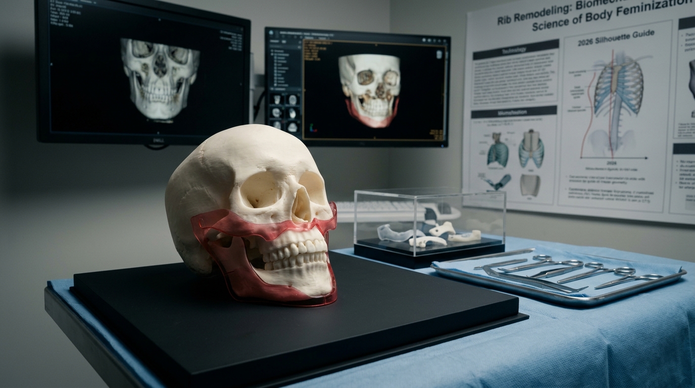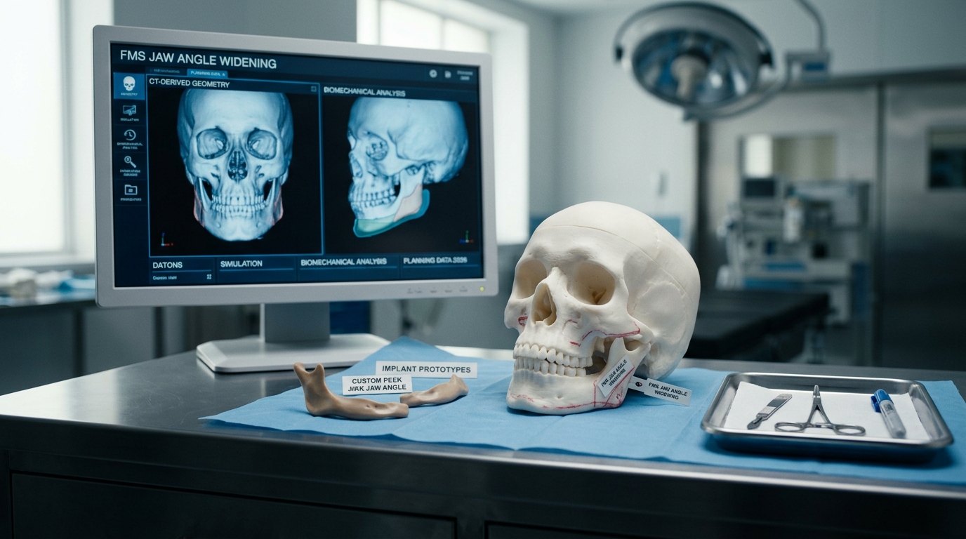
In modern facial feminization surgery, type 3 cranioplasty ct measurement analysis serves as the essential diagnostic standard for safe frontal bone remodeling. The frontal sinus anatomy varies dramatically among individuals, making blind surgical intervention hazardous. Patients planning upper-facial gender affirmation can examine our comprehensive forehead contouring procedures to discover how virtual surgical simulation guarantees protective sinus wall preservation.
Table of Contents
Biomechanical Thresholds in Type 3 Cranioplasty CT Measurement
In feminine craniofacial remodeling, type 3 cranioplasty ct measurement dictates whether simple bone shaving (Type 1) or complete anterior sinus wall osteotomy and setback (Type 3) is indicated. Sub-millimeter thin-slice computed tomography (0.5 mm to 0.75 mm collimation) with 3D bone window reconstructions allows exact mapping of bone density. According to craniofacial surgical metrics established by the National Institutes of Health (NIH), preserving mucosal vascularity while osteotomizing the frontal table eliminates secondary osteonecrosis risks.
When type 3 cranioplasty ct measurement identifies anterior table thickness exceeding 4.5 mm with minimal pneumatization, modest burring may suffice. However, in over 85% of assigned male at birth (AMAB) crania, substantial sinus volume demands true segment osteotomy to push the bony prominence backward safely.
Step-by-Step Surgical Execution and Osteotomy Protocols
A rigorous surgical plan driven by type 3 cranioplasty ct measurement follows standardized safety checkpoints across each operative phase:
- High-Resolution Pre-Surgical Mapping: Direct volumetric assessment of the inter-sinus septum and supraorbital nerve exit notches.
- Piezosurgical Osteotomy: Ultrasonic bone cutters incise the anterior wall cleanly without tearing the underlying sinus lining.
- Sinus Floor Inspection: Mucosal debridement and patency verification of the frontonasal duct prevent post-operative mucocele formation.
- Rigid Internal Micro-Fixation: The recessed bone flap is secured with 1.0 mm titanium or resorbable plates flush against the frontal bone plane.
Adhering strictly to type 3 cranioplasty ct measurement parameters prevents accidental intracranial entry and ensures symmetrical supraorbital margin transition across both orbits.
Long-Term Telemetry and Aesthetic Cranial Longevity
Clinical telemetry following type 3 cranioplasty ct measurement guidance demonstrates zero palpable step-offs and complete bone re-consolidation within 12 weeks. When combined with simultaneous hairline lowering or endoscopic browpexy, the resulting forehead contour reflects harmonious feminine curves without visible hardware.
Patients seeking feminizing frontal surgery must verify that their surgical provider utilizes calibrated type 3 cranioplasty ct measurement and virtual surgical planning prior to scheduling.
How CT Measurement Supports Type 3 Cranioplasty Planning
Type 3 cranioplasty CT measurement supports planning by showing frontal bone thickness, sinus boundaries, orbital relationships, and the available setback corridor. The scan is not a substitute for clinical examination. It is one dataset used to decide whether a setback, reconstruction, contouring, custom implant, or combined approach is appropriate. Image quality, slice thickness, patient positioning, and interpretation all influence the usefulness of the plan.
A responsible consultation should explain how the frontal sinus is protected, how the anterior table is managed, how fixation is selected, and which findings could change the operative plan. Patients should also receive clear information about swelling, numbness, scar care, infection, contour irregularity, revision surgery, and the limits of prediction from a three-dimensional model. The target is facial balance, not an isolated measurement.
Patients researching type 3 cranioplasty CT measurement can review the broader forehead contouring guide and bring their imaging questions to a qualified craniofacial surgeon. Treatment decisions should follow an individual examination and imaging review rather than a generic thickness threshold.
Ask whether the imaging review includes the frontal sinus, orbital rims, scalp soft tissues, and any previous reconstruction. These details help the team explain realistic contour limits, protect nearby structures, and select the least aggressive option that meets the patient’s goals. Written instructions should be provided.


Why is CT used for type 3 cranioplasty planning?
CT can show frontal bone thickness, frontal sinus boundaries, orbital relationships, and the available contouring corridor. It supports planning but does not replace examination.
Does one CT measurement determine candidacy for type 3 cranioplasty?
No. Candidacy depends on the complete anatomy, sinus position, bone quality, soft tissues, previous surgery, symptoms, goals, and the surgeon’s assessment.
What complications should patients discuss?
Discuss swelling, numbness, infection, contour irregularity, scar care, sinus-related concerns, revision surgery, and the limits of prediction from a three-dimensional model.