Canthopexy Overcorrection Prevention: 2026 Vector Guide

Canthopexy overcorrection prevention eyelid anatomy

In aesthetic periorbital and cat eye surgery, canthopexy overcorrection prevention strategies represent the primary safeguard against post-operative eyelid malposition and unnatural ocular distortion. Elevating the lateral canthus to achieve an exotic almond or feline tilt requires nuanced vector alignment. Patients considering delicate periorbital enhancements can explore our dedicated cat eyes surgery protocols to understand how canthopexy coordinates with temporal brow lifting.

Biomechanical Mechanics of Canthopexy Overcorrection Prevention

Proper canthopexy overcorrection prevention relies on anchoring the lateral retinacular tendon to the internal periosteum of Whitnall tubercle, located 3 mm to 4 mm posterior to the lateral orbital rim. Fixing sutures too anteriorly pulls the lower eyelid away from the ocular globe, inducing nocturnal lagophthalmos and chronic dry eye syndrome. According to periorbital reconstructive safety literature cataloged by the National Institutes of Health (NIH), preserving posterior vector tension is vital for ocular surface protection.

Surgeons must account for natural tissue relaxation. While mild 1 mm immediate post-operative overcorrection is intentional to compensate for gravity, true canthopexy overcorrection prevention avoids acute angulation exceeding 15 degrees relative to the horizontal intercanthal axis.

Technical Steps for Controlled Vector Suspension

Surgical success in canthopexy overcorrection prevention involves meticulous micro-dissection protocols:

  • Retinacular Release: Controlled mobilization of the inferior crus of the lateral canthal tendon without detaching superior ligamentous attachments.
  • Posterior Orbital Rim Anchoring: Placement of non-absorbable 4-0 or 5-0 braided sutures directly through the periosteum inside the lateral orbital margin.
  • Ocular Globe Coaptation Check: Verifying that the lower lid margin maintains snug, frictionless contact against the bulbar conjunctiva upon forced upward gaze.
  • Orbicularis Muscle Resuspension: Supporting the lateral orbicularis muscle strap prevents secondary gravitational lower lid descent.

By executing these measures, canthopexy overcorrection prevention preserves natural blinking dynamics while delivering crisp canthal obliquity.

Long-Term Telemetry and Aesthetic Harmony

Longitudinal clinical audits demonstrate that structured canthopexy overcorrection prevention reduces revision rates for lateral ectropion and canthal webbing to under 1.2%. When paired with modern deep temporal lifting, the eye displays energetic, youthful rejuvenation without a startled surgical appearance.

Patients seeking periorbital refinement should discuss canthopexy overcorrection prevention and vector simulations during in-depth pre-surgical consultations.

Patient Selection and Follow-Up for Canthopexy Overcorrection Prevention

Canthopexy overcorrection prevention begins before surgery. Assessment should include eyelid laxity, lower-lid tone, globe prominence, tear-film symptoms, previous blepharoplasty, scar tissue, facial asymmetry, and the patient’s desired canthal angle. A conservative plan respects the eyelid’s relationship with the globe and avoids treating a photographic trend as a universal anatomical target.

Follow-up focuses on closure, corneal comfort, swelling, wound healing, lid position, and symmetry. Temporary asymmetry can occur while tissues settle, but increasing pain, marked redness, vision changes, inability to close the eye, or severe dryness requires prompt clinical assessment. Recovery instructions should cover lubrication, sleep position, activity limits, sun protection, and the timing of aesthetic evaluation.

Canthopexy overcorrection prevention should be individualized rather than reduced to a single angle or millimeter value. Patients can review the wider cat eyes surgery program to understand how canthopexy relates to brow, temple, and lower-eyelid planning. A qualified oculoplastic or craniofacial surgeon should confirm candidacy and explain revision options.

Photographs, ocular-surface symptoms, previous operations, and baseline eyelid position should be recorded before treatment. This documentation supports safer planning and makes postoperative changes easier to interpret during follow-up. Emergency contact details should be clear.


Canthopexy overcorrection prevention vector diagram
Canthopexy overcorrection prevention clinical follow-up

What does canthopexy overcorrection prevention involve?

It involves preoperative eyelid assessment, conservative vector planning, stable fixation, protection of ocular-surface function, and structured postoperative follow-up.

Which symptoms need prompt postoperative assessment?

Vision changes, inability to close the eye, severe dryness, increasing pain, marked redness, or rapidly worsening swelling require prompt clinical assessment.

Can one canthal angle guarantee a good result?

No. Aesthetic targets must be individualized to eyelid laxity, globe prominence, tear-film symptoms, asymmetry, previous surgery, and facial proportions.


Type 3 Cranioplasty CT Measurement: 2026 Guide

Type 3 cranioplasty CT measurement medical planning

In modern facial feminization surgery, type 3 cranioplasty ct measurement analysis serves as the essential diagnostic standard for safe frontal bone remodeling. The frontal sinus anatomy varies dramatically among individuals, making blind surgical intervention hazardous. Patients planning upper-facial gender affirmation can examine our comprehensive forehead contouring procedures to discover how virtual surgical simulation guarantees protective sinus wall preservation.

Biomechanical Thresholds in Type 3 Cranioplasty CT Measurement

In feminine craniofacial remodeling, type 3 cranioplasty ct measurement dictates whether simple bone shaving (Type 1) or complete anterior sinus wall osteotomy and setback (Type 3) is indicated. Sub-millimeter thin-slice computed tomography (0.5 mm to 0.75 mm collimation) with 3D bone window reconstructions allows exact mapping of bone density. According to craniofacial surgical metrics established by the National Institutes of Health (NIH), preserving mucosal vascularity while osteotomizing the frontal table eliminates secondary osteonecrosis risks.

When type 3 cranioplasty ct measurement identifies anterior table thickness exceeding 4.5 mm with minimal pneumatization, modest burring may suffice. However, in over 85% of assigned male at birth (AMAB) crania, substantial sinus volume demands true segment osteotomy to push the bony prominence backward safely.

Step-by-Step Surgical Execution and Osteotomy Protocols

A rigorous surgical plan driven by type 3 cranioplasty ct measurement follows standardized safety checkpoints across each operative phase:

  • High-Resolution Pre-Surgical Mapping: Direct volumetric assessment of the inter-sinus septum and supraorbital nerve exit notches.
  • Piezosurgical Osteotomy: Ultrasonic bone cutters incise the anterior wall cleanly without tearing the underlying sinus lining.
  • Sinus Floor Inspection: Mucosal debridement and patency verification of the frontonasal duct prevent post-operative mucocele formation.
  • Rigid Internal Micro-Fixation: The recessed bone flap is secured with 1.0 mm titanium or resorbable plates flush against the frontal bone plane.

Adhering strictly to type 3 cranioplasty ct measurement parameters prevents accidental intracranial entry and ensures symmetrical supraorbital margin transition across both orbits.

Long-Term Telemetry and Aesthetic Cranial Longevity

Clinical telemetry following type 3 cranioplasty ct measurement guidance demonstrates zero palpable step-offs and complete bone re-consolidation within 12 weeks. When combined with simultaneous hairline lowering or endoscopic browpexy, the resulting forehead contour reflects harmonious feminine curves without visible hardware.

Patients seeking feminizing frontal surgery must verify that their surgical provider utilizes calibrated type 3 cranioplasty ct measurement and virtual surgical planning prior to scheduling.

How CT Measurement Supports Type 3 Cranioplasty Planning

Type 3 cranioplasty CT measurement supports planning by showing frontal bone thickness, sinus boundaries, orbital relationships, and the available setback corridor. The scan is not a substitute for clinical examination. It is one dataset used to decide whether a setback, reconstruction, contouring, custom implant, or combined approach is appropriate. Image quality, slice thickness, patient positioning, and interpretation all influence the usefulness of the plan.

A responsible consultation should explain how the frontal sinus is protected, how the anterior table is managed, how fixation is selected, and which findings could change the operative plan. Patients should also receive clear information about swelling, numbness, scar care, infection, contour irregularity, revision surgery, and the limits of prediction from a three-dimensional model. The target is facial balance, not an isolated measurement.

Patients researching type 3 cranioplasty CT measurement can review the broader forehead contouring guide and bring their imaging questions to a qualified craniofacial surgeon. Treatment decisions should follow an individual examination and imaging review rather than a generic thickness threshold.

Ask whether the imaging review includes the frontal sinus, orbital rims, scalp soft tissues, and any previous reconstruction. These details help the team explain realistic contour limits, protect nearby structures, and select the least aggressive option that meets the patient’s goals. Written instructions should be provided.

Type 3 cranioplasty CT measurement anatomical diagram
Type 3 cranioplasty CT measurement clinical follow-up

Why is CT used for type 3 cranioplasty planning?

CT can show frontal bone thickness, frontal sinus boundaries, orbital relationships, and the available contouring corridor. It supports planning but does not replace examination.

Does one CT measurement determine candidacy for type 3 cranioplasty?

No. Candidacy depends on the complete anatomy, sinus position, bone quality, soft tissues, previous surgery, symptoms, goals, and the surgeon’s assessment.

What complications should patients discuss?

Discuss swelling, numbness, infection, contour irregularity, scar care, sinus-related concerns, revision surgery, and the limits of prediction from a three-dimensional model.

Rib Remodeling Body Feminization: 2026 Silhouette Guide

rib remodeling body feminization complete guide 2026

Achieving an accentuated hourglass silhouette often requires targeting thoracic frame width, where rib remodeling body feminization represents the most advanced minimally invasive surgical innovation for dramatic waist narrowing. Transgender and cisgender women with wider lower rib cages frequently encounter anatomical plateaus with soft tissue liposuction alone. Patients evaluating comprehensive body sculpting options can explore our dedicated body feminization surgery procedures to understand how skeletal remodeling combines with high-definition liposculpture.

rib remodeling body feminization architecture and process flow diagram 2026

Biomechanical Science of Rib Remodeling Body Feminization

Historic body sculpting relied on radical rib resection, which removed segments of the 11th and 12th floating ribs entirely. In contemporary rib remodeling body feminization protocols, surgeons utilize ultrasonic piezosurgery. Ultrasonic acoustic cavitation enables selective cortical weakening without damaging the adjacent neurovascular bundle or parietal pleura. According to clinical reconstructive safety data cataloged by the National Institutes of Health (NIH), ultrasonic bone sculpting significantly minimizes periosteal hematoma formation and soft tissue trauma.

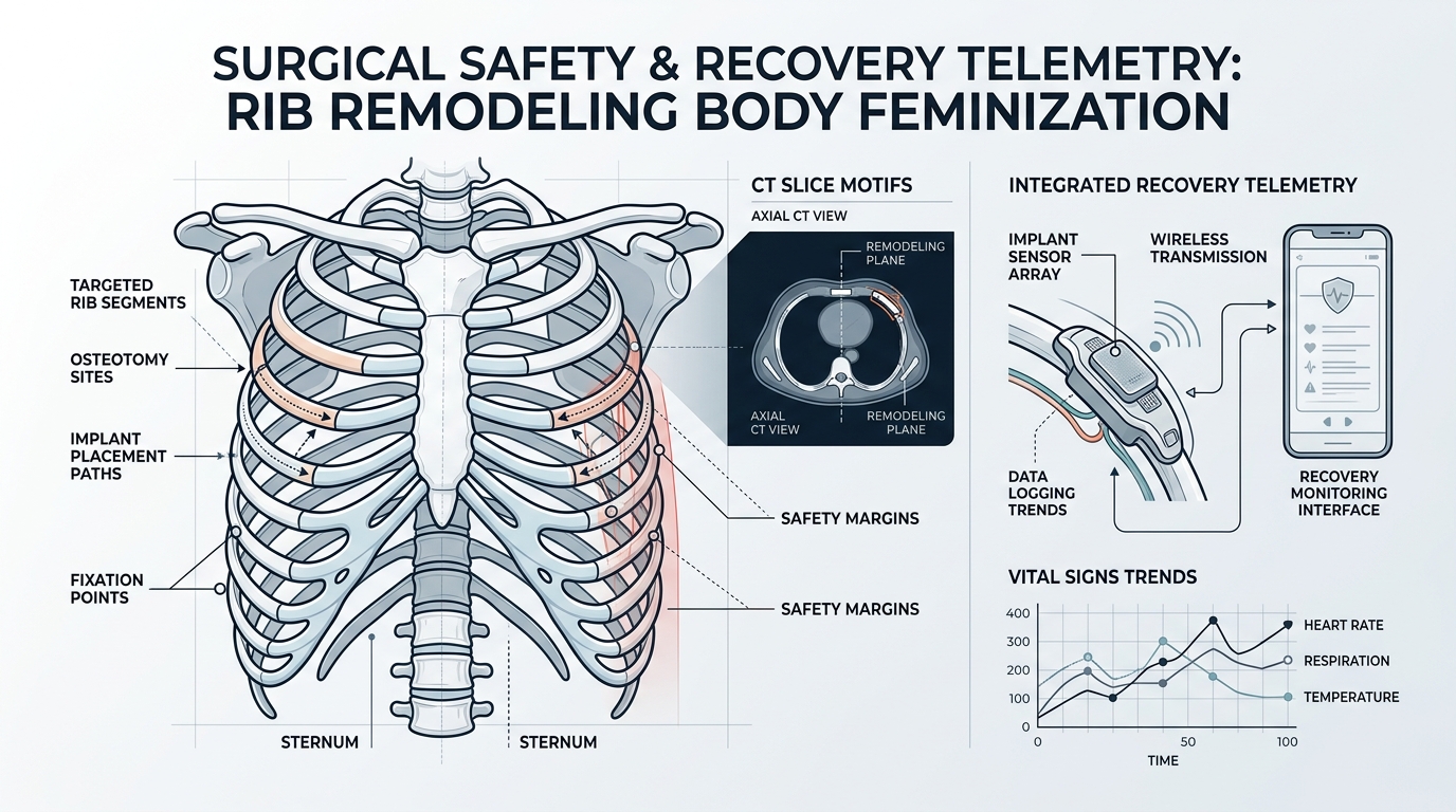
By producing partial osteotomies (greenstick remodeling), the costal cartilages can be flexed inward by 3 to 7 centimeters bilaterally. This preserve vital organ protection while creating a permanently tapered lateral silhouette.

Surgical Safety and Recovery Telemetry: Rib Remodeling Body Feminization

Comparative surgical analysis between traditional resection and ultrasonic rib remodeling body feminization highlights compelling clinical advantages:

  • Micro-Incisions: Access requires only tiny 1.5 cm dorsal puncture incisions hidden along natural skin crease lines.
  • Pleural Integrity: Preservation of the inner costal cortex drastically reduces pneumothorax risk down to under 0.2%.
  • Pain Management: Intact intercostal nerve pathways allow patients to mobilize comfortably within 24 hours post-operatively.
  • Consolidation Phase: Controlled external compression stabilizes osseous union within 60 to 90 days.

Post-operative compliance is paramount during rib remodeling body feminization. Wearing an orthopedic waist compression device guarantees that the ribs mend in their refined inward contour.

Aesthetic Integration with Gluteal and Hip Contouring

rib remodeling body feminization performance metrics and analytics dashboard 2026

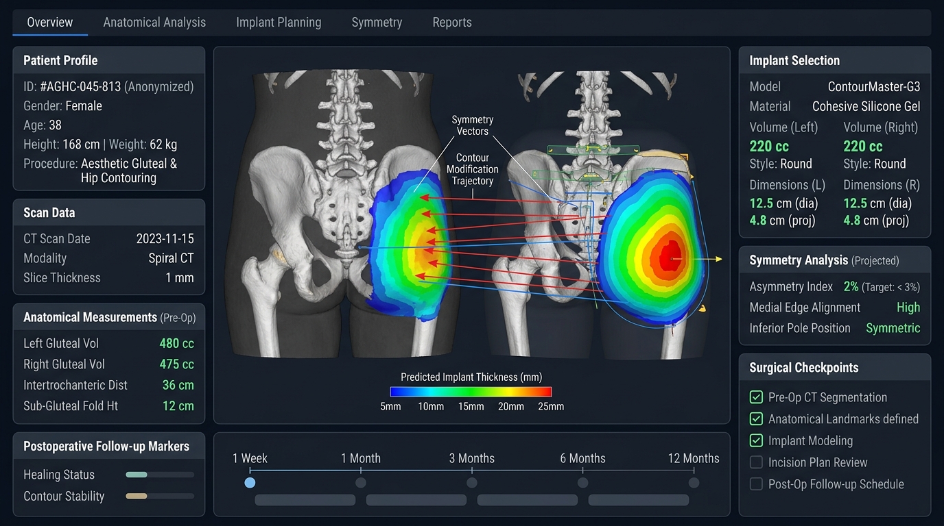
The true power of rib remodeling body feminization lies in its compounding effect on overall waist-to-hip ratio (WHR). When paired with autologous fat transfer (BBL) and lateral hip dip filling, narrowing the lower ribcage creates an optical and volumetric transition that soft tissue work alone cannot duplicate. Candidates considering rib remodeling body feminization should consult board-certified craniofacial and body specialists to assess costal anatomy via low-dose chest CT.

Safety Planning for Rib Remodeling Body Feminization

Rib remodeling body feminization requires careful distinction between contour refinement and removal of structural bone. A conservative remodeling plan aims to influence the lower thoracic silhouette while protecting chest-wall stability, breathing mechanics, intercostal structures, and the soft-tissue envelope. The appropriate technique depends on rib anatomy, cartilage flexibility, prior surgery, body composition, and the target waist-to-hip proportion.

Preoperative planning should document the patient’s medical history, respiratory status, scars, asymmetry, and expectations. Ask how imaging is used, how pain is managed, what activity restrictions apply, and which symptoms require urgent review. Recovery varies, and visible swelling can temporarily obscure the final silhouette. Long-term assessment should consider comfort, contour stability, breathing, sensation, and whether additional body-feminization procedures are being staged.

Rib remodeling body feminization should be discussed with a surgeon experienced in chest-wall anatomy and gender-affirming contour surgery. The broader body feminization surgery guide provides context for combining waist, hip, and soft-tissue procedures without treating one measurement as the sole definition of a successful result.

Rib remodeling body feminization clinical planning

What is the goal of rib remodeling in body feminization?

The goal is controlled contour refinement while respecting chest-wall stability, breathing mechanics, intercostal structures, and the soft-tissue envelope.

Is rib remodeling the same as removing ribs?

No. The techniques and extent of treatment differ. The exact approach depends on anatomy, cartilage flexibility, medical history, and the planned silhouette.

What should recovery planning include?

Recovery planning should cover pain control, activity restrictions, breathing symptoms, swelling, wound care, warning signs, and the timing of final contour assessment.

FMS Jaw Angle Widening Peek: 2026 Definitive Guide

fms jaw angle widening peek complete guide 2026

Achieving a prominent, sharply defined lower facial contour requires structured planning, and fms jaw angle widening peek surgery represents the premier clinical solution for transmasculine and cisgender male patients seeking structural lower-third definition. A well-projected gonial angle establishes the foundation for facial masculinity. Patients exploring comprehensive craniofacial transformations can review our advanced facial masculinization surgery protocols to understand holistic lower jaw sculpting.

fms jaw angle widening peek architecture and process flow diagram 2026

Biomechanical Advantages of FMS Jaw Angle Widening Peek

When engineering mandibular lateralization, fms jaw angle widening peek techniques utilize computer-aided design and virtual surgical planning (VSP). Polyetheretherketone is an osteoconductive-compatible biomaterial with a compressive strength closely mirroring human mandibular cortical bone. Based on craniofacial reconstructive principles documented by the National Institutes of Health (NIH), rigid internal fixation of patient-matched implants prevents the bony resorption commonly induced by older elastomer devices.

Unlike off-the-shelf silicone blocks, each fms jaw angle widening peek prosthesis is milled to interlock flawlessly with the ascending ramus and mandibular base. This precise anatomical seating neutralizes hardware step-offs, ensuring that masseteric contractions do not dislodge the implant over time.

Surgical Execution and Masseteric Reattachment Protocols

fms jaw angle widening peek performance metrics and analytics dashboard 2026

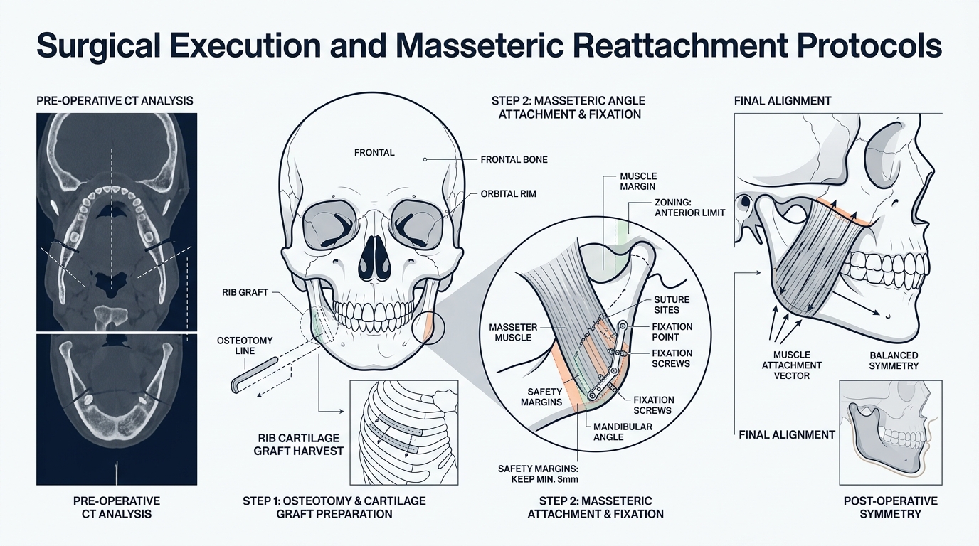
Executing an fms jaw angle widening peek procedure demands rigorous surgical precision through an entirely intraoral approach:

  • Transoral Incision: Lateral vestibular incisions preserve mental nerve sensation while exposing the masseteric sling without external cutaneous scars.
  • Subperiosteal Pocket Creation: Dissection extends strictly along the subperiosteal plane to protect the facial artery and marginal mandibular branch of the facial nerve.
  • Dual Titanium Screw Fixation: Low-profile bicortical micro-screws secure the PEEK implant firmly against the gonial angle to prevent mechanical micromotion.
  • Pterygomasseteric Sling Suspension: Careful resuturing of the masseter muscle over the expanded implant lateral border ensures full masticatory power and seamless soft tissue drape.

This systematic methodology ensures that fms jaw angle widening peek interventions yield immediate structural definition with minimal morbidity.

Long-Term Telemetry and Aesthetic Harmony: FMS Jaw Angle Widening Peek

fms jaw angle widening peek implementation and case study 2026

Longitudinal surgical telemetry confirms that patient cohorts undergoing fms jaw angle widening peek experience high satisfaction rates exceeding 95%. Because PEEK exhibits zero radiopacity artifacts on magnetic resonance imaging, routine surveillance remains crystal clear throughout the patient lifetime.

The strategic deployment of fms jaw angle widening peek delivers durable lateral jaw flare, transforming an obtuse or rounded jawline into an authoritative, squared masculine profile. Specialized surgical consultation with pre-operative 3D cephalometric analysis is critical to tailoring lateral and vertical vectors to individual cranial proportions.

Planning FMS Jaw Angle Widening With Custom PEEK

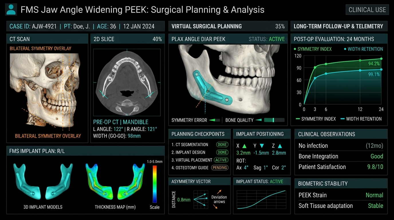
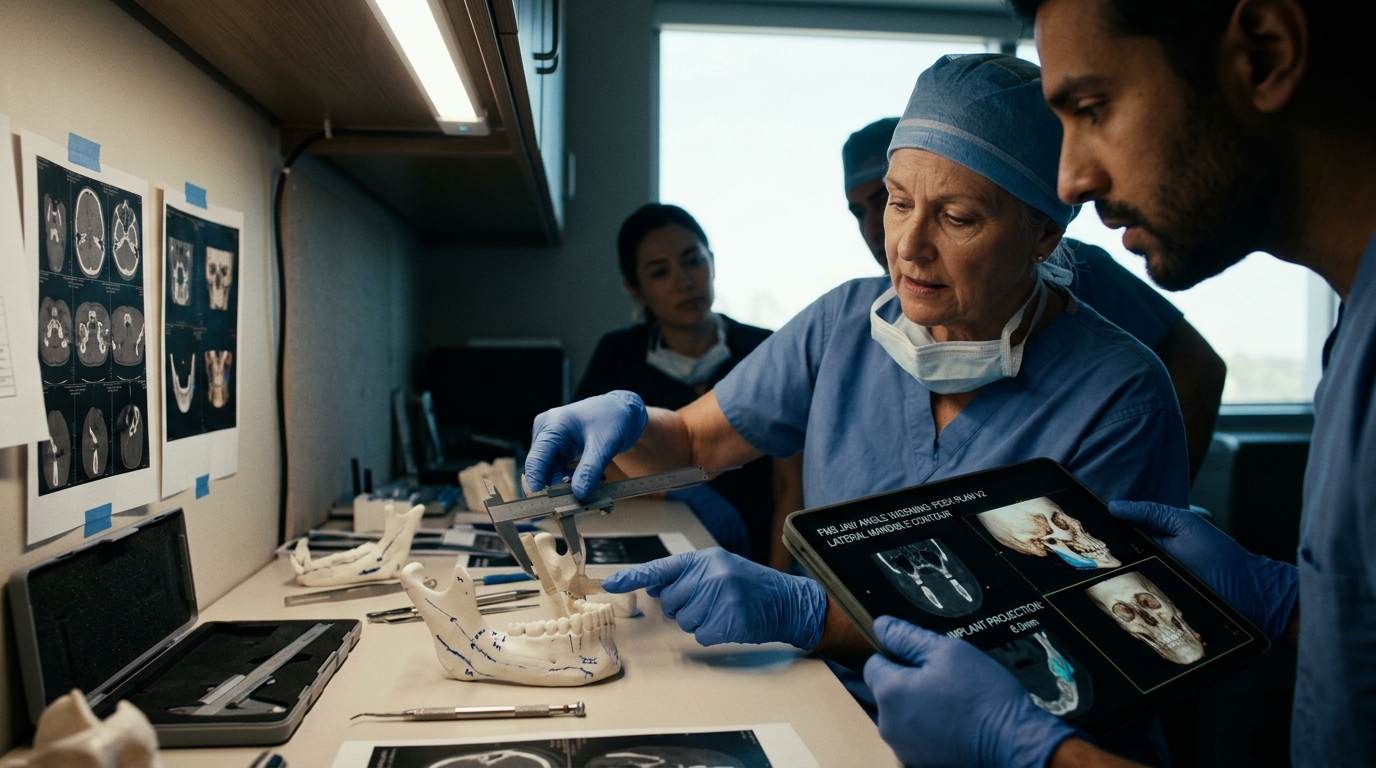
FMS jaw angle widening with custom PEEK is planned around the patient’s existing mandibular width, gonial angle, masseter contour, occlusion, and facial proportions. A custom implant should improve the transition from the ramus to the lower border without creating a sharp or isolated corner. Three-dimensional imaging helps the surgical team compare symmetry, projection, fixation points, and the relationship between the implant and nearby nerves.
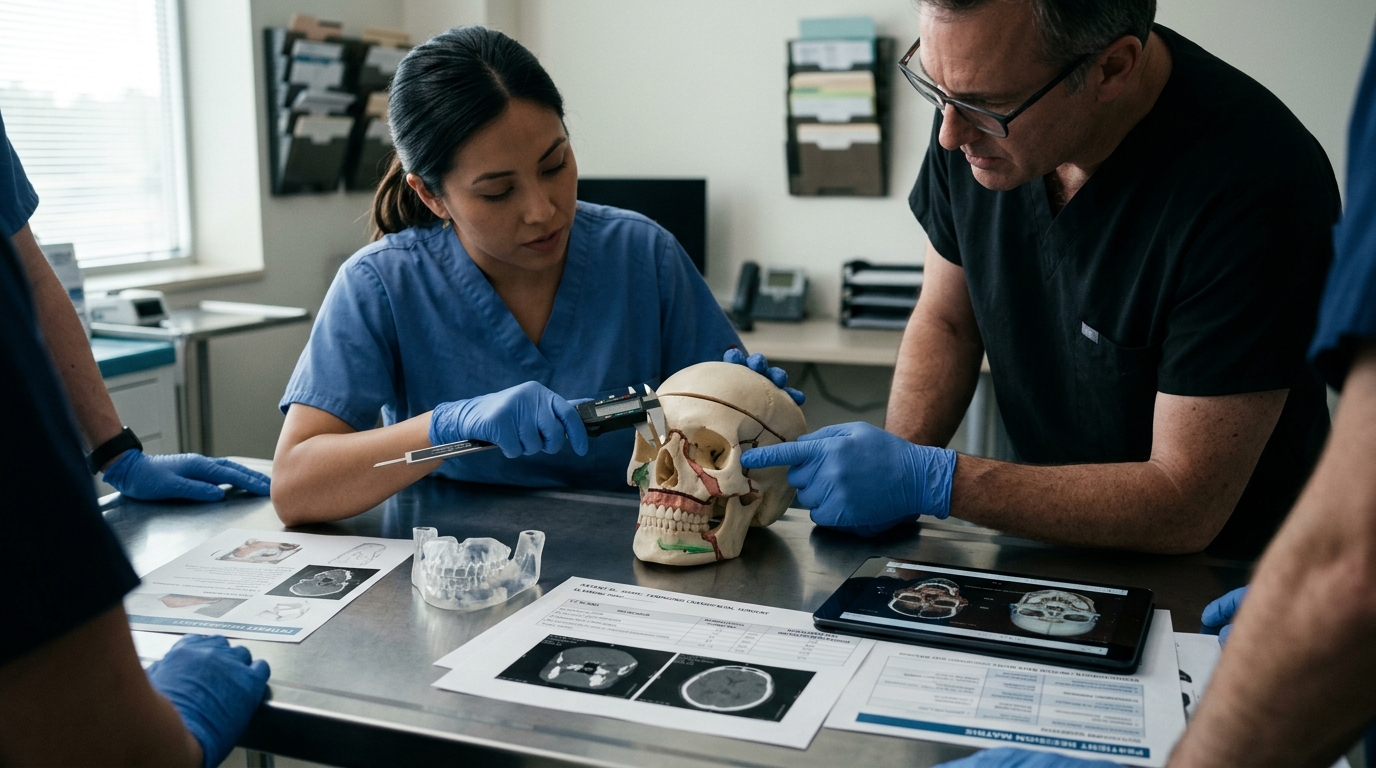
During consultation, discuss implant thickness, edge transitions, incision access, fixation, swelling, sensory changes, infection prevention, and revision options. The desired masculine contour is not defined by maximum width alone. Balanced lateral projection, a controlled gonial angle, and harmony with the chin and midface generally produce a more coherent result. Pre-existing asymmetry should be documented before surgery so that expectations remain realistic.

Custom PEEK is a planning option, not a guarantee of a specific appearance. Final selection depends on anatomy, medical history, imaging, and the surgeon’s assessment. Patients can review the broader facial masculinization surgery program to understand how jaw angle widening may fit into a coordinated treatment plan.

What does custom PEEK add to FMS jaw angle widening?

Custom PEEK allows patient-specific planning of lateral projection, gonial angle, symmetry, and edge transitions using three-dimensional imaging.

Does wider always mean a more masculine jaw?

No. A balanced mandibular contour also depends on chin projection, facial width, gonial angle, soft tissue, and pre-existing asymmetry.

What risks should be discussed before jaw angle implant surgery?

Discuss infection, sensory changes, implant movement, contour asymmetry, swelling, fixation, recovery limits, and possible revision surgery with the treating surgeon.

Titanium Mesh vs PEEK FFS: 2026 Biocompatibility Guide

titanium mesh vs peek ffs complete guide 2026

In modern facial feminization surgery, evaluating titanium mesh vs peek ffs implants is one of the most critical structural decisions for achieving natural, long-lasting facial balance and skeletal symmetry. Facial gender affirmation requires millimeter precision, particularly across the upper third and lower jawline. Patients preparing for facial gender affirmation surgery can explore our comprehensive facial feminization surgery procedures to understand how craniofacial biomaterials integrate into comprehensive multi-stage treatment plans.

titanium mesh vs peek ffs architecture and process flow diagram 2026

Biomechanical Properties of Titanium Mesh vs PEEK FFS

When analyzing titanium mesh vs peek ffs applications, biomaterial engineering dictates surgical longevity. High-grade titanium provides unmatched tensile malleability, allowing intraoperative micro-adaptation across complex three-dimensional contours. According to biomaterial research indexed by the National Institutes of Health (NIH), titanium facilitates direct fibrovascular osseointegration through micro-porous architectural lattices, substantially reducing late implant migration risks.

Conversely, polyetheretherketone (PEEK) is a high-performance semi-crystalline thermoplastic with an elasticity modulus remarkably close to human cortical bone. In the clinical realm of titanium mesh vs peek ffs, custom patient-specific PEEK implants are milled using high-resolution 3D CT reconstructions. This guarantees seamless anatomical margins without the risk of palpable step-offs along the supraorbital rim or temporal fossa.

Long-Term Revision Rates: Titanium Mesh vs PEEK FFS in Practice

titanium mesh vs peek ffs performance metrics and analytics dashboard 2026

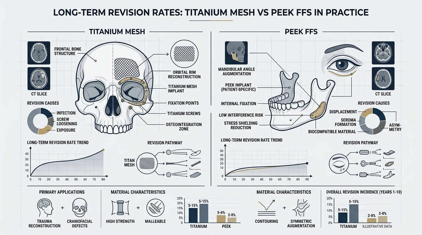
Long-term surgical telemetry reveals distinct performance profiles between titanium mesh vs peek ffs reconstructions over 5 to 10-year follow-up intervals across international centers:

  • Radiological Imaging: PEEK implants remain completely radiolucent on post-operative MRI and CT scans, preventing scatter artifacts during routine cranial imaging.
  • Thermal Conductivity: PEEK serves as a natural thermal insulator, eliminating winter cold sensations often reported with metallic craniofacial hardware.
  • Structural Load Bearing: Titanium mesh delivers exceptional crush resistance over reconstructed frontal sinus cavities when managing extensive posterior wall bone loss.
  • Infection and Extrusion Rates: Both biomaterials demonstrate remarkably low infection rates (under 1.5%) when placed beneath well-vascularized pericranial flaps.

Craniofacial teams evaluate patient soft tissue thickness before finalizing titanium mesh vs peek ffs implant geometries. When subgaleal dissection reveals an ultra-thin dermis, custom feather-edged PEEK reduces visible contour boundaries. In contrast, traumatic or secondary cases with significant anterior wall comminution benefit from the rigidity of pre-formed titanium mesh fixation.

Surgical Execution and Fixation Protocols

titanium mesh vs peek ffs implementation and case study 2026

Fixation dynamics differ considerably between these systems. Titanium mesh is anchored using self-drilling 1.0mm to 1.5mm micro-screws, creating immediate rigid fixation across bony ledges. With PEEK implants, low-profile counter-sunk screw holes ensure hardware remains impalpable even under high-tension coronal closures. The choice between titanium mesh vs peek ffs therefore impacts operative timing, with custom PEEK reducing operative shaping time substantially.

Biocompatibility studies continue to affirm that both options promote stable soft tissue adherence when subperiosteal vascularity is respected. Virtual surgical planning enables surgical simulation before entering the operating suite, optimizing aesthetic curves while protecting sinus mucosa.

Patient Decision Matrix for Feminizing Biomaterials

Prospective candidates should evaluate anatomical criteria, budget considerations, and long-term functional goals. While custom computer-aided design for PEEK requires advance manufacturing lead time, its anatomical fidelity is unparalleled. Meanwhile, titanium mesh offers exceptional versatility in emergent revisions. Consulting experienced craniofacial specialists ensures the correct material aligns with your unique forehead anatomy.

Choosing Between Titanium Mesh and PEEK in FFS

The choice between titanium mesh and PEEK in FFS depends on anatomy, defect size, soft-tissue coverage, imaging findings, and the surgeon’s reconstructive plan. Titanium offers a strong, thin framework and is commonly considered when rigid contour support is required. PEEK allows patient-specific computer-aided design and a stiffness profile closer to cortical bone, which may be useful when a custom contour is the priority. Neither material is automatically safer for every patient.

Planning should begin with a high-resolution CT assessment and a review of the frontal sinus, orbital rims, bone thickness, prior hardware, and available soft-tissue coverage. The consultation should also address infection risk, palpability, fixation, revision access, expected swelling, and the possibility that the final contour may require staged treatment. Material selection is only one part of the result; accurate design, sterile handling, stable fixation, and appropriate follow-up matter equally.

Patients should compare titanium mesh and PEEK in FFS with a qualified craniofacial surgeon rather than choosing by material name alone. A useful consultation includes the proposed implant design, alternatives, limitations, recovery milestones, and a clear revision plan. For broader procedure context, review the facial feminization surgery pillar before making a treatment decision.

How are titanium mesh and PEEK selected in FFS?

Selection depends on CT anatomy, defect size, soft-tissue coverage, prior surgery, fixation needs, and the surgeon’s reconstructive plan. Neither material is universally best.

Is PEEK automatically safer than titanium for facial feminization?

No. PEEK offers patient-specific design, while titanium provides rigid structural support. Infection, fixation, contour, and revision risks depend on the patient and the complete surgical technique.

What should a consultation for a custom FFS implant include?

The consultation should cover CT findings, implant design, fixation, alternatives, recovery, warning symptoms, expected limitations, and a revision plan.