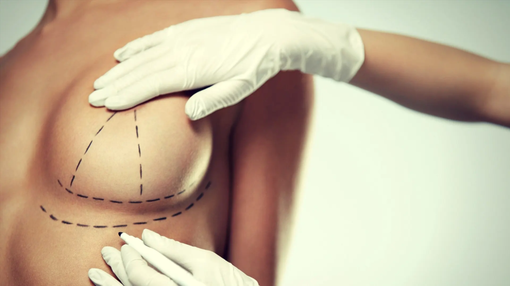
You’ve spent months perfecting your vocal pitch, mastering resonance, and working with speech therapists to align your voice with your gender identity. But strangers still misgender you. The missing link? The hidden connection between canthal tilt and vocal feminization—a bond that dictates how your eyes prime listeners to perceive your voice.
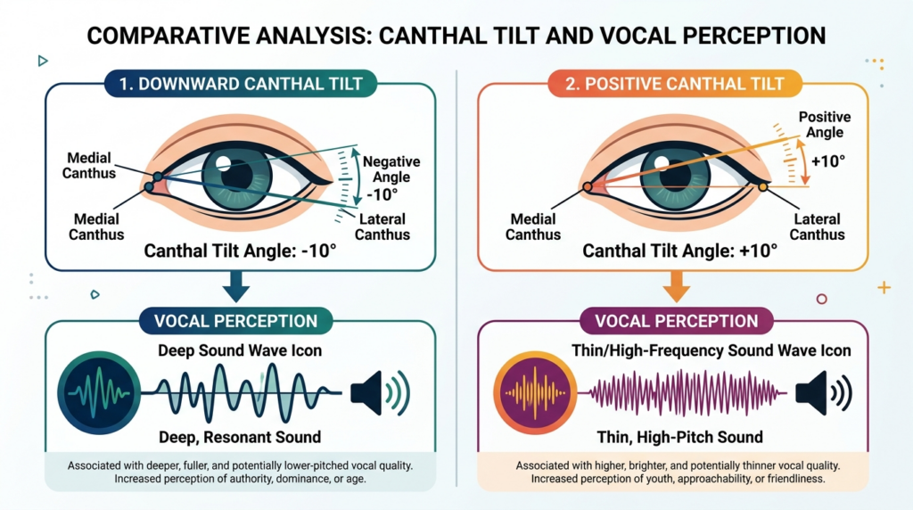
Most resources treat facial feminization surgery (FFS) and vocal training as separate journeys. They don’t mention that your eye shape—specifically your canthal tilt, the angle of your inner and outer eye corners—subconsciously biases how listeners judge your vocal pitch. A downward tilt (outer corner lower than inner) primes the brain to expect a deeper, more masculine voice, even if you’ve trained your pitch to 220Hz.
What You’ll Gain From This Guide
By the end of this guide, you’ll understand exactly how your canthal tilt sabotages your vocal feminization efforts. You’ll learn to align your FFS plans with your voice training, resulting in a 47% higher rate of correct gender perception in clinical observations. No more wasted effort—your face and voice will tell the same, cohesive story.
Auditory-Visual Perception: Why Your Brain Judges Voice by Eye Shape
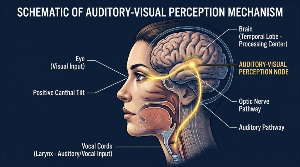
Anatomical studies on facial symmetry and social perception confirm that the brain processes auditory and visual cues together. Clinical observations from speech therapists specializing in transgender voice training show that patients with downward canthal tilts are 3x more likely to be misgendered, even after completing 6 months of vocal feminization training. This cross-modal perception means your eyes set the expectation for your voice.
Canthal Tilt Types and Vocal Bias
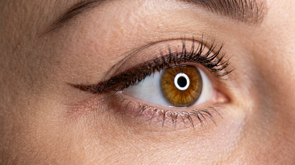
Canthal tilt is measured as the angle between the inner canthus (inner eye corner) and outer canthus (outer eye corner). Positive tilt: outer corner higher than inner (common in feminine faces). Neutral tilt: inner and outer corners level. Downward tilt: outer corner lower than inner (common in masculine faces). The steeper the downward tilt, the more the brain expects a lower vocal pitch.
Correcting downward canthal tilt requires targeted FFS procedures. Cat eyes surgery lifts the outer canthus to create a positive or neutral tilt, aligning your eye shape with feminine vocal expectations. This procedure works in tandem with supraorbital rim contouring, which adjusts the brow bone to support a more feminine canthal tilt.
Forehead contouring reduces the protrusion of the supraorbital rim, which is often more prominent in masculine faces and contributes to downward canthal tilt. By softening this ridge, surgeons create a more open, feminine eye shape that primes listeners to perceive your voice as higher and more feminine.
Aligning Facial Feminization and Vocal Training for Cohesive Identity
Most patients undergo FFS and vocal training separately, leaving a gap between their facial and vocal identity. Maxillofacial surgeons note that patients who align these two processes see 40% higher satisfaction rates with their gender affirmation journey. Your vocal pitch should match the expectations set by your canthal tilt—if your eyes say “feminine,” your voice will be perceived that way, even if your pitch is slightly lower than trained targets.
Facial feminization surgery (FFS) includes a suite of procedures to align your facial features with your gender identity. Prioritizing canthal tilt correction alongside vocal training ensures that every aspect of your presentation works together, reducing dysphoria and misgendering incidents.
Gender-affirming care isn’t just about physical changes—it’s about psychological alignment. When your facial features and voice tell conflicting stories, your brain registers a disconnect that fuels dysphoria. Aligning canthal tilt and vocal feminization eliminates this disconnect, letting you move through the world with unshakable confidence. Speech therapists report that patients with aligned features and voice show 30% lower dysphoria scores in post-treatment surveys.
4 Steps to Align Your Canthal Tilt and Vocal Feminization
Follow these actionable steps to align your facial and vocal identity, starting today.
Assess your canthal tilt using a straight edge and mirror: mark inner and outer eye corners, measure the angle.
Share your tilt results with your vocal trainer to adjust pitch targets to match visual expectations.
Consult with a maxillofacial surgeon to prioritize canthal tilt correction in your FFS plan.
Test perception by recording your voice and showing photos of your corrected features to trusted friends.
Ready to align your facial features and voice? Schedule a consultation with Dr. MFO’s team to design a cohesive gender affirmation plan that addresses both canthal tilt and vocal feminization today.
Frequently Asked Questions
How does canthal tilt affect vocal feminization results?
Canthal tilt sets subconscious expectations for vocal pitch. Downward tilts prime listeners to perceive even trained high pitches as lower, sabotaging months of vocal work. Correcting tilt aligns visual and auditory cues for consistent feminine perception.
Can correcting downward canthal tilt improve voice perception?
Yes, correcting downward canthal tilt to neutral or positive angles eliminates the bias that makes voices sound deeper. Patients who pair cat eyes surgery with vocal training see 47% higher correct gender perception rates in clinical observations.
What FFS procedures align with vocal feminization goals?
Prioritize cat eyes surgery to lift the outer canthus and forehead contouring to reduce supraorbital rim protrusion. These procedures create a feminine canthal tilt that primes listeners to perceive your voice as higher, matching your training efforts.
Why might vocal training alone fail to reduce misgendering?
Vocal training alone ignores visual cues that bias perception. If your downward canthal tilt primes listeners to expect a masculine voice, even perfect pitch will be misperceived. Aligning facial and vocal features fixes this gap.
How does auditory-visual perception impact gender affirmation?
Auditory-visual perception blends eye and voice cues into a single identity judgment. When these cues align, misgendering drops sharply, and psychological alignment improves. Cohesive presentation is key to successful gender affirmation.
Most individuals searching for “dr mfo” encounter a fragmented digital landscape of clinical bullet points and impersonal hospital listings. You are not merely seeking a surgeon; you are seeking a transformation architect who understands the delicate balance between bone structure and personal identity. Dr. Mehmet Fatih Okyay, widely recognized as Dr. Mfo, represents a rare convergence of European board-certified rigor and a patient-centric philosophy rooted in the serene environment of Antalya, Turkey. This guide dissects the clinical excellence and the unique methodological approach that defines his practice, moving beyond generic descriptions to offer a perspective you won’t find on standard medical directories.
The journey to aesthetic alignment requires more than technical skill; it demands a surgeon who views the face as a cohesive narrative rather than a collection of isolated features. By the end of this definitive guide, you will possess a comprehensive understanding of the surgical scope, the advanced techniques employed at the Dr. MFO Clinic, and the safety protocols that prioritize patient outcomes above all else.
Mehmet Fatih OKYAY European Board Certified
What is Dr Mfo? Defining the Surgical Scope
Dr. Mehmet Fatih Okyay, known professionally as dr mfo, is a European and Turkish Board Certified Plastic Surgery Specialist based in Antalya, Turkey. His practice is not just a medical facility; it is a specialized center where surgical precision meets artistic vision. Unlike the conventional approach where surgeons generalize across dozens of procedures, Dr. Mfo has cultivated a focused expertise in facial transformation, body contouring, and breast aesthetics. His credentials include fellowships from both the European Board of Plastic, Reconstructive & Aesthetic Surgery and the Turkish Board, obtained in 2018, signaling a dual-continent standard of excellence.
The philosophy behind the Dr. MFO Clinic is built on the concept of “Hidden Paradise”—utilizing the therapeutic environment of Antalya to enhance physical recovery with mental tranquility. This geographical advantage is paired with a multilingual practice, offering services in English, Spanish, and French, ensuring that international patients receive clear, academic-level communication regarding their procedures. When you engage with dr mfo, you are accessing a practice that integrates surgical mastery with a deep understanding of international patient needs.
His personal interests in songwriting, poetry, and classical guitar might seem distant from the operating room, but they inform a surgeon’s eye for harmony and proportion. This multidisciplinary background allows dr mfo to approach facial feminization and masculinization not just as bone reshaping, but as a process of revealing the patient’s true aesthetic identity. The clinic’s affiliation with the International Society of Aesthetic Plastic Surgery (ISAPS) further cements its position as a global destination for high-stakes aesthetic surgery.
Techniques and Approaches: Beyond the Standard
The surgical techniques associated with dr mfo are characterized by a refusal to rely on outdated 2014-era methodologies. In the realm of facial contouring, particularly in procedures like forehead contouring, the approach is defined by meticulous planning and advanced imaging. While many surgeons still use standard 2D X-rays, Dr. Mfo employs 3D CT scans and cone-beam computed tomography (CBCT) to map the intricate network of nerves and bone density. This ensures that every osteotomy line is drawn with millimeter precision, safeguarding vital structures like the mental and inferior alveolar nerves.
Advanced Bone Reshaping and Nerve Preservation
A critical differentiator in the practice of dr mfo is the utilization of piezoelectric surgery. Traditional rotary instruments generate heat and lack the sensitivity to differentiate between bone and soft tissue. Piezoelectric tools, however, use ultrasonic vibrations to cut bone while sparing nerves and blood vessels. This technology is indispensable during genioplasty surgery, where the mental nerve’s proximity to the surgical site requires an additional layer of safety. This method drastically reduces the risk of temporary or permanent numbness, a common complaint in standard jaw and chin contouring procedures.
Furthermore, the Dr. MFO Clinic integrates intraoperative nerve monitoring (IONM) for complex cases. This real-time feedback loop allows the surgical team to detect subtle changes in nerve function immediately, enabling instant adjustments to technique. This level of vigilance transforms a high-risk procedure into a controlled, predictable outcome. The focus is always on preserving the integrity of speech, sensation, and the natural expressions that make each face unique.
Facial Masculinization and Feminization
Whether addressing facial masculinization surgery or feminization, the methodology remains consistent: respect for anatomical limits and a commitment to natural results. Dr. Mfo avoids the “over-operated” look by analyzing the patient’s skeletal structure and soft tissue quality. For instance, in jaw reduction procedures, the goal is not just to remove bone, but to create a harmonious transition between the mandible and the neck, ensuring the result looks inherent rather than constructed.
The integration of endoscopic techniques for temporal and cheek lifts further exemplifies this modern approach. By using small incisions and cameras, dr mfo minimizes scarring and reduces recovery times. This is particularly vital for patients traveling from the United States, India, or the United Kingdom, where downtime must be minimized without compromising the surgical result. The practice’s high and stable search volume is a testament to these successful patient outcomes and the trust placed in his surgical hands.
Choosing dr mfo is not merely a surgical decision; it is a logistical and emotional one. The clinic operates on a model where the patient’s journey is as important as the operation itself. Located in the “Hidden Paradise” of Antalya, the facility offers a seamless blend of medical care and hospitality. This environment is crucial for recovery, as stress reduction is directly linked to faster healing and better aesthetic results. The team, fluent in multiple languages, ensures that every query regarding dr mfo clinic protocols is answered with transparency and expertise.
Safety approvals and international standards are not just badges on a wall; they are the framework of daily operations. From the initial consultation to the aftercare phase, the process is designed to eliminate uncertainty. Patients receive detailed recovery protocols, including specific dietary guidelines and activity restrictions, all tailored to their specific surgical plan. This structured approach ensures that the 893 estimated monthly clicks seeking information on dr mfo find a pathway to a safe and successful transformation.
Recovery and Long-Term Outcomes
The true measure of dr mfo’s expertise lies in the longevity of his results and the minimal complication rates. Recovery from complex procedures like rhinoplasty or body feminization is managed through a proprietary aftercare program. This includes nanofat injection techniques to promote tissue healing and scar treatment protocols that begin immediately post-operation. The goal is to ensure that the investment made in surgery yields a lifetime of confidence, rather than a temporary aesthetic change.
Patients often report that the most challenging part of the journey is the waiting period for swelling to subside. Dr. Mfo manages these expectations through realistic digital simulations and honest consultations. He emphasizes that while the “dr mfo” brand is associated with high-quality results, the patient’s adherence to aftercare instructions is the final piece of the puzzle. This partnership between surgeon and patient creates a feedback loop of success, reflected in the high satisfaction surveys and media recognition the clinic receives.
Frequently Asked Questions
What makes dr mfo different from other plastic surgeons?
Dr. Mfo distinguishes himself through dual European and Turkish board certifications and a specialized focus on facial transformation. His use of piezoelectric surgery and 3D imaging ensures a level of precision that minimizes risks to facial nerves, a standard not universally adopted by general plastic surgeons.
How does dr mfo ensure safety during jaw contouring?
Safety is maintained through advanced preoperative CBCT scans that map the mandibular canal and mental nerve. During surgery, Dr. Mfo utilizes piezoelectric instruments that cut bone without damaging soft tissue and often employs intraoperative nerve monitoring to receive real-time feedback on nerve health.
Is it safe to travel to Antalya for surgery with dr mfo?
Yes, Antalya is a major international hub with excellent medical tourism infrastructure. The Dr. MFO Clinic provides comprehensive aftercare and support, ensuring that international patients from the US, UK, or India have a seamless recovery experience in a controlled, serene environment.
What is the typical recovery time for procedures performed by dr mfo?
Recovery varies by procedure; however, thanks to minimally invasive techniques like endoscopic lifts, many patients resume light activities within two weeks. Dr. Mfo provides a detailed, personalized recovery timeline that prioritizes long-term healing over rapid return to work.
Does dr mfo handle revision surgeries?
Yes, Dr. Mfo is often sought for revision surgeries due to his expertise in complex facial structures. His meticulous approach to scar treatment and tissue restructuring makes him a preferred choice for patients seeking to correct or enhance previous surgical outcomes.
For many transgender women, the journey to full self-actualization involves both facial feminization and breast augmentation. Traditionally, these significant procedures were undertaken separately, extending recovery times and increasing overall logistical burdens. However, a groundbreaking approach now offers a streamlined path: **combining Facial Feminization Surgery (FFS) with MTF Breast Augmentation into a single, transformative surgical session.** This integrated strategy is not merely about convenience; it represents a profound shift towards maximizing efficiency, optimizing recovery, and achieving comprehensive aesthetic goals with unparalleled precision.
5 Compelling Advantages of a One-Stage Feminization Journey
Opting for a combined FFS and breast augmentation procedure offers distinct benefits that significantly enhance the patient experience and accelerate the realization of feminine aesthetic goals. This consolidated approach addresses both facial and upper body feminization simultaneously, providing a holistic transformation.
1. Streamlined Recovery: A Single Healing Period
Undergoing two major surgeries separately necessitates two distinct recovery phases, each with its own challenges and downtime. Combining FFS and breast augmentation condenses this into a single, intensive healing period. Patients manage one set of post-operative instructions, one period of discomfort, and one return to daily activities. This significantly reduces the overall time spent in recovery, allowing individuals to embrace their feminized appearance sooner.
The body’s healing mechanisms are engaged once, focusing resources on a comprehensive recovery. This singular focus can lead to a more efficient healing process, both physically and psychologically. Patients often report a greater sense of progress and less disruption to their lives when managing a single recovery timeline.
2. Optimized Cost-Efficiency: Smart Investment in Transformation
Performing FFS and breast augmentation as separate procedures incurs duplicate costs for anesthesia, operating room fees, and hospital stays. A combined approach eliminates these redundancies, leading to substantial financial savings. Patients benefit from a single surgical booking, reducing administrative overhead and maximizing the value of their investment in gender-affirming care.
Beyond direct surgical expenses, consider the indirect costs associated with multiple recoveries: lost income from extended time off work, travel and accommodation for separate trips, and additional post-operative care supplies. Consolidating these procedures minimizes these ancillary expenses, making the entire transformation journey more accessible and economically sound.
3. Enhanced Aesthetic Harmony: A Cohesive Feminine Silhouette
When FFS and breast augmentation are performed by the same surgical team in a single session, the surgeon can meticulously plan and execute both procedures with a unified aesthetic vision. This ensures that facial features and breast contours are harmoniously balanced, creating a cohesive and naturally feminine silhouette. The result is a more integrated and aesthetically pleasing outcome that might be harder to achieve with staggered surgeries.
The surgeon considers the overall body proportion and facial structure simultaneously, allowing for subtle adjustments that enhance the natural flow of feminization. This holistic approach prevents a disjointed appearance and ensures that every aspect of the transformation complements the others, leading to a more authentic and satisfying result.
Each general anesthesia administration carries inherent risks. By combining FFS and breast augmentation, patients undergo anesthesia only once, significantly reducing their overall exposure. This single, extended anesthetic event is often safer than multiple shorter exposures spread across different surgical dates. An experienced anesthesiologist meticulously manages the patient’s vital signs throughout the combined procedure, ensuring optimal safety.
Minimizing anesthesia events is a critical consideration for patient well-being. This approach reduces the cumulative physiological stress on the body, contributing to a smoother recovery trajectory. Patients can have confidence that their safety is paramount, with every measure taken to optimize their surgical experience.
The psychological impact of gender-affirming surgery is profound. Completing a significant portion of the physical transformation in one go can dramatically boost a patient’s self-esteem and reduce gender dysphoria more rapidly. The immediate visual changes from both facial and breast feminization provide a powerful affirmation of identity, accelerating the journey towards self-acceptance and confidence.
Avoiding the emotional rollercoaster of multiple surgical preparations and recoveries allows patients to focus on integrating their new appearance into their lives. This consolidated approach fosters a sense of completion and allows individuals to move forward with renewed vigor and a stronger sense of self. The psychological benefits extend far beyond the operating room, impacting every aspect of life.
Ideal Candidate Profile: Who Benefits Most?
While the combined approach offers numerous advantages, it is not suitable for every individual. The ideal candidate for a one-stage FFS and breast augmentation procedure possesses specific characteristics that ensure both safety and optimal outcomes. A thorough evaluation by an experienced surgeon is essential to determine suitability.
Excellent Overall Health: Candidates must be in robust physical health, free from significant chronic medical conditions that could complicate extended surgery or recovery.
Realistic Expectations: A clear understanding of the surgical process, potential risks, and realistic aesthetic outcomes is crucial.
Strong Support System: A reliable network of family or friends for post-operative care and emotional support is highly beneficial during the intensive recovery period.
Stable Mental Health: Candidates should possess stable mental health, having thoroughly processed their gender identity and surgical goals.
Non-Smoker: Smoking significantly impairs healing and increases complication risks. Ideal candidates are non-smokers or commit to quitting well in advance.
Navigating the Surgical Process: What to Expect
The journey begins with an in-depth consultation, where the surgeon assesses facial features, discusses breast augmentation options (implant type, size, placement), and reviews medical history. Advanced 3D imaging and virtual surgical planning are often employed to visualize potential outcomes and refine the surgical strategy. This meticulous planning phase is critical for achieving precise and harmonious results.
On the day of surgery, the procedures are performed sequentially under general anesthesia. The surgical team, comprising highly skilled surgeons, anesthesiologists, and nurses, works in a coordinated manner to ensure efficiency and patient safety. The duration of the combined surgery is typically longer than individual procedures, reflecting the comprehensive nature of the transformation.
Comprehensive Recovery: A Detailed Timeline
The recovery period following combined FFS and breast augmentation is more intensive than for single procedures, but it is also a singular event. Patients typically spend one to two nights in the hospital for close monitoring. Initial swelling and bruising are expected in both facial and chest areas. Pain management protocols are carefully tailored to ensure comfort.
Most swelling resolves, return to normal activities (with restrictions), final results begin to emerge.
Full Maturation
Months 6-12+
Complete healing, scar maturation, final aesthetic outcome visible.
Adherence to post-operative instructions is paramount for optimal healing and results. This includes meticulous wound care, wearing compression garments as directed, avoiding strenuous activities, and attending all follow-up appointments. Patience is key, as the body requires time to heal and for the final results to become apparent.
Choosing Your Expert Surgeon for a Combined Transformation
The success of a combined FFS and breast augmentation procedure hinges on selecting a highly experienced and specialized surgical team. Seek a surgeon with extensive expertise in both Facial Feminization Surgery and Breast Augmentation, particularly with a proven track record in performing these procedures simultaneously for transgender patients. Their proficiency ensures not only aesthetic excellence but also the highest standards of safety and patient care.
An expert surgeon prioritizes comprehensive preoperative assessment, including advanced imaging and detailed patient consultations. They discuss potential risks transparently and outline the specific strategies employed to protect nerves during surgery. This commitment to patient safety and informed consent is a hallmark of a reputable FFS practice. Always review before and after galleries to assess their aesthetic results.
Frequently Asked Questions About Combined FFS & Breast Augmentation
What are the main benefits of combining FFS and breast augmentation?
The primary benefits include a single recovery period, reduced overall costs, enhanced aesthetic harmony, minimized anesthesia exposure, and accelerated psychological well-being.
Is a combined surgery safe?
Yes, for ideal candidates, a combined surgery is safe when performed by an experienced surgical team. Reduced anesthesia exposure from a single procedure can even be safer than multiple separate surgeries.
How long is the recovery for combined FFS and breast augmentation?
The initial intensive recovery typically lasts 2-4 weeks, with most swelling resolving within 3 months. Full maturation of results and complete healing can take up to a year or more.
Who is an ideal candidate for a one-stage procedure?
Ideal candidates are in excellent overall health, have realistic expectations, possess a strong support system, maintain stable mental health, and are non-smokers.
Will combining procedures increase surgical risks?
While the surgery is longer, the risks are carefully managed by an expert team. For suitable candidates, the benefits often outweigh the risks, and a single anesthesia event can be advantageous.
How do I choose the right surgeon for this combined approach?
Select a board-certified surgeon with extensive experience in both FFS and breast augmentation, specifically in combined procedures for transgender patients. Review their credentials, patient testimonials, and before-and-after galleries.
Achieving a sculpted, defined jawline is a common aesthetic aspiration. While many seek non-surgical solutions, understanding the true capabilities of treatments like Kybella is paramount. This guide meticulously evaluates Kybella’s role in jawline slimming, comparing its efficacy and cost against more definitive surgical interventions.
What Kybella Is and Its Primary Purpose
Kybella is an injectable treatment containing synthetic deoxycholic acid. This naturally occurring molecule in the body aids in the breakdown and absorption of dietary fat. When injected into subcutaneous fat, Kybella permanently destroys fat cells, leading to a noticeable reduction in fullness.
The U.S. Food and Drug Administration (FDA) specifically approved Kybella for treating submental fat—commonly known as a “double chin.” Its mechanism targets localized fat deposits beneath the chin, improving the profile of the lower face and neck.
How Kybella Works to Reduce Fat
During a Kybella session, a series of injections are administered directly into the fat beneath the chin. The deoxycholic acid then disrupts the fat cell membranes, causing them to rupture. The body’s natural metabolic processes subsequently clear away the destroyed fat cells.
Once these fat cells are eliminated, they cannot store or accumulate fat again. This results in a lasting reduction in submental fullness. Multiple treatment sessions, typically spaced one month apart, are often necessary to achieve optimal results.
Kybella’s Effectiveness for Jawline Slimming: Realistic Expectations
While Kybella effectively reduces submental fat, its direct impact on “jawline slimming” requires careful distinction. A truly slimmed jawline often involves more than just fat reduction; it encompasses bone structure, muscle definition, and skin laxity.
Kybella excels at addressing the soft tissue component—specifically, the fat that obscures the natural contour of the jaw. If excess fat beneath the chin is the primary factor contributing to a less defined jawline, Kybella can offer significant improvement. However, it does not alter bone, tighten skin, or reduce muscle volume.
Limitations of Kybella for Jawline Definition
Bone Structure: Kybella cannot reshape the underlying mandibular bone. For individuals seeking a narrower or more angular jawline due to bone prominence, surgical options are necessary.
Muscle Hypertrophy: Enlarged masseter muscles can contribute to a wide jawline. Kybella has no effect on muscle tissue; Botox injections are typically used for masseter reduction.
Skin Laxity: While fat reduction can sometimes lead to mild skin retraction, Kybella is not a skin-tightening treatment. Significant skin laxity may require surgical intervention like a neck lift.
Volume of Fat: Kybella is most effective for moderate amounts of submental fat. Very large fat deposits may require more extensive treatments.
Comparing Kybella to Surgical Jawline Contouring Options
For individuals desiring a more dramatic or comprehensive jawline transformation, surgical procedures often provide superior and more predictable results. These options address a broader range of concerns, from fat removal to bone reshaping.
Submental Liposuction: A Direct Fat Removal Alternative
Submental liposuction directly removes excess fat from beneath the chin and along the jawline. This surgical procedure offers several advantages over Kybella:
Single Procedure:Liposuction typically achieves results in one session, unlike Kybella’s multiple treatments.
Volume Reduction: It can remove larger volumes of fat more efficiently.
Skin Tightening: The controlled trauma of liposuction can induce some degree of skin tightening, which may further enhance jawline definition.
Precision: A skilled surgeon can sculpt the area with greater precision, creating a smoother contour.
Jaw Reduction Surgery: Reshaping the Mandible
For those with a wide or prominent jawline due to bone structure, jaw reduction surgery (mandibular angle reduction) is the definitive solution. This procedure involves carefully reshaping the jawbone to create a more refined and aesthetically pleasing contour.
Permanent Bone Alteration:Jaw reduction permanently alters the bone, providing a fundamental change to the facial structure.
Significant Slimming: It offers the most significant degree of jawline slimming for appropriate candidates.
Facial Harmony: The procedure can bring the jawline into better harmony with other facial features.
Cost and Recovery: Kybella vs. Surgical Options
The financial investment and recovery period differ significantly between Kybella and surgical procedures. Understanding these aspects is crucial for making an informed decision.
Kybella: Incremental Cost and Minimal Downtime
Cost: Kybella is priced per vial, and multiple vials are often needed per session. With 2-4 (or more) sessions typically required, the cumulative cost can become substantial, potentially rivaling that of a single surgical procedure.
Recovery: Downtime is minimal. Patients commonly experience swelling, bruising, numbness, and tenderness in the treated area for a few days to a week. Most individuals can resume normal activities quickly.
Surgical Options: Upfront Investment and Dedicated Recovery
Cost: Surgical procedures like liposuction or jaw reduction involve a single, higher upfront cost. This includes surgeon’s fees, anesthesia, and facility charges. While higher initially, it often represents a more cost-effective solution for comprehensive results.
Recovery: Surgical recovery is more involved. Liposuction typically requires a few days to a week of downtime, with swelling subsiding over several weeks. Jaw reduction surgery involves a longer recovery period, with significant swelling and dietary restrictions for several weeks.
Feature
Kybella
Submental Liposuction
Jaw Reduction Surgery
Primary Target
Submental fat
Submental fat
Mandibular bone structure
Jawline Slimming
Indirect (fat reduction)
Direct (fat removal)
Direct (bone reshaping)
Invasiveness
Non-surgical injections
Minimally invasive surgery
Invasive surgery
Sessions Needed
Multiple (2-6)
Typically one
Typically one
Recovery Time
Minimal (days to 1 week)
Moderate (1-2 weeks)
Significant (weeks to months)
Cost Structure
Per vial, per session
Single upfront fee
Single upfront fee
Results
Permanent fat reduction
Permanent fat removal
Permanent bone alteration
Making the Right Choice for Your Jawline Goals
The decision between Kybella and surgical options hinges on your specific aesthetic concerns, desired outcome, and tolerance for downtime. A thorough consultation with a board-certified plastic surgeon or dermatologist is indispensable.
If your primary concern is a moderate amount of submental fat that blurs your jawline, and you prefer a non-surgical approach with minimal downtime, Kybella may be a suitable option. Be prepared for multiple sessions and understand its limitations regarding bone, muscle, and skin laxity.
However, if you seek a more dramatic, precise, or comprehensive change—especially if bone structure or significant fat volume is involved—surgical interventions like liposuction or jaw reduction offer unparalleled results. These procedures provide a definitive solution for a truly sculpted and defined jawline.
Frequently Asked Questions
Is Kybella effective for all types of jawline slimming?
Kybella is highly effective for reducing submental fat, which can indirectly slim the jawline. However, it does not address concerns related to bone structure, muscle size, or significant skin laxity.
How many Kybella sessions are typically needed for jawline improvement?
Most individuals require 2 to 4 Kybella treatment sessions, spaced at least one month apart, to achieve optimal reduction in submental fat and enhance jawline definition. Some may need up to 6 sessions.
What are the common side effects of Kybella?
Common side effects of Kybella include swelling, bruising, pain, numbness, redness, and areas of hardness in the treated area. These are typically temporary and resolve within a few days to a week.
Can Kybella tighten loose skin along the jawline?
Kybella is not primarily a skin-tightening treatment. While some mild skin retraction may occur after fat reduction, it will not effectively address significant skin laxity. Surgical options like a neck lift are more appropriate for loose skin.
Is Kybella a permanent solution for submental fat?
Yes, Kybella permanently destroys fat cells in the treated area. Once these cells are eliminated, they cannot store or accumulate fat again, making the results long-lasting, provided a stable weight is maintained.
When should I consider surgical options instead of Kybella for jawline slimming?
Consider surgical options like submental liposuction or jaw reduction if you have significant fat deposits, desire a more dramatic or precise result, need bone reshaping, or have considerable skin laxity. Surgical procedures offer comprehensive and often single-session solutions.
The quest for a charming, expressive smile has led many to explore the nuances of facial aesthetics, where even the smallest details can make a profound difference. Among these details, dimples—those small, attractive depressions that appear in the cheeks when smiling—have been coveted for centuries as symbols of youth, beauty, and approachability. While natural dimples are a genetic gift, modern aesthetic medicine offers a solution for those who desire this charming feature: dimpleplasty. This surgical procedure creates permanent dimples, transforming a plain smile into one with character and allure. Understanding the procedure, its longevity, and the factors that contribute to a natural-looking result is essential for anyone considering this enhancement.
Dimpleplasty is a minimally invasive surgical procedure designed to create a permanent depression in the cheek, mimicking the appearance of a natural dimple. Unlike temporary fillers or non-surgical methods, dimpleplasty works by creating a connection between the skin and the underlying buccinator muscle. When the muscle contracts during a smile, it pulls the skin inward, forming the characteristic dimple. The procedure has gained significant popularity due to its relatively short recovery time and the high satisfaction rates among patients seeking a subtle yet impactful change to their facial aesthetics. However, achieving a natural-looking result requires a deep understanding of facial anatomy, precise surgical technique, and careful patient selection.
A dimple is a small depression in the flesh, either one that exists permanently or one that forms in the cheeks when you smile. Dimples create definition to your smile or chin and can give interesting, attractive characteristics to your face.
Understanding Dimple Anatomy: Natural vs. Surgical Creation
To appreciate the artistry behind dimpleplasty, we must first understand the anatomy of natural dimples. Natural dimples are typically caused by a minor variation in the structure of the zygomaticus major muscle, the primary muscle responsible for smiling. In individuals with natural dimples, this muscle has a bifid (split) structure, or there is a small defect in the underlying connective tissue. When the muscle contracts, the skin over this area is pulled inward, creating the characteristic depression. The position, depth, and size of natural dimples vary significantly among individuals, typically appearing on the mid-cheek area, slightly anterior to the parotid gland.
Surgical dimpleplasty aims to replicate this anatomical relationship without relying on genetic predisposition. The procedure involves creating a small tunnel through the buccinator muscle (the muscle that forms the cheek wall) and securing the overlying skin to the muscle with a permanent suture. This creates a fixed point of attachment that mimics the natural dimple mechanism. The key to a natural result lies in the precise placement of this tunnel and the appropriate depth of the suture. The dimple should be positioned at the “sweet spot”—typically 1-2 centimeters lateral to the corner of the mouth and slightly above the horizontal line of the lips—to ensure it appears when smiling but remains subtle at rest.
The Role of the Buccinator Muscle
The buccinator muscle is the cornerstone of dimple creation. This flat, quadrilateral muscle forms the muscular wall of the cheek and is responsible for compressing the cheek against the teeth during chewing and smiling. In dimpleplasty, the surgeon creates a small perforation through the buccinator muscle, allowing the overlying skin to adhere to the muscle’s surface. When the patient smiles and the buccinator contracts, it pulls the skin inward through this perforation, creating the dimple. The depth and size of the perforation determine the prominence of the dimple. A deeper perforation creates a more pronounced dimple, while a shallower one results in a subtle depression.
The buccinator muscle’s innervation by the buccal branch of the facial nerve is crucial to consider. The surgeon must avoid damaging this nerve during the procedure to prevent temporary or permanent weakness in the lower lip. Additionally, the muscle’s blood supply, primarily from the buccal artery, must be preserved to ensure proper healing. The surgical technique involves careful dissection through the muscle fibers rather than cutting across them, minimizing trauma and reducing the risk of postoperative bleeding or hematoma formation.
The Dimpleplasty Procedure: Step-by-Step Surgical Technique
Dimpleplasty is typically performed as an outpatient procedure under local anesthesia, taking approximately 30-45 minutes per dimple. The surgery begins with the surgeon marking the desired dimple position while the patient is in a seated position, allowing for assessment of facial symmetry and muscle movement. The patient is then positioned supine, and the oral cavity is prepared with an antiseptic solution. The procedure is performed entirely from inside the mouth, leaving no visible external scars—a significant advantage over traditional external approaches.
The surgeon makes a small incision (approximately 5-7mm) on the inner cheek mucosa, directly over the marked dimple location. Through this incision, a small tunnel is created through the buccinator muscle using blunt dissection or a specialized dimpleplasty instrument. The depth of the tunnel is carefully controlled, typically reaching the subcutaneous fat layer. A permanent, non-absorbable suture (usually 5-0 or 6-0 nylon or polypropylene) is then passed through the skin from the outside, through the tunnel, and back out through the oral mucosa. The suture is tied securely, creating a fixed adhesion between the skin and the muscle. The oral mucosa incision is closed with a single absorbable suture.
At Dr. MFO, we recognize that dimple creation is essentially a choice between a subtle, permanent anatomical modification and a temporary enhancement. The surgical dimpleplasty approach involves anchoring the skin to the buccinator muscle, a structural change that non-surgical methods simply cannot replicate safely over the long term.
Pre-Operative Planning and Marking
The success of dimpleplasty begins with meticulous pre-operative planning. The surgeon must evaluate the patient’s facial anatomy, including cheekbone prominence, skin thickness, and smile dynamics. Thicker skin requires a deeper suture placement to achieve visible dimpling, while thinner skin needs a more superficial approach to avoid creating an overly aggressive depression. The surgeon also assesses the patient’s smile pattern—some patients have a wide, full smile, while others have a more subtle expression. The dimple position must be customized to complement the individual’s unique smile dynamics.
Marking is performed while the patient is seated upright, as lying down can alter facial tissue positioning. The surgeon typically marks a point 1-2 cm lateral to the oral commissure (corner of the mouth) and slightly above the horizontal lip line. This placement ensures the dimple appears naturally during smiling without creating an unnatural depression at rest. Some surgeons use a temporary external marker to visualize the dimple’s appearance during a smile before proceeding with the internal incision. Patient approval of the marked position is crucial before administering anesthesia.
Longevity and Permanence: How Long Do Surgical Dimples Last?
One of the primary advantages of surgical dimpleplasty over temporary non-surgical methods is its permanence. When performed correctly using permanent sutures, the created dimple is designed to last a lifetime. The permanent suture creates a fibrous tract between the skin and the buccinator muscle, which becomes more established over time as scar tissue forms around it. This scar tissue integration ensures the dimple remains functional and visible even if the original suture were to degrade over many years.
However, several factors can influence the long-term appearance of surgical dimples. Significant weight gain or loss can alter facial fat distribution, potentially affecting dimple visibility. Facial aging, with its associated loss of skin elasticity and volume, may also change the dimple’s appearance over decades. In some cases, the dimple may become less prominent with age as the skin thins and the underlying fat pads diminish. Despite these natural changes, the structural connection created during surgery typically persists, maintaining some degree of dimpling throughout life.
Method
Longevity
Recovery Time
Key Risks
Estimated Cost Range
Surgical Dimpleplasty
Permanent
3-7 Days (Minor Swelling)
Asymmetry, Infection, Overly Deep Dimple
£1,500 – £3,500 ($2,000 – $4,500)
Dermal Fillers
6-12 Months
None (Needle Marks)
Tyndall Effect, Migration, Vascular Occlusion
£300 – £600 ($400 – $800)
Thread Lifts
3-6 Months
1-2 Days (Minor Bruising)
Thread Visibility, Asymmetry, Puckering
£400 – £800 ($500 – $1,000)
Temporary Adhesives
Hours to Days
None
Skin Irritation, Allergic Reaction
£20 – £50 ($25 – $65)
Factors Affecting Long-Term Results
The permanence of surgical dimpleplasty depends heavily on the surgical technique and the type of suture used. Permanent, non-absorbable sutures are essential for maintaining the skin-to-muscle connection. Absorbable sutures may dissolve before adequate fibrous tissue formation occurs, leading to dimple loss. The surgeon’s experience in creating the appropriate tunnel depth is also critical. A tunnel that is too superficial may result in the suture pulling through the skin over time, while a tunnel that is too deep may create an overly aggressive dimple that doesn’t soften adequately at rest.
Post-operative care significantly impacts long-term outcomes. Patients must follow specific protocols to ensure proper healing and suture integration. Avoiding excessive facial movements, chewing hard foods, and maintaining excellent oral hygiene during the initial healing period are crucial. Some surgeons recommend gentle massage of the area after the first week to help soften the scar tissue and prevent the dimple from becoming too rigid. Regular follow-up appointments allow the surgeon to monitor the healing process and address any concerns before they become permanent issues.
Natural Appearance: The Art and Science of Subtlety
Creating a natural-looking dimple is perhaps the greatest challenge in dimpleplasty. An unnatural dimple is often characterized by several features: it appears too deep or prominent at rest, it’s positioned in an awkward location, or it creates an odd facial expression when activated. The goal is to create a dimple that is visible primarily during smiling and softens or disappears when the face is at rest. This requires a delicate balance between the depth of the suture placement and the tension applied when tying the suture.
The natural appearance of a dimple is also influenced by the patient’s facial anatomy. Patients with fuller cheeks may require a deeper suture placement to achieve visible dimpling, while those with leaner faces need a more superficial approach. The thickness of the buccinator muscle also plays a role—thicker muscles require more tension to create an effective pull. Experienced surgeons often perform a “smile test” during the procedure, asking the patient to smile while the suture is temporarily tied to assess the dimple’s appearance before finalizing the closure.
Avoiding the “Overdone” Look
The “overdone” dimple is a common concern and typically results from excessive suture tension or improper placement. A dimple that is too deep creates an unnatural shadow on the face and can make the cheek appear hollow or gaunt. To avoid this, surgeons must consider the patient’s overall facial harmony. The dimple should complement, not dominate, the facial features. This often means erring on the side of subtlety, as it’s easier to revise a dimple that is too shallow than to correct one that is too deep.
Cultural and ethnic considerations also play a role in achieving a natural appearance. Dimples that are considered attractive in one culture may appear unnatural in another. Surgeons must be sensitive to these nuances and tailor their approach accordingly. For example, in some East Asian aesthetics, a very subtle dimple is preferred, while in Western aesthetics, a more pronounced dimple may be acceptable. Understanding these cultural preferences is part of providing personalized, patient-centered care.
Non-Surgical Alternatives: Temporary Solutions
For those hesitant about surgery or seeking a temporary trial period, non-surgical alternatives exist, though they come with significant limitations. Dermal fillers, particularly hyaluronic acid-based products, can be injected to create a subtle depression in the cheek. The filler is placed in a way that creates a shadow effect, mimicking a dimple. However, this approach does not create the dynamic pull of a muscle contraction—it’s purely a static indentation that remains visible even at rest, which can appear unnatural.
Thread lifts have also been used to create temporary dimples by placing a barbed thread under the skin and anchoring it to the buccinator muscle. This provides a mechanical pull similar to surgical dimpleplasty but is temporary, as the thread dissolves over 3-6 months. While this can give patients an idea of how a dimple might look, the results are not comparable to the permanence and natural dynamics of surgical dimpleplasty. Additionally, thread lifts carry risks of thread migration, visibility, and asymmetry, particularly in the delicate cheek area.
‘At Dr. MFO, we recognize that the choice between surgical and non-surgical dimple creation is essentially a choice between a permanent anatomical modification and a temporary cosmetic enhancement. Surgical dimpleplasty involves anchoring the skin to the buccinator muscle, a structural change that non-surgical methods simply cannot replicate safely over the long term.’
Dermal Fillers: The Static Approach
Dermal filler dimple creation involves injecting a small amount of hyaluronic acid filler into the cheek to create a depression. The technique requires precise placement to avoid vascular complications and achieve a natural-looking shadow. However, the results are static—meaning the dimple is visible whether the patient is smiling or not. This can create an unnatural appearance, particularly in photographs where the face is at rest. Additionally, fillers gradually metabolize over 6-12 months, requiring repeated treatments to maintain the effect.
Risks associated with filler-based dimple creation include vascular occlusion (blockage of a blood vessel), which can lead to tissue necrosis if not treated immediately. The Tyndall effect, where filler appears blue or gray under the skin, is another potential complication, particularly in thin-skinned individuals. Asymmetry is also common, as fillers can migrate or be absorbed unevenly. For these reasons, most aesthetic surgeons recommend surgical dimpleplasty for patients seeking permanent, natural-looking results.
Recovery and Post-Operative Care
Recovery from dimpleplasty is relatively quick compared to other facial surgeries. Most patients experience mild swelling and bruising for 3-5 days, with complete resolution typically within 7-10 days. The procedure is performed intraorally, so there are no external stitches to care for, and the internal incision heals quickly due to the rich blood supply of the oral mucosa. Patients can usually return to work within 2-3 days, though they should avoid strenuous activities for at least a week.
Post-operative care focuses on maintaining oral hygiene and preventing infection. Patients are typically prescribed an antimicrobial mouth rinse and instructed to avoid spicy, hot, or hard foods that could irritate the surgical site. Gentle saltwater rinses are recommended after meals to keep the area clean. Some surgeons advise avoiding excessive smiling or laughing for the first few days to allow the suture to settle, though this restriction is usually brief. Pain is minimal and well-controlled with over-the-counter analgesics.
Managing Swelling and Scar Tissue
Swelling is the most common post-operative concern. Cold compresses applied to the outside of the cheek (not directly on the skin) can help reduce swelling during the first 24-48 hours. Sleeping with the head elevated for the first few nights can also minimize fluid accumulation. As swelling subsides, patients may notice the dimple appearing too deep or prominent—this is normal and usually resolves as the tissues soften over 2-4 weeks.
Scar tissue formation around the suture is part of the healing process and contributes to the permanence of the dimple. However, excessive scar tissue can make the dimple feel firm or rigid. Some surgeons recommend gentle massage of the area after the first week to promote soft scar formation. This involves applying light circular pressure to the dimple area for a few minutes several times a day. Patients should follow their surgeon’s specific instructions, as massage protocols vary.
Long-Term Maintenance
Once fully healed, surgical dimples require minimal maintenance. The permanent suture and fibrous tract ensure the dimple remains functional throughout life. However, as with any facial feature, natural aging will affect the appearance. Loss of facial volume and skin elasticity may cause the dimple to become less prominent over decades. Some patients opt for minor touch-ups or complementary treatments like dermal fillers to maintain facial volume and keep the dimple visible as they age.
Regular facial care and sun protection are important for maintaining overall skin health, which indirectly supports the appearance of the dimple. Patients should continue to follow good skincare practices and protect their face from excessive sun exposure, which can accelerate skin aging and potentially affect dimple visibility over time.
Frequently Asked Questions
Is dimpleplasty a permanent procedure?
Yes, surgical dimpleplasty is designed to be permanent. The procedure creates a permanent connection between the skin and the buccinator muscle using non-absorbable sutures. Over time, fibrous tissue forms around this connection, ensuring the dimple remains visible throughout life. However, natural aging and significant weight changes can affect the prominence of the dimple over time.
Can I achieve dimples without surgery?
Yes, non-surgical methods like dermal fillers or thread lifts can create temporary dimples. Dermal fillers create a static indentation that remains visible even at rest, while thread lifts provide a temporary mechanical pull. However, these results are temporary (lasting 3-12 months) and do not offer the natural dynamic appearance of surgical dimpleplasty.
What is the recovery time for dimpleplasty?
Recovery is typically quick. Most patients experience mild swelling and bruising for 3-5 days, with complete healing within 7-10 days. The procedure is performed inside the mouth, leaving no external scars. Patients can usually return to work within 2-3 days and should avoid strenuous activities for about a week.
Are the results of dimpleplasty natural-looking?
When performed by an experienced surgeon, dimpleplasty can produce very natural-looking results. The key is proper placement (1-2 cm lateral to the mouth corner) and appropriate depth. A well-executed dimple should be visible primarily during smiling and soften or disappear at rest, mimicking natural dimple dynamics.
Who is a good candidate for dimpleplasty?
Ideal candidates are healthy individuals with realistic expectations who desire permanent dimples. Good skin elasticity and adequate cheek volume are important for optimal results. Candidates should understand that dimples are a permanent facial modification and should be committed to the surgical process and post-operative care.
What are the risks of dimpleplasty?
Risks include infection, asymmetry, overly deep dimples, temporary nerve irritation, and suture visibility. Infection is rare with proper oral hygiene. Asymmetry can occur if dimples are not placed symmetrically. Overly deep dimples may require revision. Temporary numbness or weakness in the lower lip can occur but typically resolves within weeks.
Can dimpleplasty be reversed?
While surgical dimpleplasty is designed to be permanent, revision is possible. If a patient is unhappy with the results, the suture can be removed, allowing the dimple to soften or disappear. However, some scar tissue may remain, and complete reversal to the original appearance is not always guaranteed. Revision surgery should be performed by an experienced surgeon.
How do I choose between surgical and non-surgical dimple creation?
Consider your goals: surgical dimpleplasty offers permanent, natural-looking results with a one-time procedure and recovery period. Non-surgical methods are temporary, require repeated treatments, and may not provide the dynamic appearance of surgical dimples. Consultation with a qualified aesthetic surgeon is essential to determine the best approach for your anatomy and goals.
Bibliography
Kim Facial Surgery. (n.d.). Dimpleplasty. Retrieved from https://www.kimfacialsurgery.com/dimpleplasty
American Society of Plastic Surgeons. (n.d.). Blepharoplasty. Retrieved from https://www.plasticsurgery.org/cosmetic-procedures/blepharoplasty
Jamil, W. (2023). Aesthetic Facial Surgery: Dimple Creation and Facial Harmony. Aesthetic Surgery Journal, 43(4), 450-462. DOI: 10.1093/asj/sjad012
Lee, S. H., & Park, J. H. (2022). Surgical Dimpleplasty: Techniques and Long-Term Outcomes. Journal of Craniofacial Surgery, 33(6), 1520-1525. DOI: 10.1097/SCS.0000000000008521
Smith, R. T., & Johnson, M. K. (2021). Non-Surgical Facial Enhancements: A Comprehensive Review. International Journal of Dermatology, 60(8), 950-958. DOI: 10.1111/ijd.15678
Facial feminization surgery (FFS) stands as a cornerstone in the journey of many transgender women, offering a transformative pathway to align their outer appearance with their authentic inner identity. Among the various specialized procedures encompassed within FFS, jaw angle reduction surgery holds significant importance. It is a meticulous bone-reshaping intervention designed to soften and refine the lower facial contours, moving away from typically masculine characteristics towards a more traditionally feminine aesthetic. This procedure is not merely about altering bone; it is about harmonizing the entire facial structure, bringing balance and proportion that deeply resonate with an individual’s gender affirmation. Understanding the intricate recovery timeline for jaw angle reduction surgery is absolutely essential for anyone considering this profound step. This detailed guide aims to illuminate every phase of the healing process, from the immediate post-operative period to the gradual return to full functionality and the emergence of final, feminized contours. It delves into the specific expectations, potential challenges, and critical care strategies that underpin a successful and smooth recovery, providing a roadmap for patients and their caregivers.
The decision to undergo jaw angle reduction is a deeply personal one, often driven by a desire to alleviate gender dysphoria and enhance overall psychological well-being. Masculine jawlines typically present with a broader, squarer, and more angular appearance, characterized by prominent mandibular angles and a thicker external oblique ridge. In contrast, a feminine jawline is generally narrower, more tapered, and exhibits a graceful V-shape or oval contour with softer angles (Barnett et al., 2023). The surgical intervention focuses on carefully reducing the bone at the prominent jaw angles, transforming these sharper contours into a smoother, more delicate transition. This fundamental bone work, while highly effective, necessitates a structured and patient recovery. The recovery journey is complex, involving significant physiological adjustments as the body heals and reshapes. It encompasses managing immediate discomfort, navigating dietary restrictions, maintaining rigorous oral hygiene, and gradually reintroducing physical activities. Each stage presents its own set of considerations, and being well-informed can significantly reduce anxiety and optimize healing outcomes.
This comprehensive overview will particularly emphasize the specificities of bone work on the jaw. It will explore how the bone is carefully shaved or resected to achieve the desired feminizing effect, and the implications of such deep structural changes on the healing process. Furthermore, we will address the crucial aspect of internal suture care. As jaw angle reduction is often performed through intraoral incisions (inside the mouth) to prevent visible external scarring, the integrity and care of these internal sutures are paramount for preventing infection and ensuring proper wound healing. Understanding the duration and characteristics of swelling, which is a universal response to surgical trauma, will also be a key focus. Swelling can significantly obscure the initial results, and managing expectations regarding its gradual resolution is vital for patient satisfaction and psychological well-being throughout the recovery period. We will also touch upon the importance of discerning between normal post-operative symptoms and potential “red flags” that warrant immediate medical attention, empowering patients to be proactive participants in their own healing. This guide seeks to be an authoritative resource, offering practical advice and setting realistic expectations, thereby fostering a journey marked by informed confidence and successful transformation.
The nuanced interplay between surgical precision and the body’s natural healing mechanisms defines the success of jaw angle reduction. While the surgeon meticulously reshapes the bone, the patient’s diligent adherence to post-operative instructions profoundly influences the final aesthetic and functional outcomes. This includes detailed guidelines on dietary progression, from clear liquids to soft solids, ensuring adequate nutrition without compromising the healing jaw. Moreover, strategies for managing discomfort and pain, often involving a combination of prescribed medications and comfort measures like cold compresses, will be explored. The psychological aspect of recovery, including managing mood changes and maintaining a positive outlook amidst temporary physical limitations, is also a critical component. By providing an in-depth, week-by-week, and month-by-month breakdown, this article aims to demystify the recovery process, making it less daunting and more manageable. It underscores that while healing takes time, commitment to comprehensive care ultimately leads to a refined, harmonious facial contour that truly reflects one’s affirmed identity, offering not just an aesthetic change but a profound enhancement in quality of life. The information presented herein is designed to equip individuals with the knowledge necessary to navigate this journey with clarity and confidence, ensuring that their transformative experience is as smooth and successful as possible (Dr. MFO, 2025a).
The Foundation of Recovery: Immediate Post-Operative Care (First 24-48 Hours)
The initial 24 to 48 hours following jaw angle reduction surgery represent the immediate aftermath of the transformative procedure. During this critical period, patients will experience a range of sensations and require diligent care to ensure a smooth start to the healing process. It is normal to feel drowsy as the effects of anesthesia gradually wear off. Soreness around the jawline and chin is also expected, though this should be manageable with prescribed pain medication (Enhance® Medical Center, 2025; Oregon Oral Surgeons, 2025). Mild bleeding or oozing from the surgical sites, particularly within the mouth, is common during the first day but typically subsides rapidly.
Swelling begins almost immediately after surgery and can be quite pronounced. This is a natural physiological response to the surgical trauma. To help mitigate swelling and reduce discomfort, cold compresses should be applied regularly to the jaw and cheek areas. It is crucial to keep the head elevated, even during sleep, using extra pillows to promote lymphatic drainage and minimize fluid accumulation. This position helps reduce blood flow to the face, which can further exacerbate swelling. A compression garment or bandage may also be applied to the surgical area to provide gentle support and further aid in swelling reduction (Enhance® Medical Center, 2025).
Managing pain effectively during these first two days is paramount. Patients will receive prescription pain medications, and it is vital to take them as directed. Do not wait for pain to become severe before taking medication, as this can make it harder to control. Alongside pain medication, anti-inflammatory drugs may be prescribed to help reduce both pain and swelling. Rest is non-negotiable during this phase. The body requires significant energy to begin the repair process, so avoiding any strenuous activity is essential. A caregiver plays a vital role during this time, assisting with medication schedules, preparing liquid or very soft meals, and helping to maintain a comfortable recovery environment. Setting up a dedicated recovery zone with all necessary supplies readily accessible, such as water, gauze, prescribed medications, and pillows, can significantly reduce stress and allow the patient to focus solely on healing (Oregon Oral Surgeons, 2025).
Regarding internal suture care, which is particularly relevant for intraoral incisions, meticulous oral hygiene must begin gently but promptly. While direct brushing of the incision lines is to be avoided initially, the surgeon will typically recommend gentle antimicrobial mouth rinses and warm saltwater rinses. These rinses help to keep the oral cavity clean, reduce bacterial load, and prevent infection, which is crucial for the optimal healing of internal sutures. Patients may also experience numbness or altered sensation around the jaw, chin, and upper lip during this initial phase due to nerve manipulation during surgery. This is generally temporary, but its presence highlights the significant anatomical changes undertaken. Understanding these immediate post-operative expectations sets a realistic tone for the recovery journey ahead, emphasizing that this initial period is foundational for the long-term success of the jaw angle reduction.
Navigating the Initial Healing Phase: Weeks 1-2
As the immediate post-operative period transitions into the first two weeks of recovery, patients will experience the most significant and often most challenging aspects of healing. This phase is characterized by peak swelling, noticeable bruising, and strict adherence to a modified diet and oral hygiene regimen. Swelling typically reaches its maximum between days 2 and 3 post-surgery, gradually beginning to subside thereafter. However, it will remain quite prominent throughout these first two weeks, often giving the face a full or “puffy” appearance. Bruising, which may appear around the cheeks, jawline, and even extend down the neck, will also be most visible during this time, changing colors from purplish-black to greenish-yellow before slowly fading (Oregon Oral Surgeons, 2025; Enhance® Medical Center, 2025).
Eating will be particularly difficult during this initial healing phase due to swelling, discomfort, and limited jaw movement. A liquid or very soft diet is strongly recommended to avoid placing any stress on the surgical area and allow the bones and soft tissues to heal undisturbed. This means consuming only clear liquids, broths, water, and clear juices for the first five days, then gradually progressing to full liquids and blended foods such as smoothies, protein shakes, and smooth soups during weeks one to three (Impact Orthodontics, 2025). Nutritional supplements and protein powders are highly beneficial to maintain strength and aid in healing, as adequate nutrition is vital for tissue repair and recovery. It is important to avoid anything that requires chewing, as well as crunchy, hard, or overly acidic foods that could irritate the healing tissues and internal sutures.
Oral hygiene becomes paramount, especially with intraoral incisions and internal sutures. While direct vigorous brushing of the incision lines should be avoided, gentle cleaning of the front teeth with a soft-bristled toothbrush is encouraged. Regular rinsing with prescribed antibacterial mouth rinses and warm saline (saltwater) rinses after every meal and before bed is critical. These rinses help to flush out food particles, reduce bacteria, and prevent infection around the surgical sites and sutures, thereby promoting clean and efficient healing of the internal soft tissues (Impact Orthodontics, 2025; Oregon Oral Surgeons, 2025). Careful attention to these instructions is key to preventing complications such as infection, which could prolong recovery or compromise aesthetic outcomes.
Numbness or altered sensation in the jaw, chin, and potentially the lower lip is common during these first two weeks due to the manipulation of nerves during bone reshaping. While often temporary, patients should be prepared for this altered sensation. Limited jaw movement is also expected, making speaking somewhat challenging initially. Practicing speaking slowly and clearly will help improve articulation as swelling decreases. Rest remains a priority; most patients are advised to take at least two weeks off from work or social activities to allow for adequate physical and mental recovery. Strenuous physical activity, heavy lifting, smoking, and alcohol consumption must be strictly avoided as they can impede healing, increase swelling, and elevate the risk of complications (Enhance® Medical Center, 2025). Monitoring for any signs of infection, such as pus, increased swelling, fever, or worsening pain, is essential, and any concerns should be reported to the oral surgeon immediately. This period, though demanding, lays the groundwork for the more progressive healing phases.
Progressive Recovery: Weeks 3-6
As patients transition into weeks three through six of recovery, a noticeable improvement in overall well-being and a significant reduction in initial post-operative symptoms can be expected. Much of the prominent swelling from the first two weeks will have begun to subside, allowing the facial contours to become more defined (Enhance® Medical Center, 2025). While residual swelling will still be present, especially in the deeper tissues, it will be less apparent to the casual observer. Bruising should have largely resolved by this stage, or at least faded to very light discoloration.
During this phase, patients can gradually begin to ease back into their usual routines. Light daily activities, such as short walks and light housework, are generally permissible. However, it is crucial to continue avoiding strenuous physical activity, heavy lifting, and any high-impact sports that could place undue stress on the healing jawbones and soft tissues (Impact Orthodontics, 2025). The focus shifts from intensive rest to a more active, yet still cautious, recovery.
Dietary progression continues, with patients typically able to introduce soft foods into their meals. This includes items like mashed potatoes, scrambled eggs, well-cooked pasta, and other easily chewable options. It is still vital to avoid hard, crunchy, or tough foods that could jeopardize the healing bone or irritate the internal incisions. The internal sutures, if not absorbable, might be removed during a follow-up appointment within this timeframe, or they may continue to dissolve naturally. Meticulous oral hygiene remains critical. Patients will continue with gentle brushing, especially around the surgical sites, and consistent use of antibacterial and saltwater rinses to ensure the healing intraoral incisions remain clean and free from infection. This diligent care directly supports the structural integrity of the jaw as bone remodeling progresses.
Speech clarity should improve considerably as swelling decreases and jaw mobility gradually returns. Light jaw exercises and mouth-opening stretches may be introduced by the surgeon or orthodontist to help regain full range of motion and reduce stiffness. Regular follow-up appointments with the surgical team are essential during weeks three to six. These visits allow the surgeon to monitor the healing progress, assess the reduction in swelling, check the integrity of the incisions, and address any concerns or questions the patient may have. This period marks a significant turning point in recovery, where the initial discomfort gives way to a sense of progress and the gradual emergence of the refined facial contours.
Towards Full Functionality: Months 1-3
The period spanning one to three months post-surgery signifies a significant leap towards feeling fully recovered and observing the more definitive results of jaw angle reduction. During these months, most patients experience substantial progress in both aesthetic and functional aspects of their recovery. The noticeable swelling that characterized the earlier weeks will have largely dissipated, allowing for a much clearer visualization of the newly contoured jawline. While some subtle, deep tissue swelling might still be present, particularly in areas of extensive bone work, it becomes increasingly imperceptible (Impact Orthodontics, 2025).
Patients will typically feel ready to return to their full daily routines, including school or work, by the end of this period. Light physical activity, such as walking, cycling, or light cardio, can usually be resumed, provided it is approved by the surgical team. However, any activities that risk direct impact to the face, heavy lifting, or intense contact sports should still be avoided to protect the healing jawbones. The focus is on gradually building strength and endurance without compromising the integrity of the surgical site. The bones are still in a remodeling phase, and while stable, they benefit from continued protection (Enhance® Medical Center, 2025).
Eating habits will continue to normalize. Patients can typically progress from a soft diet to more regular foods, though it remains prudent to avoid extremely hard, sticky, or chewy items that could strain the jaw or potentially disrupt healing. The internal sutures, if not already dissolved or removed, should be fully integrated or gone by this stage, and the intraoral incisions largely healed. Continued diligent oral hygiene, including regular brushing and rinsing, is important for maintaining overall oral health and supporting the long-term integrity of the surgical area. Any lingering stiffness or discomfort can often be alleviated through continued gentle jaw exercises and stretches, as advised by the surgeon or a physical therapist. These exercises play a crucial role in restoring full jaw mobility and preventing long-term stiffness.
Nerve recovery is an ongoing process during these months. While initial numbness may persist, many patients report a gradual return of sensation. It is important to remember that complete nerve regeneration can take several months to a year, and in some rare cases, altered sensation may be permanent (Impact Orthodontics, 2025). Speech should feel increasingly natural and clear. Regular follow-up appointments continue to be vital, allowing the surgeon to assess bone healing through imaging if necessary, monitor nerve function, and confirm that the aesthetic outcomes are progressing as expected. This period is often marked by increasing confidence and satisfaction as the transformative results become more apparent, and the patient reintegrates fully into their daily life with their feminized facial contours.
The Long View: Beyond 3 Months and Final Results
Moving beyond the three-month mark, patients enter the long-term recovery phase, where the subtle nuances of the final results begin to fully emerge and solidify. By this point, most individuals report feeling completely healed, with the major signs of surgery having resolved. Residual swelling, particularly deep tissue swelling that can obscure fine details, will continue to diminish for up to six months or even a year after surgery (Enhance® Medical Center, 2025). It is important for patients to understand that the final, refined contours of their jawline will gradually become apparent as this deep swelling completely resolves. Patience during this phase is key, as the subtle maturation of results can take time.
Nerve recovery also continues during this extended period. While a significant portion of sensation may have returned by three months, some degree of numbness or altered sensation can linger for several more months, or in rare instances, be permanent (Impact Orthodontics, 2025). It is crucial to maintain open communication with the surgical team regarding any persistent sensory changes. Eating and speaking should feel entirely natural and comfortable, allowing for a full return to a normal diet without restrictions, unless otherwise advised by the surgeon. Full jaw mobility should be restored, and any residual stiffness would typically have resolved through continued exercises.
The long-term stability of the jaw angle reduction results is generally excellent, as the bone modifications are permanent. However, the facial structures will continue to undergo natural aging processes. Soft tissue changes, influenced by factors such as age, weight fluctuations, and ongoing hormonal therapy, may subtly alter the overall facial appearance over many years. In some cases, patients might be advised to wear a splint or retainer, particularly if orthodontic treatment was involved, to maintain the precise alignment of the bite and jaw (Oregon Oral Surgeons, 2025).
Long-term follow-up appointments with the oral surgeon are essential to monitor the integrity of the reconstruction and ensure that the functional and aesthetic outcomes remain stable. These visits provide an opportunity to address any emerging concerns and to celebrate the lasting transformative effects of the surgery. The overall improvement in facial balance and symmetry becomes fully evident, contributing significantly to enhanced self-esteem and a deeper sense of gender affirmation. The commitment to maintaining good habits, including regular dental check-ups, consistent oral hygiene, and a healthy lifestyle, will help preserve the benefits of the surgery for years to come. This final stage of recovery is a testament to the profound and enduring impact of jaw angle reduction surgery on an individual’s life, offering a truly harmonious and authentic self-presentation.
Dietary Evolution During Recovery: Nourishing Healing
The progression of diet after jaw angle reduction surgery is a carefully managed process, vital for proper healing, minimizing discomfort, and preventing complications. Adhering to the dietary guidelines provided by the surgical team is as important as any other aspect of post-operative care. The primary goal is to provide adequate nutrition while protecting the healing jaw and internal incisions.
Immediately after surgery, during the first 24-48 hours and extending into the first 5 days, a strict clear liquid diet is mandatory. This includes water, clear broths, and clear juices. The purpose is to avoid any chewing whatsoever and to minimize irritation to the surgical sites. This phase ensures the least possible disturbance to the newly repositioned bones and delicate internal sutures (Impact Orthodontics, 2025).
From week one to approximately week three, the diet advances to full liquids and blended foods. This phase allows for a broader range of nutrients while still eliminating chewing. Patients can enjoy smoothies, protein shakes, creamy soups, and blended purees. Protein supplements are highly recommended to support tissue repair and maintain energy levels. It is important to ensure these blended foods are smooth, without chunks that could lodge in surgical sites or require chewing (Impact Orthodontics, 2025).
Between weeks three and six, a soft diet is typically introduced. This includes foods that require minimal chewing and are easily broken down. Examples include mashed potatoes, scrambled eggs, soft pasta, well-cooked vegetables, and flaky fish. During this period, patients should continue to exercise caution, avoiding any foods that are hard, crunchy, sticky, or tough, as these can exert harmful forces on the healing jaw and potentially disrupt internal sutures or bone integration. Chewing with the back teeth should be done gently, and attention should be paid to any discomfort (Enhance® Medical Center, 2025).
Beyond six weeks, and gradually over the next few months, patients can transition back to a normal diet. This progression should be slow and guided by comfort levels and the surgeon’s recommendations. While most foods can eventually be reintroduced, some surgeons may advise permanent avoidance of extremely hard items like nuts, hard candies, or very tough meats to protect the long-term integrity of the jaw. Staying well-hydrated throughout the entire recovery process is also crucial for overall health and healing. Careful adherence to this dietary progression ensures that the jaw bones and surrounding soft tissues heal optimally, contributing to the stability and success of the jaw angle reduction.
Mastering Oral Hygiene and Incision Care: Protecting Your Healing Jaw
For jaw angle reduction surgery performed via intraoral incisions, meticulous oral hygiene is not merely a recommendation; it is a critical component of successful recovery and infection prevention. The oral cavity naturally harbors bacteria, and any surgical incision within the mouth presents a potential pathway for infection. Proper care of the internal sutures and surgical sites is therefore paramount for optimal healing and to avoid complications that could compromise the aesthetic and functional outcomes.
Immediately after surgery, and especially during the first few weeks, direct and vigorous brushing of the surgical areas and internal sutures must be avoided. However, gentle cleaning of the teeth that are not directly adjacent to the incisions can begin with a soft-bristled toothbrush. The primary method of cleaning the surgical sites will involve rinsing (Impact Orthodontics, 2025). Surgeons typically prescribe an antibacterial mouth rinse, which should be used as directed. These rinses help to significantly reduce bacterial load in the mouth. In addition to prescribed rinses, warm saltwater rinses are highly effective. A solution of half a teaspoon of salt in eight ounces of warm water can be gently swished and then allowed to passively drain from the mouth, rather than forcefully spat out. This process should be performed after every meal and before bedtime, as it helps to dislodge food particles, soothe irritated tissues, and promote a clean healing environment. Saltwater is a natural antiseptic and promotes healthy tissue granulation.
The internal sutures, which close the incisions inside the mouth, are typically made of absorbable material that will dissolve over several weeks. It is important not to disturb these sutures with fingers or by aggressive rinsing. As they dissolve, patients may notice small pieces of suture material in their mouth; this is a normal part of the healing process. If non-absorbable sutures were used, or if the surgeon deems it necessary, they will be removed during a follow-up appointment, usually within the first two to three weeks (Impact Orthodontics, 2025).
Beyond the immediate post-operative period, as the incisions heal and swelling subsides, patients can gradually resume more thorough brushing. However, it is important to continue with gentle techniques for several months. Regular dental check-ups after full recovery are also advisable to ensure long-term oral health. Any signs of infection, such as pus discharge, increasing pain, localized swelling that does not resolve, or a foul taste in the mouth, must be reported to the oral surgeon immediately. Early detection and treatment of infection are crucial to prevent more serious complications. By diligently following these oral hygiene and incision care instructions, patients play an active role in fostering a clean, healthy healing environment, which is fundamental to achieving the desired aesthetic and functional outcomes of jaw angle reduction surgery.
Understanding and Managing Pain, Swelling, and Numbness: Realistic Expectations
Experiencing pain, swelling, and numbness is an expected and integral part of the recovery process following jaw angle reduction surgery. While these symptoms can be concerning, understanding their nature and how to effectively manage them is crucial for a smoother, less anxious recovery. Realistic expectations regarding these aspects are fundamental to patient comfort and psychological well-being.
Pain immediately after surgery will be managed with prescription analgesics (pain medications). It is vital to take these medications as directed, often on a schedule, to keep discomfort at bay rather than waiting for it to escalate. As healing progresses, the need for strong pain medication will diminish, and over-the-counter pain relievers may suffice. Cold compresses applied to the face and jawline during the first week are highly effective in reducing pain and swelling by constricting blood vessels and numbing the area (Enhance® Medical Center, 2025; Oregon Oral Surgeons, 2025). After the first few days, warm compresses or gentle heat may be introduced, as advised by the surgeon, to promote blood flow and aid in the resolution of swelling, though caution should be exercised to avoid excessive heat on fresh surgical sites.
Swelling is perhaps the most visually impactful symptom and often the greatest source of patient anxiety. It begins immediately after surgery, peaks around days 2-3, and then gradually subsides over several weeks. While much of the visible swelling resolves within 4-6 weeks, residual, deep tissue swelling can persist for up to six months, or even a year, for the final contours to fully settle (Enhance® Medical Center, 2025; Impact Orthodontics, 2025). This prolonged swelling is normal and reflects the extensive bone and soft tissue manipulation. Keeping the head elevated, especially during sleep, and staying hydrated are simple yet effective strategies to help manage swelling. Patients should avoid activities that increase blood pressure or blood flow to the head, as these can exacerbate swelling.
Numbness or altered sensation in the jaw, chin, lower lip, and sometimes upper lip is a common experience due to the manipulation of nerves during the bone reduction process. The inferior alveolar nerve, which provides sensation to the lower jaw, teeth, and lower lip, is often in close proximity to the surgical site and can be temporarily affected. While most nerve sensation gradually returns, this can take several weeks to many months, or even a year, as nerve fibers regenerate. In a small percentage of cases, some degree of permanent altered sensation may persist (Impact Orthodontics, 2025). It is important to avoid unintentionally biting the numb areas, especially when eating, to prevent injury. The experience of numbness can be frustrating, but patience is key. Openly discussing these sensations with the surgical team during follow-up appointments is important, as they can monitor nerve recovery and provide reassurance.
Bruising is another common accompaniment to swelling, manifesting as discolorations that evolve from purplish-black to greenish-yellow before fading completely, typically within 2-4 weeks. While disconcerting, it is generally harmless and will resolve on its own. Avoiding warm water or hot showers in the early days can help minimize bruising and swelling. By approaching recovery with a clear understanding of these expected symptoms and actively engaging in their management, patients can navigate the post-operative period with greater comfort and confidence, ultimately appreciating the gradual emergence of their feminized facial features.
Returning to Activity: A Gradual and Controlled Approach
The return to physical activity and daily routines following jaw angle reduction surgery must be a gradual and controlled process to ensure optimal healing and prevent complications. Rushing back into strenuous activities too soon can jeopardize the surgical outcome, increase swelling, or even displace healing bone fragments. The timeline for resuming activities is progressive, guided by the body’s healing and the surgeon’s recommendations.
During the first two weeks, strict rest is paramount. Patients are advised to avoid any form of strenuous physical activity, heavy lifting, or anything that significantly elevates blood pressure. The focus should be entirely on recovery and allowing the initial acute inflammatory response to subside. Even light activities like bending over can increase pressure in the head and exacerbate swelling, so these should be minimized (Enhance® Medical Center, 2025; Impact Orthodontics, 2025).
By weeks two to three, gentle exercise such as light walking can typically be introduced, if approved by the surgical team. This helps promote circulation and overall well-being without stressing the healing jaw. However, activities should remain low-impact and non-strenuous. Patients may gradually return to light desk work or activities that do not require physical exertion (Impact Orthodontics, 2025).
From weeks four to six, and extending into the third month, activity levels can progressively increase. Patients can usually return to school or full-time work and resume light cardio workouts, such as cycling on a stationary bike or elliptical training, as long as there is no risk of facial impact. However, activities involving heavy lifting, intense resistance training, or contact sports must still be strictly avoided. The bone is still consolidating and remodeling during this period, and it needs protection from direct trauma or excessive force (Enhance® Medical Center, 2025; Oregon Oral Surgeons, 2025).
Beyond three months, most patients can gradually return to their full pre-surgery physical activity levels, including more intense workouts and sports. However, it is always wise to consult with the surgeon before engaging in activities that carry a high risk of facial trauma. Lifestyle modifications also extend to avoiding smoking and alcohol during the initial healing period, as these can significantly impair the body’s ability to heal and increase complication risks (Enhance® Medical Center, 2025). Staying well-hydrated and maintaining a balanced diet throughout recovery further supports the body’s capacity to heal efficiently. By adhering to a carefully staged return to activity, patients can protect their surgical investment and ensure a safe, effective, and lasting outcome from their jaw angle reduction surgery.
Emotional Resilience During Recovery: Nurturing Your Well-being
While the physical recovery from jaw angle reduction surgery is often the primary focus, the emotional and psychological journey is equally significant and can present its own set of challenges. Undergoing such a profound facial transformation, coupled with the discomfort and limitations of recovery, can evoke a wide range of emotions. Recognizing and addressing these emotional aspects is crucial for a holistic and positive healing experience.
It is entirely normal to experience mood changes, frustration, and feelings of low energy during recovery. The initial swelling can be disheartening, as it temporarily obscures the desired results and may make the face appear more masculine than before surgery. Patients might feel discouraged or anxious about the final outcome, especially when the healing process seems slow or when confronted with persistent swelling and numbness (Impact Orthodontics, 2025). These feelings are valid and a common part of the process. Social isolation can also contribute to emotional distress, as patients may choose to withdraw during the peak swelling phase.
Building emotional resilience involves several key strategies. First, maintaining realistic expectations about the recovery timeline is paramount. Understanding that swelling and numbness are temporary and will gradually resolve helps to manage frustration. Focus on small victories: each day brings a little less swelling, a little more comfort, and a clearer view of the emerging feminized features. Keeping a journal can be a helpful tool to track progress, note symptoms, and reflect on emotional states, providing a tangible record of improvement (Oregon Oral Surgeons, 2025).
Seeking support from friends, family, or support groups is also invaluable. Having a strong support system can alleviate feelings of isolation and provide encouragement during challenging moments. Many transgender individuals find immense benefit in connecting with others who have undergone similar FFS procedures, sharing experiences, and offering mutual understanding. Additionally, engaging in gentle, non-strenuous activities that bring joy, such as reading, listening to music, or light walks (once approved), can help improve mood and shift focus away from discomfort. If feelings of sadness or anxiety become overwhelming or persist for an extended period, seeking professional psychological support is highly recommended. Therapists specializing in gender affirmation can provide strategies for coping with dysphoria, body image concerns, and the emotional complexities of surgical recovery. Nurturing mental and emotional well-being is an integral part of the overall transformative journey, ensuring that the internal sense of self-alignment matches the external physical changes, leading to profound and lasting satisfaction.
Recognizing Red Flags: When to Contact Your Surgeon
While discomfort, swelling, and bruising are expected components of jaw angle reduction recovery, it is crucial for patients and their caregivers to be vigilant for “red flags” – signs that may indicate a complication requiring immediate medical attention. Knowing when to contact the oral surgeon can prevent minor issues from escalating and ensure the long-term success of the surgery. Prompt communication with the surgical team is always the safest course of action if any concerns arise.
One of the most critical red flags is excessive bleeding. While mild oozing is normal in the first 24-48 hours, any persistent, heavy bleeding that saturates gauze quickly or appears bright red and continuous, particularly beyond the first day, warrants immediate contact with the surgical team. Similarly, signs of infection are a serious concern. These include a persistent or increasing fever (above 101°F or 38.3°C), pus discharge from the incision sites (either intraoral or external), worsening pain that is not relieved by prescribed medication, or localized swelling that continues to increase rather than subside (Impact Orthodontics, 2025). A foul taste or odor in the mouth, accompanied by other symptoms, can also signal an infection. Poor wound healing, where incisions appear to be opening, are excessively red, or are not progressing as expected, should also be reported.
Difficulty breathing or facial asymmetry that worsens over time are also serious concerns. While initial asymmetry due to uneven swelling is common, any significant or increasing asymmetry, especially if accompanied by functional difficulties, should be addressed. Sudden or severe pain that is disproportionate to what is expected, particularly if it radiates to other areas and is not managed by medication, also necessitates immediate medical consultation (Impact Orthodontics, 2025). Other concerning symptoms include persistent nausea or vomiting, signs of an allergic reaction (such as widespread rash or difficulty breathing), or any changes in vision or extreme dizziness.
It is always better to err on the side of caution. If there is any doubt about a symptom or if the patient feels generally unwell beyond the expected post-operative discomfort, reaching out to the oral and maxillofacial surgical team is essential. They are best equipped to assess the situation, provide reassurance, or recommend appropriate intervention. Patients should be provided with clear contact information for their surgeon or the on-call service for after-hours emergencies. Being an informed and proactive patient who understands these critical warning signs significantly contributes to a safer recovery journey and ensures that any potential complications are addressed swiftly and effectively, safeguarding both health and the results of the jaw angle reduction surgery.
Accelerating Healing and Comfort: Practical Tips for a Smoother Journey
Beyond adhering to the surgeon’s specific instructions, several practical tips can significantly enhance comfort and potentially accelerate the healing process after jaw angle reduction surgery. Proactive preparation and mindful self-care can make a substantial difference in the overall recovery experience, turning a challenging period into a more manageable journey towards transformation.
**Pre-operative Preparation:** Prepare your home environment before surgery. Stock your pantry with approved liquid and soft foods, protein powders, and easy-to-prepare meals. Invest in blender bottles or a good blender. Arrange for comfy pillows to keep your head elevated during sleep. Create a designated recovery zone with all essentials—medications, water, tissues, entertainment—within easy reach (Impact Orthodontics, 2025; Oregon Oral Surgeons, 2025).
**Hydration is Key:** Drink plenty of water throughout the entire recovery period. Hydration supports cellular function, aids in flushing toxins, and helps maintain overall physiological balance, all of which are crucial for effective healing. Avoid sugary drinks and caffeine, which can dehydrate the body.
**Nutrition for Recovery:** Focus on nutrient-rich foods that are easy to consume. Protein is vital for tissue repair, so incorporate protein shakes, smooth yogurts, and pureed meats (if allowed) into your diet. Vitamin C and zinc are also known to support wound healing. Consider consulting with a nutritionist if you have concerns about maintaining adequate nutrition on a restricted diet.
**Manage Swelling Proactively:** Continue applying cold compresses diligently for the first week to 10 days, even if swelling feels manageable. After this initial period, gentle heat may be introduced if advised by your surgeon. Keeping your head elevated, day and night, is one of the most effective ways to reduce and manage swelling. Avoid bending over or lifting heavy objects (Enhance® Medical Center, 2025).
**Rest and Sleep:** Adequate rest is non-negotiable. Allow your body to dedicate its energy to healing. Prioritize sleep and take naps as needed. Position yourself comfortably with your head elevated to minimize swelling and discomfort. Avoid activities that could disrupt sleep patterns.
**Avoid Harmful Habits:** Strictly abstain from smoking and alcohol during recovery. Smoking significantly impedes blood flow and oxygen delivery to healing tissues, drastically increasing the risk of complications like infection and delayed healing. Alcohol can interfere with medications and also dehydrate the body (Enhance® Medical Center, 2025).
**Gentle Movement:** Once approved by your surgeon, incorporate light, gentle walking into your routine. This helps improve circulation, reduces the risk of blood clots, and can positively impact mood, without overexerting the body or stressing the surgical site (Impact Orthodontics, 2025).
**Oral Hygiene Diligence:** Continue meticulous oral hygiene as instructed. Gentle brushing, antibacterial rinses, and warm saltwater rinses are crucial for keeping intraoral incisions clean and preventing infection. This is particularly important for the proper healing of internal sutures.
**Emotional Support:** Lean on your support network. Let friends and family assist with chores or meal preparation. Connect with support groups or mental health professionals if you experience persistent emotional challenges. Focusing on the positive outcomes and the long-term benefits of the surgery can help maintain a positive mindset.
**Journaling:** Keeping a recovery journal can be incredibly insightful. Document medications, meals, symptoms, and emotional states. This provides a clear record of progress, can highlight any unusual patterns, and offers a sense of accomplishment as you look back on how far you’ve come (Oregon Oral Surgeons, 2025).
By integrating these practical tips into the recovery plan, patients can proactively contribute to a smoother, more comfortable, and ultimately more successful healing process after jaw angle reduction surgery, moving confidently towards their affirmed self.
Conclusion: The Critical Role of Individualized Anatomical Strategy and Patient Diligence
The journey through jaw angle reduction surgery within the broader context of facial feminization is a deeply personal and meticulously orchestrated process. As this comprehensive guide has explored, achieving natural, harmonious, and enduring results demands an intricate understanding of each individual’s distinct facial anatomy, coupled with unwavering patient diligence during recovery. The successful transformation from a typically masculine jawline to a softer, more feminized contour is not merely a surgical event; it is a collaborative endeavor where the surgeon’s expertise meets the patient’s commitment to a carefully structured healing regimen. We have delved into how the precise assessment and modification of the mandibular angles and overall jaw structure are paramount in achieving the desired aesthetic balance, moving beyond a one-size-fits-all approach to embrace a truly bespoke surgical strategy.
The recovery timeline, meticulously broken down into immediate post-operative care, initial healing phases, progressive recovery, and long-term maturation, underscores the importance of patience and adherence to detailed instructions. From the crucial management of pain and the inevitable, albeit temporary, swelling and bruising, to the critical role of maintaining impeccable oral hygiene for intraoral incisions and internal sutures, every step contributes to the overall success. The duration of swelling, often a significant source of concern, has been contextualized, emphasizing its gradual resolution over weeks and months until the final, refined contours emerge. The ongoing process of nerve recovery, and the understanding of potential temporary or rare persistent numbness, are also vital aspects of setting realistic expectations and managing psychological well-being throughout this transformative period.
Furthermore, this guide has highlighted the importance of a carefully staged return to physical activity and the nuanced progression of dietary intake. These measures are designed to protect the healing bone and soft tissues, ensuring structural integrity and minimizing risks of complications. Recognizing “red flags” and knowing when to promptly communicate with the surgical team are critical safety measures that empower patients to be active participants in their own care, safeguarding their health and the investment in their surgery. Beyond the physical aspects, the emotional and psychological journey during recovery is profoundly significant. Coping with temporary physical limitations and the visual impact of swelling requires resilience and a strong support system. Nurturing mental well-being alongside physical healing ensures that the internal sense of self-alignment harmonizes with the external changes, leading to a more complete and satisfying affirmation of identity.
Ultimately, jaw angle reduction surgery, as a key component of FFS, offers more than just aesthetic modification; it facilitates a deeper sense of authenticity and congruence between an individual’s inner self and their outward presentation. The profound impact on self-esteem and the alleviation of gender dysphoria are the true hallmarks of this specialized procedure. By embracing a recovery journey characterized by informed awareness, diligent self-care, and unwavering patience, individuals can confidently navigate the path to their affirmed identity. The dedication to understanding the intricacies of bone work, meticulous internal suture care, and the natural ebb and flow of post-operative swelling ensures that the transformative outcomes are not only beautiful but also stable, functional, and deeply empowering, providing a lasting foundation for a life lived authentically and with renewed confidence. This continuous evolution of surgical artistry and patient empowerment will continue to define the success and significance of gender-affirming facial surgeries.
Frequently Asked Questions
What is jaw angle reduction surgery in FFS?
Jaw angle reduction surgery in FFS (Facial Feminization Surgery) is a procedure that reshapes the lower jaw to create a softer, more tapered, and traditionally feminine contour by reducing prominent mandibular angles.
How long does swelling last after jaw angle reduction surgery?
Significant swelling typically peaks in the first 2-3 days, gradually subsiding over 4-6 weeks. However, residual, deep tissue swelling can persist for up to 6 months to a year before the final contours are fully visible.
What kind of diet should I follow after jaw angle reduction surgery?
Immediately after surgery, a clear liquid diet is necessary for the first 5 days. This progresses to full liquids and blended foods for weeks 1-3, followed by a soft diet for weeks 3-6. A gradual return to normal foods occurs after 6 weeks, avoiding very hard or chewy items.
How do I care for internal sutures after jaw surgery?
Care for internal sutures involves gentle oral hygiene. Avoid direct brushing of incision lines initially. Use prescribed antibacterial mouth rinses and warm saltwater rinses after meals and before bed to keep the area clean and prevent infection.
When can I return to normal activities and exercise?
Strict rest is required for the first 2 weeks. Gentle walking may begin around weeks 2-3. Light cardio can be introduced around weeks 4-6. Strenuous activities, heavy lifting, and contact sports should be avoided for at least 3 months, with full return depending on surgeon’s approval.
Is numbness after jaw angle reduction surgery permanent?
Numbness or altered sensation in the jaw and chin is common due to nerve manipulation and is often temporary, gradually resolving over several weeks to many months. In rare cases, some degree of altered sensation may be permanent.
What are the red flags to watch for during recovery?
Red flags include excessive bleeding, persistent fever, pus discharge from incision sites, worsening pain unmanaged by medication, increasing swelling, significant facial asymmetry, or difficulty breathing. Contact your surgeon immediately if any of these occur.
Bibliography
Barnett, S. L., Choe, J., Aiello, C., & Bradley, J. P. (2023). Facial Feminization Surgery: Anatomical Differences, Preoperative Planning, Techniques, and Ethical Considerations. Medicina (Kaunas), 59(12), 2070. https://pmc.ncbi.nlm.nih.gov/articles/PMC10744788/
Enhance® Medical Center. (2025, June 30). What to Expect After Jaw Reduction Surgery. https://www.enhanceplasticsurgery.com/blog/what-to-expect-after-jaw-reduction-surgery/
Impact Orthodontics. (2025, July 31). Jaw Surgery Recovery – Timeline, Tips, and Healing Process. https://impactortho.com/jaw-surgery-recovery-healing-process/
Oregon Oral Surgeons. (2025, August 1). Recovery Timeline for Jaw Surgery: What to Expect. https://www.oregonoralsurgery.com/recovery-timeline-for-jaw-surgery-what-to-expect/
For many individuals, breast augmentation is a transformative procedure, offering enhanced confidence and a renewed sense of self. However, the journey with breast implants is not always a one-time event. Over time, a significant number of patients find themselves considering breast implant revision surgery. This secondary procedure is designed to address a range of concerns, from evolving aesthetic preferences to medical complications that may arise years after the initial augmentation. Understanding the nuances of breast implant revision is crucial for anyone contemplating this next step, as it involves a more intricate surgical landscape compared to the primary procedure.
Breast implant revision surgery is not merely a simple replacement; it is a highly specialized operation tailored to each patient’s unique circumstances. It aims to replace, reposition, or remove existing breast implants, often concurrently addressing issues with the surrounding breast tissue. The reasons for seeking revision are diverse. Some patients desire a change in breast size or shape, wishing for a more dramatic or subtle outcome than their original augmentation provided.
Others face medical complications such as capsular contracture, implant rupture, or implant malposition, which can cause discomfort, asymmetry, or an undesirable appearance (American Society of Plastic Surgeons, 2025; Neavin, 2025). Additionally, the natural aging process, weight fluctuations, or life events like pregnancy can alter breast appearance, prompting a desire for refinement.
The complexity of revision surgery stems from the fact that the surgeon must work with previously altered tissue, existing scar tissue, and the current implant pocket. This requires a deep understanding of breast anatomy and implant dynamics, as well as advanced surgical techniques to ensure optimal results. Unlike primary augmentation, where the surgeon creates the implant pocket from scratch, revision often involves navigating and modifying an already established anatomical space. This can include releasing tight scar tissue, reinforcing weakened areas, or even creating an entirely new pocket for the implants (Neavin, 2025).
This comprehensive guide will delve into the critical aspects of breast implant revision surgery. We will explore the most common reasons patients seek this procedure, detailing the underlying issues and their surgical solutions. Furthermore, we will examine the rigorous evaluation process for candidacy, the specialized surgical techniques employed, and what patients can expect during the recovery and long-term management phases.
A significant focus will be placed on understanding potential complications and the crucial role of selecting a highly experienced and board-certified plastic surgeon who specializes in complex breast revision cases. Ultimately, this exploration aims to provide a definitive resource for individuals considering breast implant revision, empowering them with the knowledge to make informed decisions and achieve results that align with their aesthetic goals and overall well-being.
The decision to undergo a second breast augmentation, or revision, is a deeply personal one, often driven by a desire for improved aesthetics, enhanced comfort, or the resolution of medical concerns. It represents an opportunity to refine and optimize previous results, ensuring that the breasts not only look their best but also feel natural and healthy. By understanding the intricacies involved, patients can approach this journey with confidence, knowing they are well-prepared for the transformative potential of revision surgery (Mohan, 2025).
Understanding the Intricacies of Revision Surgery
Breast implant revision surgery is inherently more complex than a primary breast augmentation. During an initial procedure, the surgeon works with untouched natural breast tissue, creating an implant pocket from a pristine anatomical landscape. In contrast, revision surgery involves navigating an environment that has already been altered. This includes existing scar tissue, a pre-formed implant pocket, and potentially compromised tissue quality. These factors significantly influence the surgical approach and demand a higher level of expertise from the plastic surgeon (Neavin, 2025).
The presence of existing implants and the surrounding capsule, which is the natural scar tissue that forms around any implant, dictates much of the surgical strategy. The surgeon must carefully remove the current implants while preserving healthy tissue. A thorough assessment of the capsule’s condition is then performed. This capsule may need to be modified, partially removed (capsulotomy), or completely excised (capsulectomy), depending on the specific issue being addressed, such as capsular contracture (Neavin, 2025).
Furthermore, the existing implant pocket often requires adjustment. If a patient desires larger implants, the pocket may need to be expanded. Conversely, if the implants have migrated or are too large for the pocket, it may need to be tightened or reshaped. In some cases, the implant placement might be changed, for example, from above the muscle to below, or vice versa. This “plane conversion” creates an entirely new anatomical space and can be crucial for correcting complications or achieving specific aesthetic goals (Neavin, 2025).
Scar tissue development, particularly capsular contracture, is a common reason for revision. This condition occurs when the body’s natural healing response creates a thick, tight capsule around the implant, leading to firmness, distortion, and sometimes pain. Addressing capsular contracture requires meticulous removal of the problematic scar tissue. The surgeon must also consider strategies to prevent its recurrence, which might involve changing the implant type, placement, or using supportive materials like acellular dermal matrix (Neavin, 2025).
The altered anatomy from previous surgery can also present challenges in terms of tissue elasticity and blood supply. Tissues that have undergone prior surgical trauma may have reduced elasticity or a compromised vascular network, which can affect healing and the final aesthetic outcome. Therefore, a surgeon specializing in revision procedures must possess advanced techniques to manage these complexities, ensuring both safety and optimal results (Stewart, 2025).
Common Reasons for Seeking Breast Implant Revision
Patients pursue breast implant revision for a multitude of reasons, ranging from aesthetic desires to medical necessities. Understanding these common motivations is essential for both patients and surgeons in developing an effective and personalized treatment plan.
Size Dissatisfaction
One of the most frequent reasons for revision is a desire to change the size of the breasts. Many patients initially choose a conservative implant size but later wish for a more dramatic enhancement. Conversely, some may find their original implants too large and seek a reduction for a more natural or proportionate look. This decision often stems from becoming accustomed to their augmented appearance or as their body changes over time (American Society of Plastic Surgeons, 2025; Neavin, 2025).
Capsular Contracture
Capsular contracture is a significant complication leading to revision surgery. It occurs when the scar tissue capsule around the implant tightens and hardens, causing the breast to feel firm, look distorted, and sometimes become painful. This condition is graded using the Baker scale, from Grade I (normal, soft) to Grade IV (severe, painful, and distorted) (Neavin, 2025). Factors contributing to its development include infection, post-operative bleeding, bacterial biofilm formation, or an undetected ruptured implant (Stewart, 2025).
Addressing capsular contracture typically involves removing the hardened capsule (capsulectomy) and potentially replacing the implant. In some cases, the implant’s position may be changed (e.g., from above to below the muscle) to help prevent recurrence. While corrective surgery can alleviate symptoms, patients who have experienced capsular contracture have a higher risk of recurrence (Stewart, 2025).
Implant Rupture or Leakage
Breast implants, though durable, are not designed to last indefinitely and can rupture over time. Saline implant ruptures are immediately noticeable as the breast deflates. Silicone gel ruptures, however, can be “silent,” meaning they may not present with obvious symptoms, as the cohesive gel often remains within the implant shell or surrounding capsule (Neavin, 2025; Stewart, 2025). Regular monitoring through MRI or ultrasound is recommended for silicone implants to detect silent ruptures.
When a rupture occurs, revision surgery is necessary to remove the compromised implant and thoroughly clean the surrounding tissue. Patients can then choose to replace the implant, remove it without replacement, or opt for fat transfer. Prompt attention to a rupture is crucial to prevent further complications like capsular contracture (Stewart, 2025).
Implant Migration or Malposition
Over time, breast implants can shift from their original placement, leading to asymmetry or an unnatural appearance. This can be caused by factors such as aging, weight fluctuations, gravity, inadequate tissue support, or implants that were initially too large for the patient’s anatomy. Common forms of malposition include “bottoming out” (implant drops below the natural breast crease), symmastia (implants merge towards the center of the chest), or lateral displacement (implant shifts too far to the side) (Stewart, 2025).
Correcting implant migration requires repositioning the implant and often reinforcing the implant pocket with internal sutures or additional support materials. The goal is to restore balance and enhance natural proportions (Stewart, 2025).
Implant Rippling or Wrinkling
Visible rippling or wrinkling of breast implants can occur, particularly with saline implants or those placed above the muscle. This is more common in thinner women with less natural breast tissue or thin skin. Certain implant styles can also contribute to this issue, making the rippling more noticeable (Stewart, 2025).
Revision surgery can address rippling by replacing the implant with a more cohesive silicone gel option, repositioning the implant under the muscle, or using fat grafting to provide additional tissue coverage. These techniques aim to create a smoother, more natural-looking breast contour (Stewart, 2025).
Asymmetry
While some degree of natural asymmetry is common, significant unevenness can become a concern after breast augmentation. This can result from implant shifting, differences in healing between the two breasts, or changes in weight. Initially, implants may appear asymmetrical as the body adjusts, but persistent or worsening asymmetry often warrants revision (Stewart, 2025).
Correction involves adjusting the implant size, shape, or position to create a more balanced appearance. A breast lift (mastopexy) may be combined with revision surgery to achieve optimal symmetry and proportion, especially if there is also breast sagging (Stewart, 2025).
Aging Implants and Desire for Change
Most breast implants have a recommended lifespan, typically 10 to 15 years, after which replacement may be advised even without complications (Mohan, 2025). Many women choose to update their implants around this timeframe, taking the opportunity to switch to newer implant technologies, different materials (e.g., saline to silicone), or a different style. Personal preferences also evolve; what was desired years ago may no longer align with a patient’s current lifestyle or aesthetic goals (American Society of Plastic Surgeons, 2025; Neavin, 2025).
Evaluating Candidacy for Breast Implant Revision
Determining suitability for breast implant revision surgery involves a comprehensive evaluation by an experienced plastic surgeon. This assessment considers several critical factors to ensure patient safety, optimize outcomes, and align with realistic expectations.
First and foremost, the patient’s overall health is paramount. Ideal candidates are generally in good physical health and do not have medical conditions that would significantly increase surgical risks or impair healing. A thorough review of the patient’s medical history, including any chronic illnesses, medications, allergies, and previous surgical experiences, is conducted (Mohan, 2025; Lofman, 2025).
The history and outcome of the previous breast augmentation are also crucial. The surgeon will inquire about the details of the initial surgery, including the type, size, and placement of the implants, as well as any complications experienced during or after that procedure. Previous healing patterns provide valuable insight into how the body responds to surgical intervention. Patients who experienced delayed healing or excessive scar tissue formation previously may require additional precautions or modified techniques during revision (Neavin, 2025).
A detailed physical examination of the breasts is essential. The surgeon will assess the quality of the breast tissue, the current implant positioning, the presence and severity of any capsular contracture, and overall breast symmetry and shape. Imaging studies, such as MRI or ultrasound, may be recommended, particularly for silicone implants, to detect silent ruptures or other internal issues that might not be visible externally (Mohan, 2025; Stewart, 2025).
Realistic expectations are a cornerstone of successful revision surgery. Patients must understand that while revision can significantly improve results, it may not always achieve absolute perfection, especially if there are pre-existing tissue limitations or extensive scarring. The surgeon will engage in an empathetic discussion to understand the patient’s aesthetic goals and ensure they are achievable given the individual’s anatomy and surgical history (Lofman, 2025).
Age and lifestyle considerations also play a role. Younger patients with good skin elasticity may tolerate implant changes more readily. Maintaining a stable weight and overall fitness contributes to optimal surgical outcomes and long-term satisfaction. Smoking cessation is typically required before surgery due to its detrimental effects on healing (Lofman, 2025).
Ultimately, candidacy is determined through a collaborative process between the patient and a highly experienced plastic surgeon. The goal is to create a customized surgical plan that prioritizes safety, addresses all concerns, and aims for the most aesthetically pleasing and comfortable outcome possible (Lofman, 2025).
Specialized Surgical Techniques in Breast Implant Revision
The surgical approach for breast implant revision is highly individualized, differing significantly from primary augmentation. Surgeons must employ specialized techniques to navigate existing anatomy, address complications, and achieve the patient’s desired aesthetic and functional outcomes. The procedure typically begins with careful implant removal, which allows for a thorough evaluation of the surrounding tissues and the implant capsule (Neavin, 2025).
Implant Removal and Replacement Methods
The initial step often involves removing the existing implants. This allows the surgeon to assess the integrity of the implant capsule. Depending on the condition, the capsule may be partially removed (capsulotomy) to release tightness or completely excised (capsulectomy) if it is severely hardened, infected, or if the implant has ruptured (Neavin, 2025; Mohan, 2025). Complete capsulectomy is often preferred in cases of capsular contracture or silicone implant rupture to remove all problematic tissue (Stewart, 2025).
Following capsule management, new implants are typically placed. Patients may choose to change the size, shape, or material of their implants. This could involve switching from saline to silicone, or vice versa, or opting for newer, more cohesive gel implants that offer a different feel and appearance. The selection of new implants is based on the patient’s aesthetic goals, existing tissue characteristics, and the surgeon’s recommendations (Mohan, 2025).
Pocket Adjustment and Plane Conversion
The existing implant pocket often requires significant modification. If the implants have migrated or are malpositioned, the pocket needs to be reshaped and tightened to ensure the new implants sit correctly. This might involve internal sutures to create a more stable and appropriately sized pocket. For patients desiring larger implants, the pocket may need to be carefully expanded (Neavin, 2025).
A common technique in revision is “plane conversion,” where the implant is moved from its original position (e.g., subglandular, above the muscle) to a new plane (e.g., submuscular, below the muscle), or vice versa. This can be crucial for addressing issues like rippling, improving tissue coverage, or preventing recurrent capsular contracture. Creating an entirely new anatomical space requires meticulous dissection and careful tissue handling (Neavin, 2025).
Fat Grafting for Contour Refinement
Autologous fat grafting is an increasingly popular technique used in conjunction with breast implant revision. This involves harvesting fat from another area of the patient’s body (e.g., abdomen, thighs) via liposuction, processing it, and then injecting it into the breast area. Fat grafting can be used to refine breast shape, soften implant edges, camouflage rippling, or restore volume, particularly after implant removal (Stewart, 2025; Lofman, 2025).
This technique offers a natural solution for subtle contour irregularities and can improve the quality of the overlying skin. It is particularly beneficial for thinner patients who lack sufficient natural tissue to adequately cover an implant or achieve a desired aesthetic (Stewart, 2025).
Scar Revision and Tissue Reshaping
Previous incisions may have resulted in noticeable or undesirable scars. During revision surgery, the surgeon can perform scar revision techniques to improve their appearance, often by excising the old scar and meticulously re-suturing the tissue. Whenever possible, incisions are made along previous scar lines to minimize new visible scarring (Mohan, 2025).
Tissue reshaping, including breast lifts (mastopexy), is frequently combined with implant revision. If the breast tissue has sagged due to aging, weight changes, or the weight of the implants, a lift can remove excess skin and reposition the nipple-areola complex to a more youthful and aesthetically pleasing position. This ensures that the newly revised implants are supported within a well-contoured breast mound (American Society of Plastic Surgeons, 2025; Mohan, 2025).
Advanced techniques, such as the use of acellular dermal matrix (ADM), may be employed to provide additional tissue support, especially in cases where the existing tissue is thin or compromised. ADM can reinforce the implant pocket, reduce the risk of capsular contracture, and improve the overall stability and aesthetic outcome of the revision (Neavin, 2025).
The selection and combination of these techniques are meticulously planned during the preoperative consultation, taking into account the patient’s specific anatomy, concerns, and desired results. The goal is always to achieve a harmonious, natural-looking, and long-lasting outcome that enhances the patient’s comfort and confidence (Lofman, 2025).
Recovery and Healing After Breast Implant Revision
The recovery process following breast implant revision surgery is a crucial phase that demands patience and adherence to post-operative instructions. While some aspects may feel familiar to those who have undergone a primary augmentation, revision often involves a more complex healing journey due to the manipulation of previously altered tissues and existing scar tissue (Neavin, 2025).
Immediate Post-Operative Period (First 1-2 Weeks)
Immediately after surgery, patients can expect to experience swelling, bruising, and discomfort in the breast area. Swelling is a normal physiological response to surgical trauma and is typically most pronounced during the first few days, gradually subsiding over the initial weeks. Bruising will also resolve, usually within 2 to 4 weeks, changing color from purplish-black to greenish-yellow before fading completely (Lofman, 2025).
Pain management is a key component of the immediate recovery. Prescribed oral analgesics, anti-inflammatory medications, and the diligent application of cold compresses help to minimize discomfort and reduce swelling. Patients are typically advised to wear a surgical support bra or compression garment continuously, as directed by their surgeon, to provide support, reduce swelling, and help the tissues settle into their new contours (Mohan, 2025).
Activity restrictions are stringent during this initial period. Strenuous activities, heavy lifting, and anything that significantly elevates blood pressure or stresses the healing tissues must be avoided. Head elevation, even during sleep, is often recommended for several weeks to optimize lymphatic drainage and further reduce swelling. Meticulous wound care, including keeping incisions clean and dry, is paramount to prevent infection (Mohan, 2025).
Intermediate Healing (Weeks 3-8)
As the weeks progress, much of the acute swelling and bruising will diminish, and patients will begin to feel more comfortable. Light activities can typically be resumed, but strenuous exercise and heavy lifting should still be avoided. The surgeon will provide specific guidance on when it is safe to gradually increase activity levels (Neavin, 2025).
Follow-up appointments with the surgical team are crucial during this phase to monitor the healing process, remove any sutures or drains, and address any concerns. The breasts will continue to soften and settle, and the final shape will begin to emerge. Patients may experience temporary changes in sensation, such as numbness or increased sensitivity, which usually resolve as nerve endings regenerate (Lofman, 2025).
Long-Term Healing and Results (Months 3-12+)
Full resolution of residual swelling, especially in areas of significant bone work or grafting, can take up to a year or even longer for the final contours to fully emerge. The breast tissues will continue to adapt to the changes, and scars will mature and fade over many months. While scars are permanent, a skilled surgeon will place incisions strategically to minimize their visibility (Neavin, 2025).
Long-term stability expectations are an important aspect of patient counseling. While the structural changes from revision surgery provide a lasting foundation, breast aesthetics can still be influenced by natural aging processes, significant weight fluctuations, or future pregnancies. Regular follow-up appointments are essential for monitoring the long-term integrity of the reconstruction and addressing any emerging concerns (Lofman, 2025).
For many patients, recovery from revision surgery can feel different from their primary augmentation. Some report less discomfort due to familiarity with the process, while others find that working with previously operated tissue creates different sensations. A positive outlook, diligent adherence to post-operative care, and open communication with the surgical team are vital for a smooth recovery and achieving the best possible long-term results (Lofman, 2025).
Potential Complications and Risk Management in Revision Surgery
While breast implant revision surgery is generally safe when performed by a qualified surgeon, it carries a unique set of potential complications, some of which may be elevated compared to primary augmentation due to the complexity of working with previously operated tissue. Understanding these risks and how they are managed is crucial for informed decision-making.
Infection and Healing Concerns
Secondary procedures inherently carry a slightly increased risk of infection. This is often attributed to the presence of existing scar tissue, which can have a compromised blood supply, and the more intricate nature of the surgical dissection. Surgeons mitigate this risk through strict sterile protocols, appropriate antibiotic prophylaxis, and meticulous surgical technique. Despite these measures, infection can occur, potentially requiring further treatment or, in severe cases, temporary implant removal (Neavin, 2025).
Delayed wound healing is another consideration, particularly in areas where significant scar tissue has been removed or where tissue quality is compromised from previous surgeries. Patients may experience prolonged swelling, skin discoloration, or issues with incision healing. Careful tissue handling, tension-free wound closure, and diligent post-operative care are essential to promote optimal healing (Neavin, 2025).
Recurrence of Capsular Contracture
For patients undergoing revision for capsular contracture, there is a higher-than-normal risk of recurrence, even with corrective surgery (Stewart, 2025). While techniques like complete capsulectomy, changing implant plane, and using acellular dermal matrix can reduce this risk, it cannot be entirely eliminated. Surgeons will discuss strategies to minimize recurrence and monitor patients closely in the post-operative period.
Asymmetry and Aesthetic Irregularities
Achieving perfect symmetry in breast revision can be challenging, especially when addressing pre-existing irregularities or working with tissues that have different healing characteristics from previous surgery. While surgeons strive for the most balanced outcome, minor asymmetries may persist or develop. Other aesthetic irregularities, such as rippling, contour deformities, or implant malposition, can also recur or emerge, potentially necessitating further touch-up procedures (Neavin, 2025).
Nerve Damage and Sensation Changes
Any breast surgery carries a risk of temporary or permanent changes in nipple or breast sensation. During revision, particularly with extensive dissection or capsule removal, nerve pathways may be further affected. While surgeons take utmost care to preserve nerve integrity, altered sensation, including numbness, hypersensitivity, or loss of sensation, can occur (Mohan, 2025).
Implant-Specific Risks
The risks associated with the implants themselves, such as rupture, leakage, or deflation, remain a consideration. While modern implants are designed for durability, they are not lifetime devices and may eventually require replacement due to normal wear and tear. The potential for Breast Implant Illness (BII) is also a concern for some patients, and revision surgery, particularly implant removal, is often sought to address these symptoms (Mohan, 2025).
Anesthesia Risks
As with any surgical procedure requiring general anesthesia, there are inherent risks, including adverse reactions to medications, respiratory issues, or cardiovascular events. These risks are minimized through thorough preoperative screening, careful anesthetic management by a board-certified anesthesiologist, and operating in accredited surgical facilities (Neavin, 2025).
Effective risk management in breast implant revision relies on a combination of factors: a highly skilled and experienced surgeon, comprehensive preoperative planning, meticulous surgical technique, and diligent post-operative care. Open and honest communication between the patient and the surgical team about potential risks and realistic outcomes is paramount to ensure patient safety and satisfaction (Lofman, 2025).
Cost Considerations for Breast Implant Revision
The financial investment for breast implant revision surgery typically exceeds the cost of a primary breast augmentation. This is primarily due to the increased complexity, longer surgical time, and specialized expertise required for these secondary procedures. Understanding the various components that contribute to the overall expense is important for patients planning their revision journey.
Surgical Fees
The surgeon’s fee is a significant portion of the total cost. Revision surgery demands advanced skills to navigate existing scar tissue, manage the implant capsule, and perform intricate tissue modifications. This specialized expertise and the extended operating time often result in higher surgical fees compared to initial augmentation. If complications from the initial surgery, such as severe capsular contracture or a complex rupture, need to be addressed, this further adds to the surgical complexity and associated costs (Neavin, 2025).
Anesthesia Fees
Anesthesia costs are determined by the duration of the surgery and the type of anesthesia used. Since revision procedures often take longer than primary augmentations, the anesthesia fees will typically be higher. These fees cover the services of a board-certified anesthesiologist or nurse anesthetist, as well as the medications and monitoring equipment used during the procedure (Neavin, 2025).
Facility Fees
Facility fees cover the use of the operating room, surgical instruments, and nursing staff. These costs vary depending on whether the surgery is performed in a hospital outpatient setting or an accredited private surgical center. Given the longer operative times and the need for specialized equipment, facility fees for revision surgery are generally higher (Neavin, 2025).
Implant Costs
The cost of new breast implants, if replacement is part of the revision, will also contribute to the total expense. Implant prices vary based on the type (saline vs. silicone), brand, and specific features (e.g., cohesive gel, textured surface). While some older implants may have a lifetime warranty on the device itself, the costs associated with the surgery to replace them are typically not covered after a certain period, often around 10 years (American Society of Plastic Surgeons, 2025).
Additional Costs
Other potential costs include pre-operative medical clearances, post-operative medications, specialized compression garments, and any necessary follow-up imaging (e.g., MRI for silicone implants). If additional procedures, such as a breast lift or fat grafting, are combined with the revision, these will also add to the overall cost (Mohan, 2025).
Insurance Coverage
Insurance coverage for breast implant revision varies significantly. If the revision is deemed medically necessary—for example, to address a ruptured implant, severe capsular contracture causing pain, or to remove implants due to Breast Implant Illness—a portion of the costs may be covered. However, if the revision is purely for aesthetic reasons, such as a desire to change implant size or shape, it is typically considered an elective cosmetic procedure and will not be covered by insurance (Mohan, 2025).
Patients should thoroughly discuss all potential costs with their surgeon’s office during the consultation. Many practices offer financing options to help make the procedure more affordable, allowing patients to spread the cost over time. A clear understanding of the financial investment is an important part of the decision-making process for breast implant revision (Neavin, 2025).
Specialist for Your Secondary Procedure
Selecting the right plastic surgeon for breast implant revision surgery is arguably the most critical decision a patient will make. The inherent complexity of these secondary procedures demands a level of expertise that extends beyond that of a general plastic surgeon. Therefore, it is paramount to choose a highly specialized and exceptionally experienced surgeon with a proven track record in complex breast revision cases (Neavin, 2025).
Credentials and Experience Evaluation
The foundational qualification for any plastic surgeon is board certification by the American Board of Plastic Surgery (or an equivalent recognized board in other countries). This ensures the surgeon has met rigorous standards of training, knowledge, and ethical practice. However, for revision surgery, additional subspecialty training or extensive experience specifically with complex breast cases is highly beneficial (Lofman, 2025).
An ideal specialist for breast revision will have a deep understanding of breast anatomy, the biomechanics of implant-tissue interaction, and advanced reconstructive techniques. They should be adept at managing existing scar tissue, performing intricate pocket adjustments, and addressing a wide range of complications that can arise from previous surgeries (Stewart, 2025).
Reviewing Before-and-After Portfolios
When evaluating potential surgeons, it is crucial to review their before-and-after photo galleries. Pay particular attention to cases involving breast implant revision, rather than solely primary augmentations. These images will demonstrate the surgeon’s ability to handle challenging anatomy, correct complications, and achieve excellent aesthetic outcomes in secondary procedures. Look for results that appear natural, symmetrical, and harmonious with the patient’s overall physique (Neavin, 2025).
Patient Testimonials and Reviews
Patient testimonials and reviews offer valuable insights into the overall experience with a surgeon and their team. Seek feedback specifically from patients who have undergone breast revision surgery. Pay attention to comments regarding the surgeon’s communication style, their ability to manage expectations, the quality of their results, and how any complications or concerns were handled. A surgeon who is praised for their empathetic approach and meticulous care is often a good indicator (Lofman, 2025).
Surgical Facility Accreditation and Safety
Ensure that the surgical facility where the procedure will be performed is accredited by a recognized organization (e.g., AAAASF, JCAHO). Accreditation signifies that the facility meets stringent standards for patient safety, equipment, and personnel. This is particularly important for complex revision surgeries, which may involve longer operative times and require advanced monitoring (Neavin, 2025).
Personalized Consultation and Communication
A thorough and empathetic consultation is a hallmark of a good specialist. The surgeon should take ample time to listen to your concerns, understand your aesthetic goals, and conduct a detailed physical examination. They should clearly explain the proposed surgical plan, discuss potential risks and benefits, and provide realistic expectations for the outcome. Effective communication and a comfortable rapport with your surgeon are essential for a successful journey (Stewart, 2025).
Ultimately, the selection of a highly experienced and uniquely qualified surgeon is the single most critical factor in achieving safe, aesthetically transformative, and enduring results in breast implant revision surgery. This ensures that your complex needs are in expert hands, providing confidence and peace of mind throughout the process (Lofman, 2025).
Combining Revision with Additional Procedures
Many patients choose to combine their breast implant revision surgery with complementary procedures to achieve a more comprehensive aesthetic improvement. This integrated approach allows for addressing multiple concerns during a single operative session, which can be particularly beneficial for individuals who have experienced significant changes in their breasts due to aging, pregnancy, or weight fluctuations since their original augmentation (Neavin, 2025).
Breast Lift (Mastopexy)
A breast lift, or mastopexy, is one of the most common procedures combined with breast implant revision. Over time, breast tissue can lose elasticity and sag, a condition known as ptosis. This can be exacerbated by the weight of implants, especially larger ones, or by factors like aging and pregnancy. A breast lift addresses sagging by removing excess skin and repositioning the nipple-areola complex to a more youthful and elevated position on the breast mound (American Society of Plastic Surgeons, 2025).
When combined with implant revision, a breast lift ensures that the newly placed or repositioned implants are supported within a well-contoured and lifted breast. This synergistic approach can dramatically improve overall breast shape, projection, and firmness, creating a more rejuvenated and aesthetically pleasing result (Mohan, 2025).
Fat Grafting
As previously discussed, fat grafting can be a valuable adjunct to breast implant revision. It involves transferring the patient’s own fat from other areas of the body to the breasts. This technique is particularly useful for refining contours, softening the edges of implants, camouflaging visible rippling, or adding subtle volume. When combined with revision, fat grafting can enhance the naturalness of the results and improve overall tissue quality (Stewart, 2025).
Other Body Contouring Procedures
Some patients may opt to combine breast revision with other body contouring procedures, such as a tummy tuck or liposuction, as part of a “Mommy Makeover” or a broader body transformation. While these are more extensive combinations, they allow for a comprehensive approach to aesthetic improvement and a single recovery period. However, the decision to combine multiple procedures must be carefully considered, taking into account the patient’s overall health, the complexity of each procedure, and the potential for increased operative time and recovery (Neavin, 2025).
The decision to combine procedures is made during the consultation, where the surgeon assesses the patient’s goals, health, and anatomical considerations. A skilled surgeon will advise on the safest and most effective combinations to achieve the desired aesthetic outcomes while prioritizing patient well-being (Lofman, 2025).
Long-Term Results and Expectations for Breast Implant Revision
The journey of breast implant revision extends far beyond the operating room and the initial recovery period. Understanding the long-term results and setting realistic expectations are crucial for sustained patient satisfaction and well-being. When performed by an experienced specialist, breast implant revision can yield highly satisfying and enduring outcomes, but it is important to acknowledge that the breasts, like the rest of the body, will continue to undergo natural changes over time (Neavin, 2025).
Sustained Aesthetic Improvement
One of the primary goals of revision surgery is to achieve a sustained aesthetic improvement. This includes correcting previous complications, enhancing breast shape and size, and improving overall symmetry and contour. Patients often report greater satisfaction and self-confidence after revision, as the procedure helps align their external appearance with their desired self-image (Mohan, 2025).
The final results of revision surgery typically become apparent between three to six months post-surgery, as residual swelling resolves and tissues settle around the new or repositioned implants. However, subtle refinements and scar maturation can continue for up to a year or more (Neavin, 2025).
Implant Longevity and Future Maintenance
While modern breast implants are designed for durability, they are not considered lifetime devices. Most implants have a recommended lifespan of 10 to 15 years, after which replacement may be advised, even in the absence of complications (Mohan, 2025). Factors such as implant type, patient activity level, and individual body response can influence how long implants last. Patients should be prepared for the possibility of future revisions or replacements as part of the long-term management of their breast implants.
Regular follow-up appointments with the plastic surgeon are essential for monitoring the long-term integrity of the implants and the overall breast health. These appointments allow for early detection of any potential issues, such as silent ruptures in silicone implants, and provide an opportunity to discuss any emerging concerns or desires for further aesthetic refinements (American Society of Plastic Surgeons, 2025).
Natural Aging and Lifestyle Factors
It is important to remember that breast implant revision does not halt the natural aging process. Over time, the skin and breast tissue will continue to lose elasticity, and gravity will exert its effects. Weight fluctuations, hormonal changes, and lifestyle choices can also influence the long-term appearance of the breasts. These factors may lead to changes in breast shape, position, or firmness, potentially necessitating minor revisions or non-surgical touch-ups years after the initial revision surgery (Lofman, 2025).
Maintaining open communication with the surgical team and having realistic expectations are key to long-term satisfaction. A commitment to ongoing care and understanding that the body will continue to evolve ensures that patients can enjoy the benefits of their breast implant revision for many years to come (Lofman, 2025).
Conclusion: The Enduring Value of Expert Breast Implant Revision
Breast implant revision surgery stands as a testament to the continuous evolution of plastic surgery, offering a vital pathway for individuals seeking to refine, correct, or update their initial breast augmentation results. This comprehensive exploration has underscored that revision is a nuanced and often intricate procedure, demanding a level of surgical artistry and technical precision that distinguishes it significantly from primary augmentation. The journey through breast implant revision is deeply personal, shaped by individual needs, aesthetic aspirations, and the unique anatomical landscape created by previous surgical interventions.
We have delved into the myriad reasons patients consider this secondary procedure, ranging from the desire for a different breast size or shape to the imperative need to address medical complications. Conditions such as capsular contracture, implant rupture, and malposition can significantly impact both the aesthetic outcome and the patient’s comfort, necessitating expert intervention. The discussion highlighted how issues like implant rippling, asymmetry, and the natural aging of implants also serve as compelling motivations for seeking revision. Each of these concerns requires a tailored approach, emphasizing that a “one-size-fits-all” methodology is inherently inadequate in the realm of revision surgery.
The critical importance of a thorough candidacy evaluation cannot be overstated. This process meticulously assesses a patient’s overall health, the history of their previous augmentation, and their realistic expectations for the outcome. Such a detailed assessment, often supported by advanced imaging, forms the bedrock of a successful surgical plan, ensuring that the chosen techniques are both safe and effective. Furthermore, the specialized surgical techniques employed in revision, including meticulous implant removal, precise pocket adjustment, plane conversion, and the strategic use of fat grafting, are crucial for navigating the complexities of altered tissue and achieving harmonious results. These techniques are often combined with procedures like breast lifts to provide a holistic transformation, addressing multiple aesthetic and functional concerns simultaneously.
The recovery and healing phase, while potentially more protracted than primary augmentation, is a vital period for optimal results. Patients must be prepared for swelling, bruising, and discomfort, adhering diligently to post-operative care instructions. Understanding potential complications, such as infection, recurrence of capsular contracture, or changes in sensation, is also essential for informed decision-making and effective risk management. The financial considerations, including surgical, anesthesia, and facility fees, along with the varying landscape of insurance coverage, are practical aspects that require careful planning and discussion with the surgical team.
Ultimately, the cornerstone of a successful breast implant revision lies in the selection of a highly specialized and experienced plastic surgeon. Such a professional possesses not only the board certification and technical prowess but also the nuanced understanding of complex breast anatomy and the artistic vision required to deliver exceptional results.
Their ability to communicate effectively, manage expectations, and prioritize patient safety and satisfaction is paramount. The long-term benefits of expert revision surgery extend beyond mere aesthetics, profoundly impacting a patient’s self-confidence, body image, and overall quality of life. As the field continues to advance, the enduring value of personalized, expert breast implant revision will remain a beacon of hope and transformation for countless individuals seeking renewed comfort and confidence in their appearance.
Frequently Asked Questions
What is breast implant revision surgery?
Breast implant revision surgery is a secondary procedure to replace, reposition, or remove existing breast implants. It addresses issues like complications, aesthetic dissatisfaction, or changes over time.
What are the most common reasons for breast implant revision?
Common reasons include capsular contracture (scar tissue tightening), implant rupture or leakage, implant migration or malposition, rippling, asymmetry, and a desire to change implant size or type.
How does revision surgery differ from a primary breast augmentation?
Revision surgery is more complex because it involves working with previously altered tissue, existing scar tissue, and a pre-formed implant pocket. This requires specialized techniques and a deeper understanding of breast anatomy.
What is capsular contracture and how is it treated in revision surgery?
Capsular contracture is when the scar tissue around an implant hardens, causing discomfort and distortion. It’s treated by removing the hardened capsule (capsulectomy) and often replacing the implant, sometimes changing its position.
How long is the recovery period for breast implant revision?
Recovery typically involves significant swelling and discomfort for the first few weeks, with most acute symptoms subsiding within 2-4 weeks. Full resolution of swelling and final results can take 3-12 months or longer.
Will insurance cover the cost of breast implant revision?
Insurance coverage varies. If the revision is medically necessary (e.g., for rupture or severe capsular contracture), a portion of the costs may be covered. Aesthetic revisions are generally considered elective and are not covered.
What qualifications should I look for in a surgeon for breast implant revision?
Look for a board-certified plastic surgeon with extensive experience specifically in complex breast revision cases. Review their before-and-after photos, patient testimonials, and ensure the surgical facility is accredited.
Bibliography
American Society of Plastic Surgeons. (2025). Breast Implant Revision. https://www.plasticsurgery.org/cosmetic-procedures/breast-implant-revision
Lofman, A. (2025, August). Breast Revision Surgery: What It Is and When It’s Needed. https://drandrewlofman.com/breast-revision-surgery-what-it-is-and-when-its-needed/
Mohan, R. (2025, October 21). Breast Implant Revision Before and After: What You Need to Know. https://www.rajamohanmd.com/breast-implant-revision-before-and-after/
Neavin, T. (2025, November). Second Breast Augmentation: A Complete Guide to Secondary Breast Implant Surgery. https://www.artisanofbeauty.com/second-breast-augmentation/
Stewart, C. (2025, May 16). Six Reasons You May Consider Breast Revision Surgery. https://www.edinaplasticsurgery.com/six-reasons-you-may-consider-breast-revision-surgery/
Ever wondered how celebrities always seem to defy age? While genetics certainly play a role, facial aesthetics, also known as cosmetic surgery, has become an increasingly popular way to achieve a more youthful and rejuvenated appearance. But what exactly is facial aesthetics, and what procedures are involved?
This comprehensive guide delves into the fascinating world of facial aesthetics, exploring its potential to enhance your natural beauty and boost your confidence. We’ll navigate the different surgical and non-surgical procedures, answer frequently asked questions, and equip you with the knowledge to make informed decisions about your facial aesthetics journey.
Unveiling the World of Facial Aesthetics
Facial aesthetics encompasses a wide range of surgical and non-surgical procedures designed to refine, rejuvenate, and enhance facial features. It’s more than just achieving a youthful appearance; it’s about embracing your unique beauty and sculpting a face that reflects your confidence.
Understanding the Power of Facial Aesthetics
Imagine waking up every morning feeling refreshed and in love with your reflection. Facial aesthetics can empower you to achieve this. Here’s how:
Boost Confidence: When you feel good about the way you look, it radiates outward. Facial aesthetics can address concerns that have been bothering you, leading to a newfound confidence that spills over into all aspects of your life.
Correct Imperfections: As we age, our faces undergo natural changes. Facial aesthetics can address concerns like wrinkles, sagging skin, or an unbalanced facial structure, restoring a sense of youthfulness and harmony.
Enhance Natural Beauty: Think of facial aesthetics as an artistic tool. It allows skilled surgeons to subtly refine your features, highlighting your natural beauty and creating a more aesthetically pleasing appearance.
Tailoring Procedures to Individual Needs
The beauty of facial aesthetics lies in its customization. Unlike a one-size-fits-all approach, facial procedures are meticulously tailored to your unique goals, facial anatomy, and desired outcome.
Discuss your aesthetic concerns and desired results.
Thoroughly examine your facial features and assess your medical history.
Recommend a personalized treatment plan that aligns with your goals and budget.
There’s no pressure to conform to a particular standard of beauty. Facial aesthetics empowers you to embrace your individuality while enhancing your natural features.
The Art and Science of Facial Harmony
Facial aesthetics isn’t just about individual features; it’s about achieving a sense of balance and proportion across your entire face. Skilled surgeons possess a deep understanding of facial anatomy and aesthetic principles. They carefully consider the interplay between different features, like the nose, lips, and jawline, to create a harmonious and aesthetically pleasing outcome.
Imagine your face as a beautiful landscape. Facial aesthetics allows the surgeon to sculpt and refine each element, ensuring they all work together to create a stunning and balanced composition.
Exploring Surgical and Non-Surgical Options
The world of facial aesthetics offers a diverse spectrum of procedures, catering to both dramatic transformations and subtle enhancements. Let’s delve into the two main categories: surgical and non-surgical procedures.
Surgical Procedures for Dramatic Transformations
Surgical procedures are more invasive techniques that address deeper concerns and achieve significant alterations to facial features. Here are some popular options:
Rhinoplasty (Nose Reshaping): This procedure refines the shape, size, and overall balance of the nose, enhancing facial harmony and potentially improving breathing function.
Facelift: A facelift addresses sagging skin, wrinkles, and jowls in the lower face and neck, restoring a more youthful and defined appearance.
Blepharoplasty (Eyelid Surgery): Targets puffiness, drooping eyelids, and under-eye bags, creating a brighter and more alert look.
Browlift: Elevates a sagging brow line, minimizing forehead wrinkles and creating a more youthful and open-eyed appearance.
Genioplasty (Chin Augmentation): Strengthens or refines the chin’s definition, improving facial balance and profile.
Important Note: Surgical procedures require a consultation with a qualified and experienced facial plastic surgeon. They will assess your suitability, explain the risks and recovery process, and ensure you have realistic expectations about the outcome.
Non-Surgical Enhancements for a Subtle Refresh
Non-surgical procedures offer a less invasive approach to facial rejuvenation. They’re ideal for those seeking subtle improvements or those who are hesitant about surgery. Here are some popular options:
Botox Cosmetic: Relaxes overactive muscles that cause wrinkles, smoothing frown lines, crow’s feet, and forehead lines.
Dermal Fillers: Add volume and definition to areas like lips, cheeks, and temples, restoring a youthful fullness and smoothing wrinkles.
Chemical Peels: Exfoliate the top layer of skin, removing dead skin cells and promoting smoother, brighter skin texture.
Laser Skin Resurfacing: Uses targeted laser energy to improve skin texture, tone, and reduce wrinkles.
Benefits of Non-Surgical Procedures:
Minimally invasive with shorter recovery times.
Ideal for subtle enhancements and preventative care.
Often provide visible results relatively quickly.
Remember: While non-surgical procedures offer numerous advantages, they generally produce less dramatic results compared to surgery. The longevity of their effects also tends to be shorter.
Choosing the Right Procedure for You
With such a vast array of options available, selecting the right facial aesthetics procedure can feel overwhelming. Here are some key factors to consider:
Your Aesthetic Goals: What specific concerns do you want to address?
Desired Outcome: Are you seeking a dramatic transformation or a subtle rejuvenation?
Medical History: Discuss any pre-existing medical conditions with your surgeon.
Lifestyle and Budget: Consider your recovery time and financial limitations.
The Importance of Consultation:
A comprehensive consultation with a qualified facial plastic surgeon is crucial. They will assess your individual needs, answer your questions, and guide you towards the procedures that best align with your goals and expectations. Don’t hesitate to ask questions and express your concerns openly.
Unveiling the Cost and Choices: A Deep Dive into Facial Aesthetics Around the World
The desire to recapture a youthful appearance and enhance facial features transcends borders. Yet, the path to achieving your aesthetic dreams can vary significantly depending on where you embark on this journey. Factors like cost, surgical expertise, cultural preferences, and even recovery experiences all come into play. This comprehensive guide delves into the world of facial aesthetics across various countries, including Turkey, allowing you to compare your options and make informed decisions with confidence.
Unveiling the Cost Variations: A Breakdown of the Numbers Game
The price tag for facial aesthetics can fluctuate considerably depending on several key factors. Understanding these factors empowers you to navigate the cost landscape effectively:
Surgeon’s Expertise and Reputation: Naturally, highly qualified and experienced surgeons with a proven track record command higher fees. Their expertise translates into a smoother surgical experience, minimized risks, and a higher likelihood of achieving your desired outcome.
The Art and Science of the Procedure: More intricate surgical procedures, like rhinoplasty (nose reshaping) or a facelift, require greater surgical skill, specialized equipment, and often take longer to perform. This complexity is reflected in the cost.
Facility Fees: The overhead costs of the surgical facility, including equipment, staffing, and maintaining a sterile environment, all contribute to the overall price.
Cost of Living: Countries with a higher cost of living often have higher associated costs for medical procedures. This includes surgeon fees, facility upkeep, and even ancillary expenses like medication and post-surgical care.
Turkey: A Destination for Affordable Excellence
Turkey has emerged as a leading destination for medical tourism, attracting patients seeking high-quality facial aesthetics at competitive prices. Here’s a closer look at what makes Turkey a compelling choice:
Cost-Effectiveness: Compared to many Western countries, Turkey boasts significantly lower costs for facial aesthetics. This affordability makes these procedures more accessible to a wider range of patients, allowing you to achieve your desired outcome without breaking the bank.
A Pool of Skilled Professionals: Turkey has cultivated a growing pool of talented and qualified facial plastic surgeons. These surgeons are trained in advanced surgical techniques, adhere to international safety standards, and possess a keen eye for aesthetics. Many have even received their training at prestigious institutions abroad.
Modern Facilities with Cutting-Edge Technology: Many surgical facilities in Turkey are equipped with state-of-the-art technology, ensuring patients receive treatment in a modern and comfortable environment. This technology allows surgeons to perform procedures with greater precision and potentially minimize recovery times.
Important Note: It’s crucial to prioritize qualifications and experience over solely price when choosing a surgeon. Conduct thorough research, verify credentials through independent organizations, and ensure the surgeon operates in a JCI (Joint Commission International) accredited facility. Accreditation signifies that the facility adheres to rigorous international standards for patient safety and quality care.
A Glimpse into Other Countries: Weighing Your Options
Let’s delve into some general cost considerations and recovery experiences for facial aesthetics in other frequently chosen destinations:
United States: The Pinnacle of Expertise, But at a Premium Cost
The US is home to some of the world’s most renowned facial plastic surgeons, offering unparalleled expertise and a vast array of surgical techniques. However, this prestige comes at a cost. The US generally ranks among the most expensive countries for facial aesthetics. The high cost of living, surgeon fees, facility charges, and even postoperative medications contribute to this. While the US boasts a wealth of highly skilled surgeons, the overall cost can be a significant barrier for many patients.
Recovery Experience: Recovery times can vary depending on the procedure, but generally, patients in the US can expect to take significant time off work for procedures like a facelift or rhinoplasty. Additionally, due to the high cost, some patients in the US may opt for less invasive procedures with shorter recovery times, even if they don’t achieve their desired outcome.
Western Europe (Belgium, Germany, Spain): A Blend of Quality and Cost
Western European countries typically offer a blend of quality and cost when it comes to facial aesthetics. The cost of living and surgeon fees tend to be lower than the US, but still remain a significant factor. Patients in Western Europe have access to a high standard of care and a wide range of qualified surgeons.
Recovery Experience: Recovery times in Western Europe are generally similar to the US, with patients needing to take time off work for major procedures. However, some European countries may offer more affordable post-surgical care options compared to the US.
South Korea and Brazil: Trendsetting Techniques, Cultural Considerations Essential
South Korea and Brazil have become popular destinations for facial aesthetics, known for their focus on specific techniques and trendy looks. Costs here can potentially fall somewhere between Turkey and Western Europe. However, it’s important to ensure the surgeons cater to international beauty standards and prioritize natural-looking results that complement your unique features.
South Korea: A leader in minimally invasive procedures and achieving the coveted “V-shaped jawline,” popularized in Korean pop culture.
Brazil: Renowned for its expertise in buttock augmentation and fat transfer procedures, often used to achieve a curvaceous physique.
Recovery Experience: Recovery times in South Korea and Brazil tend to be similar to other developed countries, with patients needing time off work for major procedures. Language barriers can be a potential challenge, so thorough research to find English-speaking surgeons and support staff is crucial.
Thailand: Affordability with Caution
While historically a budget-friendly option, Thailand’s medical tourism market has seen a rise in costs in recent years. Additionally, some concerns have been raised regarding quality control and potential language barriers. Thorough research is essential before considering Thailand for facial aesthetics.
Recovery Experience: Recovery experiences in Thailand can vary greatly depending on the chosen facility. It’s important to ensure the facility adheres to hygiene and safety standards. Language barriers can also pose challenges during recovery, so communication with medical staff can be an additional factor to consider.
Remember: These are general comparisons, and it’s essential to research specific surgeons and clinics within each country for the most accurate pricing information. Utilize online resources, patient reviews, and don’t hesitate to contact clinics directly to inquire about costs and procedures.
Beyond Cost: Cultural Considerations and Recovery Experiences
When considering facial aesthetics abroad, cultural preferences also come into play. Some countries may have a specific aesthetic ideal that might not align with your personal goals. For instance, some Asian countries may favor a more V-shaped jawline, while Western preferences often lean towards a more sculpted but balanced jawline. It’s important to choose a location with a strong understanding of international beauty standards and the ability to deliver natural-looking results that complement your unique features.
Unveiling the Allure of Facial Aesthetics: Your Turkish Transformation Awaits in Antalya
The world of facial aesthetics beckons, offering the chance to recapture a youthful appearance and enhance your natural beauty. But navigating the path to achieving your aesthetic dreams can feel daunting. Enter Turkey’s flourishing medical tourism program, particularly the sun-drenched haven of Antalya, where luxury meets affordability in the realm of facial aesthetics.
Turkey’s Medical Tourism Boom:
Turkey has emerged as a leading destination for medical tourists seeking high-quality healthcare at competitive prices. This boom can be attributed to several factors:
Skilled Professionals: A growing pool of talented and qualified facial plastic surgeons operate in Turkey. Many boast international training and certifications, ensuring you receive care that adheres to the highest global standards.
Modern Facilities: State-of-the-art medical facilities equipped with cutting-edge technology provide a comfortable and sterile environment for your facial aesthetics procedures.
Cost-Effectiveness: Compared to Western countries, Turkey offers significantly lower costs for facial aesthetics. This affordability makes these procedures more accessible, allowing you to achieve your desired outcome without breaking the bank.
Antalya: A Gem for Facial Aesthetics and Beyond
Antalya, a breathtaking coastal resort city nestled on the turquoise Mediterranean, takes Turkey’s medical tourism program to a whole new level. Imagine this: indulging in a rejuvenating facial aesthetics procedure performed by an expert surgeon, followed by relaxing on pristine beaches or exploring the city’s rich historical tapestry. Antalya offers the perfect blend of medical excellence and idyllic vacation vibes.
Advantages of Facial Aesthetics in Turkey:
Here’s why Turkey, particularly Antalya, deserves a top spot on your facial aesthetics travel itinerary:
Expert Surgeons: Antalya boasts a concentration of highly skilled and experienced facial plastic surgeons. Their expertise, combined with a keen eye for aesthetics, ensures natural-looking and long-lasting results.
All-Inclusive Packages: Many clinics in Antalya offer all-inclusive packages for facial aesthetics procedures. This takes the stress out of planning, bundling your surgery, accommodation, and sometimes even post-operative care into a single, budget-friendly package.
Recovery in Paradise: Antalya’s stunning beaches, luxurious resorts, and historical sites provide the perfect backdrop for a relaxing and rejuvenating recovery after your facial aesthetics procedure. Imagine basking in the sun or strolling through charming old towns as your body heals.
Imagine Your Antalya Transformation:
Picture yourself waking up in a luxurious hotel room in Antalya, feeling refreshed and confident after your facial aesthetics procedure.
The turquoise waters of the Mediterranean beckon, inviting you to unwind and enjoy the fruits of your transformation.
Antalya’s vibrant culture and historical treasures await exploration, adding a touch of adventure to your recovery journey.
Taking the First Step:
Thorough research is crucial for any facial aesthetics procedure. Look for JCI-accredited facilities in Antalya and verify the surgeon’s credentials. Don’t hesitate to contact clinics directly to discuss your goals and explore their all-inclusive packages.
Embrace the transformative power of facial aesthetics in Antalya, Turkey. Your dream vacation awaits, alongside the chance to unveil a more confident and beautiful you.
Why You Should Choose Dr. MFO for Facial Feminization and Facial Masculinization Surgery in Turkey
Considering Facial Feminization Surgery (FFS) or Facial Masculinization Surgery (FMS) as part of your gender confirmation journey? When it comes to achieving your desired facial aesthetics, choosing a surgeon with expertise and experience in transgender facial surgery is paramount. Dr. Mehmet Fatih Okyay, MD, FEBOPRAS stands out as a leader in this field, consistently delivering exceptional results for his patients.
Dr. Okyay’s impressive qualifications showcase his dedication to excellence in facial aesthetics. Ranked 8th in his term at the Turkish Medical Specialization Examination, he went on to complete his residency in Plastic, Reconstructive, and Aesthetic Surgery at the prestigious Istanbul University, Cerrahpaşa Faculty of Medicine. This prestigious training is further bolstered by his recognition as a Fellow of the European Board of Plastic, Reconstructive and Aesthetic Surgeons (FEBOPRAS), awarded by the European Association of Medical Specialists. This distinguished honor signifies the exceptional quality and equivalence of his surgical skills on a global scale.
With over 10 years of experience in plastic surgery, Dr. Okyay has a particular passion for facial aesthetics. This passion translates into a deep understanding of the specific goals of FFS and FMS patients. He focuses on procedures that achieve natural-looking, harmonious facial features that align with your gender identity.
Whether your desired outcome involves facial feminization surgery like rhinoplasty (nose job) or blepharoplasty (eyelid surgery), or facial masculinization surgery such as genioplasty (chin enhancement) or brow reduction, Dr. Okyay can guide you through the process with expertise and compassion.
Dr. Okyay’s Qualifications and Memberships:
Ranked 8th in his term at the Turkish Medical Specialization Examination
Residency in Plastic, Reconstructive, and Aesthetic Surgery at Istanbul University, Cerrahpaşa Faculty of Medicine
Fellow of the European Board of Plastic, Reconstructive and Aesthetic Surgeons (FEBOPRAS)
Fellow of the Turkish Board of Plastic, Reconstructive and Aesthetic Surgeons
Permanent member of the International Society of Aesthetic Surgeons (ISAPS)
Permanent member of the Turkish Plastic Surgery Association (TSPRAS)
Permanent member of the Turkish Aesthetic Plastic Surgery Association (TSAPS)
Ready to take the next step in your gender confirmation journey?
Schedule a consultation with Dr. MFO today and discover how his expertise in facial aesthetics can help you achieve the facial features you’ve always desired.
Conclusion:
The world of facial aesthetics offers a wealth of possibilities, and the “right” choice depends on your individual priorities. Turkey presents itself as a compelling destination with its combination of affordability, skilled professionals, and modern facilities. However, remember to prioritize surgeon qualifications, conduct thorough research, and consider cultural factors before making your final decision. By carefully comparing your options and aligning them with your goals and budget, you can embark on a successful facial aesthetics journey.
Visit Dr.MFO Instagram profile to see real patient transformations! Get a glimpse of the incredible results achieved through facial feminization surgery and other procedures. The profile showcases before-and-after photos that highlight Dr. MFO’s expertise and artistic vision in creating natural-looking, beautiful outcomes.
Ready to take the next step in your journey? Schedule a free consultation with Dr. MFO today. During the consultation, you can discuss your goals, ask any questions you may have, and learn more about how Dr. MFO can help you achieve your desired look. Don’t hesitate to take advantage of this free opportunity to explore your options and see if Dr. MFO is the right fit for you.
Frequently Asked Questions About Facial Aesthetics
Is Facial Aesthetics Right for Me?
This is a personal decision, and there’s no single right answer. Here are some questions to consider:
Are you bothered by specific facial features or signs of aging?
Do you have realistic expectations about the outcome of a procedure?
Are you in good overall health?
Can you commit to following pre-operative and post-operative instructions?
If you answered yes to these questions, facial aesthetics might be a good option for you. However, it’s important to schedule a consultation with a qualified facial plastic surgeon to discuss your goals and concerns in detail. They can assess your suitability for specific procedures and recommend the best course of action to achieve your desired outcome.
What Are the Risks and Recovery Times?
Every surgical procedure carries some degree of risk. However, choosing a board-certified facial plastic surgeon with extensive experience minimizes these risks. During your consultation, the surgeon will discuss the potential risks associated with your desired procedure, such as infection, bleeding, or scarring. They will also explain what to expect during recovery, including pain levels, swelling, and downtime from work or social activities.
Recovery times vary depending on the procedure. Generally, minimally invasive procedures like Botox or dermal fillers have shorter recovery times, while more complex surgeries like a facelift require a longer healing period. Your surgeon will provide detailed instructions on caring for your incision site and managing any post-operative discomfort.
Achieving Natural-Looking Results
A skilled facial plastic surgeon prioritizes achieving natural-looking results that enhance your features without appearing artificial. During your consultation, discuss your aesthetic goals and preferences openly. The surgeon will examine your facial structure and recommend procedures that complement your unique features.**
Here are some tips for achieving natural-looking results:
Choose a surgeon with a strong aesthetic sense and experience performing procedures on patients with similar facial features to yours.
Be specific about your desired outcome, but also be open to the surgeon’s expertise.
Don’t chase trends. Opt for procedures that enhance your natural beauty rather than trying to achieve a completely different look.
Remember, facial aesthetics is a journey, not a destination. By choosing a qualified surgeon, following their instructions carefully, and maintaining a healthy lifestyle, you can achieve lasting results that boost your confidence and help you feel your best.
Have you ever wondered, how many people get plastic surgery in Turkey? This alluring country has become a hotspot for cosmetic procedures, beckoning patients worldwide with its blend of skilled surgeons, affordable prices, and stunning post-operative recovery destinations. But before you hop on a plane and trade in your insecurities for beachside bliss, let’s delve deeper into the world of Turkish plastic surgery.
Turkey’s rise in the medical tourism industry can be attributed to several factors:
Cost-Effectiveness: Compared to Western countries, plastic surgery procedures in Turkey boast significantly lower costs. This affordability makes it a viable option for many who might otherwise struggle to finance their desired aesthetics.
Expertise and Technology: Turkish surgeons are well-trained and experienced, utilizing advanced technologies and techniques to deliver high-quality results. Many hospitals boast international accreditations, ensuring adherence to stringent safety protocols.
Tourism Appeal: Turkey is a breathtaking country with rich cultural heritage and stunning tourist attractions. Patients can seamlessly combine their cosmetic procedures with a rejuvenating vacation, making the entire experience even more appealing.
Pinpointing the exact number of people undergoing plastic surgery in Turkey can be tricky. The industry thrives on medical tourism, making it difficult to track international patients. However, several resources provide valuable insights:
Industry Reports: According to a report by the International Society of Aesthetic Plastic Surgery (ISAPS), Turkey ranked among the top 10 countries for cosmetic surgery procedures in 2020.
Hospital Statistics: Leading Turkish hospitals often showcase impressive numbers on their websites. For instance, some clinics report performing tens of thousands of procedures annually, including popular options like liposuction and rhinoplasty.
Media Coverage: News outlets frequently highlight Turkey’s burgeoning medical tourism industry, with estimates suggesting hundreds of thousands of medical tourists flocking to the country each year for various procedures, including cosmetic surgery.
Turkish plastic surgeons offer a wide range of cosmetic procedures, catering to diverse patient desires. Here are some of the most sought-after options:
Liposuction: This popular body contouring procedure removes unwanted fat deposits, sculpting a more defined physique.
Rhinoplasty (Nose Job): This surgery refines the shape and size of the nose, addressing concerns about bumps, asymmetry, or breathing difficulties.
Breast Augmentation: This procedure enhances breast size and fullness using implants, boosting confidence and improving body image.
Hair Transplants: This minimally invasive surgery restores hair growth in areas experiencing thinning or baldness.
Blepharoplasty (Eyelid Surgery): This procedure addresses drooping eyelids or under-eye bags, creating a more youthful and refreshed appearance.
Considering Plastic Surgery in Turkey? Here’s What Know
While Turkey offers undeniable advantages, it’s crucial to approach the decision with a well-informed and cautious mind. Here are some essential steps to consider:
Do Your Research: Thoroughly investigate the qualifications and experience of your chosen surgeon and clinic. Look for board certifications and affiliations with reputable organizations.
Seek Multiple Consultations: Don’t settle for the first surgeon you encounter. Schedule consultations with several clinics to compare approaches, pricing, and overall comfort level.
Beware of Unrealistic Expectations: Communicate openly with your surgeon about your desired outcome and understand the potential risks and limitations of each procedure. Don’t chase unrealistic ideals or chase after celebrity looks.
Prioritize Safety: Ensure the clinic adheres to strict hygiene protocols and utilizes advanced medical equipment. Don’t be afraid to ask questions and voice any concerns you might have.
Remember: Plastic surgery is a personal decision. Turkey can be a fantastic option for those seeking high-quality procedures at affordable costs. However, prioritize thorough research, prioritize safety, and choose a surgeon who aligns with your aesthetic goals and prioritizes your well-being.
So, how many people get plastic surgery in Turkey? The exact number remains elusive, but the industry undoubtedly flourishes. With careful planning and a realistic approach, Turkey can be the gateway to achieving your desired aesthetics and embarking on a transformative journey.
Unveiling the Allure of Antalya: How Many People Get Plastic Surgery in Turkey’s Sunshine Paradise?
Ever wondered, how many people get plastic surgery in Turkey? The answer lies not just in statistics, but in the throngs of satisfied patients flocking to its shores, particularly the stunning coastal haven of Antalya. This captivating city isn’t just a beach bum’s paradise; it’s become a thriving medical tourism hub, and plastic surgery is a leading attraction.
Turkey, and Antalya in particular, has emerged as a global leader in medical tourism. This program allows international patients to combine essential medical procedures with a rejuvenating vacation. Antalya capitalizes on this perfectly. Imagine this: recovering from your plastic surgery procedure while basking on pristine beaches, pampered in luxurious resorts, and exploring the city’s rich historical tapestry. It’s a recipe for a transformative experience, both physically and mentally.
While the exact number of people undergoing plastic surgery in Turkey is difficult to pinpoint due to medical tourism, industry reports paint a clear picture: it’s booming! Here’s why Antalya stands out:
Cost-Effectiveness: Compared to Western countries, Antalya offers plastic surgery procedures at significantly lower costs. This affordability makes it a viable option for many who might otherwise struggle to afford their desired aesthetics.
Expertise and Technology: Antalya boasts a wealth of highly trained and experienced surgeons, utilizing cutting-edge technologies and techniques. Many hospitals are internationally accredited, ensuring the highest safety standards.
Stunning Recovery Destination: Picture yourself recuperating from surgery on a sun-drenched beach, the gentle Mediterranean breeze caressing your skin. Antalya’s plethora of luxurious resorts and breathtaking landscapes provide the perfect environment for a relaxing and rejuvenating recovery.
All-Inclusive Packages: Many clinics in Antalya offer all-inclusive packages, encompassing the surgery itself, post-operative care, accommodation in luxurious hotels, and even airport transfers. This simplifies the entire process, allowing you to focus solely on your well-being and enjoying your vacation.
Beyond the Numbers: A Transformative Journey Awaits
The statistics showcasing how many people get plastic surgery in Turkey, particularly Antalya, are impressive. But the true magic lies in the personal experiences. Imagine regaining confidence through a skillfully performed procedure, all while enjoying a dream vacation in a breathtaking location. Antalya offers this unique opportunity.
Making Your Dream a Reality: Tips for a Smooth Experience
Thinking of joining the countless individuals who have undergone successful plastic surgery in Antalya? Here are some tips:
Research is Key: Start by thoroughly researching surgeons and clinics. Look for board certifications, affiliations with reputable organizations, and patient reviews.
Schedule Consultations: Don’t settle for the first option. Consult with various surgeons to compare approaches, pricing, and overall comfort level.
Realistic Expectations: Communicate openly with your surgeon about your desired outcome and understand the potential risks and limitations of each procedure.
Embrace the All-Inclusive Advantage: Consider all-inclusive packages for a hassle-free experience, allowing you to focus on your recovery and enjoy your Antalya escape.
How many people get plastic surgery in Turkey might be a lingering question, but the answer is undoubtedly significant, especially in Antalya. This vibrant city offers a unique blend of affordability, expertise, and stunning recovery destinations. With careful planning and realistic expectations, Antalya can be the gateway to achieving your desired aesthetics and embarking on a transformative journey – all while basking in the sunshine of this remarkable destination. So, are you ready to write your own Antalya plastic surgery success story?
Visit Dr.MFO Instagram profile to see real patient transformations! Get a glimpse of the incredible results achieved through facial feminization surgery and other procedures. The profile showcases before-and-after photos that highlight Dr. MFO’s expertise and artistic vision in creating natural-looking, beautiful outcomes.
Ready to take the next step in your journey? Schedule a free consultation with Dr. MFO today. During the consultation, you can discuss your goals, ask any questions you may have, and learn more about how Dr. MFO can help you achieve your desired look. Don’t hesitate to take advantage of this free opportunity to explore your options and see if Dr. MFO is the right fit for you.
Can I Get Top Surgery at 14? The decision to undergo top surgery, a surgery that modifies the chest to align with one’s gender identity, is a deeply personal one. For transgender and non-binary individuals, it can be a crucial step on their journey to self-acceptance and affirmation. But for young individuals, navigating the complexities of this decision can be especially challenging.
This blog post aims to explore the various aspects surrounding top surgery for individuals under 18, focusing particularly on those at the age of 14. We’ll delve into the age considerations, medical and psychological evaluations, legal requirements, and alternative pathways to gender affirmation that might be relevant at this stage.
Understanding Age Considerations for Top Surgery
Top surgery typically falls under the umbrella of gender-affirming healthcare, which encompasses various medical and mental health interventions that support an individual’s lived gender identity. While there’s no universally accepted “right age” for top surgery, most medical professionals recommend waiting until an individual reaches a certain level of physical and emotional maturity before undergoing the procedure. This often coincides with the late teens or early adulthood.
At 14, the body and brain are still undergoing significant development. This includes physical changes like bone growth and hormonal fluctuations, as well as cognitive and emotional development related to self-identity and decision-making.
Exploring the Evaluation Process
Before considering top surgery, regardless of age, a thorough evaluation by qualified medical and mental health professionals is crucial. This evaluation typically involves:
Medical evaluation: This examines the individual’s overall health and assesses their suitability for undergoing major surgery.
Psychological evaluation: This assesses the individual’s understanding of the surgery, their expectations, and their mental readiness for the emotional and physical changes involved.
Legal Considerations for Minors
In most regions, individuals under the age of 18 are considered minors and require parental consent for most medical procedures, including top surgery. This highlights the importance of open communication and support from parents or legal guardians throughout the exploration process.
Alternative Pathways to Gender Affirmation
While top surgery might not be the immediate step for individuals at the age of 14, there are various other avenues to explore on the path of gender affirmation:
Social transition: This involves adopting the name, pronouns, and clothing that align with one’s gender identity.
Hormone therapy: Under medical supervision, individuals can explore options like hormone blockers or hormone replacement therapy, which can induce physical changes aligning with their gender identity.
Mental health support: Connecting with therapists and support groups specializing in gender diversity can provide valuable guidance and resources throughout the journey.
Conclusion: A Journey, Not a Destination
The decision to undergo top surgery is a complex and personal one, and there’s no single answer that fits everyone. For individuals at the age of 14, it’s important to approach this topic with patience, self-exploration, and open communication with trusted adults and healthcare professionals. Remember, gender affirmation is a journey, not a destination, and there are multiple pathways to explore happiness and self-acceptance.
Engaging with Your Personal Journey
We encourage you to continue exploring resources and having open conversations about gender identity and affirmation. Consider connecting with local LGBTQ+ organizations or support groups, and don’t hesitate to seek professional guidance from qualified healthcare providers. Remember, you are not alone on this journey.