In aesthetic periorbital and cat eye surgery, canthopexy overcorrection prevention strategies represent the primary safeguard against post-operative eyelid malposition and unnatural ocular distortion. Elevating the lateral canthus to achieve an exotic almond or feline tilt requires nuanced vector alignment. Patients considering delicate periorbital enhancements can explore our dedicated cat eyes surgery protocols to understand how canthopexy coordinates with temporal brow lifting.
Biomechanical Mechanics of Canthopexy Overcorrection Prevention
Proper canthopexy overcorrection prevention relies on anchoring the lateral retinacular tendon to the internal periosteum of Whitnall tubercle, located 3 mm to 4 mm posterior to the lateral orbital rim. Fixing sutures too anteriorly pulls the lower eyelid away from the ocular globe, inducing nocturnal lagophthalmos and chronic dry eye syndrome. According to periorbital reconstructive safety literature cataloged by the National Institutes of Health (NIH), preserving posterior vector tension is vital for ocular surface protection.
Surgeons must account for natural tissue relaxation. While mild 1 mm immediate post-operative overcorrection is intentional to compensate for gravity, true canthopexy overcorrection prevention avoids acute angulation exceeding 15 degrees relative to the horizontal intercanthal axis.
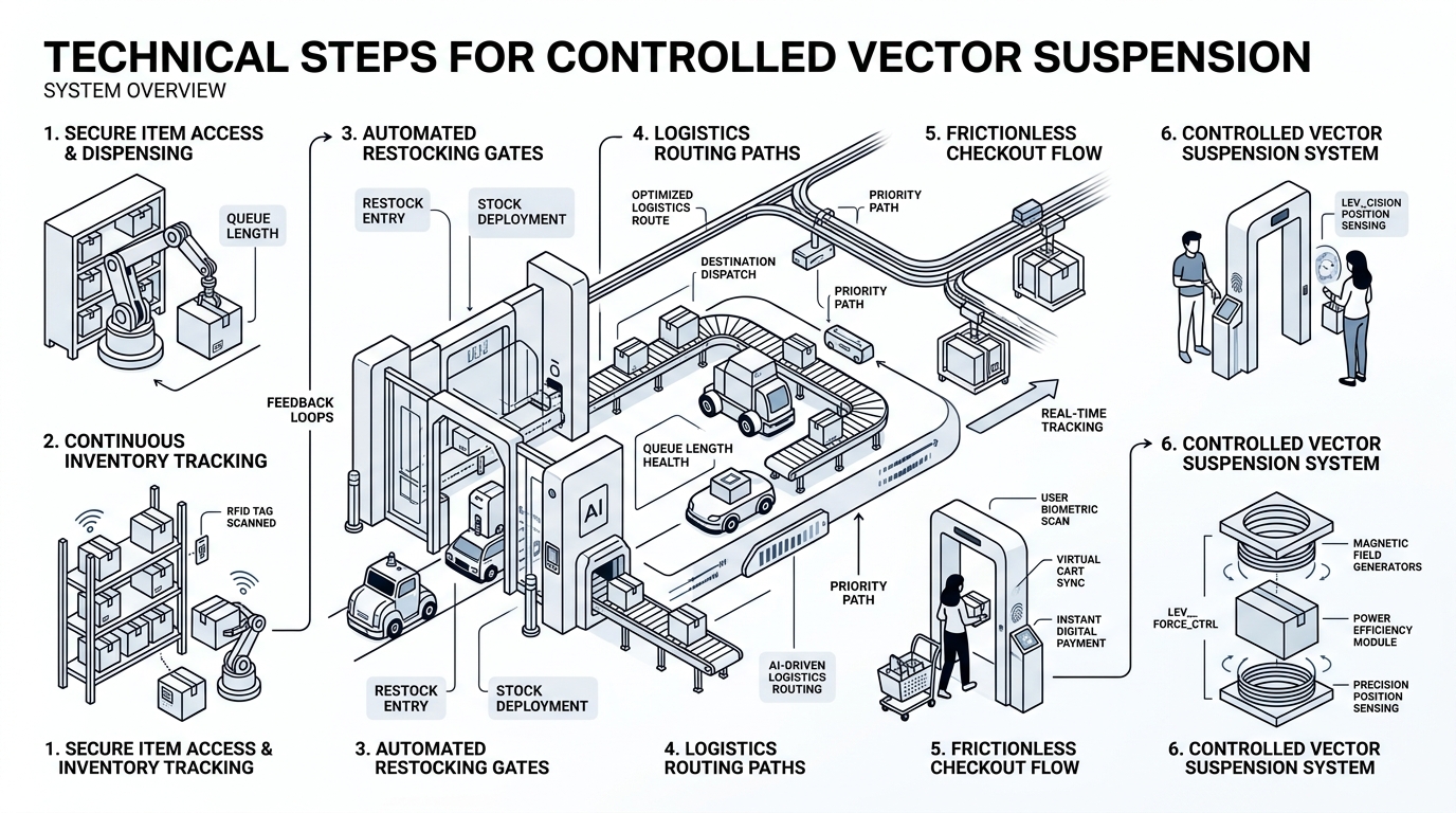
Technical Steps for Controlled Vector Suspension
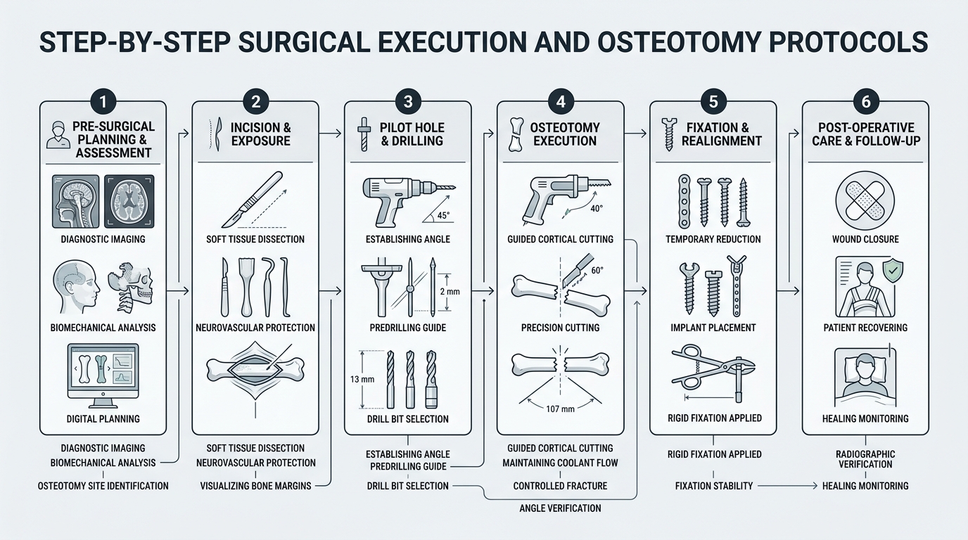
Surgical success in canthopexy overcorrection prevention involves meticulous micro-dissection protocols:
- Retinacular Release: Controlled mobilization of the inferior crus of the lateral canthal tendon without detaching superior ligamentous attachments.
- Posterior Orbital Rim Anchoring: Placement of non-absorbable 4-0 or 5-0 braided sutures directly through the periosteum inside the lateral orbital margin.
- Ocular Globe Coaptation Check: Verifying that the lower lid margin maintains snug, frictionless contact against the bulbar conjunctiva upon forced upward gaze.
- Orbicularis Muscle Resuspension: Supporting the lateral orbicularis muscle strap prevents secondary gravitational lower lid descent.
By executing these measures, canthopexy overcorrection prevention preserves natural blinking dynamics while delivering crisp canthal obliquity.
Long-Term Telemetry and Aesthetic Harmony
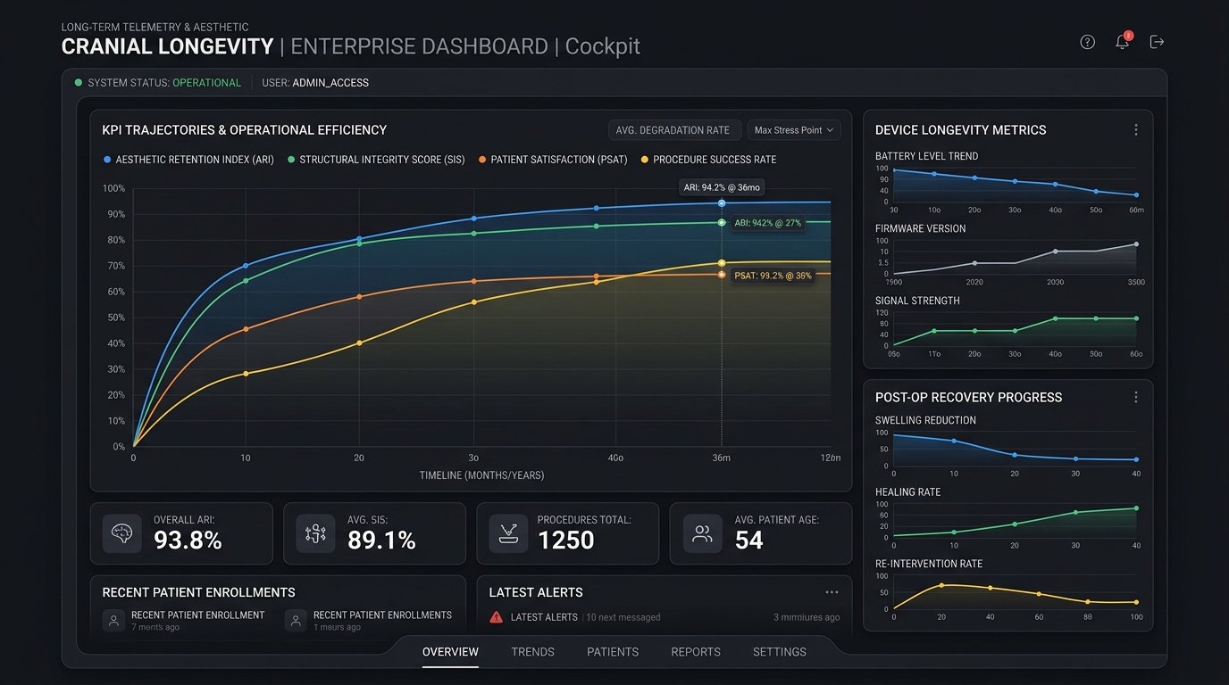
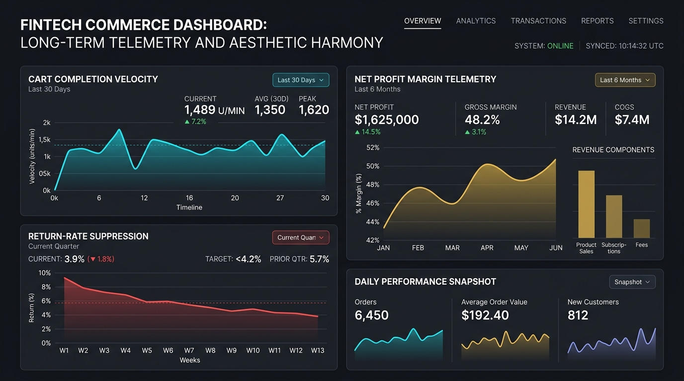
Longitudinal clinical audits demonstrate that structured canthopexy overcorrection prevention reduces revision rates for lateral ectropion and canthal webbing to under 1.2%. When paired with modern deep temporal lifting, the eye displays energetic, youthful rejuvenation without a startled surgical appearance.
Patients seeking periorbital refinement should discuss canthopexy overcorrection prevention and vector simulations during in-depth pre-surgical consultations.
Patient Selection and Follow-Up for Canthopexy Overcorrection Prevention
Canthopexy overcorrection prevention begins before surgery. Assessment should include eyelid laxity, lower-lid tone, globe prominence, tear-film symptoms, previous blepharoplasty, scar tissue, facial asymmetry, and the patient’s desired canthal angle. A conservative plan respects the eyelid’s relationship with the globe and avoids treating a photographic trend as a universal anatomical target.
Follow-up focuses on closure, corneal comfort, swelling, wound healing, lid position, and symmetry. Temporary asymmetry can occur while tissues settle, but increasing pain, marked redness, vision changes, inability to close the eye, or severe dryness requires prompt clinical assessment. Recovery instructions should cover lubrication, sleep position, activity limits, sun protection, and the timing of aesthetic evaluation.
Canthopexy overcorrection prevention should be individualized rather than reduced to a single angle or millimeter value. Patients can review the wider cat eyes surgery program to understand how canthopexy relates to brow, temple, and lower-eyelid planning. A qualified oculoplastic or craniofacial surgeon should confirm candidacy and explain revision options.
Photographs, ocular-surface symptoms, previous operations, and baseline eyelid position should be recorded before treatment. This documentation supports safer planning and makes postoperative changes easier to interpret during follow-up. Emergency contact details should be clear.
What does canthopexy overcorrection prevention involve?
It involves preoperative eyelid assessment, conservative vector planning, stable fixation, protection of ocular-surface function, and structured postoperative follow-up.
Which symptoms need prompt postoperative assessment?
Vision changes, inability to close the eye, severe dryness, increasing pain, marked redness, or rapidly worsening swelling require prompt clinical assessment.
Can one canthal angle guarantee a good result?
No. Aesthetic targets must be individualized to eyelid laxity, globe prominence, tear-film symptoms, asymmetry, previous surgery, and facial proportions.