

Understanding how to fix negative canthal tilt has become one of the most prominent aesthetic goals in contemporary periorbital and craniofacial surgery. Canthal tilt defines the angle of the palpebral fissure, drawn between the inner medial and outer lateral corners of the eye. When the lateral canthus descends below the horizontal plane of the medial canthus, individuals exhibit a negative canthal tilt, often projecting a tired, sad, or prematurely aged expression that patients seek to correct through specialized surgical interventions.

Why Patients Seek How to Fix Negative Canthal Tilt
Learning how to fix negative canthal tilt allows patients to restore youthful, energetic, and open eye positioning. In aesthetic oculoplastic practice, knowing how to fix negative canthal tilt provides both reconstructive and cosmetic enhancements, transforming a droopy lateral eyelid margin into an attractive almond or cat eye shape.
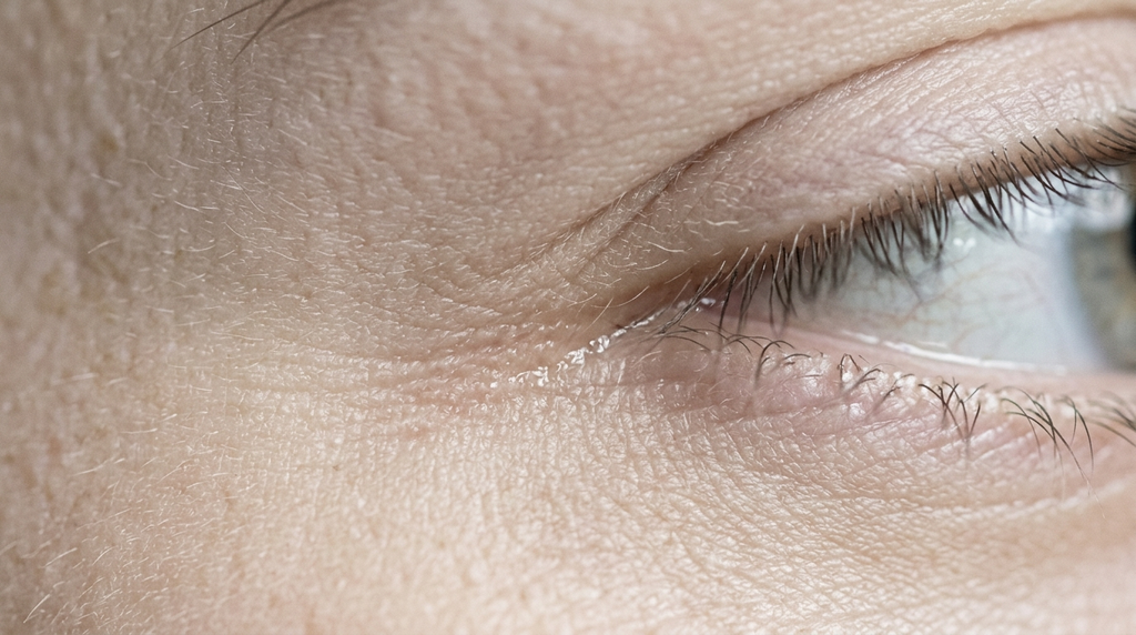
Anatomy and Vectors of the Canthal Tilt
The palpebral aperture relies on dynamic lateral retinaculum attachments, comprising the lateral canthal tendon, lateral horn of the levator aponeurosis, and pretarsal orbicularis muscle. In youthful, vibrant eyes, the lateral canthus rests two to four millimeters superior to the medial canthus, representing a positive canthal tilt of roughly four to eight degrees.
A downward slant arises from three distinct anatomic factors that guide surgeons on how to fix negative canthal tilt:
- Genetic Skeletal Morphology: Hypoplastic or retruded zygomatic-orbital rims provide insufficient bony projection, causing lateral soft tissues to hang posteriorly and downward.
- Laxity of the Lateral Canthal Tendon: Congenital ligamentous looseness or age-related tissue degradation detaches the lateral retinaculum from Whitnall’s tubercle.
- Lower Eyelid Retraction: Vertical eyelid shortness or prior aggressive blepharoplasty pulls the lateral corner inferolaterally.
Understanding these anatomical nuances is thoroughly detailed in our clinical diagnostic overview of negative canthal tilt facts. Furthermore, peer-reviewed clinical data published on PubMed Oculoplastic Surgery details precise retinaculum transposition vectors on how to fix negative canthal tilt.

Can Non-Surgical Treatments Fix Negative Canthal Tilt?
Popular social media trends frequently promote facial exercises, face tape, or neurotoxin injections when asking how to fix negative canthal tilt. However, non-surgical approaches cannot remodel ligamentous insertions. Dermal fillers along the temple can subtly mask shallow orbital margins, but they cannot physically elevate the canthal apex.
To achieve a definitive and permanent repositioning of the palpebral axis, patients require surgical lateral retinaculum anchoring through procedures outlined in our cat eyes surgery guide, providing the premier answer for how to fix negative canthal tilt.
Surgical Approaches: Canthopexy vs. Lateral Canthoplasty
Depending on eyelid tension and the severity of downward tilt, Dr. Mehmet Fatih Okyay selects between two reconstructive techniques when planning how to fix negative canthal tilt:
1. Canthopexy (Tendon Tightening & Suspension)
Canthopexy reinforces the existing lateral canthal tendon without detaching it from its anatomical insertion. High-tensile permanent sutures anchor the tendon to the inner periosteum of the lateral orbital rim at Whitnall’s tubercle, providing moderate two to three millimeter vertical elevation on how to fix negative canthal tilt.
2. Lateral Canthoplasty (Tendon Division & Transposition)
For pronounced downward tilt or significant horizontal laxity, lateral canthoplasty represents the definitive surgical answer for how to fix negative canthal tilt. The inferior crus of the lateral canthal tendon is completely divided (cantholysis), the lateral tarsal strip is tailored and shortened, and the newly mobilized tendon is anchored superior-posteriorly inside the orbital rim.
To analyze where your eye angle falls along the spectrum, review our surgical guide to canthal tilt types before deciding on how to fix negative canthal tilt.

Combination with Endoscopic Temporal Fascia Brow Lift
In many patients, downward canthal slant coexists with lateral brow ptosis and hooding over the orbital margin. Performing an isolated procedure without addressing descending temple tissues can limit surgical outcomes when considering how to fix negative canthal tilt.
By pairing lateral canthoplasty with an endoscopic temporal lift, surgeons release the deep temporal fascia, elevating the brow tail and outer eye corner in a harmonious upward-diagonal vector that yields an exquisite feline almond contour while resolving how to fix negative canthal tilt.
Incision Design and Hidden Scar Management
Modern canthoplasty techniques prioritize invisible healing. Incisions are placed precisely within a tiny horizontal laughter line at the outer eye corner, measuring no more than four to six millimeters. Alternatively, transconjunctival approaches access the lateral retinaculum from inside the lower eyelid, leaving zero external skin scars when addressing how to fix negative canthal tilt.
Subcutaneous anchoring with non-absorbable Gore-Tex or Ethibond sutures ensures that elevated tendon vectors resist gravity and muscular blinking forces throughout the patient’s lifetime after learning how to fix negative canthal tilt.
Postoperative Healing Timeline and Recovery Expectations
Understanding recovery ensures realistic expectations during initial healing after discovering how to fix negative canthal tilt:
- Days 1 to 5: Peak eyelid swelling, minor subconjunctival ecchymosis, and temporary eye dryness. Prescribed lubricating eye drops and nocturnal ointments protect the cornea.
- Day 7: Removal of delicate canthal skin sutures. Visual acuity remains clear, and patients comfortably wear sunglasses in public after learning how to fix negative canthal tilt.
- Weeks 2 to 3: Resolution of acute swelling. Minor initial overcorrection softens into an open, natural almond tilt.
- Months 2 to 3: Complete tendon consolidation against the orbital bone, providing stable long-term aesthetic correction following how to fix negative canthal tilt.
Individuals ready to correct downward-slanting eyes and restore vibrant periorbital harmony are encouraged to contact now to discuss personalized surgical options with Dr. MFO’s team in Istanbul.
Frequently Asked Questions
Can canthoplasty permanently fix negative canthal tilt?
Yes. Lateral canthoplasty permanently alters the physical insertion of the canthal tendon against the orbital bone, providing lifelong structural correction that does not reverse with time.
Will canthoplasty surgery leave visible scars on my face?
No. The surgical incision is meticulously placed within a natural skin crease at the outer eye corner measuring only 4 to 6 millimeters, fading into an imperceptible micro-line within weeks.
How long does recovery take after canthal tilt surgery?
Most patients resume daily routines and remote work within 7 to 10 days once initial eyelid swelling and bruising subside, with full tissue softening achieved by week six.