Facial Feminization Surgery (FFS) is a transformative set of procedures designed to soften masculine facial features and enhance those typically associated with femininity. For many transgender individuals, FFS is a crucial step in aligning their external appearance with their internal gender identity, significantly contributing to psychological well-being and reducing gender dysphoria. While FFS encompasses a wide array of procedures targeting different facial regions, this comprehensive guide zeroes in on the complex interplay between nose and cheek procedures when performed in conjunction.
The nose, often the central focal point of the face, and the cheeks, which define the midface contour and overall facial shape, are intimately connected both aesthetically and anatomically. Understanding the intricate anatomical considerations for combined nose and cheek procedures is paramount for achieving harmonious, natural-looking results and ensuring patient safety. This guide will delve into the foundational principles of FFS, the specific anatomical structures of the nose and cheeks, and critically, the synergistic planning required when these two highly impactful procedures are performed together.
Foundations of Facial Feminization Surgery: Goals and Principles
Facial Feminization Surgery is not merely about making a face “prettier”; it is about re-sculpting the skeletal and soft tissue structures to align with societal perceptions of femininity. The goals extend beyond individual feature alteration to encompass overall facial harmony and balance, crucial for gender affirmation.

Table of Contents
Defining Facial Feminization Surgery (FFS)
FFS is a highly specialized field within plastic surgery that aims to modify facial features to create a more feminine appearance. This involves a range of surgical techniques, from bone reduction and reshaping to soft tissue augmentation and redistribution. The overarching objective is to achieve a softer, more rounded, and less angular facial structure, which are common characteristics of feminine faces, thereby facilitating social transition and improving quality of life for transgender women.
Key Facial Features in Gender Affirming Care
Several facial features contribute significantly to gender perception. In feminine faces, key characteristics often include a less prominent brow bone, a shorter upper lip, a more rounded jawline, a smaller chin, and critically, specific nasal and cheek contours. The nose and cheeks are particularly impactful because they occupy the central and mid-facial regions, heavily influencing overall facial balance and perceived age. Alterations to these areas can dramatically shift facial aesthetics towards a more feminine ideal.
The Role of Nasal Contouring in Feminization
Nasal feminization, or FFS rhinoplasty, aims to refine and reduce the size of the nose to achieve a more delicate appearance. Typically, masculine noses are perceived as having a more prominent dorsal hump, a wider bridge, and a broader, less refined tip. Specific aims of nasal procedures in FFS include:
- Reducing Dorsal Height: Lowering the nasal bridge to create a softer profile.
- Refining the Tip: Making the nasal tip smaller, more defined, and often slightly rotated upwards.
- Narrowing the Alar Base: Reducing the width of the nostrils for a more delicate base.
- Addressing Nasal Length: Shortening an overly long nose to create better facial proportions.
These modifications help create a nose that harmonizes with other feminized facial features, avoiding an overly strong or large appearance.
The Significance of Cheekbone Augmentation and Midface Contouring
Cheekbones are a cornerstone of feminine facial beauty, often appearing more rounded and prominent in women compared to men. Augmenting cheekbones and contouring the midface contributes to a more feminine facial shape by:
- Creating Volume and Projection: Enhancing the malar (cheekbone) area to create a desirable convexity.
- Addressing Infraorbital Hollowing: Filling in sunken areas beneath the eyes to create a smoother transition from the lower eyelid to the cheek.
- Achieving a “Heart-Shaped” or “Oval” Face: Contributing to the overall desired facial shape, moving away from a more square or angular masculine midface.
- Lifting Effect: Augmenting the midface can have a subtle lifting effect on surrounding soft tissues, improving overall facial youthful appearance.
These procedures often involve implants, fat grafting, or osteotomies (bone reshaping) to achieve the desired projection and contour.

Anatomical Considerations for Nasal Procedures in FFS
A thorough understanding of nasal anatomy is non-negotiable for successful FFS rhinoplasty. The nose is a complex three-dimensional structure comprising bone, cartilage, and a soft tissue envelope, all supported by a rich neurovascular network.
Skeletal Anatomy of the Nose
The nasal framework is a combination of bone and cartilage, providing its shape and support. The upper part of the nose is bony, while the lower part is cartilaginous.
Nasal Bones and Septum
The nasal bones form the superior part of the nasal bridge, extending from the forehead. The radix is the deepest point of the nasal bridge, typically at the level of the pupils. The nasal dorsum extends from the radix to the tip, encompassing both bony and cartilaginous components. The nasal septum, a central wall of bone and cartilage, divides the nasal cavity and provides crucial support to the dorsum. In FFS, reduction of the nasal bones often involves rasping or osteotomies (controlled bone cuts) to lower the dorsal height and narrow the bridge.
Cartilaginous Framework (Upper and Lower Lateral Cartilages)
Below the nasal bones, the nasal framework consists of several cartilages:
- Upper Lateral Cartilages: These attach to the caudal (lower) edge of the nasal bones and form the middle third of the nose. They are crucial for maintaining the patency of the internal nasal valve.
- Lower Lateral Cartilages (Alar Cartilages): These paired cartilages form the tip and alar (nostril) rims. They consist of a medial crus (forming the columella), an intermediate crus, and a lateral crus (forming the alar rim). Manipulation of these cartilages is essential for tip refinement, projection, and rotation, as well as for modifying alar width. Techniques include excisions, sutures, and grafts to reshape the tip for a more feminine appearance.
The intricate relationship between these cartilages dictates the final shape and stability of the nasal tip and nostrils.
Soft Tissue Envelope and Skin Thickness
The soft tissue envelope (STE) of the nose comprises the skin, subcutaneous fat, muscle (nasalis), and fibrous tissue. The thickness and elasticity of the STE significantly impact rhinoplasty outcomes. Thin skin will reveal underlying cartilaginous and bony changes more readily, while thick skin can obscure fine details and may limit the degree of achievable refinement. Understanding skin thickness, particularly in the nasal tip, is crucial for predicting results and planning surgical maneuvers. Excessive soft tissue or sebaceous glands in the tip can hinder adequate definition.
Vascular Supply and Innervation of the Nose
The nose has a rich vascular supply, primarily from branches of the ophthalmic and facial arteries. Key arteries include the dorsal nasal artery (branch of ophthalmic), angular artery (terminal branch of facial), and septal branch of the superior labial artery. Venous drainage largely parallels the arterial supply. Sensory innervation is provided by branches of the trigeminal nerve (V1 – ophthalmic, V2 – maxillary), particularly the external nasal nerve (branch of V1) for the tip and dorsum. Careful dissection is required to preserve these structures, minimizing bleeding and nerve damage, which could lead to numbness or altered sensation.
Anatomical Considerations for Cheek and Midface Procedures in FFS
The midface, encompassing the cheeks, infraorbital region, and upper jaw, is pivotal in defining facial femininity. Its complex anatomy includes significant bony structures, multiple soft tissue layers, and vital neurovascular pathways.
Skeletal Anatomy of the Zygoma and Maxilla
The bony framework of the midface is dominated by the zygoma (cheekbone) and maxilla (upper jawbone). These bones dictate the projection, width, and overall contour of the midface.
Zygomatic Arch and Body
The zygoma consists of a body and several processes, including the temporal process which forms the zygomatic arch with the temporal bone. The zygomatic body forms the most prominent part of the cheekbone. In FFS, augmentation of the zygomatic body often involves placing implants directly on the bone or performing zygomatic osteotomies (bone cuts) to reposition or reshape the bone, creating a more outward and forward projection characteristic of feminine cheekbones. Reduction of excessively prominent zygomas, though less common in FFS, might also be considered.
Infraorbital Rim and Maxillary Bone
The infraorbital rim is the lower border of the eye socket, formed by the zygoma and maxilla. The maxilla forms the central part of the midface, housing the upper teeth and contributing to the infraorbital region. In FFS, contouring around the infraorbital rim is crucial for addressing hollowness or flatness, often by augmenting the area with fat grafts or small implants to create a smooth transition from the lower eyelid to the cheek. This area is also critical due to the infraorbital nerve exiting through the infraorbital foramen.
Soft Tissue Structures of the Cheeks and Midface
The soft tissues of the midface comprise various layers that contribute to its volume and contour.
Malar Fat Pad and Subcutaneous Fat
The malar fat pad is a superficial fat collection located over the cheekbone. Its size and position greatly influence the fullness and projection of the mid-cheek. Subcutaneous fat is distributed throughout the cheek, and its thickness varies among individuals. In FFS, fat grafting is a common technique used to augment areas lacking volume, such as the malar region or infraorbital hollows, providing a natural-looking augmentation and improving skin quality. The manipulation of these fat pads contributes significantly to creating a softer, more rounded feminine cheek contour.
Facial Muscles (e.g., Zygomaticus Major/Minor)
Numerous facial muscles are present in the midface, responsible for facial expressions. The zygomaticus major and minor muscles originate from the zygoma and insert into the upper lip and corner of the mouth, being primarily responsible for smiling. While not typically the direct targets of FFS midface procedures, their location and innervation must be respected during dissection to avoid injury, which could lead to asymmetry or functional deficits in expression.
Key Neurovascular Structures in the Midface
The midface is densely populated with critical nerves and blood vessels that require meticulous identification and preservation during surgery to prevent complications such as numbness, paralysis, or significant bleeding.
Infraorbital Nerve and Vessels, Facial Artery and Vein
- Infraorbital Nerve and Vessels: The infraorbital nerve, a branch of the maxillary nerve (V2), exits the infraorbital foramen, located approximately 1 cm below the orbital rim, supplying sensation to the lower eyelid, side of the nose, upper lip, and cheek. Accompanying it are the infraorbital artery and vein. Protection of this neurovascular bundle is paramount during any midface procedure, especially infraorbital rim augmentation or zygoma manipulation, as injury can lead to permanent numbness in its distribution.
- Facial Artery and Vein: The facial artery and vein course across the face, typically anterior to the masseter muscle and then superiorly towards the medial canthus. They provide major blood supply and drainage to the midface and lower face. While typically deeper, surgical planes in cheek procedures must be carefully considered to avoid damage to these vessels, which could result in significant bleeding or hematoma formation.
Synergistic Anatomical Planning for Combined Nose and Cheek FFS Procedures
Performing nose and cheek FFS procedures concurrently requires an integrated surgical strategy that recognizes their profound anatomical and aesthetic interdependence. The face is a unified aesthetic unit; changes in one area invariably impact the perception and harmony of adjacent structures. This synergistic approach is not merely about combining two operations but about orchestrating them to achieve a superior, more balanced feminine outcome while meticulously mitigating shared surgical risks.
Interdependence of Nasal and Midface Aesthetics
The nose, as the central facial landmark, significantly influences how the surrounding midface (cheeks and infraorbital area) is perceived, and vice versa. For instance, a prominent or overly large nose can make the midface appear flat or recessed, even if the cheekbones have adequate projection. Conversely, well-projected, feminine cheekbones can visually enhance and balance a nose that might otherwise seem slightly less refined. When performing FFS, understanding this visual dialogue is crucial.
Reducing a nasal dorsal hump and refining the tip can make the cheekbones appear more prominent and defined without direct cheek augmentation. Similarly, augmenting the cheekbones can provide a more harmonious backdrop for a feminized nose, making it seem less isolated or out of proportion. Integrated aesthetic planning considers these visual relationships, often allowing for more subtle changes in one area to achieve a significant overall improvement in facial harmony, reducing the extent of surgery needed in another, or guiding the specific degree of modification.
Shared Surgical Approaches and Dissection Planes
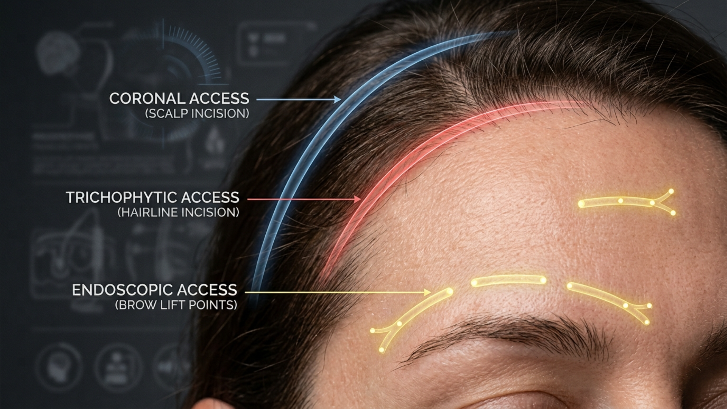
One of the primary anatomical advantages and challenges of combining nose and cheek procedures lies in their potential to share or overlap surgical approaches and dissection planes. For example, open rhinoplasty utilizes an incision across the columella and internal incisions, allowing for direct visualization of the nasal skeletal and cartilaginous framework. When cheek augmentation is performed using intraoral incisions (inside the mouth), the dissection plane can extend superiorly to access the infraorbital rim and zygomatic body.
Depending on the specific techniques employed, these dissection planes can become continuous or closely related, especially in the infraorbital area where the nose and midface converge. This contiguity requires the surgeon to have an exceptional three-dimensional understanding of the anatomy to ensure safe and efficient access while maintaining tissue integrity and minimizing trauma across both operative fields. For instance, the piriform aperture, the bony opening of the nose, is a shared landmark when addressing both the nasal base and the medial cheek/maxilla.
Overlapping Incisions and Access Routes
Strategic planning of incisions and access routes is vital in combined procedures. While nose procedures typically use columellar or endonasal incisions, and cheek procedures often use intraoral incisions (sublabial or buccal sulcus), there are areas where these approaches might converge or where a single incision might provide access to both regions.
For example, if a patient requires both alar base reduction and midface augmentation that extends medially towards the nose, the close proximity of the intraoral access for cheek implants and the alar base itself requires careful management to avoid compromising blood supply or creating an overly extensive dissection. In some cases, transconjunctival (through the lower eyelid) or external incisions might be considered for cheek work, which are distinct from nasal incisions but still impact the overall facial healing and swelling. The surgeon must choose incisions that provide optimal exposure for both procedures while minimizing visible scarring and respecting the vascularity of the tissues.
Managing Soft Tissue Redundancy and Drape
Changes to the underlying bony and cartilaginous framework of the nose and cheeks inevitably affect the overlying soft tissue envelope. When bone is removed (e.g., dorsal hump reduction) or added (e.g., cheek implants), the skin and soft tissues must redrape smoothly over the new contours. In combined procedures, this becomes more complex. For instance, reducing the nasal dorsum creates excess nasal skin. Simultaneously, augmenting the cheeks stretches the midface skin.
The surgeon must anticipate how these changes will interact. If the nasal reduction is significant, it might lead to some skin redundancy around the radix or nasal sidewalls. At the same time, the upward and outward projection from cheek augmentation can create a subtle “lift” in the midface, potentially affecting the lower eyelids and nasolabial folds. Careful management involves precise soft tissue redraping, sometimes with the need for very minor excisions or tightening sutures, to ensure a smooth, natural contour and to avoid a “stretched” or “bunched” appearance. The inherent elasticity of the skin plays a significant role here, and patient-specific factors like age and skin quality must be considered.
Protecting Shared Neurovascular Structures
As detailed earlier, both the nose and midface are rich in critical neurovascular structures. The infraorbital nerve and vessels, emanating from the infraorbital foramen on the maxilla, are particularly vulnerable during midface augmentation. The angular artery and vein, terminal branches of the facial artery, ascend alongside the nose. When both areas are operated on, the risk of compromising these shared or proximally located bundles increases.
For example, during infraorbital rim augmentation, the dissection plane is perilously close to the infraorbital nerve. Simultaneously, a septorhinoplasty will involve manipulation of areas supplied by branches of the facial artery. The surgeon must have an intimate knowledge of the exact anatomical course of these structures and employ meticulous dissection techniques, often using subperiosteal (under the bone lining) planes where safe, to shield nerves and vessels from direct injury, compression, or excessive stretch. Intraoperative nerve monitoring, though not routine, can be considered in high-risk cases. Communication with the anesthesia team regarding blood pressure management is also crucial to minimize bleeding and enhance visibility of these delicate structures.
Preventing Complications Specific to Combined Procedures
While each procedure carries its own set of risks (e.g., breathing issues for rhinoplasty, nerve damage for cheek augmentation), combining them introduces unique challenges and potential complications. Increased operative time can elevate the risk of prolonged anesthesia-related issues. The extensive dissection in adjacent areas might predispose to greater swelling, ecchymosis (bruising), and hematoma formation due to disruption of a larger network of small vessels and lymphatics. Infection risk, though low in both procedures individually, can theoretically increase with larger surgical fields. Furthermore, aesthetic complications like asymmetry or an unnatural “operated” look can be more pronounced if the two procedures are not harmoniously balanced. Preventing these requires:
- Rigorous Preoperative Planning: Detailed 3D imaging, virtual surgical planning, and precise measurements.
- Experienced Surgical Team: A surgeon highly proficient in both rhinoplasty and midface contouring for FFS.
- Meticulous Hemostasis: Careful control of bleeding during surgery.
- Prophylactic Measures: Appropriate antibiotics, drains if indicated, and post-operative compression.
Understanding these unique combined risks allows for proactive strategies to minimize their occurrence.
Ensuring Adequate Blood Supply and Lymphatic Drainage
The entire face has a robust blood supply, which generally aids in healing. However, extensive subperiosteal dissection across the nasal and midface regions can potentially compromise smaller perforating vessels or lymphatic channels. While major vessel injury is rare with careful technique, broad tissue elevation can disrupt lymphatic drainage, contributing to prolonged swelling. The surgeon must plan dissection pathways that preserve as much blood supply as possible to the elevated tissues, particularly if fat grafting or tissue repositioning is performed. Ensuring that the tissues remain well-vascularized is critical for optimal healing, prevention of skin compromise, and reduction of post-operative edema. Gentle tissue handling and avoiding excessive tension on flaps are paramount.
Addressing Potential for Swelling and Ecchymosis
Swelling (edema) and bruising (ecchymosis) are expected after any facial surgery, but they tend to be more pronounced and prolonged when combined procedures like nose and cheek FFS are performed. The larger operative field, extensive tissue manipulation, and disruption of more blood and lymphatic vessels contribute to this. Patients must be thoroughly educated about the expected duration and severity of swelling. Strategies to minimize and manage post-operative swelling and bruising include:
- Intraoperative Measures: Meticulous hemostasis, judicious use of vasoconstrictors, and controlled hypotensive anesthesia where appropriate.
- Post-operative Care: Head elevation, cold compresses, compression garments, and in some cases, lymphatic massage.
- Medication: Anti-inflammatory drugs as prescribed.
Realistic patient expectations are key, as resolving the combined swelling can take several weeks to months, with subtle residual swelling persisting for up to a year or more.
Conclusion
The successful integration of nose and cheek procedures in Facial Feminization Surgery hinges upon an unparalleled understanding of their complex, synergistic anatomy. From the foundational principles of feminization to the intricate layers of bone, cartilage, soft tissue, and neurovascular networks, every element plays a critical role. When performed together, these procedures offer the potential for profound facial harmony and gender affirmation, but they also demand a heightened level of surgical expertise, meticulous planning, and an awareness of the unique challenges posed by their combined nature. Surgeons must not only be adept at individual techniques but also possess the foresight to anticipate the interplay between these adjacent regions, ensuring both aesthetic excellence and patient safety.
Key Takeaways
- Combined nose and cheek FFS requires integrated aesthetic and anatomical planning due to their profound interdependence.
- Shared surgical planes and proximity of critical neurovascular structures necessitate meticulous dissection and preservation.
- Managing soft tissue redraping and anticipating increased swelling are crucial for optimal, harmonious outcomes.
- An experienced surgeon with a deep understanding of both rhinoplasty and midface contouring is essential for safe and effective combined procedures.
FAQ
How do nose and cheek procedures influence each other’s aesthetic outcome in FFS?
Changes in nasal contour directly impact how the midface (cheeks) is perceived, and vice versa. For instance, reducing a prominent nose can make the cheeks appear more projected. Conversely, augmenting the cheeks can visually balance the nose. Integrated planning ensures both features harmonize, creating overall facial balance and a more feminine appearance.
What are the main risks associated with performing nose and cheek procedures together?
Beyond individual procedure risks, combining nose and cheek FFS can lead to increased operative time, potentially greater swelling and bruising, higher risk of hematoma, and heightened complexity in protecting shared neurovascular structures like the infraorbital nerve. Meticulous surgical planning and execution are crucial to mitigate these risks.
Is it always necessary to perform nose and cheek procedures concurrently in FFS?
No, it is not always necessary. The decision to combine procedures depends on the individual patient’s specific anatomical needs, aesthetic goals, overall health, and recovery preferences. Sometimes, staging procedures can be beneficial, particularly for patients who prefer a more gradual recovery or have complex medical considerations. However, combining them can offer the advantage of a single recovery period and a more immediate, unified aesthetic outcome.
How does recovery differ when nose and cheek procedures are combined versus separate?
When combined, recovery typically involves more pronounced and prolonged swelling and bruising across a larger facial area. Patients may experience greater discomfort initially, and the overall recovery period for the majority of swelling to subside can be longer (several weeks to months) compared to undergoing each procedure separately. Management often involves more intensive use of cold compresses, head elevation, and possibly drains.
What type of surgeon is most qualified to perform combined nose and cheek FFS?
A plastic surgeon or maxillofacial surgeon with extensive specialized experience and documented expertise in Facial Feminization Surgery is most qualified. They should have a deep understanding of both rhinoplasty and midface contouring techniques within the specific context of gender-affirming care, as well as a comprehensive knowledge of facial anatomy and aesthetic principles to ensure harmonious results and patient safety.
Visit Dr.MFO Instagram profile to see real patient transformations! Get a glimpse of the incredible results achieved through facial feminization surgery and other procedures. The profile showcases before-and-after photos that highlight Dr. MFO’s expertise and artistic vision in creating natural-looking, beautiful outcomes.
Ready to take the next step in your journey? Schedule a free consultation with Dr. MFO ( Best Facial Feminization Surgeon for You) today. During the consultation, you can discuss your goals, ask any questions you may have, and learn more about how Dr. MFO can help you achieve your desired look. Don’t hesitate to take advantage of this free opportunity to explore your options and see if Dr. MFO is the right fit for you.