
The forehead is arguably the most critical anatomical region in Facial Feminization Surgery (FFS), serving as the primary canvas upon which gender perception is painted. When patients search for “Type 1, 2, or 3 forehead,” they are encountering a classification system that dictates the complexity, invasiveness, and ultimate outcome of their surgical journey. This terminology refers to the three distinct surgical approaches for forehead contouring, each designed to address specific anatomical variations in the frontal bone, brow ridge, and hairline. Understanding these classifications is not merely an academic exercise; it is the foundational step in making an informed decision that aligns with your skeletal structure, aesthetic goals, and tolerance for surgical intervention.
As a surgeon specializing in Facial Feminization Surgery (FFS), I consistently emphasize that the forehead is one of the most significant features in determining whether a face is perceived as masculine or feminine. A prominent brow ridge (often called brow bossing or frontal bossing), a sloped forehead, and a lower hairline are typically associated with masculine foreheads, while a smoother, more vertically oriented, and gently rounded forehead with a higher hairline is characteristic of feminine foreheads. Source
The confusion surrounding these terms often stems from the fact that they are clinical classifications rather than marketing terms. Type 1 generally refers to a less invasive approach involving only soft tissue or minimal bone work. Type 2 introduces the concept of osteotomy (bone cutting) to reshape the frontal bone. Type 3 is the most comprehensive, involving significant bone removal and reconstruction. This guide will dissect the anatomical realities behind each type, the surgical techniques involved, and the recovery protocols required, ensuring you possess the knowledge to navigate your consultation with confidence.
Reconstructing the scalp and forehead is a challenging endeavor frequently undertaken by facial plastic and reconstructive surgeons. There are many anatomical factors to be considered in this area, including multiple neurovascular structure present that should be identified and preserved.
— PMC Article on Forehead Reconstruction
Table of Contents
Defining the Anatomy: The Frontal Bone and Brow Ridge
To understand the difference between Type 1, 2, and 3 forehead reconstruction, one must first understand the anatomy of the forehead. The forehead is composed of the frontal bone, which forms the anterior cranial vault. In biological males, the frontal bone often exhibits a prominent supraorbital ridge (the brow ridge) and a more sloped angle from the brow to the hairline. In biological females, the frontal bone is typically smoother, with a more vertical inclination and a less pronounced or absent brow ridge.
The “Type” classification system is essentially a measure of how much of this bone needs to be altered to achieve a feminine contour. The decision is based on the thickness of the bone, the degree of bossing, and the position of the frontal sinus (the air-filled cavity behind the forehead). The frontal sinus is a critical variable; it determines how much bone can be safely shaved or reduced without compromising the structural integrity of the skull or the health of the overlying soft tissue.

Type 1 Forehead Reconstruction: The Soft Tissue Approach
Type 1 forehead reconstruction is the least invasive of the three classifications. It is typically indicated for patients who have a relatively smooth frontal bone but possess a low hairline or significant soft tissue volume in the forehead region. In this procedure, the surgeon does not perform any bone reduction. Instead, the focus is on the soft tissue envelope and the hairline.
The primary technique involved in a Type 1 classification is a forehead lift (brow lift) combined with hairline advancement. If the brow is ptotic (drooping), lifting it can create the illusion of a higher, smoother forehead. Additionally, if the hairline is low, a hairline advancement procedure (also known as a forehead reduction) removes a strip of skin at the hairline and advances the scalp forward to lower the hairline. This is often performed in conjunction with hair transplantation to create a natural hairline shape.
The Role of Endoscopic Assistance
Many Type 1 procedures utilize endoscopic techniques. Small incisions are made within the hairline, and a camera is used to visualize the underlying structures. This allows the surgeon to release the brow from its bony attachments and lift it without a large coronal incision. While Type 1 does not address prominent bone, it is highly effective for patients whose masculine appearance is driven by a heavy brow or low hairline rather than skeletal bossing.
However, Type 1 is strictly for soft tissue correction. If a patient has a significant brow ridge, a Type 1 procedure will fail to feminize the forehead because the underlying bone remains prominent. Therefore, accurate pre-operative assessment using 3D CT imaging is essential to determine if the bone structure is suitable for this minimal approach.
Type 2 Forehead Reconstruction: The Osteotomy Approach
Type 2 forehead reconstruction introduces the concept of osteotomy—the surgical cutting of bone. This classification is designed for patients with a moderate degree of brow bossing but where the frontal sinus is either small or absent. In a Type 2 procedure, the surgeon creates a controlled fracture in the frontal bone to reshape it without removing the entire bony segment.
The procedure typically involves making a coronal incision hidden within the hairline. The scalp is elevated to expose the frontal bone. The surgeon then uses a surgical saw or osteotome to make precise cuts (osteotomies) in the bone. These cuts allow the surgeon to mobilize the bony segment containing the brow ridge. Once mobilized, the bone is carefully reshaped or contoured, and then fixed back into place using small titanium plates and screws.

Advantages of Type 2 Osteotomy
The main advantage of a Type 2 osteotomy is that it preserves the natural contour of the forehead while reducing the prominence of the brow ridge. Because the bone is not completely removed, the structural integrity of the forehead is maintained. This technique is particularly useful for patients who have a “steep” forehead slope that needs to be flattened or verticalized without creating a concave appearance.
Furthermore, Type 2 osteotomy allows for the correction of asymmetry. If one side of the brow ridge is more prominent than the other, the osteotomies can be adjusted to balance the forehead. This level of precision is difficult to achieve with simple bone shaving (which is sometimes mistakenly called Type 1 but is actually a different technique entirely).
Type 3 Forehead Reconstruction: The Type III Forehead Reconstruction
Type 3 forehead reconstruction is the most extensive and invasive approach, reserved for patients with significant frontal bossing and a large, pneumatized frontal sinus. In this procedure, the surgeon removes the anterior wall of the frontal sinus entirely, reshapes the bone, and reconstructs the forehead using bone grafts or synthetic materials.
The procedure begins with a coronal incision. The surgeon carefully dissects down to the bone, preserving the pericranium (the membrane covering the bone) to ensure blood supply. The anterior wall of the frontal sinus is removed using a surgical drill or osteotome. The mucosa lining the sinus is completely removed to prevent the formation of mucoceles (fluid-filled cysts). The posterior wall of the sinus (the dura) is left intact.
Reconstruction and Contouring
Once the anterior wall is removed, the surgeon has direct access to the brow ridge. The bone is shaved down to a smooth, feminine contour. To reconstruct the forehead, the surgeon may use split-thickness calvarial bone grafts (harvested from the parietal bone) or titanium mesh covered with hydroxyapatite cement. This creates a new, smooth anterior wall for the frontal sinus.
Type 3 reconstruction offers the most dramatic change in forehead shape. It is the only technique that can significantly reduce a very prominent brow ridge and create a completely smooth, vertical forehead. However, it carries the highest risk profile due to the complexity of the surgery and the potential for complications such as CSF leaks or infection.
| Forehead Type | Primary Indication | Surgical Technique | Frontal Sinus Involvement | Recovery Time |
| Type 1 | Low hairline, soft tissue volume, mild ptosis | Forehead lift, hairline advancement, endoscopic brow lift | None (Soft tissue only) | 1-2 Weeks |
| Type 2 | Moderate brow bossing, small/absent sinus | Osteotomy (bone cutting), contouring, fixation | Minimal (Bone reshaping) | 2-3 Weeks |
| Type 3 | Severe frontal bossing, large pneumatized sinus | Anterior wall removal, bone shaving, reconstruction with grafts | Significant (Full removal and reconstruction) | 3-4 Weeks |
The Role of Hairline Lowering and Transplantation
Regardless of whether a patient undergoes Type 1, 2, or 3 forehead reconstruction, the hairline plays a pivotal role in the final aesthetic. A masculine hairline is often characterized by recession at the temples (creating an M-shape) and a low, straight hairline across the forehead. Feminine hairlines are typically higher, rounded, and lack temporal recession.
In Type 1 procedures, hairline advancement is the primary tool. In Type 2 and 3 procedures, hairline advancement is often performed simultaneously with the bone work. However, bone reduction can sometimes lower the hairline unintentionally. To counteract this, surgeons often perform a hairline advancement to maintain or raise the hairline to a feminine height.
Hair Transplantation Integration
For patients with significant temporal recession, hair transplantation is often required. This can be done simultaneously with forehead surgery or as a staged procedure. The goal is to create a soft, rounded hairline that frames the face. In some cases, the surgeon may use the strip of skin removed during hairline advancement to harvest grafts for transplantation into the temples, maximizing the use of available tissue.
Anatomical Constraints and Patient Suitability
Not every patient is a candidate for every type of forehead reconstruction. The anatomy of the frontal sinus is the primary limiting factor. If the frontal sinus is very large (pneumatized), Type 2 osteotomy may not provide enough reduction, necessitating a Type 3 approach. Conversely, if the sinus is small or absent, a Type 3 procedure is unnecessary and overly aggressive.
The thickness of the bone is also a consideration. Thin bone is more prone to fracture during osteotomy, requiring extreme precision. Thick bone can be difficult to shave and may require specialized instrumentation. Additionally, the position of the supraorbital nerve (which provides sensation to the forehead and scalp) must be identified and preserved in all three types to prevent permanent numbness.
Surgical Pathways: Incisions and Fixation
The surgical approach for forehead reconstruction almost always begins with a coronal incision hidden within the hairline. This provides the necessary exposure for all three types. However, the dissection depth and the handling of the pericranium differ. In Type 1, the dissection is subgaleal (under the scalp muscle). In Type 2 and 3, the dissection is subperiosteal (under the bone membrane) to allow for bone manipulation.
Fixation is a critical component of Type 2 and Type 3 surgeries. After bone reshaping, the segments must be stabilized to prevent movement during healing. Titanium microplates and screws are the standard of care. These are biocompatible and usually left in place permanently. In Type 3 reconstruction, where the anterior wall is removed, the reconstruction may involve titanium mesh or bone grafts secured with screws.
The Role of Hydroxyapatite Cement
In Type 3 procedures, where bone grafts are not used, hydroxyapatite cement is often employed to smooth out irregularities. This is a bone substitute that hardens upon contact with fluid, allowing the surgeon to sculpt a smooth forehead contour. It integrates with the surrounding bone over time, providing a natural feel and appearance.
Recovery Protocols: Type 1 vs. Type 2 vs. Type 3
Recovery varies significantly between the three types. Type 1 procedures, being soft tissue only, typically have the shortest recovery. Patients can expect swelling and bruising around the eyes and forehead, which usually subsides within 1-2 weeks. Pain is generally manageable with oral medication.
Type 2 and Type 3 procedures involve bone manipulation, leading to a more prolonged recovery. Swelling is more significant and can persist for 2-3 weeks. Patients often experience a sensation of tightness or pressure in the forehead. Numbness in the forehead and scalp is common initially but usually resolves over several months as the nerves regenerate.
Managing Post-Operative Edema
Head elevation is mandatory for the first week to reduce orbital edema (swelling around the eyes). Cold compresses can help, but they must be used carefully to avoid damaging the skin. Patients should avoid bending over, heavy lifting, and strenuous activity for at least 4-6 weeks to prevent increased blood pressure to the head, which could cause bleeding.
For Type 3 patients, there is a specific risk of CSF leak (cerebrospinal fluid leak) if the dura is inadvertently injured. While rare, this requires immediate medical attention. Patients are usually advised to avoid blowing their nose or sneezing forcefully for several weeks to prevent pressure changes in the sinuses.
Comparative Analysis: Risks and Longevity
When comparing the three types, the trade-off is between invasiveness and the degree of change. Type 1 offers the least risk but also the least skeletal change. It is ideal for patients who are close to their ideal forehead shape but need refinement. Type 2 offers a balance, providing significant bone reduction with moderate risk. Type 3 offers the most dramatic transformation but carries the highest surgical risk.
Longevity is excellent for all three types when performed correctly. Bone reshaping is permanent. However, soft tissue aging continues. A Type 1 forehead lift may need to be repeated after 10-15 years as gravity takes its toll. Type 2 and Type 3 bone changes are permanent, but the overlying skin will still age.
| Procedure Type | Longevity | Recovery Time | Key Risks | Estimated Cost Range |
| Type 1 (Soft Tissue) | 10-15 Years (Soft tissue aging) | 1-2 Weeks | Asymmetry, Scarring, Hair loss at incision | $4,000 – $8,000 |
| Type 2 (Osteotomy) | Permanent (Bone) | 2-3 Weeks | Non-union of bone, Numbness, Infection | $8,000 – $15,000 |
| Type 3 (Reconstruction) | Permanent (Bone) | 3-4 Weeks | CSF Leak, Infection, Implant visibility | $15,000 – $25,000+ |
Face Shape Suitability: Which Type Fits Your Anatomy?
The choice of forehead type is heavily dependent on your overall facial architecture. A Type 3 reconstruction on a petite face with a small sinus might be overkill, while a Type 1 on a face with severe bossing will be ineffective. The goal is to harmonize the forehead with the rest of the facial thirds.
Round and Square Faces
For round or square faces, a vertical forehead (achieved via Type 2 or 3) helps elongate the face. A sloped forehead (Type 1 or no surgery) can make a round face appear wider. Therefore, patients with these face shapes often benefit from the bone reduction provided in Type 2 or 3 procedures.
Long and Oval Faces
For long or oval faces, adding vertical height to the forehead is undesirable. These patients should aim for a Type 1 procedure or a very conservative Type 2 osteotomy that maintains the natural slope. Over-reduction can elongate the face further, creating an unbalanced look.

Frequently Asked Questions
What is the difference between Type 1, 2, and 3 forehead surgery?
Type 1 involves soft tissue manipulation only (lifts/advancements). Type 2 involves cutting and reshaping the bone (osteotomy) without removing the sinus wall. Type 3 involves removing the frontal sinus wall, shaving the bone, and reconstructing the forehead.
How do I know which type I need?
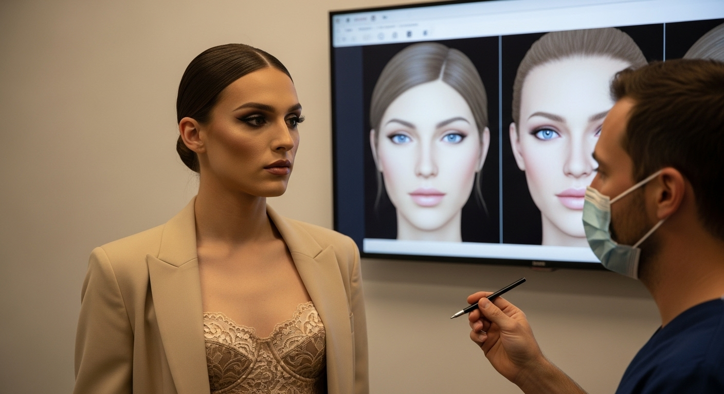
This is determined by a 3D CT scan during your consultation. The scan reveals the thickness of your brow ridge and the size of your frontal sinus. A surgeon will classify your anatomy based on these measurements.
Is Type 3 forehead surgery dangerous?
Type 3 is the most complex procedure and carries higher risks, such as CSF leak or infection. However, when performed by an experienced FFS surgeon, it is safe and offers the most dramatic feminization for prominent foreheads.
Can I combine forehead surgery with hair transplants?
Yes, it is very common. Hairline advancement (Type 1) and hair transplants can be done simultaneously with Type 2 or 3 surgeries to create a feminine hairline shape.
Will I have visible scars?
The incision is typically hidden within the hairline (coronal incision). If hairline advancement is performed, the scar is hidden just behind the new hairline. Scars are usually well-concealed.
How long until I see the final result?
While initial swelling subsides in 3-4 weeks, the final contour of the forehead, especially for Type 2 and 3, takes 6-12 months to fully settle as the bone heals and soft tissue redrapes.
Can forehead surgery be combined with other FFS procedures?
Absolutely. Forehead surgery is often combined with rhinoplasty, jaw reduction, and tracheal shave in a single operation to achieve comprehensive facial feminization.
What is the cost difference between the types?
Type 1 is generally the least expensive due to its simplicity. Type 2 is moderately priced, while Type 3 is the most expensive due to the complexity, time, and materials (implants/grafts) required.
Bibliography
- Dr. MFO. (n.d.). Forehead Contouring: Type 1, 2, 3 Difference. Retrieved from https://www.dr-mfo.com/ffs-forehead-contouring-type-1-2-3-difference/
- PubMed Central. (n.d.). Reconstructing the Scalp and Forehead. Retrieved from https://pmc.ncbi.nlm.nih.gov/articles/PMC5951698/
- Spiegel, J. H., & DeRosa, J. (2005). The Forehead in Facial Feminization Surgery: Aesthetic and Surgical Considerations. Aesthetic Surgery Journal, 25(4), 389-396.
- Altman, K. (2012). Facial Feminization Surgery: Current State of the Field. Plastic and Reconstructive Surgery, 130(6), 1361-1368.
- Ousterhout, D. K. (2008). Feminization of the Forehead: A Comparative Study of Aesthetic Surgery Techniques. Journal of Craniofacial Surgery, 19(5), 1234-1240.
- Capitán, L., et al. (2017). Facial Feminization Surgery: A Comprehensive Review. Plastic and Reconstructive Surgery – Global Open, 5(9), e1522.