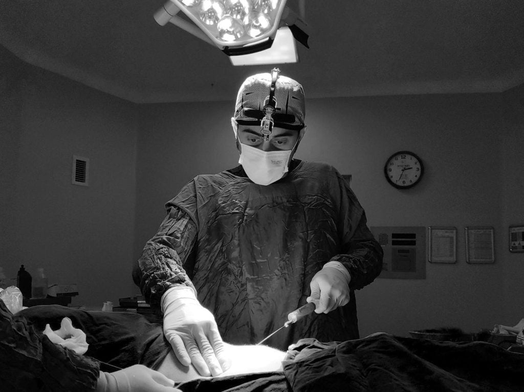
To truly appreciate the transformative potential of forehead contouring in Facial Feminization Surgery (FFS), it’s essential to look beneath the surface and understand the underlying anatomy of the forehead. Just as an artist must know the canvas to create a masterpiece, an FFS surgeon must possess a deep and nuanced understanding of the forehead’s intricate bony and soft tissue structures to achieve natural, feminine, and harmonious results.
This article serves as a guided tour of forehead anatomy relevant to feminization surgery. By understanding the bones, muscles, nerves, and other key components, you’ll gain a richer appreciation for the complexities involved in forehead contouring and the expertise required for successful outcomes.

Table of Contents
The Bony Framework: Building Blocks of the Forehead
The foundation of the forehead’s shape is the frontal bone, a single bone that forms the anterior (front) part of the cranium and the skeletal base of the forehead. Within the frontal bone, several key features are crucial for FFS:
1. Supraorbital Ridges (Brow Bones/Brow Bossing)
- Description: These are bony prominences located just above the eye sockets (orbits). In anatomical terms, they are also known as the superciliary arches or brow ridges. In FFS, they are most commonly referred to as brow bones or when prominent, brow bossing/supraorbital ridge bossing.
- Significance in FFS: The supraorbital ridges are the primary cause of the masculine brow bossing. Their prominence is a key sexually dimorphic feature. Feminization surgery often focuses on reducing or eliminating brow bossing to create a flatter, more feminine brow and upper face.
- Understanding: Surgeons must understand the precise 3D anatomy and thickness of the brow bones to perform safe and effective brow bone reduction (Type 3 foreheadplasty).
2. Glabella
- Description: The smooth bony prominence located directly between the eyebrows and above the nasal bridge.
- Significance in FFS: The glabella is often more projecting in masculine foreheads. Feminization surgery often includes smoothing and flattening the glabella region to create a more feminine contour that flows smoothly into the brow and nose.
- Understanding: Knowledge of the glabella’s bony protrusion and its relationship to the brow bones and nose is essential for achieving harmonious facial contours.
3. Frontal Sinus
- Description: An air-filled cavity located within the frontal bone, just behind the brow bones and glabella.
- Significance in FFS: The frontal sinus lies directly in the surgical field during brow bone reduction (Type 3 foreheadplasty). Surgeons must carefully navigate around and preserve the integrity of the frontal sinus during surgery. Damage to the frontal sinus is a potential complication to be avoided.
- Understanding: Detailed preoperative imaging (like CT scans) and anatomical knowledge are vital to safely perform brow bone work while protecting the frontal sinus.
4. Frontal Eminences (Forehead Bosses)
- Description: Slightly rounded prominences located on either side of the midline of the forehead, above the brow bones. These contribute to the gentle convexity of a typical forehead.
- Significance in FFS: While less prominent than brow bossing, the frontal eminences contribute to the overall shape of the forehead. FFS aims to create a balanced and gently curved forehead contour, taking these eminences into account.
- Understanding: Surgeons consider the frontal eminences when planning overall forehead shaping and contouring to achieve natural-looking results.
5. Temporal Lines
- Description: Subtle bony ridges that curve upwards and backwards from the outer corners of the eye sockets along the sides of the forehead towards the temples. They mark the attachment point of the temporalis muscle (a chewing muscle).
- Significance in FFS: The temporal lines define the lateral (side) borders of the forehead and contribute to the overall width and shape of the upper face. In some cases, subtle contouring of the temporal area might be considered during FFS to refine the forehead shape and transition smoothly to the temples.
- Understanding: Surgeons are aware of the temporal lines as anatomical boundaries during forehead contouring and consider their influence on overall facial shape.
6. Orbital Rims (Superior Orbital Rim)
- Description: The bony edges that form the upper boundary of the eye sockets (orbits), directly beneath the brow bones.
- Significance in FFS: The shape and projection of the orbital rims contribute to the overall appearance of the eye area and forehead. Brow bone reduction surgery directly affects the supraorbital rim.
- Understanding: Surgeons must understand the anatomy of the orbital rims to ensure safe and aesthetically pleasing changes to the brow bone and surrounding eye area.

Soft Tissue Layers: Skin, Muscles, and More
Lying over the bony framework are the soft tissues of the forehead, which also play a role in its appearance and are considered in FFS.
1. Skin
- Description: The outermost layer, the skin of the forehead varies in thickness and elasticity between individuals.
- Significance in FFS: Skin elasticity affects how well the skin redrapes after bone reshaping procedures. Incision placement (e.g., hairline incision) is planned considering skin tension and scar healing. Skin quality and thickness can also influence the visibility of underlying bony changes.
- Understanding: Surgeons consider skin characteristics during surgical planning and technique selection.
2. Subcutaneous Fat
- Description: A layer of fat beneath the skin, varying in thickness.
- Significance in FFS: Subcutaneous fat contributes to the overall contour and softness of the forehead. Significant changes in the bony structure will also indirectly affect the overlying soft tissues.
- Understanding: The thickness of subcutaneous fat can influence the final visible outcome after bony procedures.
3. Muscles of the Forehead
- Frontalis Muscle: A broad muscle that covers much of the forehead. It raises the eyebrows and wrinkles the forehead horizontally.
- Corrugator Supercilii Muscles: Small muscles located deep to the frontalis, above the inner corners of the eyebrows. They draw the eyebrows together and downwards, creating vertical frown lines (often referred to as “11 lines”).
- Procerus Muscle: A small muscle located in the glabella region, between the eyebrows. It draws down the medial part of the eyebrows and wrinkles the skin over the bridge of the nose horizontally.
- Significance in FFS: While FFS primarily focuses on bony structures in forehead contouring, these muscles are encountered during surgical dissection. Understanding their anatomy is important to avoid nerve damage and to potentially address forehead wrinkles as a secondary benefit in some cases. However, FFS forehead contouring is not primarily about wrinkle removal; its focus is bone reshaping for feminization.
- Understanding: Surgeons are aware of the location and function of these muscles during forehead surgery. While muscle work is not the primary aim of FFS forehead contouring, understanding muscle anatomy is crucial for safe surgery.
4. Periosteum
- Description: A thin membrane that covers the outer surface of the frontal bone.
- Significance in FFS: The periosteum is carefully elevated during forehead bone surgery to access the underlying bone. It plays a role in bone healing after osteotomies (bone cuts) and repositioning.
- Understanding: Periosteal management is a standard surgical technique in bone surgeries like forehead contouring.
5. Scalp
- Description: The hair-bearing skin covering the upper part of the head, extending down to the forehead hairline.
- Significance in FFS: The scalp is involved in hairline lowering procedures and coronal incisions for brow bone reduction. Scalp elasticity, hairline position, and hair density are all considered in surgical planning.
- Understanding: Surgeons account for scalp anatomy when planning incisions, hairline adjustments, and considering potential scar visibility.

Nerves and Blood Vessels: Key Structures to Protect
Navigating the forehead safely during FFS requires precise understanding of the neurovascular anatomy.
1. Supraorbital and Supratrochlear Nerves and Vessels
- Description: These are sensory nerves and blood vessels that emerge from the eye socket above the eye, supplying sensation to the forehead and scalp.
- Significance in FFS: These nerves and vessels run through the surgical field during forehead contouring. Care must be taken to identify and preserve them during surgery to minimize nerve damage and bleeding. Temporary numbness of the forehead and scalp after surgery is common due to manipulation of these small nerves.
- Understanding: Surgeons have detailed anatomical knowledge of these nerves and vessels and use careful surgical techniques to avoid permanent damage.
Sexual Dimorphism Reflected in Forehead Anatomy
The foundation for forehead contouring in FFS lies in recognizing and addressing the sexual dimorphism present in forehead anatomy.
- Masculine Forehead: Characterized by prominent brow bossing, a sloped forehead profile, a more projecting glabella, and often a higher hairline.
- Feminine Forehead: Characterized by a flatter brow, a more vertical or slightly convex forehead profile, a smoother glabella, and typically a lower, rounded hairline.
FFS forehead contouring techniques are specifically designed to transform masculine forehead anatomy towards a more feminine configuration by addressing these key sexually dimorphic features.
How Anatomical Knowledge Informs FFS Forehead Procedures
A surgeon’s deep knowledge of forehead anatomy directly dictates the surgical approach and techniques used in FFS forehead contouring:
- Type 3 Brow Bone Reduction: Relies on precise osteotomies and bone reshaping techniques, requiring detailed understanding of brow bone anatomy, frontal sinus location, and nerve pathways.
- Type 1 Forehead Contouring: Requires skill in selectively shaving and contouring bone without damaging underlying structures, guided by anatomical expertise.
- Hairline Lowering: Demands precise incision placement, scalp undermining, and closure techniques, considering scalp elasticity and hairline anatomy for optimal scar outcome and hairline position.

The Expertise of the FFS Surgeon: An Anatomical Artist
Successful forehead contouring in FFS is not just about mechanics; it’s about artistry grounded in anatomical expertise. The FFS surgeon acts as an anatomical artist, understanding the underlying structures and then skillfully sculpting and reshaping them to create a beautifully feminine and natural-looking forehead.
Conclusion – Anatomy: The Blueprint for Feminine Forehead Transformation
Understanding the intricate anatomy of the forehead is foundational to achieving successful and aesthetically pleasing forehead contouring in FFS. From the bony structures that define the underlying shape to the soft tissues and neurovascular elements, every component plays a role. By appreciating this anatomical complexity, you can better understand the skill and expertise of your FFS surgeon and the transformative journey that forehead contouring offers in facial feminization.
Ready to delve deeper into your FFS journey? Schedule consultations with specialist Facial Feminization Surgeons today to discuss your facial anatomy, feminization goals, and how anatomical expertise can help you achieve your desired results.

Visit Dr.MFO Instagram profile to see real patient transformations! Get a glimpse of the incredible results achieved through facial feminization surgery and other procedures. The profile showcases before-and-after photos that highlight Dr. MFO’s expertise and artistic vision in creating natural-looking, beautiful outcomes.
Ready to take the next step in your journey? Schedule a free consultation with Dr. MFO ( Best Facial Feminization Surgeon for You) today. During the consultation, you can discuss your goals, ask any questions you may have, and learn more about how Dr. MFO can help you achieve your desired look. Don’t hesitate to take advantage of this free opportunity to explore your options and see if Dr. MFO is the right fit for you.


