Ideal Face Shape and Transgender Feminization: A Surgical Perspective on What’s Possible

A brunette woman with an elegant updo hairstyle and natural makeup, smiling gently while standing by a window with greenery in the background.

Facial Feminization Surgery, often abbreviated as FFS, represents a suite of surgical procedures designed to modify masculine facial features to align more closely with societal perceptions of feminine aesthetics. For transgender women (MTF), FFS can be a crucial step in their gender affirmation journey, alleviating gender dysphoria and fostering a greater sense of self and well-being. As a surgeon specializing in this field, I aim to provide a comprehensive understanding of the possibilities and limitations of FFS in achieving a more feminine facial structure, particularly in relation to the concept of an “ideal” face shape.

It’s important to preface this discussion by emphasizing that the notion of an “ideal” face shape is subjective and culturally influenced. What is considered feminine varies across different societies and time periods. However, certain general characteristics are often associated with feminine faces, including softer contours, smoother transitions between facial planes, a more delicate jawline and chin, and a less prominent brow bone.
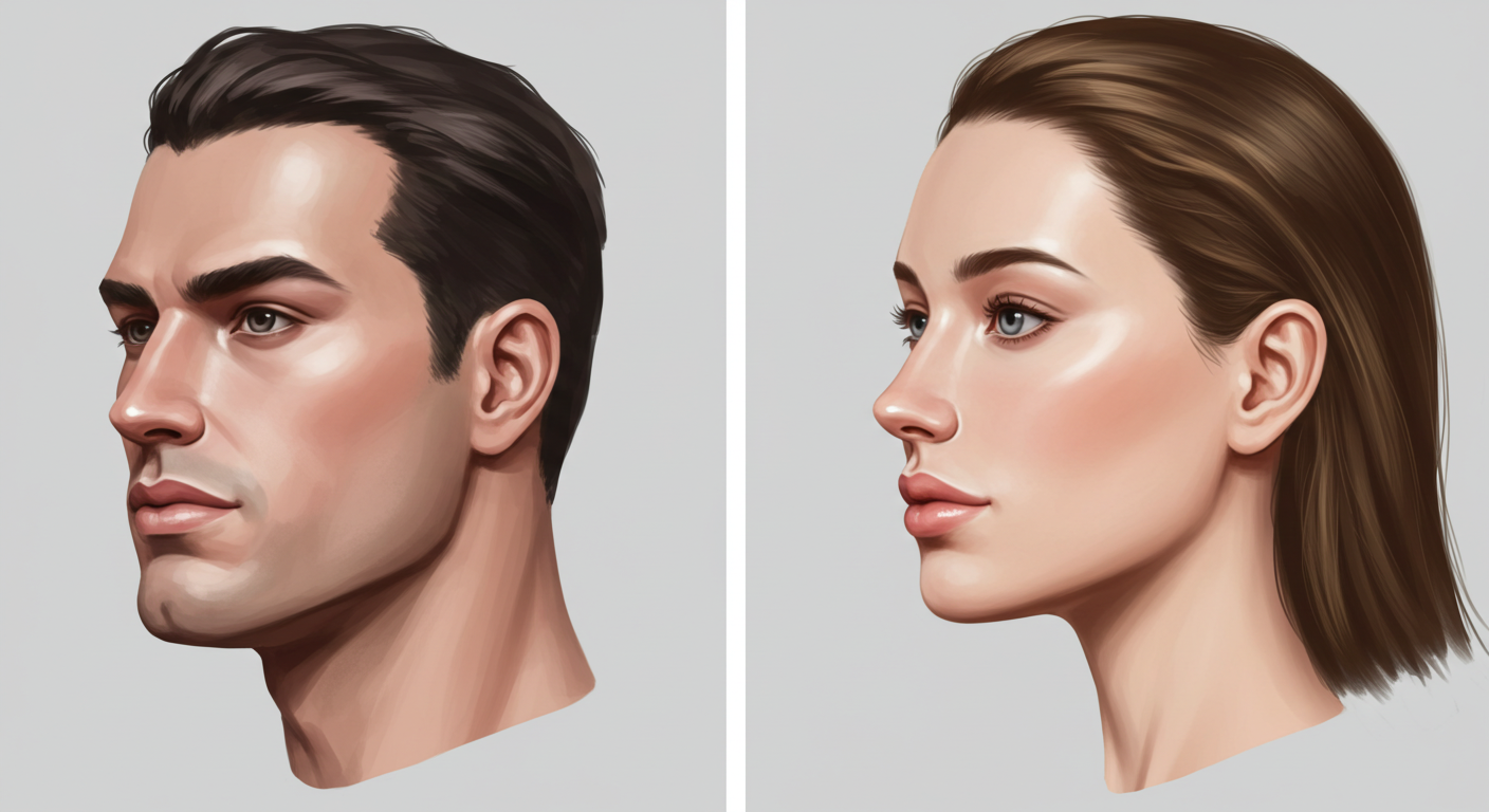
An illustration of a man and a woman with distinct facial features, showing their profiles in a side view. The man has short dark hair, a defined jawline, and a serious expression. The woman has long, straight hair, prominent cheekbones, and a neutral expression. Both figures are rendered in a realistic style with attention to detail in their facial structures.

Key Anatomical Differences Between Masculine and Feminine Faces

To understand how FFS can create a more feminine appearance, it’s essential to recognize the typical anatomical differences between masculine and feminine facial structures. These differences are largely influenced by hormonal exposure during puberty.

Skeletal Structures

  • Forehead and Brow Bone: Masculine foreheads often present with a more prominent brow bone (the bony ridge above the eyes) and a flatter, more receding slope. Feminine foreheads tend to be smoother, more vertical, and have a less pronounced or absent brow bone.
  • Midface: The midface, encompassing the cheekbones and the area beneath the eyes, can differ in projection and contour. Feminine faces often exhibit higher and more anteriorly projected cheekbones, contributing to a softer, more heart-shaped appearance.
  • Jawline and Chin: Masculine jawlines are typically wider and more angular, with a broader and often more projecting chin. Feminine jawlines are generally more tapered and delicate, with a narrower and often more pointed or rounded chin.
  • Nasal Structure: While nasal shape varies greatly among individuals, masculine noses can sometimes be larger and more prominent with a wider bridge. Feminine noses often appear more refined and delicate.

Soft Tissues

  • Fat Distribution: Hormonal differences influence fat distribution in the face. Estrogen tends to promote fat deposition in the cheeks, contributing to a fuller, rounder appearance, while testosterone can lead to less subcutaneous fat in these areas.
  • Muscle Mass: The masseter muscles (involved in chewing), located at the angle of the jaw, can be more developed in individuals with higher testosterone levels, contributing to a wider jawline.
An artist using a digital drawing tablet to create a detailed portrait of a woman's face, with precise lines and shading for realistic skin texture.

Surgical Procedures in Facial Feminization

FFS encompasses a range of surgical procedures targeting both the underlying skeletal structures and the overlying soft tissues. The specific procedures performed are highly individualized, based on the patient’s unique anatomy, desired outcome, and the surgeon’s assessment.

Forehead Contouring (Foreheadplasty)

Forehead contouring aims to create a smoother, more feminine forehead by addressing the brow bone prominence and the slope of the forehead. Several techniques can be employed:

  • Brow Bone Reduction (Type 1): This involves directly shaving down the prominent brow bone. It is suitable for individuals with a less pronounced bossing (protrusion).
  • Forehead Reconstruction (Type 3): For more significant brow bone protrusion, this technique involves carefully separating the outer table of the frontal bone, reshaping it, and then securing it back in a more feminized position with screws or plates. This allows for a more substantial reduction of the brow bone and can also address a receding hairline by advancing it forward.
  • Forehead Augmentation: In some cases, the forehead may be augmented with bone cement or other biocompatible materials to create a more convex and feminine contour.

Rhinoplasty (Nose Reshaping)

Rhinoplasty is a common FFS procedure to refine the size and shape of the nose, creating a more delicate and feminine appearance. This can involve narrowing the nasal bridge, refining the tip, and reducing overall projection.

Cheek Augmentation

Augmenting the cheeks with silicone implants or fat grafting can enhance the midface volume and create higher, more prominent cheekbones, contributing to a softer, more feminine contour. Fat grafting involves harvesting fat from other areas of the body (e.g., abdomen, thighs), processing it, and then injecting it into the cheeks.

Jawline Contouring (Mandibuloplasty)

Jawline contouring aims to narrow and soften the masculine, angular jawline. This typically involves:

  • Angle Reduction: Surgically reducing the bony angle of the mandible (lower jaw).
  • Body Reduction: Narrowing the sides of the jawbone.

Chin Contouring (Genioplasty)

Genioplasty addresses the shape and projection of the chin. A masculine chin is often wider and more projecting. Feminization can involve:

  • Chin Reduction: Reducing the overall size and projection of the chin.
  • Chin Narrowing: Shaping the chin to a more pointed or rounded form.
  • Chin Augmentation: In some cases, the chin may be augmented to achieve a more balanced and feminine profile.

Lip Lift and Augmentation

A lip lift can shorten the distance between the base of the nose and the upper lip, exposing more of the upper teeth and creating a more youthful and feminine appearance. Lip augmentation with fillers or fat grafting can increase lip volume and enhance their shape.

Tracheal Shave (Laryngoplasty)

While not directly related to facial shape, a tracheal shave reduces the prominence of the Adam’s apple, a feature often associated with masculinity.

Achieving the “Ideal” Face Shape: A Realistic Perspective

While FFS can achieve remarkable transformations and significantly feminize facial features, it’s crucial to have realistic expectations regarding the outcome. Achieving a specific “ideal” face shape is often not entirely possible due to individual anatomical limitations and the inherent complexities of surgical procedures.

Anatomical Constraints

Each individual’s underlying bone structure and soft tissue composition are unique. Factors such as bone density, skin elasticity, and the initial degree of masculinization can influence the extent of achievable feminization. Surgeons work within these anatomical constraints to create the most harmonious and feminine result possible for each patient.

Surgical Limitations

Like any surgical procedure, FFS carries inherent risks and limitations. While advancements in surgical techniques have significantly improved outcomes, complete replication of a cisgender female face is not always achievable. The goal is to create a natural-looking, feminized face that aligns with the patient’s gender identity and reduces dysphoria.

The Importance of Harmony and Balance

Rather than focusing solely on achieving a specific “ideal” shape, a skilled FFS surgeon prioritizes creating facial harmony and balance. This involves considering the proportions of different facial features in relation to each other to achieve an aesthetically pleasing and natural-looking feminine result.

The Role of Soft Tissues

It’s important to remember that soft tissues (skin, fat, muscles) play a significant role in facial appearance. While surgery can reshape the underlying bone structure, the way the soft tissues drape and heal can influence the final outcome. Factors like skin thickness and elasticity can affect the subtlety and longevity of the results.

The Consultation Process: Defining Feminine Goals

The initial consultation with a qualified and experienced FFS surgeon is a critical step. During this consultation, the surgeon will:

  • Thoroughly assess your facial anatomy.
  • Discuss your feminization goals and desired outcomes.
  • Explain the various surgical options available and their potential benefits and limitations.
  • Review your medical history and assess your suitability for surgery.
  • Provide realistic expectations regarding the achievable results.
  • Develop a personalized surgical plan tailored to your individual needs and goals.

It is essential for patients to openly communicate their desires and concerns during the consultation. Providing photographs of facial features that you find feminine can be helpful in conveying your aesthetic goals. However, it’s crucial to understand that aiming for an exact replica of someone else’s face is unrealistic and may not yield the most natural or harmonious result for your own unique anatomy.

Post-Surgical Considerations and Healing

The recovery period after FFS varies depending on the specific procedures performed. Swelling, bruising, and discomfort are common in the initial days and weeks following surgery. It is crucial to follow the surgeon’s post-operative instructions carefully to ensure proper healing and minimize the risk of complications.

Over several months, the swelling will gradually subside, and the final results of the surgery will become more apparent. It’s important to be patient during the healing process.

The Psychological Impact of FFS

For many transgender women, FFS can have a profound and positive impact on their psychological well-being. By aligning their facial appearance more closely with their gender identity, FFS can:

  • Reduce gender dysphoria.
  • Increase self-confidence and self-esteem.
  • Improve social interactions and reduce instances of misgendering.
  • Enhance overall quality of life.

However, it’s important to recognize that FFS is just one aspect of the gender affirmation journey. Mental health support, hormone therapy, and social transition also play crucial roles.

A close-up portrait of a woman with brown hair and green eyes

Conclusion: Empowering Feminine Transformation

Facial Feminization Surgery offers a powerful set of tools for transgender women seeking to feminize their facial features. While the concept of an “ideal” face shape is subjective, FFS aims to create a more harmonious and feminine appearance that aligns with the individual’s gender identity. By understanding the anatomical differences between masculine and feminine faces, the various surgical techniques available, and the realistic possibilities and limitations of these procedures, patients can make informed decisions and work collaboratively with their surgeons to achieve meaningful and empowering transformations. The focus should always be on creating a natural, balanced, and authentically feminine result that enhances the individual’s well-being and self-expression.

Visit Dr.MFO Instagram profile to see real patient transformations! Get a glimpse of the incredible results achieved through facial feminization surgery and other procedures. The profile showcases before-and-after photos that highlight Dr. MFO’s expertise and artistic vision in creating natural-looking, beautiful outcomes.

Ready to take the next step in your journey? Schedule a free consultation with Dr. MFO ( Best Facial Feminization Surgeon for You) today. During the consultation, you can discuss your goals, ask any questions you may have, and learn more about how Dr. MFO can help you achieve your desired look. Don’t hesitate to take advantage of this free opportunity to explore your options and see if Dr. MFO is the right fit for you.

Midfacial Harmony for MTF Transition: A Surgical Perspective on Facial Feminization

A professional portrait of a woman with long wavy brown hair, wearing a black top and silver jewelry, standing indoors in a well-lit room.

Facial feminization surgery (FFS) encompasses a range of procedures designed to soften masculine facial features and create a more feminine appearance. While many focus on the upper (forehead, brow) and lower (jaw, chin) thirds of the face, achieving true harmony often hinges on the often-underestimated middle third, or midface. From a surgical standpoint, midfacial harmony in the context of male-to-female (MTF) transition involves addressing the bony and soft tissue structures that contribute to a typically masculine midfacial appearance and reshaping them to align with feminine aesthetic ideals. This intricate process requires a deep understanding of craniofacial anatomy, sex-based skeletal dimorphism (differences between male and female skulls), and the interplay between bone and overlying soft tissues like fat and muscle.

The Significance of the Midface in Feminine Aesthetics

The midface plays a crucial role in overall facial perception and gender identification. Certain features are often associated with masculinity, such as a flatter cheekbone contour, a wider nasal base, and a less defined infraorbital (below the eye socket) region. Conversely, a feminine midface typically exhibits well-defined and projecting cheekbones, a narrower and more refined nose, and a smooth, convex contour beneath the eyes. Achieving midfacial harmony isn’t just about individual procedures; it’s about creating a balanced and aesthetically pleasing transition between the upper and lower facial thirds.

A profile view of a woman with dark hair pulled back, wearing elegant dangling earrings and a black dress.

Anatomical Considerations: The Bony Framework of the Midface

A surgeon approaching midfacial feminization must have a comprehensive understanding of the underlying bony structures. These include:

The Maxilla: Foundation of the Midface

The maxilla, or upper jaw bone, forms the central scaffolding of the midface. It contributes to the floor and lateral walls of the nasal cavity, the inferior (lower) and medial (inner) walls of the orbits (eye sockets), and supports the upper teeth. Masculine maxillae tend to be broader and less projected anteriorly (forward) compared to feminine ones.

The Zygomatic Bones: Defining Cheekbone Projection

The zygomatic bones, commonly known as cheekbones, are paired bones that articulate (connect) with the maxilla, temporal bone (side of the skull), and frontal bone (forehead). They are key determinants of midfacial width and projection. Masculine zygomatic arches (the sideways sweep of the cheekbone) are often more laterally flared (wider), and the overall projection may be less pronounced in the anterior-medial (forward and inward) direction, which is often desired for a feminine appearance.

The Nasal Bones: Shaping the Bridge and Dorsum

The nasal bones, two small rectangular bones, form the bridge of the nose. Their length, width, and the angle at which they meet the frontal bone influence the nasal profile. Masculine noses can have a more prominent dorsal hump (a bump along the bridge) and a wider overall structure.

The Infraorbital Rim and Pyriform Aperture: Subtle but Significant Details

The infraorbital rim, the lower edge of the eye socket, and the pyriform aperture, the pear-shaped opening of the nasal cavity, also contribute to the overall midfacial appearance. Masculine features may include a more pronounced or angular infraorbital rim and a wider pyriform aperture.

A close-up portrait of a woman with striking blue eyes, long brown hair, and a thoughtful expression. She is resting her chin on her hand, with a subtle smile. The lighting highlights her facial features, creating a soft and serene atmosphere.

Surgical Techniques for Midfacial Feminization

Several surgical techniques can be employed to address the bony and soft tissue aspects of the midface during MTF transition. The specific procedures chosen will depend on the individual’s anatomy and desired aesthetic goals.

Zygomatic Augmentation: Enhancing Cheekbone Projection

Zygomatic augmentation aims to create more defined and projecting cheekbones, a hallmark of feminine facial aesthetics. This can be achieved through the placement of implants made of biocompatible materials like silicone or porous polyethylene.

Implant Selection and Placement

The choice of implant size and shape is crucial for achieving a natural and harmonious result. Surgeons consider factors such as the patient’s existing bone structure, the desired degree of projection, and the overall facial balance. Implants are typically placed through intraoral incisions (inside the mouth) or sometimes through small incisions in the temporal hairline, minimizing visible scarring. The implants are positioned on the anterior and lateral aspects of the zygomatic body to enhance forward and slightly lateral projection.

Zygomatic Arch Reduction: Narrowing the Midface

In some individuals, the zygomatic arches may be significantly flared, contributing to a wider, more masculine midface. Zygomatic arch reduction involves carefully reshaping the bone to create a narrower and more feminine contour.

Osteotomies and Fixation

This procedure typically involves performing osteotomies (controlled bone cuts) along the zygomatic arch. Segments of bone may be removed or repositioned to achieve the desired narrowing. The bone segments are then stabilized using small titanium plates and screws, which are biocompatible and generally remain in place permanently.

Nasal Feminization (Rhinoplasty): Refining the Nasal Structure

Rhinoplasty, or nose reshaping surgery, is a cornerstone of facial feminization. It addresses various aspects of the nose to create a more refined and feminine appearance.

Dorsal Hump Reduction and Tip Refinement

Masculine noses often have a dorsal hump, which can be reduced or eliminated through careful rasping (shaping with a surgical file) or osteotomies. The nasal tip can be refined by reshaping the cartilage to create a narrower, more upturned, and less bulbous appearance.

Nasal Base Narrowing

A wider nasal base can be feminized by performing osteotomies at the base of the nasal bones and repositioning them medially (inward). This creates a more delicate and feminine nasal width.

Infraorbital Rim Contouring: Smoothing the Under-Eye Area

A more pronounced or angular infraorbital rim can be subtly softened through bone contouring techniques. This involves carefully burring (shaving) the bone to create a smoother and more convex transition from the cheek to the lower eyelid.

Pyriform Aperture Reduction: Narrowing the Nasal Base

While often addressed during rhinoplasty, the pyriform aperture (the bony opening for the nose) can be directly narrowed in some cases through limited bone removal and reshaping. This contributes to a more refined nasal base.

Soft Tissue Considerations and Adjunctive Procedures

While bony reshaping forms the foundation of midfacial feminization, the overlying soft tissues play a significant role in the final aesthetic outcome.

Fat Grafting: Restoring Feminine Volume

Fat grafting, also known as lipofilling, involves harvesting fat from other areas of the body (e.g., abdomen, thighs) and injecting it into the midface to restore youthful volume and create softer contours. In the context of feminization, fat grafting can enhance cheek projection, smooth the transition under the eyes, and add subtle fullness to areas that may appear gaunt or hollow.

Dermal Fillers: Non-Surgical Volume Enhancement

Dermal fillers made of hyaluronic acid or other biocompatible substances can be used to temporarily enhance midfacial volume and contour. While not a permanent solution, fillers can be a valuable tool for patients exploring surgical options or for those seeking subtle enhancements.

Thread Lifts: Subtle Tissue Repositioning

Thread lifts involve inserting dissolvable surgical threads beneath the skin to lift and tighten sagging tissues. While their impact on bony structures is negligible, they can provide a subtle lift to the midface and improve overall contour.

The Surgical Process: From Consultation to Recovery

The journey of midfacial feminization surgery involves several key stages:

Initial Consultation and Assessment

A thorough consultation with a qualified and experienced facial feminization surgeon is paramount. During this meeting, the surgeon will assess the patient’s facial anatomy, discuss their aesthetic goals, and explain the available surgical options, including the benefits and risks of each procedure. Advanced imaging techniques, such as 3D CT scans, may be used to evaluate the underlying bony structures.

Pre-Operative Planning

Based on the consultation and assessment, a personalized surgical plan is developed. This plan outlines the specific procedures to be performed, the type of implants (if any), and the anticipated outcomes. The surgeon will also provide detailed pre-operative instructions, including guidelines on medication, smoking cessation, and other factors that can influence the surgical outcome and recovery.

The Surgical Procedure

Midfacial feminization surgeries are typically performed under general anesthesia. The duration of the surgery will vary depending on the complexity and the number of procedures being performed. Incisions are strategically placed to minimize visible scarring, often within the mouth, along the hairline, or in natural skin creases. The surgeon will meticulously reshape the bony structures and address the soft tissues according to the pre-operative plan.

Post-Operative Recovery

The recovery period following midfacial feminization surgery involves swelling, bruising, and discomfort. Pain medication will be prescribed to manage discomfort. Patients will be provided with specific post-operative instructions, including guidelines on wound care, activity restrictions, and follow-up appointments. Swelling and bruising gradually subside over several weeks to months, and the final results of the surgery become apparent as the tissues settle.

Potential Risks and Complications

As with any surgical procedure, midfacial feminization carries potential risks and complications. These can include:

  • Infection: Although rare with proper surgical technique and post-operative care.
  • Bleeding and Hematoma: Collection of blood under the skin, which may require drainage.
  • Nerve Damage: Temporary or, in rare cases, permanent numbness or weakness in the facial area.
  • Implant-Related Issues: Capsular contracture (scar tissue tightening around the implant), implant displacement, or infection.
  • Asymmetry: Subtle differences between the two sides of the face.
  • Poor Aesthetic Outcome: Dissatisfaction with the surgical results, which may require revision surgery.
  • Breathing Issues: Particularly after rhinoplasty.
  • Prolonged Swelling or Bruising: Individual healing times can vary.

Choosing a board-certified and experienced surgeon who specializes in facial feminization surgery can significantly minimize these risks.

Achieving Harmony: A Holistic Approach to Facial Feminization

Midfacial feminization is not an isolated set of procedures but rather an integral part of a comprehensive facial feminization plan. Achieving true facial harmony requires careful consideration of the proportions and relationships between all facial features. The surgeon must have an artistic eye and a deep understanding of feminine facial aesthetics to create a natural and balanced result that aligns with the individual’s gender identity and desired appearance.

A woman with wavy hair and a teal top stands in profile against a softly blurred natural background, her expression calm and contemplative.

Conclusion: Empowering Transition Through Midfacial Feminization

Midfacial harmony is a critical element in achieving a successful and aesthetically pleasing facial feminization outcome for MTF individuals. By addressing the bony and soft tissue characteristics of the midface through a combination of surgical techniques, surgeons can create a more feminine and harmonious facial appearance, contributing significantly to the individual’s gender affirmation journey and overall well-being. A thorough understanding of the underlying anatomy, meticulous surgical planning, and a holistic approach to facial aesthetics are essential for achieving optimal results and empowering individuals to live authentically.

Visit Dr.MFO Instagram profile to see real patient transformations! Get a glimpse of the incredible results achieved through facial feminization surgery and other procedures. The profile showcases before-and-after photos that highlight Dr. MFO’s expertise and artistic vision in creating natural-looking, beautiful outcomes.

Ready to take the next step in your journey? Schedule a free consultation with Dr. MFO ( Best Facial Feminization Surgeon for You) today. During the consultation, you can discuss your goals, ask any questions you may have, and learn more about how Dr. MFO can help you achieve your desired look. Don’t hesitate to take advantage of this free opportunity to explore your options and see if Dr. MFO is the right fit for you.

Lateral Orbital Rim Contouring Facial Feminization MTF: Achieving Feminine Eye Shape

A close-up photograph of a human eye with long eyelashes and a visible green-hazel iris. The image focuses on the delicate details of the eyelid, eyelashes, and the iris, highlighting the texture and color of the eye.

For transgender women (MTF) undergoing facial feminization surgery (FFS), subtle yet impactful changes around the eyes can significantly contribute to a more feminine appearance. While procedures like blepharoplasty (eyelid surgery) and brow lift address the soft tissues, the underlying bony structure of the orbits (eye sockets) also plays a crucial role in defining the shape and perceived gender of the eyes. Lateral orbital rim contouring is a specialized surgical technique that focuses on reshaping the outer edges of the eye sockets to create a more feminine and often rounder or almond-shaped eye. This comprehensive guide, written from a surgeon’s perspective, delves into the anatomical considerations, surgical methods, and expected outcomes of lateral orbital rim contouring in MTF facial feminization.

A close-up portrait of a young woman with brown hair and brown eyes, looking directly at the camera with a soft expression.

Understanding the Lateral Orbital Rim and its Influence on Eye Shape

The orbit is the bony cavity in the skull that houses the eyeball and its associated structures. The orbital rim is the bony edge surrounding this cavity. The lateral orbital rim specifically refers to the outer bony edge of the eye socket, formed by the zygomatic bone and the frontal bone. Its shape and projection contribute to the overall contour of the periorbital (around the eyes) region and influence the perceived shape of the eye itself.

Anatomical Differences in Male and Female Lateral Orbital Rims

  • Projection and Definition: Male lateral orbital rims tend to be more prominent and sharply defined. They often exhibit a more angular or squared-off appearance.
  • Smoothness and Curvature: Feminine lateral orbital rims are typically smoother and exhibit a more gentle, often slightly curved contour. The transition from the upper to the lateral rim is usually less angular.
  • Width: While less of a primary focus, the overall width of the lateral orbital rim can contribute to the perceived width of the upper face around the eyes.

How the Lateral Orbital Rim Affects Eye Shape Perception

The shape of the lateral orbital rim influences how the soft tissues around the eye drape and contour. A more prominent and angular lateral rim can contribute to a more hooded or less open appearance of the outer eye. Conversely, a smoother and less projecting rim can create a more open, rounder, or almond-shaped eye, often considered more feminine.

Limited Impact of HRT on Bony Orbital Structures

Similar to other bony structures of the face, hormone replacement therapy (HRT) has minimal effect on the shape and projection of the lateral orbital rims in adults. Surgical intervention is typically required to achieve a noticeable and lasting change in this area.

X-ray image of a human skull with a highlighted area indicating a possible medical condition.

Surgical Techniques for Lateral Orbital Rim Contouring

Lateral orbital rim contouring is typically performed as part of a more comprehensive facial feminization surgery, often in conjunction with foreheadplasty and other orbital rim procedures. The primary technique involves direct bony modification.

Lateral Orbital Rim Ostectomy and Osteoplasty

  • Technical Explanation: The primary surgical technique for lateral orbital rim contouring involves a combination of ostectomy (surgical removal of bone) and osteoplasty (surgical reshaping of bone). This is typically performed through the same coronal incision used for foreheadplasty, providing adequate access to the upper and lateral orbital rims.
    1. Exposure: The scalp and soft tissues are elevated to expose the frontal bone, supraorbital rims (upper), and lateral orbital rims (formed by the zygomatic and frontal bones).
    2. Ostectomy: Using specialized surgical burs and osteotomes (chisels), small amounts of bone are carefully removed from the lateral orbital rim. The focus is on reducing the projection and softening any sharp angles or squared-off edges. The amount of bone removed is meticulously planned based on the individual’s anatomy and desired outcome.
    3. Osteoplasty: After bone removal, the remaining bony edges are smoothed and contoured using surgical burs to create a more gentle and curved transition. This helps to eliminate any residual angularity and create a more feminine contour.
  • Colloquial Explanation: Imagine carefully and gently shaving down the sharp or protruding outer edges of the bone around your eyes with a specialized surgical tool, making them smoother and more rounded. The surgeon essentially sculpts the bone to create a softer, more feminine curve.
  • Ideal Candidates: Individuals with prominent and angular lateral orbital rims that contribute to a less feminine eye shape. This is often considered in conjunction with other forehead and orbital contouring procedures.
  • Considerations: The surgeon must exercise extreme caution to avoid injury to the surrounding soft tissues, including the temporalis muscle and the periorbital fat. The amount of bone that can be safely removed is limited by the need to maintain structural integrity and protect the underlying tissues.

Adjunctive Procedures to Enhance Feminine Eye Shape

While lateral orbital rim contouring directly addresses the bony structure, other FFS procedures can complement its effects in achieving a more feminine eye shape:

  • Brow Lift (Forehead Lift): Elevating the eyebrows to a higher, more arched position can open up the eyes and create a more feminine appearance, complementing the smoother contour of the lateral orbital rim.
  • Upper Blepharoplasty (Eyelid Lift): Removing excess skin and fat from the upper eyelids can make the eyes appear more open and less hooded, enhancing the effect of lateral orbital rim contouring.
  • Canthoplasty/Canthopexy: These procedures address the outer corner (canthus) of the eye. Canthopexy involves tightening the tendon supporting the lower eyelid, while canthoplasty involves surgically repositioning the canthus to create a more almond or upturned shape, which is often considered feminine. While not directly related to the bony rim, these procedures significantly impact the overall shape of the eye opening.
  • Fat Grafting to the Periorbital Region: Injecting small amounts of fat into the upper eyelids or the area around the lateral orbital rim can soften contours and create a more youthful and feminine appearance.

The Surgical Process: Planning and Execution of Lateral Orbital Rim Contouring

Lateral orbital rim contouring is a delicate procedure that requires meticulous planning and precise surgical execution.

Pre-Surgical Planning and Imaging

  • Comprehensive Facial Analysis: The surgeon will assess the patient’s overall facial structure, paying close attention to the shape and projection of the orbital rims and their relationship to the forehead and midface.
  • High-Resolution 3D Computed Tomography (CT) Scans: These scans provide detailed images of the underlying bony anatomy of the orbits, allowing the surgeon to precisely plan the amount and location of bone removal.
  • Surgical Simulation: In some cases, surgeons may use 3D modeling software to simulate the planned bone contouring and visualize the potential outcome.
  • Anthropometric Measurements: Precise measurements of the orbital dimensions help guide the surgical plan towards achieving feminine proportions.

The Surgical Procedure

Lateral orbital rim contouring is typically performed under general anesthesia, often as part of a larger FFS procedure.

  • Incision: The most common incision is a coronal incision, made across the scalp behind the hairline. This provides excellent access to the frontal bone, supraorbital rims, and lateral orbital rims. In some limited cases, small, direct incisions near the lateral orbital rim may be considered, but these can result in more visible scarring.
  • Exposure of the Lateral Orbital Rim: The scalp and soft tissues are carefully elevated to expose the lateral aspect of the orbital rim, formed by the junction of the zygomatic and frontal bones.
  • Bone Reshaping: Using specialized surgical burs and small osteotomes, the surgeon carefully removes small amounts of bone to reduce the lateral projection and soften any sharp angles. The bone is then smoothed and contoured to create a more gentle curve. Extreme care is taken to avoid injury to the surrounding soft tissues, including the temporalis muscle and the periorbital fat pads.
  • Closure: The scalp incision is closed in layers with sutures or surgical staples. If direct incisions were used near the lateral orbital rim, these are closed with fine sutures.

Post-Operative Recovery

Recovery after lateral orbital rim contouring is similar to that of other bony FFS procedures, with swelling and bruising being the primary immediate effects.

  • Immediate Post-Op: Expect swelling and bruising around the eyes and forehead. Pain medication will be prescribed. A compression dressing may be applied to the head. Cold compresses are essential to minimize swelling.
  • First Few Weeks: Swelling and bruising will gradually subside. Sutures or staples from the coronal incision are typically removed within 7-14 days. Direct incisions near the lateral orbital rim will also have their sutures removed within this timeframe. Avoid strenuous activity and heavy lifting. Keep the head elevated.
  • Mid-Term Recovery (Weeks to Months): Residual swelling may persist for several months. The final contour of the lateral orbital rim and the resulting eye shape will become more apparent as the swelling resolves. Numbness or tingling in the scalp is common and usually improves gradually.
  • Long-Term Recovery (Months to a Year): The tissues will continue to settle, and any scars will mature and fade over time.

Potential Risks and Complications of Lateral Orbital Rim Contouring

While lateral orbital rim contouring is generally a safe procedure when performed by an experienced surgeon, potential risks and complications include:

  • Infection: Although rare with proper surgical technique and post-operative care.
  • Bleeding and Hematoma: Collection of blood under the skin requiring drainage.
  • Nerve Damage: Potential for temporary or permanent numbness or weakness in the forehead or temple area due to manipulation of sensory nerves during the procedure.
  • Asymmetry: Despite meticulous planning, subtle asymmetries in the orbital rims can occur.
  • Temporal Hollowing: Excessive bone removal in the lateral orbital rim region could potentially contribute to a hollowing appearance in the temples. This is usually avoided with careful surgical planning.
  • Unsatisfactory Aesthetic Outcome: The final eye shape may not fully meet the patient’s expectations, potentially requiring revision surgery.
  • Injury to Surrounding Soft Tissues: Careful surgical technique is essential to avoid damage to the temporalis muscle, periorbital fat, and other delicate structures around the eye.

Choosing a board-certified facial feminization surgeon with specific experience in bony contouring of the orbits is crucial to minimize these risks and achieve optimal results.

A close-up side profile of a woman with blonde hair and dangling earrings, wearing makeup with subtle highlights on her cheekbone.

The Role of Lateral Orbital Rim Contouring in Comprehensive Facial Feminization

Lateral orbital rim contouring, while often a subtle aspect of FFS, can contribute significantly to the overall feminization of the upper face and the perceived shape of the eyes. When combined with other procedures targeting the forehead, brows, and eyelids, it helps to create a more harmonious and feminine periorbital region. The goal is to create a smoother, less angular transition from the forehead to the midface, resulting in a softer and often rounder or almond-shaped eye, which are frequently associated with feminine aesthetics.

Conclusion: Refining the Feminine Eye Shape Through Bony Contouring

Lateral orbital rim contouring is a specialized surgical technique within the broader field of facial feminization that focuses on reshaping the outer edges of the eye sockets to achieve a more feminine eye shape. By carefully reducing bony prominence and softening sharp angles, surgeons can create a more open, rounder, or almond-shaped eye, contributing significantly to the overall feminization of the face. Understanding the anatomical considerations, surgical methods, and potential risks and benefits is crucial for MTF individuals considering this procedure.

Choosing a skilled and experienced facial feminization surgeon who is proficient in bony orbital contouring is paramount for a safe and successful journey towards a more authentic and affirmed facial identity. This targeted approach to refining the bony framework around the eyes represents a vital step in achieving a comprehensive and harmonious feminine facial appearance.

Visit Dr.MFO Instagram profile to see real patient transformations! Get a glimpse of the incredible results achieved through facial feminization surgery and other procedures. The profile showcases before-and-after photos that highlight Dr. MFO’s expertise and artistic vision in creating natural-looking, beautiful outcomes.

Ready to take the next step in your journey? Schedule a free consultation with Dr. MFO ( Best Facial Feminization Surgeon for You) today. During the consultation, you can discuss your goals, ask any questions you may have, and learn more about how Dr. MFO can help you achieve your desired look. Don’t hesitate to take advantage of this free opportunity to explore your options and see if Dr. MFO is the right fit for you.

Supraorbital Rim Reduction Facial Feminization MTF: Your Guide to Feminine Brow Contouring

Close-up photograph of a woman's face, focusing on her eyebrows and forehead. The image highlights her well-groomed, dark eyebrows and smooth, clear skin. The lighting is soft, emphasizing natural beauty and facial features.

For transgender women (MTF) seeking facial feminization surgery (FFS), achieving a softer and more feminine brow and upper orbital region is often a key objective. The supraorbital rims, the bony arches located above the eye sockets, play a significant role in the perceived masculinity or femininity of the upper face. Masculine supraorbital rims tend to be more prominent, angular, and project forward more noticeably than their feminine counterparts, which are typically smoother, less defined, and blend more seamlessly with the forehead. Learn more about sinus setback and brow bossing reduction in our full guide to forehead contouring.

Supraorbital rim reduction is a surgical procedure designed to address these bony differences, contributing significantly to overall facial feminization. This comprehensive guide, written from a surgeon’s perspective, delves into the intricacies of supraorbital rim reduction, explaining the anatomical considerations, surgical techniques, and expected outcomes in detail.

A profile view of a woman with short blonde hair, looking to the side with a soft, focused expression. The background is blurred, emphasizing her facial features and the lighting highlights her skin tone and hair texture.

Understanding the Supraorbital Rim and its Role in Facial Gender

The supraorbital rim is the bony edge of the frontal bone that forms the upper border of the eye socket (orbit). Its shape and projection are crucial in defining the contour of the brow and the transition between the forehead and the eyes.

Anatomical Differences Between Male and Female Supraorbital Rims

  • Projection: Male supraorbital rims often exhibit a more pronounced anterior (forward) projection, creating a noticeable “shelf” or ridge above the eyes. This is often associated with a more prominent frontal sinus located beneath this bony prominence.
  • Angularity: Masculine brow bones tend to be more angular and less curved than feminine ones. The lateral (outer) aspect of the male supraorbital rim can be particularly sharp or defined.
  • Smoothness: Feminine supraorbital rims typically have a smoother, more continuous contour that blends gently with the forehead without a distinct ridge.

The Influence of Supraorbital Rims on Facial Perception

The prominence and shape of the supraorbital rims significantly contribute to the overall masculine appearance of the upper face. Reducing this prominence and creating a smoother contour can have a profound feminizing effect, softening the brow area and making the eyes appear more open and feminine.

Hormonal Influence on Supraorbital Rims

It is crucial to understand that while hormone replacement therapy (HRT) induces significant changes in soft tissues, it has minimal impact on the underlying bony structure of the skull, including the supraorbital rims, in adults. Therefore, surgical intervention is typically necessary to achieve a noticeable and lasting reduction in the prominence of the brow bones.

Surgical Techniques for Supraorbital Rim Reduction

Several surgical techniques can be employed to reduce the prominence and feminize the contour of the supraorbital rims. The specific approach depends on the individual’s anatomy, the degree of reduction desired, and whether the prominence is primarily due to the bone itself or the underlying frontal sinus.

Direct Bone Burring (Osteoplasty)

  • Technical Explanation: For individuals with mild prominence of the supraorbital rims where the projection is primarily due to the thickness of the bone itself and the frontal sinus does not significantly contribute, direct bone burring (osteoplasty) can be an effective technique. This involves using high-speed surgical burs (rotating instruments with abrasive surfaces) to carefully shave down and smooth the outer layer of the bone, reducing its projection and angularity.
  • Colloquial Explanation: Imagine using a specialized sanding tool to gently file down the prominent and sharp edges of the bone above your eyes, making them smoother and less protruding.
  • Ideal Candidates: Those with relatively thin brow bones and minimal frontal sinus projection.
  • Limitations: The amount of bone that can be safely removed is limited by the proximity to the frontal sinus and the need to maintain structural integrity.

Anterior Table Setback (Cranioplasty with Bone Remodeling)

  • Technical Explanation: When the prominence of the supraorbital rims is significantly influenced by the projection of the anterior table (outer layer) of the frontal bone overlying the frontal sinus, a more involved technique called anterior table setback may be necessary. This is often performed as part of a Type 2 or Type 3 foreheadplasty. The procedure involves:
    1. Exposure: A coronal incision (across the scalp behind the hairline) is made to expose the frontal bone and supraorbital rims.
    2. Osteotomy: A precise cut (osteotomy) is made in the frontal bone anterior to the frontal sinus.
    3. Anterior Table Elevation and Reshaping: The anterior table of the frontal bone, which forms the prominent brow ridge, is carefully elevated. It can then be thinned, contoured, or even partially removed and reshaped to reduce its projection and angularity.
    4. Setback and Fixation: The reshaped anterior table is then set back (moved posteriorly) to create a smoother transition from the forehead to the eyes. It is secured in its new position using small titanium plates and screws.
    5. Supraorbital Rim Contouring: The exposed bony edges of the supraorbital rims can be further contoured by burring to refine their shape and smoothness.
  • Colloquial Explanation: Imagine the bone above your eyes has an outer “shell” that’s sticking out too much because of an air pocket (frontal sinus) underneath. The surgeon carefully removes this outer shell, reshapes it to be flatter and less angular, and then puts it back in a more recessed position, essentially pushing it inwards. They also smooth down any remaining sharp edges of the bone.
  • Ideal Candidates: Individuals with more significant brow bossing due to frontal sinus projection and angular supraorbital rims.
  • Advantages: Allows for a more substantial reduction in brow bone prominence and more comprehensive contouring of the supraorbital rims.

Subcutaneous Fat Grafting to the Forehead and Brow

  • Technical Explanation: While not a direct reduction of the bony supraorbital rims, the injection of autologous (from the patient’s own body) fat into the subcutaneous tissue of the forehead and brow area can help to soften the contours of the underlying bone. Liposuction is used to harvest fat from other areas of the body (e.g., abdomen, thighs), which is then processed and injected into the desired areas.
  • Colloquial Explanation: Think of using soft filler (your own fat) to gently fill in the areas around the bony brow ridge, making the transition to the forehead smoother and less sharp.
  • Ideal Candidates: Individuals who desire a subtle softening of the brow contours and may not require or be candidates for significant bony reduction. This can also complement bony reduction procedures.
  • Limitations: The amount of fat that can be grafted is limited, and some of the injected fat may be reabsorbed over time. Multiple sessions may be needed. This technique primarily addresses the soft tissue appearance rather than directly altering the bone.

Combination Approaches

Often, a combination of these techniques is used to achieve the desired feminine brow contour. For example, a patient might undergo anterior table setback to address significant brow bossing, followed by direct bone burring to refine the edges of the supraorbital rims and fat grafting to further soften the transition to the forehead.

Close-up of a person's forehead and eyebrows, highlighting smooth skin and well-defined eyebrow shape.

The Surgical Process: From Consultation to Recovery for Supraorbital Rim Reduction

Undergoing supraorbital rim reduction surgery involves a similar multi-stage process as other facial feminization procedures.

Initial Consultation and Assessment

A thorough consultation with a board-certified plastic surgeon experienced in facial feminization is crucial. The surgeon will:

  • Review your medical history: To identify any contraindications or risk factors.
  • Understand your goals and expectations: To determine your desired brow contour and discuss the feasibility of achieving it through surgery.
  • Perform a detailed facial analysis: Assessing your brow bone prominence, forehead shape, and overall facial harmony. This may involve physical examination and review of 3D CT scans.
  • Explain the different surgical options: Discussing the most suitable techniques based on your individual anatomy and goals.
  • **Outline the risks, benefits, and limitations of the procedure.
  • Provide information about the anesthesia, surgical facility, and cost.

Pre-Operative Preparation

Following the consultation, you will receive detailed pre-operative instructions, which may include:

  • Optimizing your health: This may involve quitting smoking and managing any underlying medical conditions.
  • Avoiding certain medications: Such as blood thinners and non-steroidal anti-inflammatory drugs (NSAIDs) for a specified period before surgery.
  • Undergoing pre-operative medical tests: To ensure you are fit for surgery.
  • Arranging for transportation and post-operative care.

The Surgical Procedure

Supraorbital rim reduction is typically performed under general anesthesia. The specific steps depend on the chosen technique.

  • Incision: A coronal incision is the most common approach for open bony reduction techniques, providing excellent access to the frontal bone and supraorbital rims.
  • Exposure of the Bone: The scalp and soft tissues are carefully elevated to expose the frontal bone and the supraorbital rims.
  • Bone Reshaping: Depending on the technique, this involves burring, osteotomies, bone removal, and setback of the anterior table.
  • Fixation (if applicable): Repositioned bone segments are secured with small titanium plates and screws.
  • Fat Grafting (if applicable): Harvested and processed fat is injected into the subcutaneous tissues of the forehead and brow.
  • Closure: The scalp incision is closed in layers with sutures or surgical staples.

Post-Operative Recovery

Recovery after supraorbital rim reduction involves swelling, bruising, and discomfort.

  • Immediate Post-Op: Expect swelling and bruising around the forehead and eyes. Pain medication will be prescribed. A compression dressing may be applied.
  • First Few Weeks: Swelling and bruising will gradually subside. Sutures or staples are typically removed within 7-14 days. Avoid strenuous activity and heavy lifting. Keep the head elevated. Numbness or tingling in the scalp is common.
  • Mid-Term Recovery (Weeks to Months): Residual swelling may persist for several months. The final contour of the brow will become more apparent as the swelling resolves. Numbness usually improves gradually.
  • Long-Term Recovery (Months to a Year): The tissues will continue to settle, and any scars will mature and fade over time. The titanium plates and screws typically remain in place without causing issues.

Potential Risks and Complications of Supraorbital Rim Reduction

  • Infection: Although rare with proper surgical technique and post-operative care.
  • Bleeding and Hematoma: Collection of blood under the skin requiring drainage.
  • Nerve Damage: Potential for temporary or permanent numbness or weakness in the forehead, scalp, or around the eyes (e.g., affecting eyebrow movement).
  • Scalp Necrosis or Hair Loss: Rare but possible, especially with extensive scalp elevation.
  • Frontal Sinus Injury or Infection (Sinusitis): More common with techniques involving the frontal sinus.
  • Asymmetry: Despite meticulous planning, some degree of asymmetry can occur.
  • Plate Palpability: The titanium plates used for fixation may be felt under the skin in thin individuals.
  • Unsatisfactory Aesthetic Outcome: The final brow contour may not fully meet the patient’s expectations, potentially requiring revision surgery.

Choosing an experienced and highly skilled facial feminization surgeon is crucial to minimize these risks and optimize outcomes.

The Role of Supraorbital Rim Reduction in Comprehensive Facial Feminization

Supraorbital rim reduction is a significant component of facial feminization surgery, often performed in conjunction with other procedures such as foreheadplasty (to address the overall forehead shape and brow bossing), rhinoplasty (nose reshaping), and jaw and chin contouring to achieve a harmonious and feminine facial appearance. Feminizing the brow and upper orbital region can have a profound impact on the overall perception of gender.

A portrait of a woman with curly hair, wearing a light-colored top and earrings, posing indoors with a soft, warm background.

Conclusion: Achieving Feminine Brow Contours

Supraorbital rim reduction is a specialized surgical procedure that plays a crucial role in feminizing the upper face for MTF individuals. By addressing the bony prominence and angularity of the brow bones, surgeons can create a smoother, more feminine contour that harmonizes with other facial features.

Understanding the different surgical techniques available, the surgical process, and the potential risks and benefits is essential for individuals considering this procedure. Choosing a board-certified facial feminization surgeon with extensive experience in supraorbital rim reduction is paramount for a safe and successful journey towards a more authentic and affirmed facial identity. This targeted approach to brow contouring represents a vital step in the broader process of gender transition and self-discovery, contributing significantly to increased self-confidence and well-being.

Visit Dr.MFO Instagram profile to see real patient transformations! Get a glimpse of the incredible results achieved through facial feminization surgery and other procedures. The profile showcases before-and-after photos that highlight Dr. MFO’s expertise and artistic vision in creating natural-looking, beautiful outcomes.

Ready to take the next step in your journey? Schedule a free consultation with Dr. MFO ( Best Facial Feminization Surgeon for You) today. During the consultation, you can discuss your goals, ask any questions you may have, and learn more about how Dr. MFO can help you achieve your desired look. Don’t hesitate to take advantage of this free opportunity to explore your options and see if Dr. MFO is the right fit for you.

Cranioplasty Facial Feminization MTF: Your Complete Guide to Feminine Cranial Contouring

Close-up portrait of a woman with smooth skin and long dark hair, showcasing a serene expression with eyes closed.

For transgender women (MTF) navigating their gender affirmation journey, facial feminization surgery (FFS) plays a pivotal role in aligning their physical appearance with their internal sense of self. Within the spectrum of FFS procedures, cranioplasty encompasses surgical techniques aimed at reshaping the bones of the skull (cranium) to create a more feminine contour. This comprehensive guide, written from a surgeon’s perspective, delves into the various types of cranioplasty procedures relevant to MTF transition, explaining the underlying anatomical considerations, surgical methodologies, and anticipated outcomes in detail. Learn more about sinus setback and brow bossing reduction in our full guide to forehead contouring.

A close-up portrait of a woman with her eyes closed, showcasing smooth skin, defined eyebrows, and glossy lips, set against a dark background.

Understanding Cranial Contributions to Facial Gender

The bony framework of the skull significantly influences the perceived masculinity or femininity of the face. While soft tissue features contribute to overall facial aesthetics, the underlying cranial structure provides the foundation for these contours. Masculine skulls often exhibit features such as a more prominent brow ridge, a sloping forehead, and less rounded contours compared to feminine skulls, which typically present with a smoother forehead, a less prominent brow, and more delicate overall cranial architecture. Cranioplasty in the context of FFS aims to address these bony differences.

Defining Cranioplasty in Facial Feminization

Cranioplasty, in the realm of MTF facial feminization, refers to surgical procedures that modify the shape of the cranial bones to achieve a more feminine appearance. This primarily involves reshaping the frontal bone (forehead) and the bones around the eyes (orbits), as these areas are key determinants of perceived facial gender. While the term “cranioplasty” can broadly refer to any surgical repair or reshaping of the skull, in FFS, it specifically focuses on aesthetic contouring.

Key Cranial Areas Targeted in Feminization

Several specific areas of the cranium are commonly addressed during facial feminization cranioplasty:

  • Frontal Bone (Forehead): Reducing the prominence of the brow ridge (frontal bossing) and reshaping the overall contour of the forehead to be smoother and more vertical.
  • Supraorbital Rims: Contouring the bony arches above the eye sockets to reduce their masculine angularity and prominence.
  • Temporal Bones: In some cases, the temporal regions (sides of the skull) may be addressed to create a smoother transition from the forehead to the midface.
  • Occipital Bone (Back of the Head): While less commonly addressed for direct feminization, the overall cranial shape and volume can be considered in comprehensive facial balancing.
A close-up portrait of a woman with dark wavy hair, wearing earrings, and resting her face on her hand.

Types of Cranioplasty Procedures for MTF Transition

Based on the specific anatomical features being addressed and the degree of feminization desired, different cranioplasty techniques are employed. These often overlap with procedures commonly referred to as foreheadplasty and orbital rim contouring.

Frontal Cranioplasty (Foreheadplasty)

As detailed in a previous guide, frontal cranioplasty, or foreheadplasty, is a cornerstone of facial feminization. It encompasses several techniques aimed at feminizing the forehead and brow bone:

  • Type 1 Foreheadplasty (Brow Bone Reduction by Burring/Osteoplasty): Involves directly shaving down the outer layer of the frontal bone to reduce mild brow bossing.
  • Type 2 Foreheadplasty (Forehead Reconstruction with Setback of the Anterior Table/Cranioplasty): The outer layer of the frontal bone overlying the frontal sinus is removed, reshaped, and repositioned posteriorly.
  • Type 3 Foreheadplasty (Forehead Reconstruction with Orbital Rim Contouring): The most comprehensive approach, involving complete separation, reshaping, and setback of the anterior frontal bone, combined with contouring of the bony edges above the eyes.

These techniques, all falling under the umbrella of frontal cranioplasty, directly address the bony contours of the forehead that contribute significantly to perceived masculinity.

Orbital Rim Contouring

Often performed in conjunction with foreheadplasty, orbital rim contouring focuses on feminizing the bony edges of the eye sockets:

  • Supraorbital Rim Reduction: This involves surgically burring down the sharp or prominent edges of the bone above the eyes, creating a smoother and more rounded contour. This is often a component of Type 3 foreheadplasty.
  • Lateral Orbital Rim Contouring: In some cases, the outer edges of the eye sockets may be subtly reshaped for a more feminine appearance.

These procedures address the bony framework surrounding the eyes, which plays a crucial role in overall facial gender perception.

Temporal Cranioplasty

While less frequently performed as an isolated procedure for feminization, the temporal regions of the skull can be addressed in certain cases:

  • Temporal Augmentation: In some individuals, hollowing in the temporal regions can contribute to a more angular or masculine appearance. Cranioplasty techniques involving the placement of biocompatible implants (e.g., silicone, porous polyethylene) can augment these areas, creating a smoother transition to the midface.
  • Temporal Reduction (Rare): True bony reduction in the temporal area for feminization is rarely indicated. Perceived temporal width is more often influenced by soft tissue and hairline.

Occipital Cranioplasty (Considerations for Overall Cranial Shape)

While direct bony reshaping of the occipital bone (the back of the skull) for feminization is uncommon, the overall shape and volume of the cranium can be a consideration in comprehensive facial balancing. This is typically addressed through soft tissue techniques and hairline adjustments rather than direct bony modification.

A close-up portrait of a woman with brown hair and a warm, natural complexion, smiling softly at the camera. Her hair is styled in loose waves, and the lighting is soft and diffused, highlighting her facial features. The background is neutral and slightly blurred, creating a serene and confident atmosphere.

The Surgical Process: Planning and Execution of Cranioplasty in FFS

Performing cranioplasty for facial feminization requires meticulous planning and precise surgical execution.

Pre-Surgical Planning and Imaging

  • Comprehensive Facial Analysis: The surgeon will assess the patient’s entire facial structure, paying close attention to the forehead, brow bones, and orbital rims.
  • High-Resolution 3D Computed Tomography (CT) Scans: These scans are essential for visualizing the underlying bony anatomy in detail, including the frontal sinus, bone thickness, and orbital structures.
  • Surgical Simulation and Planning Software: Advanced software allows surgeons to manipulate 3D reconstructions of the patient’s skull, plan osteotomy cuts (bone cuts), simulate bone repositioning, and even create custom surgical guides.
  • Anthropometric Measurements: Precise measurements of facial bone structures help guide the surgical plan towards achieving feminine proportions.

The Cranioplasty Procedure

Cranioplasty for facial feminization is typically performed under general anesthesia. The specific steps depend on the type of cranioplasty being performed.

  • Incision: The most common incision for forehead and orbital cranioplasty is a coronal incision, made across the scalp behind the hairline. This provides excellent access to the frontal bone and orbits. In some cases, endoscopic techniques with smaller incisions may be used for limited burring.
  • Exposure of the Cranial Bones: The scalp and soft tissues are carefully elevated to expose the frontal bone, brow bones, and superior orbital rims.
  • Bone Reshaping Techniques:
    • Burring (Osteoplasty): High-speed surgical instruments are used to carefully shave down and smooth bony prominences.
    • Osteotomies: Precise cuts are made in the bone to mobilize and reposition segments. This is crucial in Type 2 and 3 foreheadplasty.
    • Bone Removal (Ostectomy): Sections of bone may be removed to reduce projection or angularity, particularly in orbital rim contouring.
    • Bone Grafting or Bone Cement: In some cases, bone grafts (taken from other areas of the skull or synthetic materials like bone cement) may be used to fill gaps or further refine contours.
    • Anterior Table Setback: In Type 2 and 3 foreheadplasty, the reshaped outer layer of the frontal bone is moved posteriorly and secured with titanium plates and screws.
  • Fixation: Repositioned bone segments are typically secured using small, biocompatible titanium plates and screws. These are usually permanent.
  • Closure: The scalp incision is closed in layers with sutures or surgical staples.
Black and white profile portrait of a woman with long dark hair, looking to the side with a neutral expression, against a plain light background.

Recovery After Cranioplasty in FFS

Recovery after cranioplasty for facial feminization involves swelling, bruising, and discomfort. The extent and duration of recovery depend on the complexity of the procedures performed.

  • Immediate Post-Operative Period: Expect significant swelling and bruising around the forehead, eyes, and cheeks. Pain medication will be prescribed. A compression dressing may be applied to the head.
  • First Few Weeks: Swelling and bruising will gradually subside. Sutures or staples are typically removed within 7-14 days. It is crucial to keep the head elevated, avoid strenuous activity, and follow the surgeon’s instructions regarding wound care. Numbness or tingling in the scalp is common.
  • Mid-Term Recovery (Weeks to Months): Residual swelling may persist for several months. The final contour of the forehead and orbital rims will become more apparent as the swelling resolves. Numbness usually improves gradually.
  • Long-Term Recovery (Months to a Year): The tissues will continue to settle, and any scars will mature and fade over time. The titanium plates and screws typically remain in place without causing issues.

Potential Risks and Complications of Cranioplasty in FFS

As with any surgical procedure, cranioplasty carries potential risks and complications:

  • Infection: Although rare with proper surgical technique and post-operative care.
  • Bleeding and Hematoma: Collection of blood under the skin requiring drainage.
  • Nerve Damage: Potential for temporary or permanent numbness or weakness in the forehead, scalp, or around the eyes (e.g., affecting eyebrow movement).
  • Scalp Necrosis or Hair Loss: Rare but possible, especially with extensive scalp elevation.
  • Frontal Sinus Injury or Infection (Sinusitis): More common with foreheadplasty techniques involving the frontal sinus.
  • Cerebrospinal Fluid (CSF) Leak: A rare but serious complication involving leakage of fluid surrounding the brain.
  • Asymmetry: Despite meticulous planning, subtle asymmetries can occur.
  • Plate Palpability or Exposure: The titanium plates and screws may be felt under the skin or, in rare cases, become exposed.
  • Unsatisfactory Aesthetic Outcome: The final contour may not fully meet the patient’s expectations, potentially requiring revision surgery.

Choosing an experienced and board-certified facial feminization surgeon who specializes in cranioplasty is paramount to minimize these risks and optimize outcomes.

A close-up portrait of a woman with blonde hair and blue eyes, smiling softly. She is wearing earrings and has subtle makeup, with a blurred background.

The Role of Cranioplasty in Comprehensive Facial Feminization

Cranioplasty, particularly addressing the forehead and orbital rims, is often considered a foundational procedure in facial feminization surgery for MTF individuals. These bony structures have a significant impact on the overall perception of facial gender. By feminizing these areas, a more harmonious and balanced facial appearance can be achieved, often complementing the effects of other FFS procedures such as rhinoplasty, jaw and chin contouring, and cheek augmentation.

Integrating Cranioplasty with Other FFS Procedures

The decision of which cranioplasty techniques to employ is highly individualized and depends on a comprehensive assessment of the patient’s unique facial anatomy and their specific feminization goals. For instance:

  • Significant brow bossing may necessitate a Type 3 foreheadplasty with orbital rim contouring.
  • Mild brow bossing might be adequately addressed with a Type 1 or Type 2 foreheadplasty.
  • A high or masculine hairline is often addressed concurrently with foreheadplasty through hairline advancement.
  • Low or flat eyebrows may be addressed with a brow lift performed at the same time.

The integration of cranioplasty with other FFS procedures allows for a holistic approach to facial feminization, addressing both the bony framework and the overlying soft tissues to create a natural and congruent feminine appearance.

A close-up portrait of a woman with smooth, radiant skin, closed eyes, and a serene expression, showcasing natural beauty and a glowing complexion.

Conclusion: Achieving Feminine Cranial Contours

Cranioplasty encompasses a range of powerful surgical techniques that play a vital role in achieving feminine cranial contours for MTF individuals. By reshaping the bones of the forehead and around the eyes, surgeons can create a smoother, more vertical forehead and reduce masculine brow prominence, contributing significantly to overall facial feminization.

Understanding the different types of cranioplasty, the surgical process, and the potential risks and benefits is crucial for individuals considering these procedures. Choosing a skilled and experienced facial feminization surgeon who specializes in cranioplasty is paramount for a safe and successful journey towards a more affirmed and authentic facial identity. This specialized aspect of facial feminization surgery offers a profound opportunity to align one’s external appearance with their internal gender identity, fostering greater self-confidence and well-being.

Visit Dr.MFO Instagram profile to see real patient transformations! Get a glimpse of the incredible results achieved through facial feminization surgery and other procedures. The profile showcases before-and-after photos that highlight Dr. MFO’s expertise and artistic vision in creating natural-looking, beautiful outcomes.

Ready to take the next step in your journey? Schedule a free consultation with Dr. MFO ( Best Facial Feminization Surgeon for You) today. During the consultation, you can discuss your goals, ask any questions you may have, and learn more about how Dr. MFO can help you achieve your desired look. Don’t hesitate to take advantage of this free opportunity to explore your options and see if Dr. MFO is the right fit for you.

Foreheadplasty Types for MTF Transition: Your Complete Guide to Feminine Forehead Contouring

A close-up photograph of a person's forehead and eyebrows. The image focuses on the smooth skin of the forehead, with subtle horizontal shadows cast by overhead lighting. The eyebrows are well-defined, and the person's eyes are partially visible, showing long eyelashes and a hint of eye color. The lighting creates a soft, glowing effect on the skin.

For transgender women (MTF) seeking facial feminization surgery (FFS), addressing the forehead and brow bone is often a pivotal step in achieving a more feminine facial aesthetic. The masculine forehead typically exhibits a more prominent brow ridge (the bony protrusion above the eyebrows) and a sloping contour, while the feminine forehead tends to be smoother, more vertical, and often slightly convex. Foreheadplasty, a specialized set of surgical techniques, aims to reshape this area. This comprehensive guide, written from a surgeon’s perspective, delves into the various types of foreheadplasty procedures available for MTF transition, providing a detailed understanding of the techniques, anatomical considerations, and expected outcomes.

A close-up image of a woman with smooth skin, long eyelashes, and well-defined eyebrows, focusing on her forehead and eye area.

Understanding the Masculine and Feminine Forehead: Bony and Soft Tissue Differences

The perceived gender of the forehead is influenced by both the underlying bony structure and the overlying soft tissues.

Bony Anatomy

  • Frontal Bone: The primary bone of the forehead, extending from the hairline to the brow bones.
  • Supraorbital Rim: The bony arch above each eye socket (orbit). In males, this rim is often more pronounced and angular.
  • Frontal Bossing: The rounded eminences on either side of the forehead, typically less prominent in males.
  • Frontal Sinus: An air-filled cavity within the frontal bone, located just above the brow ridge. Its size and projection can significantly contribute to brow bossing.

Soft Tissue

  • Scalp and Hairline: The position and shape of the hairline can influence the perceived height and shape of the forehead. A higher, more rounded hairline is often considered feminine.
  • Eyebrows: The shape, thickness, and position of the eyebrows also contribute to facial gender cues. Feminine eyebrows typically have a higher arch and are thinner than masculine eyebrows.
  • Subcutaneous Fat: The distribution of fat beneath the skin can soften bony contours.

Hormonal Influence vs. Surgical Intervention

While hormone replacement therapy (HRT) can cause some subtle changes in facial fat distribution and skin thickness, it has minimal impact on the underlying bony structure of the forehead in adults. Therefore, surgical intervention through foreheadplasty is often necessary to achieve significant and lasting feminization of this area.

Close-up portrait of a young woman with short dark hair, wearing subtle makeup, and looking directly at the camera against a dark background.

Types of Foreheadplasty Procedures for MTF Transition

Based on the individual’s anatomy and the degree of feminization desired, different foreheadplasty techniques can be employed. These are often categorized into “Types” for clarity in surgical planning and communication.

Type 1 Foreheadplasty: Brow Bone Reduction by Burring (Osteoplasty)

  • Technical Explanation: Type 1 foreheadplasty, also known as brow bone reduction by osteoplasty, involves the direct abrasion (wearing down) of the anterior surface of the frontal bone, specifically the prominent brow ridge. This is achieved using surgical burs (high-speed rotating instruments).
  • Colloquial Explanation: Imagine sanding down a raised bump on a piece of wood. In this case, the “bump” is the bony brow ridge, and the “sandpaper” is a special surgical tool that gently shaves down the bone.
  • Ideal Candidates: This technique is suitable for individuals with mild brow bossing where the prominence is primarily due to the outer table (outer layer) of the frontal bone and the frontal sinus is not significantly projecting.
  • Limitations: The amount of reduction achievable is limited by the thickness of the outer bone layer and the underlying frontal sinus. Attempting to remove too much bone can risk entering the sinus.

Type 2 Foreheadplasty: Forehead Reconstruction with Setback of the Anterior Table (Cranioplasty)

  • Technical Explanation: Type 2 foreheadplasty, or setback cranioplasty, involves making a coronal incision (across the scalp, behind the hairline) to expose the frontal bone. An osteotomy (bone cut) is performed anterior to the frontal sinus. The anterior table (the outer layer of bone overlying the frontal sinus) is then carefully elevated, reshaped by thinning or contouring, and set back (moved posteriorly) to reduce the brow ridge prominence. It is then secured in its new position using titanium plates and screws.
  • Colloquial Explanation: Think of the brow bone as having an outer “shell” that’s sticking out too much. In this surgery, the surgeon carefully removes this outer shell, reshapes it to be flatter, and then puts it back in a more recessed position, like pushing it inwards.
  • Ideal Candidates: This technique is appropriate for individuals with more significant brow bossing caused by a prominent anterior table of the frontal sinus.
  • Advantages: Allows for a greater degree of brow bone reduction compared to Type 1 while preserving the integrity of the frontal sinus.
  • Considerations: Requires careful elevation and manipulation of the bone flap to avoid damage to the underlying sinus lining.

Type 3 Foreheadplasty: Forehead Reconstruction with Orbital Rim Contouring

  • Technical Explanation: Type 3 foreheadplasty is the most comprehensive approach for significant forehead feminization. It involves a coronal incision and exposure of the frontal bone and the superior orbital rims (the bony edges above the eye sockets). The anterior table of the frontal bone is completely separated from the posterior table (the inner layer) and the frontal sinus. This free-floating bone segment is then extensively reshaped – often involving thinning, contouring, and sometimes even piecemeal reconstruction – to create a smooth, more vertical, and often slightly convex feminine forehead contour. Simultaneously, the supraorbital rims are contoured by burring or ostectomy (bone removal) to reduce their masculine angularity. The reshaped anterior table is then set back and secured with titanium plates and screws. The gaps created by the bone removal may be filled with bone cement or bone grafts.
  • Colloquial Explanation: Imagine the entire front part of your forehead bone is like a thick puzzle piece that’s sticking out and has sharp edges around the eyes. In this surgery, the surgeon carefully takes this puzzle piece out, reshapes it completely to be smoother and less protruding, and then puts it back in a more feminine position. They also smooth down the sharp edges above your eyes.
  • Ideal Candidates: This is indicated for individuals with significant brow bossing involving both the frontal sinus projection and the angularity of the supraorbital rims. It is often considered the gold standard for achieving maximal forehead feminization.
  • Advantages: Provides the most dramatic and comprehensive feminization of the forehead and brow bone.
  • Considerations: This is a more complex procedure with a potentially longer recovery period and a slightly higher risk of complications compared to Types 1 and 2. Meticulous surgical technique is crucial to achieve a smooth and natural contour.

Hairline Advancement (Scalp Advancement)

  • Technical Explanation: While not strictly a foreheadplasty technique involving bone reshaping, hairline advancement is often performed in conjunction with foreheadplasty to address a high or masculine hairline. This involves making an incision along the existing hairline and dissecting the scalp forward to lower the hairline to a more feminine position. The advanced scalp is then secured to the underlying tissues. Excess forehead skin is excised (removed).
  • Colloquial Explanation: Think of moving your scalp and hairline forward like pulling down a curtain on your forehead. This makes your forehead appear shorter and more feminine.
  • Ideal Candidates: Individuals with a high hairline or a hairline shape that contributes to a masculine facial appearance.
  • Benefits: Can significantly improve the overall proportions of the forehead and complement the feminizing effects of brow bone reduction.
  • Considerations: Can result in a visible scar along the hairline, although this typically fades over time. The amount of advancement achievable is limited by scalp elasticity.

Brow Lift (Forehead Lift)

  • Technical Explanation: A brow lift, or forehead lift, aims to elevate the eyebrows to a more feminine arch and position. This can be achieved through various techniques, including a coronal lift (incision across the scalp), an endoscopic lift (using small incisions and a camera), or a temporal lift (incisions near the temples). The underlying tissues are repositioned and secured to elevate the eyebrows.
  • Colloquial Explanation: Imagine gently pulling up your eyebrows to give them a more curved and feminine shape. This surgery tightens the skin and muscles in your forehead to lift your eyebrows.
  • Ideal Candidates: Individuals with low-set or flat eyebrows that contribute to a masculine appearance.
  • Benefits: Enhances the feminine appearance of the eyes and complements forehead contouring.
  • Considerations: The choice of technique depends on the individual’s anatomy and desired outcome. Coronal lifts can result in a longer scar compared to endoscopic or temporal lifts.

The Surgical Process: Planning and Execution of Foreheadplasty

Successful foreheadplasty requires meticulous planning and precise surgical execution.

Pre-Surgical Planning and Imaging

  • Detailed Facial Analysis: The surgeon will assess the patient’s forehead shape, brow bone prominence, hairline position, and eyebrow shape.
  • 3D Computed Tomography (CT) Scans: These scans provide detailed images of the underlying bony structures, including the frontal sinus, which is crucial for planning the type of foreheadplasty.
  • Surgical Simulation: Using specialized software, surgeons can simulate the planned bone reshaping and hairline advancement to visualize the potential outcome.
  • Creation of Surgical Guides: In complex cases, custom surgical guides may be created based on the 3D planning to ensure precise bone cuts.

The Surgical Procedure

Foreheadplasty is typically performed under general anesthesia. The specific steps depend on the type of foreheadplasty being performed.

  • Incision: A coronal incision is the most common approach for open foreheadplasty techniques (Types 2 and 3), providing wide access to the frontal bone. Hairline incisions may be used for hairline advancement. Endoscopic techniques involve several small incisions in the scalp.
  • Exposure of the Bone: The scalp and soft tissues are carefully elevated to expose the frontal bone and supraorbital rims.
  • Bone Reshaping: Depending on the type of foreheadplasty, this involves burring, osteotomies, bone removal, reshaping, and setback of the anterior table.
  • Fixation: Reshaped bone segments are secured with small titanium plates and screws.
  • Hairline Advancement (if applicable): The scalp is advanced, and excess forehead skin is removed.
  • Brow Lift (if applicable): The eyebrows are elevated and secured in a more feminine position.
  • Closure: Incisions are closed with sutures.

Recovery After Foreheadplasty

Recovery after foreheadplasty involves swelling, bruising, and discomfort.

  • Immediate Post-Op: Expect swelling and bruising around the forehead and eyes. Pain medication will be prescribed. A compression dressing may be applied.
  • First Few Weeks: Swelling and bruising will gradually subside. Sutures are typically removed within one to two weeks. Avoid strenuous activity and heavy lifting. Keep the head elevated.
  • Mid-Term Recovery (Weeks to Months): Residual swelling may persist for several months. Numbness or altered sensation in the scalp is common and usually resolves gradually. The hairline may feel tight initially.
  • Long-Term Recovery (Months to a Year): The final contour of the forehead will become more apparent as the swelling fully resolves. Scars will continue to fade.

Potential Risks and Complications of Foreheadplasty

  • Infection: Although rare with proper surgical technique and post-operative care.
  • Bleeding and Hematoma: Collection of blood under the skin requiring drainage.
  • Nerve Damage: Potential for temporary or permanent numbness or weakness in the forehead, scalp, or eyebrows (e.g., affecting forehead movement).
  • Scalp Necrosis or Hair Loss: Rare but possible, especially with extensive scalp elevation.
  • Frontal Sinus Injury or Infection (Sinusitis): More common with Type 2 and 3 foreheadplasty.
  • Asymmetry: Despite careful planning, some degree of asymmetry can occur.
  • Plate Palpability: The titanium plates used for fixation may be felt under the skin in thin individuals.
  • Unsatisfactory Aesthetic Outcome: The final contour may not fully meet the patient’s expectations, potentially requiring revision surgery.

Choosing an experienced and highly skilled facial feminization surgeon is crucial to minimize these risks and optimize outcomes.

A side profile portrait of a woman with wavy hair, wearing elegant pearl earrings, highlighted by soft lighting.

The Role of Foreheadplasty in Comprehensive Facial Feminization

Foreheadplasty is often a cornerstone of facial feminization surgery, as the forehead and brow bone are prominent features that significantly contribute to facial gender perception. Addressing this area can create a more harmonious and feminine balance with other facial features. It is often performed in conjunction with other FFS procedures such as rhinoplasty, jaw and chin contouring, and cheek augmentation.

Conclusion: Achieving a Feminine Forehead Contour

Foreheadplasty encompasses a range of surgical techniques designed to reshape the masculine forehead and brow bone into a more feminine contour. Understanding the different types of foreheadplasty – from simple burring to complex reconstruction – is essential for MTF individuals considering this procedure. By addressing the bony framework and often combining it with hairline advancement and brow lift, significant and lasting feminization of the upper face can be achieved.

Choosing a board-certified facial feminization surgeon with extensive experience in these intricate procedures is paramount for a safe and successful journey towards a more authentic and affirmed facial identity. This comprehensive approach to forehead contouring represents a vital step in the broader process of gender transition and self-discovery, helping individuals align their external appearance with their internal sense of self.

Visit Dr.MFO Instagram profile to see real patient transformations! Get a glimpse of the incredible results achieved through facial feminization surgery and other procedures. The profile showcases before-and-after photos that highlight Dr. MFO’s expertise and artistic vision in creating natural-looking, beautiful outcomes.

Ready to take the next step in your journey? Schedule a free consultation with Dr. MFO ( Best Facial Feminization Surgeon for You) today. During the consultation, you can discuss your goals, ask any questions you may have, and learn more about how Dr. MFO can help you achieve your desired look. Don’t hesitate to take advantage of this free opportunity to explore your options and see if Dr. MFO is the right fit for you.

Osteotomy and Face Feminization Surgery: A Comprehensive Guide for MTF Transitions

Close-up portrait of a woman with curly brown hair smiling at the camera.

For transgender women (MTF) seeking to align their physical appearance with their gender identity, facial feminization surgery (FFS) encompasses a range of procedures designed to soften masculine facial features and create a more feminine aesthetic. Among the powerful tools in the FFS surgeon’s armamentarium is osteotomy – a surgical procedure involving the cutting and reshaping of bone. This comprehensive guide, written from a surgeon’s perspective, delves into the critical role of osteotomies in achieving significant and harmonious facial feminization, explaining the techniques, considerations, and expected outcomes in detail.

A woman with long dark hair and a bright smile, looking directly at the camera with a friendly expression. Her makeup is subtle, emphasizing her natural features. The background is softly lit, highlighting her face.

Understanding the Bony Framework of Facial Gender

The perception of facial masculinity or femininity is significantly influenced by the underlying bony structure. While hormonal transition induces changes in soft tissues such as fat distribution and skin texture, the fundamental shape and proportions of the skull remain largely unchanged in adulthood. Male skulls typically exhibit features such as a more prominent brow ridge, a wider and more angular jawline, a more projecting chin, and less prominent cheekbones compared to female skulls. Osteotomy allows for direct modification of these bony elements, providing a foundational transformation upon which soft tissue changes can further enhance feminization.

The Significance of Osteotomies in FFS

Osteotomies are not always necessary in FFS, as some individuals may achieve their desired aesthetic through soft tissue procedures alone. However, when significant bony remodeling is required to achieve a harmonious and feminine facial structure, osteotomies become indispensable. These procedures can address:

  • Forehead and Brow Bone: Reducing the prominence of the frontal bossing (the bony protrusions above the eyebrows) and reshaping the supraorbital rims (the bony arches above the eyes) to create a smoother, more vertical, and feminine forehead contour.
  • Jawline: Narrowing the width and reducing the vertical height of the mandible (lower jaw bone), as well as refining the angle of the gonion (the corner of the jaw) to create a softer, more tapered jawline.
  • Chin (Genioplasty): Reshaping the chin by reducing its projection, narrowing its width, or altering its vertical length to achieve a more feminine and balanced lower facial third.
  • Cheekbones (Zygomatic Osteotomy): Augmenting the projection and repositioning the zygomatic bones (cheekbones) to create higher, more prominent cheekbones and enhance the midface fullness characteristic of feminine faces.
  • Nose (Rhinoplasty): While technically not always classified under the same umbrella as other FFS osteotomies, rhinoplasty often involves osteotomies of the nasal bones to reshape the bridge and tip of the nose for a more feminine appearance.

Differentiating Osteotomy from Other FFS Techniques

It’s important to distinguish osteotomy from other FFS procedures that primarily target soft tissues. Procedures like blepharoplasty (eyelid surgery), lip augmentation, and fat grafting can complement the results of osteotomies by addressing the skin, fat, and other soft tissue components of the face. However, osteotomy directly alters the underlying skeletal framework, providing a more fundamental and often more impactful change in facial shape and proportions.

A close-up portrait of a smiling woman with wavy hair, wearing pearl earrings and nude lipstick, with soft lighting against a dark background.

Types of Osteotomies Performed in Facial Feminization Surgery

Depending on the specific facial features being addressed, different types of osteotomies are employed in FFS.

Forehead Osteotomy and Contouring

Masculine foreheads often exhibit a prominent brow ridge, which can be addressed through several osteotomy techniques:

  • Type 1 Foreheadplasty (Brow Bone Reduction): This involves burring down the outer layer of the frontal bone to reduce the projection of the brow ridge. It is suitable for cases where the frontal sinus (an air-filled cavity within the frontal bone) is not significantly protruding.
  • Type 2 Foreheadplasty (Setback Cranioplasty): This technique involves making an osteotomy cut anterior to the frontal sinus, removing the anterior table (outer layer) of the bone, reshaping it, and then securing it in a more posterior (receded) position. This effectively reduces a more prominent brow ridge while preserving the integrity of the frontal sinus.
  • Type 3 Foreheadplasty (Forehead Reconstruction with Orbital Rim Contouring): This is the most comprehensive approach for significant brow bossing. It involves separating the anterior table of the frontal bone, reshaping it extensively to create a smoother, more feminine contour, and then setting it back. Simultaneously, the supraorbital rims (the bony edges above the eye sockets) are often contoured and thinned for a more feminine appearance. This may involve osteotomies and reshaping of the bone around the orbits.

Mandibular Osteotomy (Jawline Feminization)

Feminizing the jawline typically involves reducing its width and angularity:

  • Angle Ostectomy (Gonionplasty): This involves surgically reshaping the angle of the mandible (gonion). Bone is removed to create a smoother, more curved, and less laterally projecting jaw angle.
  • Body Ostectomy (Jawline Narrowing): In cases of a very wide jaw, a segment of the mandibular body (the horizontal part of the jawbone) can be removed to narrow the overall jawline. This is a more complex procedure.
  • Vertical Reduction Osteotomy: If the vertical height of the jaw is considered masculine, a portion of the lower border of the mandible can be removed.

Genioplasty (Chin Osteotomy)

The chin plays a significant role in facial gender perception. Genioplasty involves osteotomies to reshape the chin:

  • Advancement Genioplasty: To increase chin projection, often not a feminizing procedure but can be used in specific cases for overall facial balance.
  • Reduction Genioplasty: To reduce chin projection, a wedge of bone is removed and the chin segment is moved posteriorly.
  • Narrowing Genioplasty: To reduce chin width, the central portion of the chin bone can be removed, and the lateral segments brought together.
  • Vertical Shortening or Lengthening Genioplasty: Osteotomies can be performed to alter the vertical height of the chin. For feminization, shortening is often desired.

Zygomatic Osteotomy (Cheekbone Feminization)

As discussed in the previous guide, zygomatic osteotomies aim to create more prominent and anteriorly projected cheekbones:

  • Zygomatic Advancement Osteotomy: The zygomatic bone is surgically cut and moved forward and potentially laterally, then fixed in the new position with plates and screws. This increases midface fullness and enhances the feminine cheekbone contour.

Nasal Osteotomy (Rhinoplasty)

While a comprehensive discussion of rhinoplasty is beyond the scope of this guide, it often involves osteotomies of the nasal bones to:

  • Narrow the nasal bridge: By fracturing and repositioning the lateral nasal bones.
  • Refine the shape of the nasal bones: To create a smoother and more feminine nasal dorsum (bridge).

The Surgical Process: Planning and Execution of Osteotomies

Performing osteotomies in FFS requires meticulous planning and precise surgical execution.

Pre-Surgical Planning and Imaging

Comprehensive pre-surgical planning is crucial for successful osteotomies. This typically involves:

  • Detailed Facial Analysis: Assessing the patient’s existing bony structure, soft tissue contours, and desired aesthetic goals.
  • Advanced Imaging: 3D Computed Tomography (CT) scans are essential for visualizing the underlying bone structure and planning the osteotomy cuts and bone repositioning in three dimensions.
  • Surgical Simulation: Using specialized software, surgeons can simulate the planned osteotomies and the expected post-operative results, allowing for fine-tuning of the surgical plan and better communication with the patient.
  • Creation of Surgical Guides: In some cases, custom surgical guides are created based on the 3D planning to ensure precise osteotomy cuts during the surgery.

The Osteotomy Procedure

Osteotomies in FFS are typically performed under general anesthesia. The specific steps involved vary depending on the type of osteotomy being performed. However, the general principles include:

  • Exposure of the Bone: Incisions are made strategically, often inside the mouth (intraoral) or within natural skin creases or the hairline, to access the underlying bone while minimizing visible scarring.
  • Precise Bone Cutting: Using specialized surgical saws and burs (rotating cutting tools), the bone is carefully cut along the pre-planned lines (osteotomy).
  • Bone Repositioning: The cut bone segments are then moved and repositioned to achieve the desired contour and projection.
  • Fixation: The repositioned bone segments are secured in their new position using small titanium plates and screws. These plates are typically biocompatible and remain in place permanently.
  • Closure: Incisions are closed with sutures.

Recovery After Osteotomy in FFS

Recovery after FFS involving osteotomies can vary depending on the extent and complexity of the procedures performed. However, some general aspects of the recovery period include:

  • Immediate Post-Operative Period: Expect swelling, bruising, and discomfort. Pain medication will be prescribed. Cold compresses are essential to minimize swelling.
  • First Few Weeks: Significant swelling and bruising will gradually subside. A soft diet is usually recommended, especially after jaw and chin osteotomies. Oral hygiene is crucial, particularly with intraoral incisions. Strenuous activity should be avoided.
  • Mid-Term Recovery (Weeks to Months): Residual swelling may persist for several months. Numbness in the surgical areas is common and typically resolves gradually. Bone healing takes time, and it’s important to follow the surgeon’s instructions regarding activity restrictions.
  • Long-Term Recovery (Months to a Year): The final results of the osteotomies will become more apparent as the swelling fully resolves and the tissues settle. Incisions will continue to fade.

Potential Risks and Complications of Osteotomies in FFS

While osteotomies are powerful tools for facial feminization, they also carry potential risks and complications, which are important to understand:

  • Infection: Although rare with proper surgical technique and post-operative care.
  • Bleeding and Hematoma: Collection of blood under the skin requiring drainage.
  • Nerve Damage: Potential for temporary or permanent numbness or weakness in the facial area, particularly affecting the nerves around the jaw and chin.
  • Malunion or Nonunion: Improper healing or failure of the bone segments to heal correctly, potentially requiring revision surgery.
  • Asymmetry: Despite careful planning, subtle asymmetries can occur.
  • Plate Palpability or Exposure: The titanium plates used for fixation may be felt under the skin or, in rare cases, become exposed.
  • Temporomandibular Joint (TMJ) Issues: Jaw osteotomies can sometimes temporarily affect the function of the TMJ, leading to pain or clicking.
  • Sinus Issues: Forehead osteotomies involving the frontal sinus can, in rare cases, lead to sinus problems.
  • Unsatisfactory Aesthetic Outcome: The final result may not fully meet the patient’s expectations, potentially requiring revision surgery.

Choosing an experienced and highly skilled facial feminization surgeon who specializes in osteotomies is crucial to minimize these risks and optimize outcomes.

The Role of Osteotomy in Comprehensive Facial Feminization

Osteotomies often form the cornerstone of comprehensive facial feminization surgery, providing the foundational bony changes necessary for a significant transformation. When combined with soft tissue procedures, they can create a harmonious and natural-looking feminine facial aesthetic that aligns with the individual’s gender identity.

Integrating Osteotomies with Soft Tissue Procedures

The decision of which osteotomies and soft tissue procedures to perform is highly individualized and based on a thorough assessment of the patient’s unique facial structure and their specific feminization goals. For example:

  • Forehead osteotomy to reduce brow bossing may be combined with scalp advancement to lower the hairline and eyebrow lift to further feminize the upper face.
  • Mandibular osteotomy to narrow the jawline and reduce the gonial angle can be complemented by chinplasty to refine the chin shape and lip augmentation to enhance lip fullness.
  • Zygomatic osteotomy to augment cheekbones can be combined with fat grafting to the cheeks for added volume and a softer contour.

The synergy between bony and soft tissue procedures allows for a holistic approach to facial feminization, addressing all aspects of facial gender presentation.

A woman in a green satin robe with a confident expression, posing against a soft pink background.

Conclusion: The Transformative Power of Osteotomy in FFS

Osteotomy is a powerful and essential surgical technique in facial feminization surgery, enabling significant and lasting changes to the underlying bony framework of the face. By precisely cutting and reshaping the skull, surgeons can address masculine features of the forehead, jawline, chin, and cheekbones, creating a more feminine and harmonious facial structure. While these procedures are more complex and involve a longer recovery than soft tissue procedures alone, the transformative impact they can have on a transgender woman’s appearance and well-being is profound. Choosing a skilled and experienced surgeon who is proficient in performing these intricate osteotomies is paramount for achieving safe, effective, and aesthetically pleasing results that align with the individual’s unique feminization goals. This comprehensive approach, often combining osteotomies with other FFS techniques, offers a pathway to a more authentic and self-affirmed facial identity.

Visit Dr.MFO Instagram profile to see real patient transformations! Get a glimpse of the incredible results achieved through facial feminization surgery and other procedures. The profile showcases before-and-after photos that highlight Dr. MFO’s expertise and artistic vision in creating natural-looking, beautiful outcomes.

Ready to take the next step in your journey? Schedule a free consultation with Dr. MFO ( Best Facial Feminization Surgeon for You) today. During the consultation, you can discuss your goals, ask any questions you may have, and learn more about how Dr. MFO can help you achieve your desired look. Don’t hesitate to take advantage of this free opportunity to explore your options and see if Dr. MFO is the right fit for you.
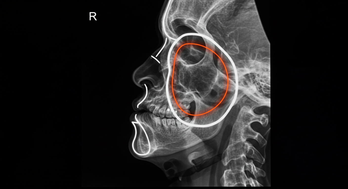
Zygomatic Bones MTF Feminization: Your Complete Guide to a Feminine Facial Structure

X-ray illustration of a human skull with highlighted jaw area, showing the temporomandibular joint (TMJ) and surrounding structures.

Achieving a feminine facial aesthetic is a significant aspect of the gender affirmation journey for many transgender women (MTF). While hormonal transition induces widespread changes, the underlying bony structure of the face plays a crucial role in perceived gender. The zygomatic bones, commonly known as the cheekbones, are key determinants of facial contour. This comprehensive guide, written from a surgeon’s perspective, delves into the nuances of zygomatic bone contouring as part of facial feminization surgery (FFS), providing an in-depth understanding of the procedure, its considerations, and expected outcomes.

A close-up image of a smiling woman with clear skin and glossy lips, wearing a diamond earring.

Understanding the Role of Zygomatic Bones in Facial Femininity

The male and female facial skeletons exhibit subtle yet significant differences. In general, female faces tend to have more prominent, anteriorly and laterally projected zygomatic bones, creating a higher and more defined cheekbone contour. This contributes to the midface fullness and the characteristic “ogee” curve of the female cheek. Conversely, male zygomatic bones are often less prominent and have a more vertical orientation.

Anatomical Considerations: The Zygomatic Complex Explained

To understand the surgical approach, it’s essential to grasp the anatomy of the zygomatic complex. This intricate structure comprises the zygomatic bone itself and its articulations (connections) with several adjacent bones:

  • Maxilla: The upper jaw bone, forming the lower and medial (towards the midline) border of the zygomatic bone.
  • Temporal Bone: Located on the side of the skull, the zygomatic bone forms the zygomatic arch, a slender bony bridge connecting to the temporal bone.
  • Sphenoid Bone: A complex butterfly-shaped bone situated at the base of the skull, contributing to the posterior (back) aspect of the orbit (eye socket) where the zygomatic bone also plays a role.
  • Frontal Bone: The forehead bone, with which the zygomatic bone articulates at the superior (upper) and lateral (outer) aspect of the orbit.

The zygomatic bone itself has several important features:

  • Body: The main bulk of the bone, contributing to cheek prominence.
  • Frontal Process: Extends upwards to meet the frontal bone.
  • Temporal Process: Extends posteriorly to form the zygomatic arch.
  • Maxillary Process: Extends medially to articulate with the maxilla.
  • Orbital Surface: Forms part of the lateral and inferior walls of the eye socket.

Hormonal Influence vs. Bony Structure

It’s crucial to understand that while estrogen plays a significant role in the feminization process by influencing soft tissues (fat redistribution, skin texture), it has minimal impact on the underlying bony structure once skeletal maturity is reached. Therefore, surgical intervention targeting the zygomatic bones is often necessary to achieve a desired degree of midface feminization.

Surgical Techniques for Zygomatic Bone Feminization

Several surgical techniques can be employed to enhance the feminine contour of the zygomatic bones. The specific approach depends on the individual’s anatomy, desired outcome, and the surgeon’s expertise.

Zygomatic Implants: Augmentation for Increased Prominence

One common method involves the placement of biocompatible implants over the existing zygomatic bones. These implants, typically made of silicone or porous polyethylene, can augment the anterior and lateral projection of the cheekbones, creating a more defined and feminine contour.

  • Material Selection: Silicone implants are smooth and can be easily removed if necessary. Porous polyethylene implants allow for tissue ingrowth, potentially leading to better long-term integration and stability.
  • Placement: Implants are typically inserted through incisions made inside the mouth (intraoral approach) or within the hairline near the temples. This minimizes visible scarring. The implant is then positioned precisely over the zygomatic body and secured if needed.
  • Customization: Implants come in various shapes and sizes, and custom-designed implants can be created based on 3D imaging of the patient’s skull for a highly tailored outcome.

Zygomatic Osteotomy and Advancement: Reshaping the Bone

In cases where augmentation alone is insufficient or a more significant change in bone position is desired, a zygomatic osteotomy and advancement may be performed. This involves surgically cutting and repositioning the zygomatic bones.

  • Osteotomy: Precise cuts are made in the zygomatic bone to mobilize it. The location and type of osteotomy depend on the desired movement. Common techniques include Le Fort I or modifications thereof to address the midface as a whole, or more localized osteotomies focusing specifically on the zygomatic body and arch.
  • Advancement and Fixation: The mobilized zygomatic segments are then moved anteriorly (forward) and potentially laterally (outward) to achieve the desired projection. They are then fixed in their new position using small titanium plates and screws.
  • Combined Procedures: Zygomatic osteotomy and advancement can be combined with bone grafting or contouring to further refine the shape and volume.

Zygomatic Arch Contouring: Refining the Lateral Aspect

The zygomatic arch contributes to the width and overall shape of the midface. In some individuals, reducing the prominence or altering the curvature of the zygomatic arch can enhance facial femininity.

  • Osteotomy and Inset: This involves making a small cut in the zygomatic arch and slightly moving a segment inward to narrow the midface.
  • Burring and Reshaping: In cases of mild prominence, the outer surface of the zygomatic arch can be carefully burred down to create a smoother and less angular contour.

The Surgical Process: From Consultation to Recovery

Undergoing zygomatic bone feminization surgery involves several key stages:

Initial Consultation and Assessment

This crucial first step involves a thorough discussion with the facial feminization surgeon. The surgeon will:

  • Review your medical history: To identify any potential contraindications or risk factors.
  • Understand your goals and expectations: To ensure a clear understanding of your desired facial outcome.
  • Perform a detailed facial analysis: Assessing your existing bone structure, soft tissue, and overall facial harmony. This may involve physical examination, photographs, and potentially 3D CT scans to visualize the underlying bone.
  • Explain the different surgical options: Discussing the most suitable techniques based on your individual anatomy and goals.
  • Outline the risks, benefits, and limitations of the procedure.
  • Provide information about the anesthesia, surgical facility, and cost.

Pre-Operative Preparation

Once you decide to proceed with surgery, you will receive detailed pre-operative instructions, which may include:

  • Avoiding certain medications: Such as blood thinners and non-steroidal anti-inflammatory drugs (NSAIDs) for a specified period before surgery.
  • Stopping smoking: As smoking can impair healing.
  • Undergoing pre-operative medical tests: To ensure you are fit for surgery.
  • Arranging for transportation and post-operative care.

The Surgical Procedure

Zygomatic bone feminization surgery is typically performed under general anesthesia. The duration of the surgery varies depending on the complexity of the techniques involved and whether it is performed in conjunction with other FFS procedures.

  • Incision Placement: As mentioned earlier, incisions are strategically placed to minimize visible scarring. Intraoral incisions are common for implant placement and some osteotomy approaches. Hairline or temporal incisions may be used for zygomatic arch work or more complex osteotomies.
  • Implant Placement or Bone Reshaping: The chosen technique is meticulously executed. For implants, they are positioned and secured. For osteotomies, the bones are cut, repositioned, and fixed with titanium plates and screws.
  • Closure: Incisions are closed with sutures.

Post-Operative Recovery

The recovery period following zygomatic bone feminization surgery involves swelling, bruising, and discomfort.

  • Immediate Post-Op: You will likely experience facial swelling, bruising around the eyes and cheeks, and numbness. Pain medication will be prescribed to manage discomfort. Cold compresses are essential to reduce swelling.
  • First Few Weeks: Swelling and bruising will gradually subside over the first few weeks. You will need to follow a soft diet and avoid strenuous activities. Oral hygiene is crucial, especially with intraoral incisions.
  • Long-Term Recovery: While significant healing occurs within the first few months, subtle changes and settling of tissues can continue for up to a year. Incisions will gradually fade.

Potential Risks and Complications

As with any surgical procedure, zygomatic bone feminization carries potential risks and complications, including:

  • Infection: Although rare with proper surgical technique and post-operative care.
  • Bleeding and Hematoma (blood collection under the skin).
  • Nerve damage: Potentially leading to temporary or, in rare cases, permanent numbness or weakness in the facial area.
  • Implant displacement or extrusion (with implant procedures).
  • Asymmetry: Despite meticulous planning, subtle asymmetries can occur.
  • Poor healing or scarring.
  • Pain or discomfort.
  • Unsatisfactory aesthetic outcome requiring revision surgery.
  • Plate palpability (feeling the titanium plates under the skin, more common in thin individuals).

It is crucial to discuss these risks thoroughly with your surgeon before proceeding with surgery.

Expected Outcomes and the Impact on Facial Femininity

Successful zygomatic bone feminization can significantly enhance the feminine appearance of the face by:

  • Creating higher and more defined cheekbones: Contributing to the classic feminine “ogee” curve.
  • Adding midface fullness: Softening the overall facial structure.
  • Improving facial harmony and balance: By bringing the midface into better proportion with other facial features.
  • Enhancing the youthful appearance: As prominent cheekbones are often associated with youthfulness.

The degree of change will depend on the chosen surgical technique and the individual’s pre-operative anatomy. It’s essential to have realistic expectations and understand that the goal is to achieve a natural and harmonious feminine facial appearance.

Combining Zygomatic Contouring with Other FFS Procedures

Zygomatic bone feminization is often performed in conjunction with other facial feminization surgeries to achieve a comprehensive transformation. These may include:

  • Forehead Contouring (Brow Bone Reduction and Foreheadplasty): To soften the upper face and create a more feminine brow shape.
  • Rhinoplasty (Nose Reshaping): To refine the size and shape of the nose.
  • Mandibular Contouring (Jaw Reduction and Genioplasty): To narrow and soften the jawline and chin.
  • Chin Augmentation or Reduction: To refine the projection and shape of the chin.
  • Lip Augmentation: To enhance lip volume and shape.
  • Tracheal Shave: To reduce the prominence of the Adam’s apple.

The combination of procedures is tailored to the individual’s specific needs and goals, aiming for overall facial harmony and balance.

A professional portrait of a woman with dark hair, wearing earrings and lipstick, smiling warmly against a dark background.

Conclusion: Embracing a More Feminine Facial Structure

Zygomatic bone feminization surgery plays a vital role in achieving a more feminine facial structure for many MTF individuals. By augmenting or reshaping the cheekbones, surgeons can create a softer, more defined, and harmonious midface contour. Understanding the anatomical considerations, surgical techniques, and the recovery process is crucial for making informed decisions and achieving satisfying outcomes. Choosing an experienced and qualified facial feminization surgeon is paramount to ensure a safe and successful procedure that aligns with your individual goals and contributes significantly to your gender affirmation journey.

Visit Dr.MFO Instagram profile to see real patient transformations! Get a glimpse of the incredible results achieved through facial feminization surgery and other procedures. The profile showcases before-and-after photos that highlight Dr. MFO’s expertise and artistic vision in creating natural-looking, beautiful outcomes.

Ready to take the next step in your journey? Schedule a free consultation with Dr. MFO ( Best Facial Feminization Surgeon for You) today. During the consultation, you can discuss your goals, ask any questions you may have, and learn more about how Dr. MFO can help you achieve your desired look. Don’t hesitate to take advantage of this free opportunity to explore your options and see if Dr. MFO is the right fit for you.

Laryngoplasty (Adam’s Apple Reduction) Cost in Turkey with Facial Feminization Surgery: A Surgeon’s Insight

A woman with short wavy hair smiling, wearing red lipstick and elegant dangling earrings with a red gemstone and diamond accents.

For the transgender woman seeking facial gender affirmation, the prominence of the Adam’s apple can be a significant source of dysphoria. Laryngoplasty, or tracheal shave, is a surgical procedure designed to reduce the size of this laryngeal prominence, contributing to a more feminine neck contour. Often considered in conjunction with broader facial feminization surgery (FFS), understanding the nuances of this procedure, particularly concerning cost in a destination like Turkey, is crucial for informed decision-making. This discourse aims to provide a comprehensive overview from a surgical perspective.

A portrait of a woman with shoulder-length wavy brown hair, wearing dangling earrings and a dark blue top, posing against a plain light gray background.

Anatomical Considerations and Surgical Technique

The Adam’s apple is the anterior projection of the thyroid cartilage, a key structural component of the larynx. Its size is largely influenced by hormonal factors, with testosterone typically leading to a more pronounced prominence during male puberty. In laryngoplasty, the surgeon meticulously reduces the anterior angle of the thyroid cartilage. This requires a precise understanding of the underlying anatomy to avoid damage to the vocal cords or other vital structures.

The procedure typically involves a small horizontal incision in a natural skin crease of the neck to minimize scarring. Subplatysmal dissection is performed to expose the thyroid cartilage. Using specialized instruments, the prominent portion of the cartilage is carefully shaved down. The edges are smoothed to create a natural contour. The platysma muscle is then reapproximated, and the skin is closed in layers. The procedure is often performed under general anesthesia, although local anesthesia with sedation may be an option in select cases.

The Role of Laryngoplasty within Facial Feminization Surgery

Facial feminization surgery encompasses a range of procedures aimed at softening masculine facial features to align them more closely with a feminine aesthetic. While various procedures address different aspects of the face – such as forehead contouring, rhinoplasty, lip augmentation, and chin reduction – laryngoplasty specifically targets the neck.

For many transgender women, the Adam’s apple is a highly visible and persistent masculine trait that can detract from their overall feminine presentation. Addressing it through laryngoplasty can significantly enhance the perceived femininity of the face and neck, contributing to a greater sense of congruence between internal gender identity and external appearance. Therefore, it is frequently integrated into a comprehensive FFS plan.

Factors Influencing Laryngoplasty Cost

The cost of laryngoplasty, whether performed in isolation or as part of FFS, is influenced by several key factors. Understanding these variables is essential for patients considering this procedure, particularly when exploring options in different geographic locations.

Surgeon’s Expertise and Reputation

The surgeon’s experience, qualifications, and reputation are significant determinants of cost. Surgeons with extensive experience in facial gender affirmation surgeries, including laryngoplasty, and a proven track record of successful outcomes typically command higher fees. Their expertise minimizes the risk of complications and maximizes the likelihood of achieving the desired aesthetic result.

Facility Fees and Anesthesia Costs

The surgical facility where the procedure is performed also contributes to the overall cost. Accredited hospitals and specialized surgical centers often have higher overhead costs compared to smaller clinics. The type of anesthesia used (general or local with sedation) and the anesthesiologist’s fees are additional factors that influence the total expense.

Geographic Location: The Case of Turkey

Turkey has emerged as a popular destination for medical tourism, including gender affirming surgeries. The cost of medical procedures, including laryngoplasty and FFS, is often significantly lower in Turkey compared to many Western countries, such as the United States, Canada, and parts of Europe. This cost difference is primarily due to factors such as lower operating costs, currency exchange rates, and competitive pricing within the Turkish healthcare market.

However, it is crucial to emphasize that cost should not be the sole deciding factor. The quality of care, the surgeon’s qualifications, the accreditation of the medical facility, and the availability of comprehensive aftercare are paramount considerations.

Inclusion within a Broader FFS Package

When laryngoplasty is performed as part of a more comprehensive FFS package, the overall cost may be structured differently. Some surgeons or clinics offer package deals that can be more cost-effective than undergoing each procedure separately. It is essential to obtain a detailed breakdown of the costs associated with each component of the FFS plan, including laryngoplasty, anesthesia, facility fees, and post-operative care.

Pre-operative and Post-operative Care

The total cost should also account for pre-operative consultations, necessary medical tests, and post-operative care, including follow-up appointments and any required medications. It is vital to clarify what is included in the quoted price to avoid unexpected expenses.

Estimating Laryngoplasty (Adam’s Apple Reduction) Cost in Turkey with FFS

Providing a precise cost estimate for laryngoplasty with FFS in Turkey is challenging without a thorough individual assessment. However, based on general trends and available information, patients can expect the cost of laryngoplasty as a standalone procedure to range from €1,500 to €3,000.

When combined with a comprehensive FFS package, the cost of laryngoplasty is typically integrated into the overall price. The total cost of FFS in Turkey can vary significantly depending on the number and complexity of the procedures involved, but it generally ranges from €5,000 to €20,000 or more.

It is strongly recommended that individuals seeking these procedures obtain personalized quotes from reputable surgeons and clinics in Turkey. These consultations will allow for a detailed discussion of the patient’s specific needs and goals, a comprehensive surgical plan, and an accurate cost estimate.

Important Considerations for Patients

Beyond the cost, several other critical factors should guide a transgender woman’s decision-making process when considering laryngoplasty and FFS in Turkey:

Surgeon Qualifications and Experience

Thoroughly research the surgeon’s credentials, experience specifically in transgender facial surgery, and before-and-after photos of their previous patients. Look for board certification and membership in relevant professional organizations.

Medical Facility Accreditation

Ensure that the surgical facility is accredited by recognized national or international bodies. This indicates adherence to certain standards of safety and quality of care.

Communication and Language

Effective communication with the surgical team is crucial. Ensure that there are clear lines of communication and that language barriers are addressed. Many reputable clinics catering to international patients have English-speaking staff.

Pre-operative Assessment and Planning

A comprehensive pre-operative assessment is essential to determine the patient’s suitability for surgery and to develop a personalized surgical plan that aligns with their aesthetic goals.

Post-operative Care and Follow-up

Understand the details of the post-operative care plan, including the duration of stay in Turkey, follow-up appointments, and how any potential complications will be managed.

Legal and Ethical Considerations

Be aware of any legal or ethical considerations related to medical tourism and ensure that the chosen clinic adheres to ethical medical practices.

A woman with wavy brown hair and blue eyes wearing a floral print top and gold earrings, looking confidently at the camera against a neutral background.

Conclusion

Laryngoplasty is a valuable procedure for transgender women seeking to feminize their neck contour and alleviate dysphoria associated with a prominent Adam’s apple. When considering this procedure as part of facial feminization surgery in Turkey, the cost can be a significant advantage. However, it is imperative to prioritize the quality of care, the surgeon’s expertise, and the safety of the medical facility above cost alone. Thorough research, open communication with the surgical team, and a comprehensive understanding of all aspects of the surgical journey are essential for a positive and successful outcome. By carefully considering these factors, transgender women can make informed decisions that align with their individual needs and contribute to their overall gender affirmation journey.

Visit Dr.MFO Instagram profile to see real patient transformations! Get a glimpse of the incredible results achieved through facial feminization surgery and other procedures. The profile showcases before-and-after photos that highlight Dr. MFO’s expertise and artistic vision in creating natural-looking, beautiful outcomes.

Ready to take the next step in your journey? Schedule a free consultation with Dr. MFO ( Best Facial Feminization Surgeon for You) today. During the consultation, you can discuss your goals, ask any questions you may have, and learn more about how Dr. MFO can help you achieve your desired look. Don’t hesitate to take advantage of this free opportunity to explore your options and see if Dr. MFO is the right fit for you.

Understanding Zygoma Reduction Procedures in Facial Feminization Surgery

A close-up portrait of a woman with smooth skin, defined eyebrows, and subtle makeup, wearing a gold hoop earring in her left ear.

The zygomatic bones, commonly known as cheekbones, are prominent structures of the midface that significantly influence facial width, contour, and the overall perception of gender. In individuals with predominantly masculine facial features, the zygomatic arches and the body of the zygoma can exhibit greater lateral projection and robusticity, contributing to a wider and more angular facial appearance. Zygoma reduction, a crucial component of Facial Feminization Surgery (FFS), aims to address these characteristics by reshaping and repositioning the zygomatic bones to create a softer, more tapered, and characteristically feminine midfacial contour.

This procedure plays a pivotal role in harmonizing the midface with other feminized features, such as the forehead, nose, and jawline, leading to a more congruent and aesthetically pleasing outcome for transgender women. A comprehensive understanding of the surgical techniques involved and the anatomical considerations of the zygoma is paramount for both the surgeon and the patient in planning and executing effective facial feminization.

Close-up portrait of a young woman with smooth skin and long dark hair, looking up with soft lighting highlighting her facial features.

Anatomical Considerations of the Zygoma in Facial Feminization

The zygomatic bone is a paired quadrilateral bone that forms the prominence of the cheek, contributes to the lateral wall and floor of the orbit, and articulates with the maxilla, temporal bone (via the zygomatic arch), sphenoid bone, and frontal bone. Key anatomical features relevant to zygoma reduction include:

  • Zygomatic Body: The main body of the cheekbone, which determines the anterior and lateral projection of the cheeks.
  • Zygomatic Arch: A slender bony arch formed by the zygomatic process of the temporal bone and the temporal process of the zygomatic bone. The lateral flare of the zygomatic arch significantly contributes to facial width.
  • Frontal Process: Extends superiorly to articulate with the frontal bone.
  • Temporal Process: Extends posteriorly to articulate with the temporal bone, forming part of the zygomatic arch.
  • Maxillary Process: Extends inferiorly to articulate with the maxilla.
  • Orbital Surface: Forms part of the lateral wall and floor of the orbit.

In masculine facial structures, the zygomatic arches tend to be more laterally flared, and the zygomatic bodies may exhibit increased anterior and lateral projection, resulting in a wider and more angular midface. Feminization often involves reducing this lateral projection and creating a smoother, more medially positioned cheekbone prominence.

Surgical Techniques for Zygoma Reduction

Several surgical techniques can be employed to reduce the prominence of the zygomatic bones, primarily involving osteotomy (bone cutting) and ostectomy (bone removal). The specific techniques used depend on the individual’s anatomy, the degree of reduction required, and the surgeon’s expertise.

Zygomatic Arch Reduction (Inward Fracture and Fixation)

This technique focuses on reducing the lateral flare of the zygomatic arches, which significantly contributes to facial width. It involves carefully fracturing the zygomatic arch and repositioning it more medially.

Surgical Procedure

Performed through incisions typically placed within the hairline in the temporal region and sometimes supplemented with intraoral incisions, the zygomatic arch is exposed. Precise osteotomies are performed to mobilize the arch. The arch is then carefully infractured (bent inward) and secured in the new, more medial position using titanium plates and screws. The surrounding soft tissues are redraped, and the incisions are closed.

Advantages of Zygomatic Arch Reduction

  • Significant Reduction in Facial Width: Effectively narrows the midface, creating a more tapered and feminine contour.
  • Relatively Predictable Outcome: The degree of inward movement can be planned and executed with precision.
  • Concealed Incisions: Hairline incisions offer excellent camouflage.

Limitations and Considerations

  • Technical Complexity: Requires precise surgical technique to avoid damage to surrounding structures, including the temporalis muscle and the facial nerve branches.
  • Risk of Temporomandibular Joint (TMJ) Issues: While rare, alterations to the zygomatic arch can theoretically affect TMJ function.
  • Swelling and Bruising: Significant swelling and bruising are expected and can take several weeks to resolve.
  • Potential for Palpable Hardware: In some cases, the fixation plates and screws may be palpable under the skin.

Zygomatic Body Reduction (Wedge Resection and Contouring)

This technique involves removing a wedge-shaped section of the zygomatic body to reduce its anterior and lateral projection.

Surgical Procedure

Typically performed through intraoral incisions in the upper buccal sulcus (the groove between the cheek and upper gum), the zygomatic body is exposed. A carefully planned wedge of bone is removed, reducing the overall projection. The remaining bony edges are then smoothed and contoured to create a more refined and feminine cheekbone shape. In some cases, additional bone removal or contouring may be performed through temporal or preauricular (in front of the ear) incisions to address the zygomatic arch and body simultaneously.

Advantages of Zygomatic Body Reduction

  • Reduces Cheekbone Prominence: Effectively decreases the anterior and lateral projection of the cheeks, creating a softer midfacial contour.
  • Can Be Combined with Arch Reduction: Allows for comprehensive reshaping of the zygoma.
  • Intraoral Incisions Minimize Visible Scarring: The primary incisions are hidden inside the mouth.

Limitations and Considerations

  • Technical Complexity: Requires meticulous surgical planning and execution to achieve symmetrical and natural-looking results.
  • Risk of Infraorbital Nerve Injury: The infraorbital nerve, which provides sensation to the lower eyelid, cheek, and upper lip, runs close to the zygomatic body and is at risk of injury during this procedure.
  • Swelling and Bruising: Significant swelling and bruising are expected and can take several weeks to resolve.
  • Potential for Contour Irregularities: Careful bone contouring is essential to avoid sharp edges or an unnatural appearance.

Combination Techniques

Often, a combination of zygomatic arch reduction and zygomatic body reduction techniques is employed to achieve the desired degree of midfacial feminization. The specific combination and extent of bone modification are tailored to the individual’s unique anatomy and aesthetic goals.

Minimally Invasive Techniques (Burring and Shaving)

In cases where only a subtle reduction in zygomatic prominence is required, minimally invasive techniques involving burring or shaving down the outer surface of the zygomatic bone may be considered. This is typically performed through small incisions.

Advantages of Minimally Invasive Techniques

  • Less Invasive: Involves less extensive bone cutting and may have a shorter recovery period.
  • Subtle Refinement: Effective for minor contour adjustments.

Limitations and Considerations

  • Limited Degree of Reduction: Not suitable for significant zygomatic prominence.
  • Less Predictable Outcome: The amount of bone removed can be less precisely controlled compared to osteotomy and resection techniques.

The Role of 3D Surgical Planning in Zygoma Reduction

Advanced 3D surgical planning software and cone-beam computed tomography (CBCT) are invaluable tools in planning zygoma reduction procedures. These technologies allow the surgeon to:

  • Accurately Assess the Zygomatic Anatomy: Visualize the zygomatic bones and their relationship to surrounding structures in three dimensions.
  • Simulate Surgical Outcomes: Plan osteotomies, bone removal, and repositioning with greater precision.
  • Create Custom Surgical Guides: Fabricate templates that guide the surgeon during the actual procedure, ensuring accurate bone cuts and achieving the desired reduction.
  • Optimize Symmetry: Plan symmetrical reductions to achieve a balanced and natural-looking result.
  • Improve Patient Communication: Provide visual representations of the planned surgical changes and expected outcomes.

3D surgical planning enhances the safety and predictability of zygoma reduction procedures, particularly complex cases involving significant bone modification.

Integration of Zygoma Reduction with Other FFS Procedures

Zygoma reduction is often performed in conjunction with other FFS procedures to achieve comprehensive facial feminization. Common adjunctive procedures include:

  • Forehead Recontouring and Brow Lift: Creating a softer upper face that harmonizes with the feminized midface.
  • Rhinoplasty: Reshaping the nose to achieve a more feminine size and contour.
  • Cheek Augmentation (Paradoxically): In some cases, subtle augmentation of the anterior cheek area after zygoma reduction can further refine the midfacial contour and create a more feminine convexity.
  • Jawline Contouring and Genioplasty: Refining the lower face to create a more feminine jawline and chin, balancing the overall facial structure.

The sequencing and combination of these procedures are carefully tailored to the individual patient’s anatomy and aesthetic goals.

Recovery and Aftercare Following Zygoma Reduction

The recovery period following zygoma reduction varies depending on the extent of the surgical intervention. Generally, patients can expect:

  • Swelling and Bruising: Significant swelling and bruising are common in the midface and cheek areas, potentially extending to the eyelids. This typically peaks within the first few days and gradually subsides over several weeks.
  • Pain Management: Oral pain medication is prescribed to manage post-operative discomfort.
  • Cold Compresses: Applying cold compresses to the face during the initial days can help reduce swelling and bruising.
  • Head Elevation: Keeping the head elevated, especially during sleep, helps to minimize swelling.
  • Dietary Restrictions: A soft diet may be recommended initially to minimize chewing and stress on the healing bones.
  • Oral Hygiene: Meticulous oral hygiene, including gentle rinsing with antiseptic mouthwash, is crucial, especially with intraoral incisions.
  • Activity Restrictions: Strenuous activity and heavy lifting should be avoided for several weeks.
  • Follow-up Appointments: Regular follow-up appointments with the surgeon are necessary to monitor healing and address any concerns.

Potential Risks and Complications of Zygoma Reduction

Zygoma reduction procedures carry potential risks and complications, including:

  • Infection: Can occur at the incision sites.
  • Bleeding and Hematoma: Collection of blood under the skin.
  • Nerve Damage: Potential injury to branches of the facial nerve (leading to temporary or, rarely, permanent weakness of facial muscles) or the infraorbital nerve (causing numbness in the cheek, lower eyelid, and upper lip).
  • Asymmetry: Uneven reduction or contour between the two sides of the face.
  • Malunion or Non-union: Improper or incomplete healing of the bone segments.
  • Temporomandibular Joint (TMJ) Issues: Although rare, changes in the zygomatic arch can potentially affect TMJ function.
  • Palpable Hardware: Fixation plates and screws may be palpable under the skin in some cases.
  • Contour Irregularities: Sharp edges or unnatural depressions in the cheekbone area.
  • Swelling and Bruising: Prolonged or excessive swelling and bruising.

A thorough discussion of these potential risks and complications with the surgeon is essential before proceeding with zygoma reduction surgery.

A close-up portrait of a woman with curly hair, wearing large gold hoop earrings and makeup. She has a warm, natural expression with subtle eye shadow and lip gloss, emphasizing her eyes and lips.

Conclusion: Achieving Feminine Midfacial Harmony through Zygoma Reduction

Zygoma reduction is a transformative procedure within Facial Feminization Surgery, directly addressing prominent cheekbones to create a softer, more feminine midfacial contour. Techniques ranging from zygomatic arch reduction to zygomatic body resection, often guided by advanced 3D surgical planning, offer effective means to narrow the face and refine cheekbone projection. When performed by a skilled and experienced surgeon, zygoma reduction can harmonize beautifully with other FFS procedures, contributing significantly to overall facial balance and a greater sense of gender congruence for transgender women.

A comprehensive understanding of the anatomical considerations, surgical options, and potential outcomes is crucial for both the patient and the surgeon in navigating this important aspect of facial gender affirmation. Choosing a surgeon specializing in FFS with extensive experience in zygoma reduction is paramount to achieving safe, effective, and aesthetically pleasing results.

Visit Dr.MFO Instagram profile to see real patient transformations! Get a glimpse of the incredible results achieved through facial feminization surgery and other procedures. The profile showcases before-and-after photos that highlight Dr. MFO’s expertise and artistic vision in creating natural-looking, beautiful outcomes.

Ready to take the next step in your journey? Schedule a free consultation with Dr. MFO ( Best Facial Feminization Surgeon for You) today. During the consultation, you can discuss your goals, ask any questions you may have, and learn more about how Dr. MFO can help you achieve your desired look. Don’t hesitate to take advantage of this free opportunity to explore your options and see if Dr. MFO is the right fit for you.