From the nuanced vantage point of a surgeon specializing in Facial Feminization Surgery (FFS), the periorbital region – encompassing the eyes and their surrounding structures – presents a critical frontier in achieving optimal aesthetic harmony and feminization. Among the myriad procedures within FFS, the reshaping of the supraorbital (brow bone) and lateral orbital (outer eye socket) rims holds paramount importance. This extensive discussion delves into the anatomical intricacies, surgical methodologies, potential complications, and expected outcomes of these procedures, presented with both medical precision and accessible explanations for the discerning patient and fellow practitioner.
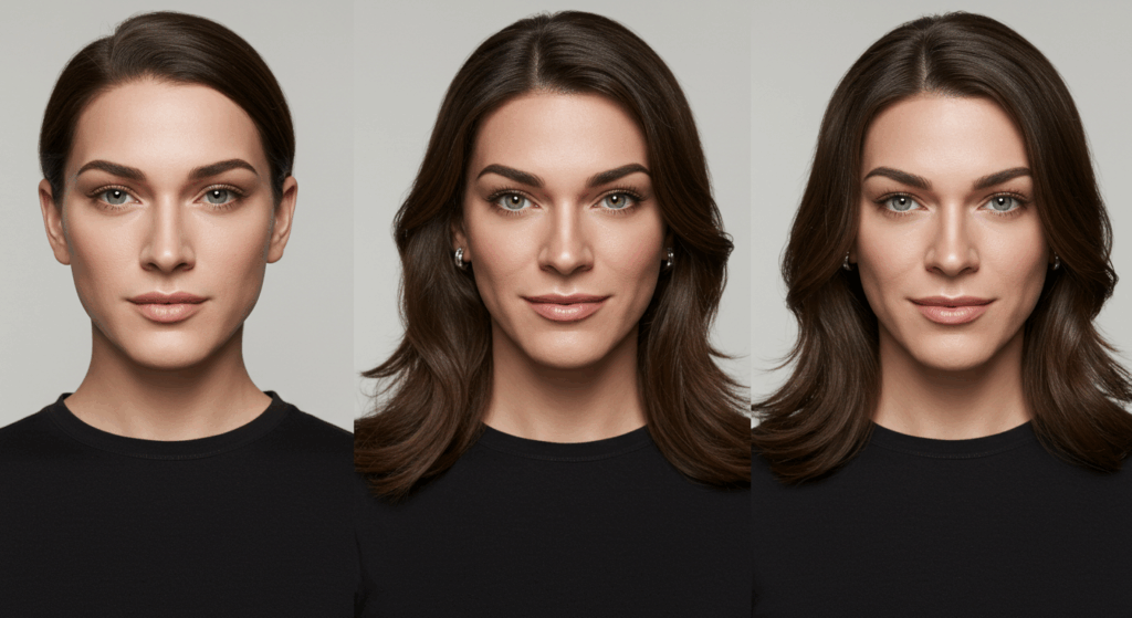
Table of Contents
Understanding the Periorbital Anatomy: A Foundation for Feminization
The periorbital area is a complex interplay of skeletal, muscular, adipose (fat), and integumentary (skin) tissues. A profound understanding of these components is not merely academic; it is the bedrock upon which successful surgical feminization is built.
The Supraorbital Rim: The Forehead’s Foundation
The supraorbital rim, colloquially known as the brow bone, forms the superior (upper) boundary of the orbit, or eye socket. In biological males, this structure tends to be more prominent and protrusive, creating a heavy, sometimes shadowed appearance to the eyes. This prominence is primarily due to the underlying frontal sinus and the thickness of the frontal bone in this region.
- Frontal Sinus: A pair of air-filled cavities located within the frontal bone, just above the nasal cavity. Their size and configuration significantly influence the projection of the brow bone.
- Glabella: The smooth, triangular area between the eyebrows and above the bridge of the nose. This is often the most prominent part of the brow bossing in males.
- Supraorbital Notch/Foramen: A small groove or opening on the supraorbital rim, transmitting the supraorbital nerve and artery. Protecting these neurovascular structures is paramount during surgery to prevent sensation loss or bleeding.
The Lateral Orbital Rim: Defining the Eye’s Outer Contour
The lateral orbital rim constitutes the outer boundary of the eye socket, extending from the temporal bone (side of the skull, near the temple) to the zygomatic bone (cheekbone). In males, this region can also appear more robust, contributing to a less open and more angular appearance of the eyes. Feminization often involves subtle contouring to soften this angle and create a more graceful transition to the temporal region.
- Zygomaticofrontal Suture: The fibrous joint connecting the zygomatic bone and the frontal bone. This is a key anatomical landmark during lateral orbital rim reduction.
- Temporal Fossa: The depression on the side of the skull, where the temporalis muscle (a chewing muscle) is located. Reshaping the lateral orbital rim often influences the aesthetic of this adjacent area.
Soft Tissue Considerations: More Than Just Bone
While skeletal reshaping is central, the overlying soft tissues are equally critical. The thickness of the skin, the presence of subcutaneous fat, and the activity of periorbital muscles (such as the orbicularis oculi, which closes the eye) all contribute to the final aesthetic outcome. A surgeon must meticulously consider how changes to the underlying bone will impact these soft tissues, ensuring a natural and harmonious result.

Surgical Indications and Patient Selection
The decision to proceed with supraorbital and lateral orbital rim reshaping is driven by specific aesthetic goals, primarily to achieve a more feminine appearance of the upper face and eyes.
Primary Indications
- Brow Bossing Reduction: Addressing a prominent supraorbital rim that casts shadows over the eyes, makes the forehead appear heavier, or contributes to a masculine facial profile.
- Orbital Reshaping: Creating a more open, less angular, and aesthetically pleasing contour of the eye sockets.
- Overall Facial Feminization: Integrating periorbital reshaping into a comprehensive FFS plan to achieve harmonious facial feminization.
Contraindications and Considerations
While these procedures are generally safe, certain factors may contraindicate or necessitate caution:
- Pre-existing Medical Conditions: Uncontrolled systemic diseases (e.g., severe cardiovascular disease, bleeding disorders) can increase surgical risks.
- Active Infections: Any active infection in the surgical field must be resolved before surgery.
- Unrealistic Patient Expectations: A thorough discussion of potential outcomes and limitations is crucial to ensure patient satisfaction.
- Insufficient Bone Thickness: In rare cases, extremely thin frontal bone may limit the extent of reduction achievable through burring alone, potentially necessitating a bone flap setback.

Surgical Techniques for Supraorbital Rim Reshaping
The approach to supraorbital rim reshaping depends largely on the degree of prominence and the underlying anatomy, particularly the size and configuration of the frontal sinus.
Type I: Burring (Shaving)
This technique involves direct reduction of the bone using a specialized surgical burr.
- Application: Ideal for cases with mild to moderate brow bossing, where the frontal sinus is relatively small or positioned favorably, allowing for sufficient bone removal without breaching the sinus cavity.
- Mechanism: The outer cortical bone (the hard, outer layer of the bone) and some diploë (spongy bone between the inner and outer layers) are carefully removed until the desired contour is achieved.
- Advantages: Less invasive than bone flap procedures, shorter recovery time, generally lower risk of complications associated with sinus entry.
- Disadvantages: Limited in the amount of reduction achievable; not suitable for prominent bossing where the frontal sinus extends significantly into the area.
Type II: Frontal Sinus Setback with Bone Flap (Ostectomy and Fixation)
This is a more complex procedure indicated for significant brow bossing, especially when the frontal sinus is large and extends into the area requiring reduction.
- Application: When burring alone would lead to perforation of the frontal sinus or would not achieve adequate feminization.
- Mechanism: A precisely cut section of the frontal bone, including the outer wall of the frontal sinus, is removed (osteotomy). This “bone flap” is then reshaped, often by burring its posterior (inner) surface to reduce its projection. The reshaped bone flap is then re-positioned backward (setback) and secured with small titanium plates and screws. The anterior wall of the frontal sinus is effectively moved backward, thereby reducing the projection of the brow bone.
- Advantages: Allows for substantial reduction of brow bossing, even in cases of large frontal sinuses.
- Disadvantages: More invasive, longer recovery, potential for complications related to sinus entry (e.g., cerebrospinal fluid leak, infection, mucocele formation), requires careful fixation of the bone flap.
Type IIa: Partial Frontal Sinus Setback
In some cases, only a portion of the frontal sinus wall needs to be set back, typically the central glabella region. This is a variation of the Type II procedure, adapted for specific anatomical needs.
Type IIb: Complete Frontal Sinus Setback
When the entire anterior wall of the frontal sinus is prominent, a complete setback may be necessary. This involves carefully elevating the dura mater (the tough membrane covering the brain) from the posterior wall of the frontal sinus to allow for the complete setback of the anterior wall. This is a delicate maneuver requiring extensive surgical experience.
Type III: Supraorbital Rim Craniotomy (Forehead Reconstruction)
This technique is less commonly employed for isolated supraorbital rim reshaping but may be necessary in extreme cases of skull asymmetry or when extensive forehead reshaping is required in conjunction with brow bossing reduction. It involves a more extensive removal and reshaping of the frontal bone, potentially extending beyond the confines of the frontal sinus.
Surgical Techniques for Lateral Orbital Rim Reshaping
Reshaping the lateral orbital rim focuses on softening the transition from the forehead to the temporal region and reducing any masculine angularity.
Lateral Orbital Rim Reduction (Burring)
- Application: Common for subtle to moderate lateral orbital rim prominence.
- Mechanism: Similar to Type I brow bone reduction, a surgical burr is used to carefully reduce the outer aspect of the lateral orbital rim, creating a smoother, more curved contour. Care is taken to avoid damage to the underlying temporal muscle and nerves.
- Advantages: Relatively straightforward, good for subtle feminization.
- Disadvantages: Limited in the amount of reduction possible; cannot address severe lateral orbital protrusion alone.
Lateral Orbital Rim Contouring with Zygomatic Arch Reduction (if indicated)
In some cases, especially when the cheekbones (zygomatic arches) are also prominent or wide, simultaneous reduction of the lateral orbital rim and the adjacent zygomatic arch may be performed. This creates a more harmonious and feminized contour from the outer eye to the midface.
- Application: When a broader midface or prominent zygomatic arch contributes to a masculine appearance.
- Mechanism: This may involve burring or even osteotomies (bone cuts) of the zygomatic bone to narrow the facial width and create a softer transition from the lateral orbital rim.
Surgical Approach and Incisions
The choice of incision for these procedures is crucial for both access and aesthetic outcome, aiming for minimal visibility of scars.
Coronal Incision (Hairline Incision)
- Location: Typically made behind the hairline, extending from ear to ear across the scalp.
- Advantages: Provides excellent access to the entire forehead, supraorbital rims, and temporal regions. The scar is well-hidden within the hair, making it nearly invisible once healed.
- Disadvantages: Longer incision, potential for temporary scalp numbness (paresthesia) due to nerve disruption.
Endoscopic Approach (Limited Incisions)
- Location: Several small incisions within the hairline are made, through which an endoscope (a small camera) and specialized instruments are inserted.
- Application: Primarily for forehead lift procedures, but can be used for limited brow bone burring in select cases.
- Advantages: Less invasive, shorter incisions, potentially faster recovery of scalp sensation.
- Disadvantages: Limited visibility and maneuverability, not suitable for extensive bone reduction or bone flap procedures.
Direct Incision (Less Common for FFS)
- Location: Directly over the brow or at the brow hairline.
- Application: Rarely used in FFS due to visible scarring. More common in traditional brow lifts.
- Disadvantages: Highly visible scar.
In the context of FFS for supraorbital and lateral orbital rim reshaping, the coronal incision is overwhelmingly the preferred approach due to the comprehensive access it provides for significant bone contouring while allowing for scar concealment.
Pre-operative Planning: The Blueprint for Success
Meticulous pre-operative planning is non-negotiable for achieving predictable and safe outcomes.
3D CT Scan Analysis
- Purpose: A high-resolution computed tomography (CT) scan provides detailed, three-dimensional images of the facial skeleton, particularly the frontal bone, frontal sinuses, and orbital rims.
- Information Gained:
- Frontal Sinus Size and Configuration: Crucial for determining whether burring (Type I) or a setback procedure (Type II) is necessary.
- Bone Thickness: Assesses the amount of bone available for reduction.
- Symmetry Analysis: Identifies any pre-existing asymmetries that need correction.
- Neurovascular Mapping: Helps identify the precise location of nerves and blood vessels to minimize injury.
- Pre-operative Simulation: Advanced software can use CT data to create 3D models and simulate potential surgical outcomes, allowing the surgeon and patient to visualize the proposed changes.
Photographic Analysis
Standardized pre-operative photographs from multiple angles (frontal, lateral, oblique, worm’s eye view) are essential for documenting the initial appearance and for post-operative comparison. These also aid in surgical planning by highlighting areas of concern.
Patient Consultation and Goal Setting
A thorough discussion with the patient is paramount. This includes:
- Understanding Patient Expectations: Aligning the patient’s aesthetic goals with realistic surgical outcomes.
- Informed Consent: Explaining the procedure in detail, including potential risks, benefits, alternatives, and recovery process.
- Addressing Concerns: Allowing the patient to voice any anxieties or questions.
The Surgical Procedure: A Step-by-Step Overview
While specific steps vary based on the chosen technique, a general overview of the surgical process provides insight into the complexity and precision involved.
Anesthesia
General anesthesia is typically administered, ensuring the patient is completely unconscious and pain-free throughout the procedure.
Incision and Flap Elevation
- The chosen incision (usually coronal) is made.
- The scalp and forehead tissues are carefully elevated in a subgaleal plane (below the galea aponeurotica, a fibrous sheet covering the scalp) or subperiosteal plane (directly on the bone) to expose the underlying frontal bone, supraorbital rims, and lateral orbital rims. This elevation must be performed meticulously to protect nerves and blood vessels.
Supraorbital Rim Reshaping
- Type I (Burring): Using a high-speed surgical burr, the prominent bone is meticulously and gradually reduced. Constant irrigation (washing with saline) is used to cool the bone and clear debris. The surgeon frequently palpates (feels) the bone to ensure a smooth, symmetric, and adequate reduction without perforating the frontal sinus.
- Type II (Frontal Sinus Setback):
- Osteotomy: Precise bone cuts are made to create the bone flap, typically using an oscillating saw.
- Flap Removal: The bone flap is carefully lifted, exposing the frontal sinus cavity.
- Sinus Management (if applicable): If the sinus is entered, the lining (mucosa) is often removed to prevent mucocele formation (a mucus-filled cyst). The posterior wall of the sinus is inspected for integrity.
- Bone Reshaping: The removed bone flap is thinned and reshaped on a separate sterile table to reduce its projection.
- Repositioning and Fixation: The reshaped bone flap is then re-positioned backward into the desired contour and secured with small, biocompatible titanium plates and screws. These plates and screws are typically permanent but are very small and generally not palpable.
Lateral Orbital Rim Reshaping
- Using a surgical burr, the outer aspect of the lateral orbital rim is carefully contoured and reduced to achieve a softer, more feminine curve. This is often done in conjunction with the supraorbital rim reshaping to ensure a harmonious transition.
Hemostasis and Irrigation
Throughout the procedure, meticulous hemostasis (control of bleeding) is maintained using electrocautery (a device that uses heat to stop bleeding) and other techniques. The surgical field is continuously irrigated with saline solution.
Closure
- The elevated tissues are redraped, and any excess scalp skin may be removed (scalp advancement) if a forehead lift is desired concurrently.
- The incision is closed in layers using sutures, typically with a drain placed temporarily to collect any fluid accumulation.
Post-operative Care and Recovery
The post-operative period is crucial for optimal healing and minimizing complications.
Immediate Post-operative Period (First Few Days)
- Pain Management: Oral pain medication is prescribed.
- Swelling and Bruising: Significant swelling and bruising around the eyes and forehead are expected. Cold compresses and elevation of the head can help minimize these.
- Drains: If placed, drains are typically removed within 24-48 hours once drainage is minimal.
- Head Dressing: A light compressive head dressing may be applied for a short period.
- Numbness: Temporary numbness of the scalp and forehead is common due to nerve disruption during tissue elevation. This typically resolves over weeks to months.
Weeks 1-6
- Reduced Swelling: Major swelling gradually subsides, though some subtle swelling may persist for several months.
- Bruising Resolution: Bruising typically resolves within 2-3 weeks.
- Activity Restrictions: Strenuous activities, heavy lifting, and bending over are restricted to prevent increased swelling or bleeding.
- Suture Removal: If non-dissolvable sutures are used, they are removed around 7-10 days.
Months 3-12 and Beyond
- Full Healing: The majority of swelling resolves, and the final contour becomes apparent.
- Nerve Regeneration: Sensation in the scalp and forehead continues to return. Complete sensation may take up to a year or longer, and some areas may have permanent subtle numbness.
- Scar Maturation: Incision lines will gradually fade over time, becoming less noticeable. Sun protection is crucial for scar healing.
Potential Complications and Risk Management
While FFS procedures are generally safe in the hands of experienced surgeons, no surgery is without risks. A comprehensive understanding of potential complications allows for proactive risk management.
General Surgical Risks
- Infection: Though rare, infection can occur at the surgical site. Prophylactic antibiotics are typically administered.
- Bleeding/Hematoma: Excessive bleeding during or after surgery can lead to a hematoma (a collection of blood under the skin), which may require drainage.
- Adverse Reaction to Anesthesia: Risks associated with general anesthesia are rare but can include allergic reactions or cardiovascular events.
- Scarring: While efforts are made to minimize scar visibility, all incisions result in a scar.
- Asymmetry: Despite meticulous planning and execution, subtle asymmetries can occur.
Specific Complications Related to Supraorbital and Lateral Orbital Rim Reshaping
- Numbness/Paresthesia: Temporary or, rarely, permanent numbness of the scalp and forehead due to injury to the supraorbital or supratrochlear nerves. Paresthesia (tingling, burning sensation) can also occur as nerves regenerate.
- Cerebrospinal Fluid (CSF) Leak: A rare but serious complication, especially with Type II frontal sinus setback, if the dura mater is perforated. This requires immediate recognition and repair to prevent meningitis (inflammation of the brain and spinal cord membranes).
- Mucocele Formation: If the frontal sinus mucosa is not completely removed during a Type II procedure, a mucocele (mucus-filled cyst) can form within the sinus, potentially causing pain or requiring further surgery.
- Sinusitis: Infection or inflammation of the frontal sinus post-operatively, particularly if the sinus was entered.
- Forehead Irregularities/Dents: Inadequate bone reduction or uneven contouring can lead to visible irregularities.
- Plate/Screw Palpability or Infection: Although rare, the fixation plates and screws can sometimes become palpable (feelable) or, even more rarely, become infected, requiring removal.
- Recurrence of Bossing (Rare): Extremely rare, but inadequate initial reduction or bone remodeling could theoretically lead to some re-projection over many years.
- Aesthetic Dissatisfaction: Despite technically successful surgery, the patient may not be entirely satisfied with the aesthetic outcome, highlighting the importance of clear communication and realistic expectations pre-operatively.
Risk Mitigation Strategies
- Detailed Pre-operative Imaging: Comprehensive 3D CT scans to map out anatomical structures and plan the safest approach.
- Experienced Surgeon: Choosing a surgeon with extensive experience specifically in FFS and complex craniofacial procedures.
- Meticulous Surgical Technique: Precise execution, careful hemostasis, and diligent protection of neurovascular structures.
- Post-operative Monitoring: Close monitoring for any signs of complications.
- Patient Education: Ensuring the patient is fully informed about potential risks and what to expect during recovery.
Expected Outcomes and Aesthetic Impact
The goal of supraorbital and lateral orbital rim reshaping is to achieve a more feminine, softened, and aesthetically harmonious appearance of the upper face and eyes.
Key Aesthetic Transformations
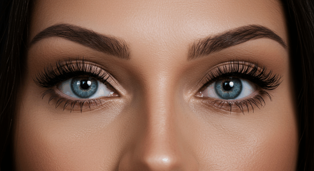
- Reduced Brow Bossing: The most significant change is the reduction of the prominent brow bone, which eliminates shadowing over the eyes, making them appear more open and larger.
- Smoother Forehead Contour: A continuous, gentler curve from the hairline to the nose, replacing the angularity often seen in male foreheads.
- Enhanced Eye Appearance: The eyes appear more prominent, brighter, and less “hooded.”
- Softer Lateral Canthus: The outer corner of the eye (lateral canthus) transitions smoothly to the temple, avoiding a harsh or bony projection.
- Overall Facial Feminization: These changes contribute significantly to the overall feminization of the face, creating a more balanced and harmonious profile.
Psychological and Emotional Benefits
Beyond the physical changes, the psychological and emotional benefits of FFS, including periorbital reshaping, are profound. Patients often report:
- Increased Self-Confidence: Feeling more comfortable and authentic in their own skin.
- Reduced Gender Dysphoria: Alleviation of distress associated with the incongruence between their gender identity and their facial features.
- Improved Social Interaction: Feeling more affirmed in their gender, leading to more positive social experiences.
Conclusion: Art and Science in Facial Feminization
Reshaping the supraorbital and lateral orbital rims is a cornerstone of Facial Feminization Surgery, combining precise anatomical knowledge with nuanced artistic judgment. From the initial detailed CT scan analysis to the meticulous bone contouring and careful post-operative management, every step is critical.
A surgeon’s commitment to excellence in this complex field involves not only mastery of surgical techniques but also a deep understanding of facial aesthetics and the profound impact these procedures have on a patient’s life. By meticulously reducing masculine bone prominence and sculpting feminine contours, we aim to unveil the true self, empowering individuals to live authentically and confidently in their feminized appearance. The journey through periorbital reshaping is a testament to the transformative power of FFS, where the art of surgical reshaping meets the science of human anatomy to redefine identity and beauty.
Visit Dr.MFO Instagram profile to see real patient transformations! Get a glimpse of the incredible results achieved through facial feminization surgery and other procedures. The profile showcases before-and-after photos that highlight Dr. MFO’s expertise and artistic vision in creating natural-looking, beautiful outcomes.
Ready to take the next step in your journey? Schedule a free consultation with Dr. MFO ( Best Facial Feminization Surgeon for You) today. During the consultation, you can discuss your goals, ask any questions you may have, and learn more about how Dr. MFO can help you achieve your desired look. Don’t hesitate to take advantage of this free opportunity to explore your options and see if Dr. MFO is the right fit for you.