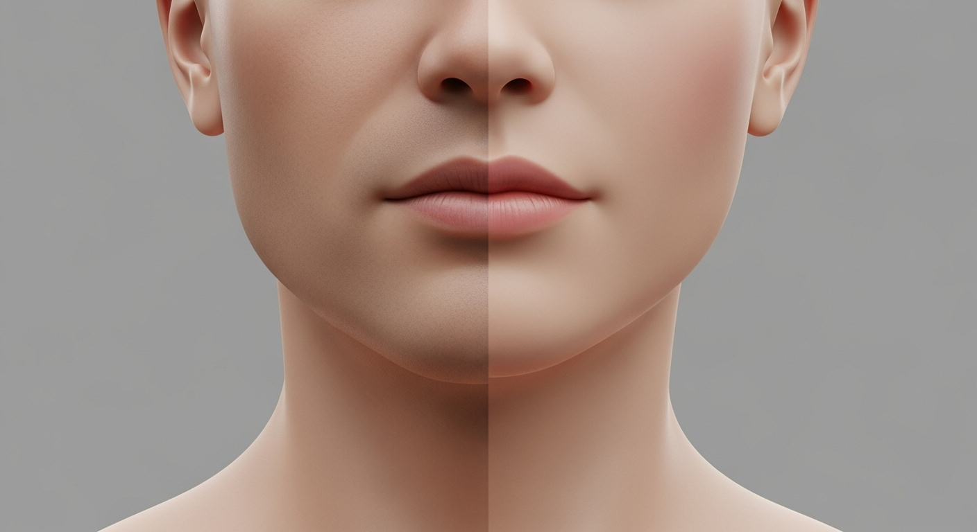
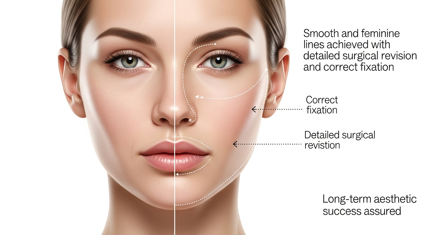
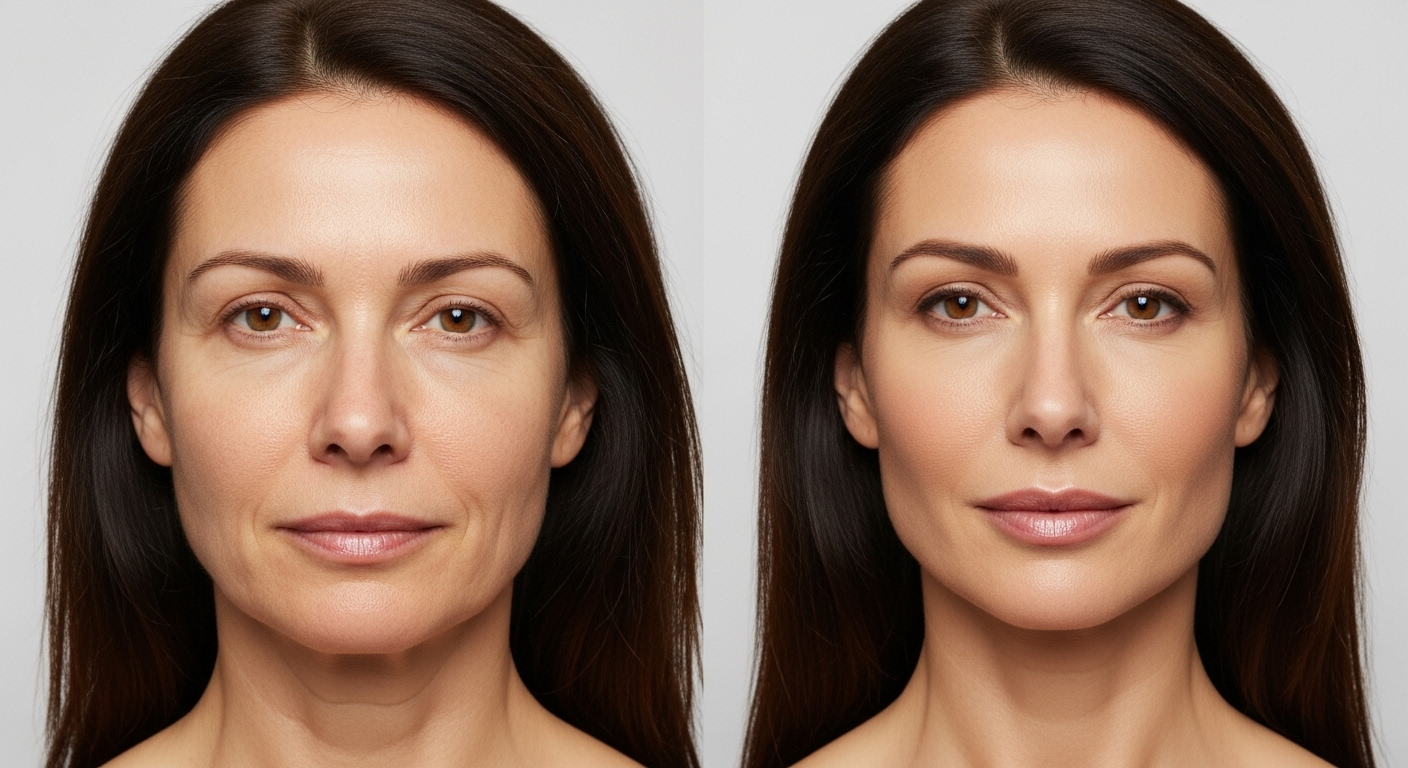
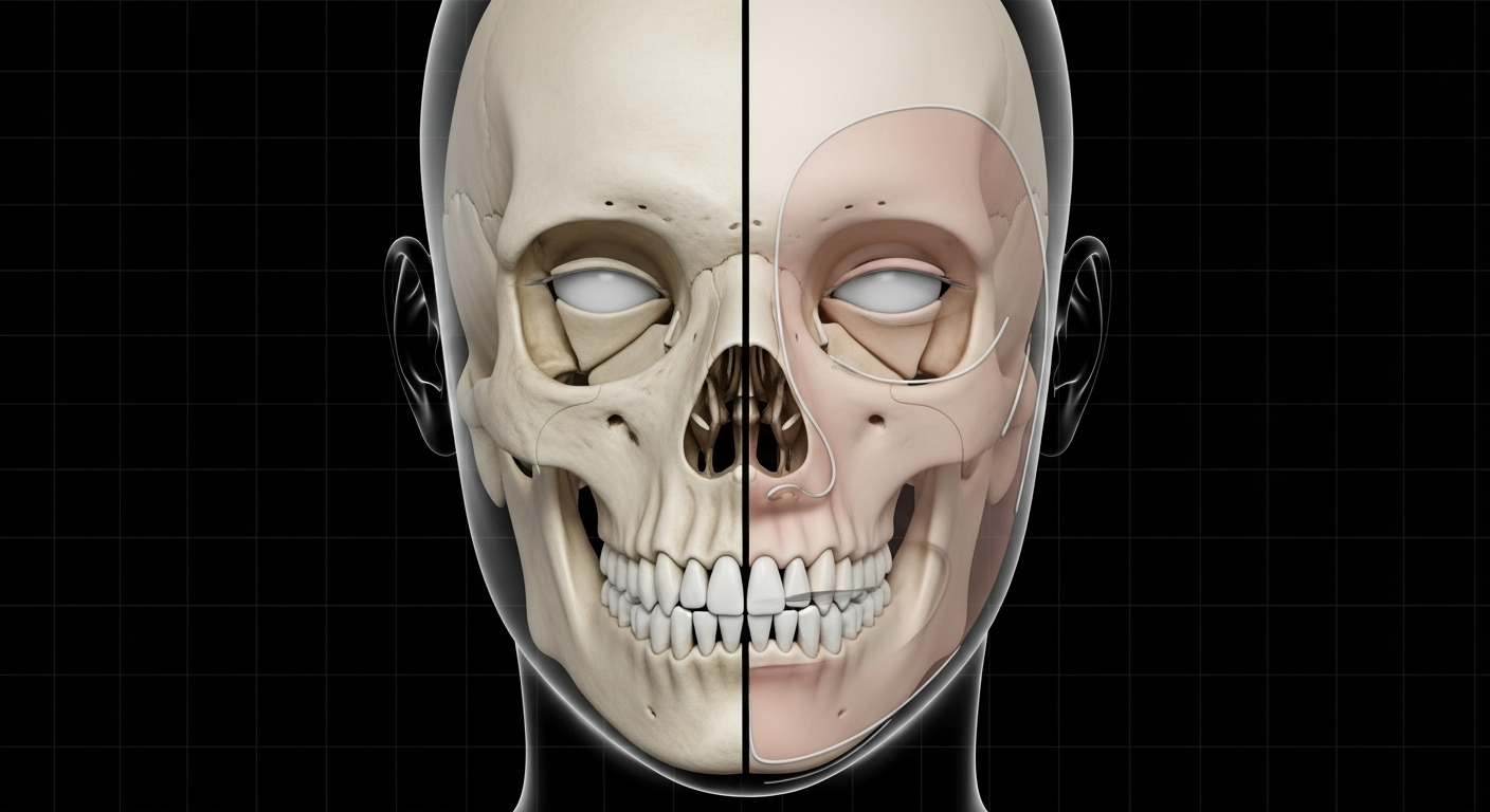
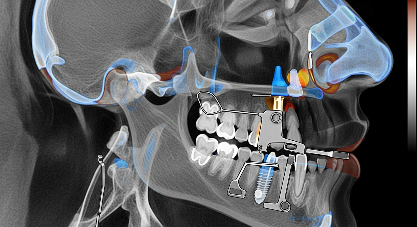
Facial Feminization Surgery (FFS) represents a transformative milestone for many transgender women, offering not just aesthetic alignment with gender identity but also profound psychological affirmation. While the immediate outcomes of FFS—such as softened facial contours, refined nasal structures, and harmonized jawlines—are often the focus of patient and surgeon discussions, the long-term stability of these results is equally critical. The durability of FFS outcomes hinges on a complex interplay of biomechanical and biological processes, including skeletal remodeling, soft tissue adaptation, and the body’s natural healing responses. Without a deep understanding of these mechanisms, even the most meticulously executed procedures may fail to deliver lasting satisfaction, leading to relapse, asymmetry, or functional complications.
This guide explores the scientific foundations of lasting FFS results, emphasizing the importance of skeletal stability, soft tissue integration, and the role of surgical precision. By examining the biomechanical principles governing bone healing, the biological processes of soft tissue adaptation, and the impact of surgical techniques on long-term outcomes, we aim to provide a comprehensive resource for both patients and practitioners. Whether you are considering FFS for the first time or seeking to refine your understanding of post-surgical stability, this guide will equip you with the knowledge to navigate your journey with confidence.
We will delve into key topics such as Wolff’s Law and its application to facial bone remodeling, the stages of bone healing specific to osteotomies, and the critical role of fixation techniques in maintaining structural integrity. Additionally, we will discuss how soft tissues—skin, muscle, and scar tissue—adapt to the new facial architecture and the strategies surgeons employ to minimize laxity and ensure harmonious contours. By the end of this guide, you will understand not only the immediate transformations achieved through FFS but also the biological and biomechanical factors that underpin their longevity.
Introduction: Beyond the Initial Transformation
Facial Feminization Surgery (FFS) is a collection of procedures designed to reshape masculine facial features into more feminine ones, addressing areas such as the forehead, nose, cheeks, jaw, and chin. While the immediate aesthetic changes are often the primary focus, the long-term stability of these results is equally vital. The durability of FFS outcomes depends on the body’s ability to adapt to the new facial architecture, a process governed by biomechanical and biological principles. Without a thorough understanding of these mechanisms, even the most skillfully performed procedures may not yield lasting results.
This section introduces the foundational concepts that underpin lasting FFS results. We will explore how bone remodeling, soft tissue adaptation, and surgical techniques contribute to the stability of FFS outcomes. By understanding these principles, patients and practitioners can make informed decisions that enhance the longevity of surgical results.
The Importance of Long-Term Stability
Long-term stability in FFS refers to the ability of the facial structure to maintain its new form over time. This stability is influenced by factors such as bone healing, soft tissue adaptation, and the body’s natural aging processes. For instance, bone remodeling—a process where bone tissue is continuously renewed—plays a critical role in maintaining the structural integrity of the face post-surgery. Similarly, soft tissues, including skin and muscle, must adapt to the new contours to prevent sagging or asymmetry.
Research indicates that the stability of FFS results is closely tied to the body’s biomechanical responses. For example, studies on Le Fort I osteotomies have shown that bone fixation techniques significantly impact long-term outcomes. The use of titanium or biodegradable plates to stabilize bone segments ensures that the new facial structure remains intact during the healing process. Additionally, the gradual adaptation of soft tissues to the altered bone structure is essential for maintaining a natural appearance.
Biomechanical and Biological Foundations
The biomechanical and biological foundations of FFS stability are rooted in the principles of bone healing and soft tissue adaptation. Bone healing, particularly in procedures involving osteotomies (surgical cuts to the bone), follows a predictable sequence: inflammation, soft callus formation, hard callus formation, and remodeling. Each stage is critical for ensuring that the bone segments fuse correctly and maintain their new position.
Soft tissue adaptation is equally important. The skin, muscles, and ligaments of the face must conform to the new skeletal framework to achieve a harmonious and natural appearance. Scar tissue formation, for instance, plays a role in maintaining the position of soft tissues, but excessive scarring can lead to complications such as restricted movement or asymmetry. Surgical techniques that minimize scarring and promote healthy tissue integration are therefore essential for long-term stability.
Skeletal Remodeling: The Body’s Adaptation to New Forms
Skeletal remodeling is a dynamic process where bone tissue is continuously renewed to adapt to mechanical stresses and changes in the body. In the context of FFS, this process is critical for maintaining the new facial contours created through surgical interventions. Understanding how bone adapts to these changes can help patients and surgeons optimize outcomes for long-term stability.
Wolff’s Law in FFS
Wolff’s Law states that bone adapts to the loads placed upon it. In FFS, this principle is particularly relevant for procedures such as forehead contouring, jaw reshaping, and chin augmentation. For example, when the jawbone is reshaped to create a softer, more feminine contour, the bone must adapt to the new mechanical stresses. This adaptation ensures that the bone remains strong and stable in its new form. Studies have shown that proper surgical techniques, such as precise osteotomies and stable fixation, are essential for guiding this adaptive process and preventing complications such as bone resorption or misalignment.
Bone Healing Mechanics
Bone healing after FFS procedures involves several stages: inflammation, soft callus formation, hard callus formation, and remodeling. During the inflammation stage, the body initiates the healing process by removing damaged tissue and preparing the site for new bone formation. The soft callus stage involves the formation of a fibrous network that bridges the bone segments, while the hard callus stage sees the deposition of mineralized bone that strengthens the union. Finally, during remodeling, the bone is reshaped to optimize its structure and function. This process can take several months to years, depending on the extent of the surgical intervention and the patient’s overall health.
Research on Le Fort I osteotomies highlights the importance of proper fixation techniques in ensuring stable bone healing. For instance, the use of titanium plates and screws provides the necessary stability for bone segments to fuse correctly. Additionally, factors such as blood supply, immobilization, and patient health play significant roles in the healing process. Poor blood supply or inadequate immobilization can lead to delayed healing or nonunion, compromising the long-term stability of the results.
Factors Influencing Bone Fusion
Several factors influence the success of bone fusion in FFS procedures. Blood supply is critical, as adequate vascularization ensures that the bone segments receive the nutrients and oxygen necessary for healing. Immobilization, achieved through fixation devices such as plates and screws, prevents movement that could disrupt the healing process. Patient health, including factors such as nutrition, smoking status, and overall physical condition, also plays a significant role. For example, smoking can impair blood flow and delay healing, while poor nutrition can weaken the body’s ability to regenerate bone tissue.
Studies have shown that patients who adhere to post-operative care instructions, including avoiding smoking and maintaining a healthy diet, experience better healing outcomes. Additionally, the use of advanced surgical techniques, such as 3D planning and computer-assisted navigation, can enhance the precision of bone cuts and fixation, further improving the stability of FFS results.
Fixation and Fusion: Anchoring the New Architecture
Fixation and fusion are critical components of FFS that ensure the stability of the new facial structure. Proper fixation techniques anchor the bone segments in their new positions, allowing for undisturbed healing and integration. This section explores the role of internal fixation devices, bone grafts, and implant biocompatibility in achieving lasting FFS results.
Role of Internal Fixation
Internal fixation devices, such as titanium plates and screws, play a vital role in stabilizing bone segments after FFS procedures. These devices provide the necessary support to prevent movement during the healing process, ensuring that the bone segments fuse correctly. Research has shown that the use of internal fixation significantly reduces the risk of complications such as nonunion or malunion, which can compromise the stability of the results. For example, a study on long-term skeletal stability in orthognathic surgery demonstrated that proper fixation techniques are essential for maintaining the position of bone segments and achieving stable outcomes.
Graft Integration
Bone grafts are often used in FFS to augment areas such as the cheeks or chin, providing additional volume and contour. The success of these grafts depends on their ability to integrate with the surrounding bone tissue. This process, known as osseointegration, involves the vascularization of the graft and its incorporation into the existing bone structure. Studies have shown that the use of autologous bone grafts, which are harvested from the patient’s own body, tends to yield better integration and long-term stability compared to synthetic or allogenic grafts. Additionally, the placement of grafts in well-vascularized areas enhances their chances of successful integration.
Implant Biocompatibility
In some FFS procedures, implants made of materials such as silicone or polyether ether ketone (PEEK) are used to enhance facial contours. The biocompatibility of these materials is crucial for ensuring that the body does not reject the implants and that they integrate smoothly with the surrounding tissues. Research has shown that implants with high biocompatibility, such as those made from medical-grade silicone, are less likely to cause adverse reactions and more likely to maintain their position over time. Additionally, the design and placement of implants play a significant role in their long-term stability. For example, implants that are securely anchored to the bone are less likely to shift or become displaced.
Soft Tissue Adaptation: Draping and Integration
Soft tissue adaptation is a critical aspect of FFS that ensures the skin, muscles, and other tissues conform to the new facial structure. This process involves the redraping of soft tissues over the altered bone framework and the maturation of scar tissue, which helps maintain the new contours. Understanding how soft tissues adapt to these changes is essential for achieving natural and lasting results.
Skin and Muscle Redraping
The redraping of skin and muscle over the new facial structure is a key component of FFS. This process involves carefully repositioning the soft tissues to ensure they conform to the altered bone contours. Techniques such as precise suturing and the use of suspension techniques help prevent sagging and maintain a smooth, natural appearance. Research has shown that proper soft tissue management during surgery can significantly enhance the aesthetic outcomes of FFS and reduce the risk of complications such as asymmetry or laxity.
Scar Tissue Maturation
Scar tissue formation is a natural part of the healing process after FFS. While scars help anchor the soft tissues to the new bone structure, excessive scarring can lead to complications such as restricted movement or asymmetry. Techniques to minimize scarring, such as precise surgical incisions and the use of anti-scarring treatments, are essential for achieving optimal results. Studies have shown that the maturation of scar tissue occurs over several months to years, during which time the tissue gradually remodels to become more flexible and integrated with the surrounding structures.
Minimizing Laxity
Minimizing laxity, or sagging of the soft tissues, is a critical goal in FFS. Surgical strategies such as precise skin redraping, the use of suspension techniques, and the placement of supportive structures help maintain the position of the soft tissues over time. Research has shown that patients who undergo FFS with careful attention to soft tissue management experience better long-term stability and aesthetic outcomes. Additionally, post-operative care, including the use of compression garments and physical therapy, can further enhance the adaptation of soft tissues to the new facial contours.
Maintaining Equilibrium: Forces and Dynamics
Maintaining equilibrium in the facial structure involves balancing the forces exerted by muscles, ligaments, and other tissues. This section explores how these forces adapt to the new facial architecture and the impact of aging on FFS results. Understanding these dynamics is essential for ensuring that the outcomes of FFS remain stable and natural over time.
Muscle and Ligament Readjustment
The muscles and ligaments of the face must adapt to the new bone positions created through FFS. This readjustment is critical for maintaining functional harmony and aesthetic balance. For example, the muscles responsible for facial expressions must conform to the new contours to ensure natural movement and prevent asymmetry. Research has shown that proper surgical techniques, such as precise bone cuts and stable fixation, facilitate this adaptation process and reduce the risk of complications such as muscle imbalance or restricted movement.
Aging and FFS
Aging is a natural process that affects all tissues of the body, including those altered through FFS. Over time, changes such as collagen loss, fat redistribution, and bone resorption can impact the stability of FFS results. Understanding these processes is essential for managing patient expectations and planning for long-term maintenance. Studies have shown that while FFS results are generally stable, the effects of aging may necessitate touch-up procedures to maintain the desired aesthetic outcomes. For example, the loss of collagen in the skin can lead to sagging, which may require additional treatments such as fillers or laser therapy.
Long-Term Monitoring
Long-term monitoring is a critical component of FFS that ensures the stability and longevity of the results. Regular follow-up appointments allow surgeons to assess the healing process, identify any potential complications, and make adjustments as needed. Research has shown that patients who adhere to a structured follow-up schedule experience better outcomes and higher satisfaction with their FFS results. Additionally, long-term monitoring provides an opportunity to address any changes due to aging or other factors, ensuring that the results remain optimal over time.
Surgical Precision and Patient Adherence: Cornerstones of Longevity
Surgical precision and patient adherence are the cornerstones of achieving lasting FFS results. This section explores the role of the surgeon’s expertise in executing precise modifications and the importance of patient compliance in post-operative care. Together, these factors ensure that the outcomes of FFS are both stable and satisfying.
Surgeon’s Role
The surgeon’s expertise is critical in achieving optimal FFS outcomes. A deep understanding of facial biomechanics and precise surgical techniques are essential for creating a stable and natural-looking result. Research has shown that surgeons with extensive experience in FFS are better equipped to anticipate and address potential complications, ensuring that the results are both aesthetically pleasing and functionally sound. Additionally, the use of advanced technologies, such as 3D imaging and computer-assisted planning, enhances the precision of surgical interventions and improves the likelihood of long-term stability.
Patient Compliance
Patient compliance is a key factor in the success of FFS. Adhering to post-operative care instructions, such as avoiding smoking, maintaining a healthy diet, and attending follow-up appointments, significantly enhances the healing process and the stability of the results. Studies have shown that patients who follow their surgeon’s recommendations experience fewer complications and better long-term outcomes. Additionally, patient compliance extends to lifestyle choices that support overall health, such as regular exercise and stress management, which can further contribute to the longevity of FFS results.
Conclusion: A Foundation for Lasting Femininity
The science of lasting FFS results is rooted in a deep understanding of biomechanical and biological processes. From skeletal remodeling to soft tissue adaptation, each aspect of the healing process plays a critical role in maintaining the stability and natural appearance of the results. By focusing on surgical precision, patient adherence, and long-term monitoring, patients and practitioners can achieve outcomes that are not only transformative but also enduring.
This guide has explored the key principles that underpin the longevity of FFS results, providing a comprehensive resource for those seeking to understand the science behind lasting transformations. By applying these principles, patients can make informed decisions that enhance their confidence and satisfaction with their FFS journey.
Key Takeaways
Skeletal remodeling and bone healing are critical for maintaining the stability of FFS results. Understanding these processes helps patients and surgeons optimize outcomes.
Fixation techniques, such as the use of titanium plates and screws, ensure that bone segments remain stable during the healing process.
Soft tissue adaptation, including skin and muscle redraping, is essential for achieving natural and harmonious results.
Patient compliance with post-operative care instructions significantly enhances the healing process and the longevity of FFS results.
Long-term monitoring allows for the early identification and management of potential complications, ensuring that the results remain optimal over time.
Next Steps
If you are considering FFS or seeking to refine your understanding of post-surgical stability, consult with a board-certified surgeon who specializes in gender-affirming procedures. Their expertise will guide you through the process, ensuring that your results are both transformative and enduring. Additionally, adhere to post-operative care instructions and attend regular follow-up appointments to support the healing process and maintain the longevity of your FFS results.
Frequently Asked Questions
What is the role of Wolff’s Law in FFS?
Wolff’s Law states that bone adapts to the mechanical stresses placed upon it. In FFS, this principle ensures that the reshaped bones, such as the jaw or forehead, adapt to their new positions, maintaining stability and strength over time.
How long does it take for bone healing to complete after FFS?
Bone healing after FFS typically involves several stages, including inflammation, soft callus formation, hard callus formation, and remodeling. This process can take several months to years, depending on the extent of the surgery and the patient’s overall health.
What are the most common fixation techniques used in FFS?
The most common fixation techniques in FFS include the use of titanium plates and screws, which provide stability to the bone segments during the healing process. These devices ensure that the bones remain in their new positions, promoting proper fusion and long-term stability.
How do soft tissues adapt to the new facial structure after FFS?
Soft tissues, including skin and muscle, adapt to the new facial structure through a process of redraping and scar tissue maturation. Proper surgical techniques and post-operative care are essential for ensuring that the soft tissues conform smoothly to the altered bone contours.
What factors influence the long-term stability of FFS results?
Several factors influence the long-term stability of FFS results, including surgical precision, patient adherence to post-operative care, and the body’s natural healing processes. Proper fixation techniques and soft tissue management also play significant roles.
How does aging affect FFS results?
Aging can impact FFS results through processes such as collagen loss, fat redistribution, and bone resorption. While FFS results are generally stable, these changes may necessitate touch-up procedures to maintain the desired aesthetic outcomes.
Why is long-term monitoring important after FFS?
Long-term monitoring is important after FFS to assess the healing process, identify potential complications, and make adjustments as needed. Regular follow-up appointments help ensure that the results remain optimal over time.
What can patients do to enhance the longevity of their FFS results?
Patients can enhance the longevity of their FFS results by adhering to post-operative care instructions, maintaining a healthy lifestyle, and attending regular follow-up appointments. These steps support the healing process and help maintain the stability of the results.
Bibliography
Shido, R., et al. (2024). Comparison of Skeletal Stability after Le Fort I Osteotomy With Bone Fixation Using Biodegradable and Titanium Systems: A Retrospective Study. Journal of Craniofacial Surgery.
Buchanan, E. P., & Hyman, C. H. (2013). LeFort I Osteotomy. Seminars in Plastic Surgery.
Jeong, W. S., et al. (2018). Large-Scale Study of Long-Term Vertical Skeletal Stability in a Surgery-First Orthognathic Approach Without Presurgical Orthodontic Treatment: Part II. Journal of Craniofacial Surgery.
Saltaji, H., et al. (2012). Long-term skeletal stability after maxillary advancement with distraction osteogenesis in cleft lip and palate patients: A systematic review. The Angle Orthodontist.
The pursuit of facial harmony and congruence with one’s deeply held gender identity is a profound journey for many individuals. Facial Feminization Surgery (FFS) stands as a cornerstone in this transformative process, meticulously reshaping facial features to align with a more feminine aesthetic. However, the path to achieving optimal results is not always linear. Despite advancements in surgical techniques and a growing understanding of gender-affirming care, a subset of patients may find themselves seeking revision FFS to address outcomes that, for various reasons, fall short of their expectations or surgical goals. This specialized area of aesthetic surgery is not merely about correcting “mistakes” but about refining, enhancing, and often completing a patient’s vision for their facial appearance, ensuring it truly reflects their authentic self.
Revision FFS encompasses a complex array of surgical methodologies designed to correct suboptimal results from initial feminization procedures. These suboptimal outcomes can stem from a multitude of factors, including the inherent unpredictability of human healing, anatomical complexities that become more apparent post-operatively, or even the evolving aesthetic preferences of the individual over time. For instance, sometimes the initial surgical corrections may not have been extensive enough, leading to an “under-correction” where masculine features are still too prominent. Conversely, an “over-correction” could result in an unnatural or overly sculpted appearance. Asymmetry, visible scarring, or incomplete feminization in certain facial zones are also common drivers for individuals to consider a revision. The Feminization Center highlights that reasons for revision can include unnatural-looking results, asymmetry, visible scarring, incomplete results, and changes in aesthetic preferences (Feminization Center, n.d.). Similarly, Facialteam acknowledges that re-operations are common in FFS due to factors like lack of skill, professionalism, or poor aftercare, though they boast a low revision rate of 3% (Facialteam, n.d.).
The necessity for a specialized surgical approach in revision FFS is underscored by the unique challenges presented by previously operated tissues. Surgeons undertaking these procedures must navigate altered anatomical planes, scar tissue, and potentially compromised vascularity, all of which demand an advanced level of expertise, meticulous planning, and a nuanced understanding of facial aesthetics. The Ultimate FFS Revision Guide by Dr. MFO emphasizes that surgeons performing redos may encounter unexpected circumstances such as asymmetrical scarring or adhesions, requiring detailed planning (Dr. MFO FFS Revision 2, 2024). This guide will delve into the intricate surgical methodologies and strategic considerations employed in advanced revision FFS, offering a detailed roadmap for understanding how surgeons address and correct less-than-optimal outcomes from prior procedures. We will explore the nuanced techniques applied to various facial regions, from the forehead and brow to the jaw, chin, nose, and midface, as well as strategies for managing soft tissue concerns and scars. Furthermore, we will emphasize the critical role of advanced diagnostic imaging, judicious graft selection, and the distinct recovery profile associated with revision cases. By providing a comprehensive, evidence-based analysis, this discourse aims to serve as an authoritative resource for individuals considering revision FFS, as well as for professionals seeking a deeper understanding of these intricate and highly specialized surgical interventions. The goal is not just to modify physical features but to achieve a profound sense of self-alignment and aesthetic harmony that significantly enhances an individual’s quality of life and confidence. The evolution of FFS has been marked by technological advancements in imaging, surgical tools, and materials, leading to minimally invasive techniques that reduce scarring and recovery time (The Spiegel Center, n.d.). This advanced landscape informs all aspects of revision FFS, making precise, patient-centered care more achievable than ever before. Understanding the interplay of surgical factors, patient physiology, evolving aesthetic goals, and unforeseen complications is paramount to approaching revision FFS with informed confidence, ultimately guiding patients toward their most authentic and harmonized facial presentation.
Advanced Revision FFS: Surgical Techniques for Correcting Suboptimal Outcomes
Forehead and Brow Revision
The forehead and brow region are paramount in determining perceived gender. Masculine foreheads typically feature a prominent brow bossing, a more defined ridge above the eyes, and often a higher or M-shaped hairline. Feminine foreheads, in contrast, are characterized by a smoother, more rounded contour and a lower, often oval-shaped hairline. Correcting suboptimal outcomes in this area during revision FFS demands exceptional precision, as the bone structure is fundamental to the overall facial impression.
One of the most frequent reasons for revision in the upper face is the **under-reduction of the brow bone**. If the initial surgery did not sufficiently reduce the supraorbital ridge, a noticeable prominence may persist, hindering the desired feminization. Revision techniques in such cases involve further burring or precise reshaping of the frontal bone to achieve a smoother, more effeminate contour. This can be a delicate procedure, as the thickness of the frontal bone varies, and excessive reduction carries risks. Dr. MFO highlights that under-reduction of the brow bone is a common reason for revision (Dr. MFO FFS Revision 1, 2025). The Gender Confirmation Center describes frontal sinus setback as a procedure that reduces or reshapes the brow bone to make the upper third of the face appear softer and less prominent (Gender Confirmation Center, n.d.).
Another significant concern requiring revision is the presence of **irregularities or depressions** on the forehead. These subtle imperfections can arise during the initial healing process, particularly following Type III cranioplasty procedures where a section of bone is removed, reshaped, and reattached. Minor bumps or depressions can become visible as swelling subsides. Revision strategies for these issues may involve targeted burring to smooth out bony irregularities. In select, more complex cases, fat grafting or specialized bone cements can be employed to meticulously fill depressions and restore a seamless contour.
**Hairline irregularities or recessions** also frequently necessitate revision. An initial hairline advancement procedure might not have achieved the optimal shape or sufficient reduction of the forehead height, leaving an irregular or still-receding hairline that detracts from the overall feminine frame. Revision can involve further hairline advancement, often through careful scalp excision, to lower the hairline and create a more natural, rounded shape. Hair grafting can also be used in conjunction with advancement to increase density or refine the hairline’s aesthetic. The Gender Confirmation Center mentions hairline advancement (also known as hairline lowering) uses an incision around the forehead to reshape the hairline, focusing on rounding the corners of M-shaped hairlines and making the forehead appear shorter and more round (Gender Confirmation Center, n.d.).
**Brow asymmetry** can be another challenging issue. While sometimes related to soft tissue positioning, persistent brow asymmetry may indicate an underlying bony discrepancy that was not fully addressed. Revision in such instances can involve targeted bone work to re-balance the orbital rims or a more refined brow lift to ensure symmetry in eyebrow height and arch. Endoscopic methods are increasingly utilized to minimize scarring and improve precision in these delicate revisions (Dr. MFO FFS Revision 1, 2025).
The anatomical complexity of the forehead and brow, coupled with its central role in gender perception, makes revision procedures in this area particularly demanding. Surgeons must possess not only a deep understanding of craniofacial anatomy but also an artistic eye to sculpt a harmonious and natural-looking feminine contour. The importance of virtual surgical planning and 3D imaging in these cases cannot be overstated, as they allow for precise pre-operative assessment and detailed surgical strategy, mitigating potential pitfalls. Facialteam emphasizes detailed assessment and planning with patient input for predictable, safe, and precise results, which significantly reduces revision rates (Facialteam, n.d.). These tools are crucial for navigating the delicate balance between effective feminization and preserving structural integrity.
Jaw and Chin Revision
The lower third of the face, comprising the jaw and chin, plays a pivotal role in distinguishing masculine from feminine features. A typically masculine jawline is often characterized by a square, broad appearance with prominent angles, and a chin that can be tall and broad with significant projection. In contrast, feminine lower facial contours are generally softer, more tapered, and often present with a “V-shaped” or rounded chin. Revision FFS in this region frequently addresses concerns such as insufficient contouring, over-reduction, or the presence of bony irregularities and asymmetries that detract from the desired feminine aesthetic.
One of the primary drivers for revision is **insufficient jaw angle reduction**. If the initial mandibular angle shaving was conservative, or if bone regrowth occurs, the jawline may retain a masculine angularity. In these instances, revision involves further osteotomy—the surgical removal of bone—to achieve a softer, more tapered, and less prominent jawline. This requires meticulous planning to ensure symmetry and avoid over-reduction, which could lead to an unnatural or “pinched” appearance. The study by Gursky et al. on genioplasty in FFS highlights that masculine chins are typically taller and broader with more pronounced projection, while feminine chins are softer, less prominent, and sometimes V-shaped (Gursky et al., 2024). Jaw contouring aims to shave down the jaw and reshape the chin for a more narrow and softer look (Gender Confirmation Center, n.d.).
**Chin asymmetry or under-correction** after an initial genioplasty is another common reason for revision. The chin’s projection, width, and vertical height are critical for facial balance. If the initial procedure did not adequately reduce a prominent chin, or if it left an uneven contour, revision surgery can address these discrepancies. This may involve further bone reshaping to correct asymmetry, increase or decrease projection as desired, or refine the chin’s shape (e.g., to be more rounded or pointed). The review by Gursky et al. indicates that reduction genioplasty is the most common technique for feminizing the chin, with sliding genioplasty also detailed, often combined with bony burring for additional contouring (Gursky et al., 2024). Dr. MFO notes that revision for the chin can involve further bone reshaping to correct asymmetry, or to adjust projection or shape if the initial genioplasty was insufficient (Dr. MFO FFS Revision 1, 2025).
**Palpable irregularities** along the resected jawline or chin are also concerns that can lead to revision. These might manifest as small bumps, steps, or unevenness in the bone contour that become apparent upon palpation or visible under certain lighting. Revision surgery focuses on carefully burring or smoothing these areas to create a seamless and natural transition along the bone, enhancing both the tactile and visual smoothness of the lower face.
The technical complexities of jaw and chin revision are often heightened by the presence of existing scar tissue and altered soft tissue adherence. Surgeons must precisely re-osteotomize or burr bone while preserving critical anatomical structures such as nerves and blood vessels. Advanced imaging techniques, including 3D CT scans, are indispensable for pre-operative planning, enabling the surgeon to visualize the underlying bony architecture, assess previous osteotomy lines, and meticulously plan the corrective actions. Facialteam emphasizes that planning and experience significantly increase the likelihood of success in FFS and help address problems from previously incorrectly implemented operations (Facialteam, n.d.). Such detailed planning is crucial for achieving predictable, safe, and precise outcomes, especially in secondary procedures where the anatomical landscape has been altered. The objective is to achieve not just a reduction or reshaping, but a harmonious integration of the lower face with the overall feminized facial aesthetic, thereby contributing to a profound sense of self-alignment.
Nasal Revision (Rhinoplasty)
Nasal morphology is a crucial determinant of facial gender perception. Masculine noses often present with a broader bridge, a more prominent dorsal hump, and a less refined tip, typically projecting further from the face. Feminine noses, conversely, are generally characterized by a narrower, more delicate bridge, a subtle or absent dorsal hump, a refined and slightly upturned tip, and a harmonious relationship with other facial features. Revision rhinoplasty, following an initial Facial Feminization Surgery (FFS), is a highly intricate procedure that aims to correct aesthetic and sometimes functional shortcomings from the primary operation. Given the nose’s central position on the face and its complex cartilaginous and bony architecture, it is among the most frequently revised facial procedures.
One of the common reasons for revision is **persistent dorsal hump or width**. If the initial FFS rhinoplasty did not adequately reduce the bridge of the nose, a remaining masculine prominence or excessive width can be observed. Revision involves further precise bone and cartilage reduction to sculpt a smoother, more delicate nasal profile that integrates harmoniously with the feminized forehead and midface. This requires careful judgment to avoid over-resection, which can lead to an undesirable “scooped” or “pinched” appearance, a more challenging issue to correct. Allure Esthetic notes that a rhinoplasty is often chosen for revision if it was not adequately thinned, feminized, or supported, with many revisions occurring years after the initial procedure due to aging and facial changes (Allure Esthetic, n.d.). Dr. MFO also lists persistent dorsal hump or width as reasons for revision (Dr. MFO FFS Revision 1, 2025).
**Tip irregularities** are another frequent concern. The nasal tip is a complex area composed of delicate cartilages, and achieving a refined, slightly upturned, and symmetrical tip is a hallmark of feminizing rhinoplasty. If the initial surgery resulted in a tip that remains too bulbous, asymmetrical, or lacks adequate projection or rotation, revision may be necessary. Techniques often involve precise suturing of existing cartilages, or the use of cartilage grafts—often harvested from the septum, ear, or rib—to augment, reshape, and support the tip, creating a more delicate and defined aesthetic.
While primarily aesthetic, **breathing issues (functional revision)** can occasionally arise or persist after FFS rhinoplasty. Less commonly, an initial feminizing rhinoplasty might inadvertently lead to or exacerbate difficulties in nasal airflow. In such cases, revision focuses on addressing internal structural issues, such as septal deviations or collapse of the internal nasal valve, to improve respiratory function while simultaneously enhancing aesthetic outcomes.
**Asymmetry** of the nose, though often subtle and inherent in all faces, can become a noticeable concern after surgery. If a significant asymmetry of the nostrils, bridge, or tip develops or persists post-operatively, revision aims to improve overall nasal symmetry, contributing to a more balanced facial appearance. The Spiegel Center highlights that Dr. Spiegel was one of the first surgeons to perform lip lifts with rhinoplasty, indicating the integrated approach in facial feminization (The Spiegel Center, n.d.).
**Over-reduction**, while less common, is a particularly complex issue in revision rhinoplasty. If too much bone or cartilage was removed during the primary surgery, resulting in an overly “scooped” dorsal profile or a “pinched” tip, rebuilding the nasal structure often requires extensive cartilage grafting. This reconstructive aspect of revision rhinoplasty demands advanced surgical skill and artistry to restore natural contours and functional integrity.
Revision rhinoplasty is technically demanding due to the scar tissue from previous surgery, which can alter tissue elasticity and vascularity. Meticulous dissection, careful preservation of remaining cartilage, and strategic grafting are crucial for successful outcomes. Pre-operative 3D imaging is invaluable for assessing the existing nasal framework and planning precise corrective maneuvers. The goal of revision rhinoplasty is to achieve a refined, natural-looking nose that harmonizes with the feminized features of the entire face, contributing significantly to a patient’s confidence and perceived gender congruence.
Cheek and Midface Revision
The midface, particularly the cheeks, is a key region in defining facial femininity. Masculine midfaces tend to be flatter, with less pronounced cheekbones, while feminine midfaces often feature fuller, higher, and more rounded cheek contours, contributing to a softer, more youthful, and often heart-shaped facial outline. Revision FFS in the cheek and midface area primarily focuses on correcting volume discrepancies, adjusting implant placement or size, and addressing any irregularities that may have arisen from previous augmentation or contouring procedures.
One of the main objectives of midface revision is **correcting volume discrepancies**. If an initial fat grafting or implant procedure resulted in uneven volume distribution, or if there was insufficient augmentation to achieve the desired feminine fullness, revision can restore balance. This often involves additional **fat grafting**, where fat is harvested from another part of the body, processed, and meticulously injected into areas needing more volume. Fat grafting is particularly advantageous as it uses the patient’s own tissue, reducing the risk of rejection and providing natural-feeling results. However, fat graft survival can be variable, sometimes necessitating touch-ups (Dr. MFO FFS Revision 1, 2025). The Gender Confirmation Center notes that cheek augmentation can involve the use of cheek implants, fillers, or analogous fat transfer (Gender Confirmation Center, n.d.).
For patients who underwent **cheek implant placement** in their primary surgery, revision may be required to adjust the placement or size of these implants. Implants that are incorrectly positioned can lead to asymmetry, an unnatural contour, or be palpable beneath the skin. Revision involves carefully repositioning existing implants, exchanging them for different sizes or shapes, or in some cases, removing them entirely if they are no longer desired or are causing complications. The complexity arises from navigating existing fibrous tissue around the implants and ensuring precise new placement for optimal aesthetic integration.
Another concern in midface revision is addressing **facial fat grafting irregularities**. While fat grafting is a versatile technique for adding volume, sometimes the grafted fat can settle unevenly, leading to small lumps, depressions, or an asymmetrical appearance. Revision for such irregularities might involve targeted massage, micro-liposuction of areas with excess fat, or additional, more carefully placed grafting to smooth contours. The Spiegel Center also notes that dermal fillers and fat grafting can be used to add volume to cheeks and lips for a more youthful and feminine look, and advancements in injectable treatments offer non-surgical options (The Spiegel Center, n.d.). This highlights a broader approach that may involve both surgical and non-surgical modalities in revision.
The midface is highly dynamic, influenced by expressions and the aging process. Therefore, revision considerations also account for how changes will integrate over time. The approach to midface revision is highly individualized, requiring a surgeon with a keen aesthetic eye and extensive experience in both soft tissue management and bone/implant adjustments. Pre-operative advanced imaging, such as 3D CT scans, assists in evaluating the underlying bone structure and the precise positioning of any previous implants or areas of fat grafting, allowing for meticulous planning. The overarching goal is to create a soft, rounded, and harmonized midface that contributes significantly to the feminized appearance, addressing any lingering sources of concern from previous procedures.
Soft Tissue Revisions
Beyond the foundational bony alterations, soft tissue procedures are integral to achieving a comprehensive feminine aesthetic in Facial Feminization Surgery (FFS). These elements, which include the skin, fat, muscles, and their collective drape, play a significant role in refining facial contours, improving texture, and managing visible signs of surgery. Revision FFS often extends to soft tissue adjustments, addressing issues such as persistent scarring, skin laxity, or inadequate soft tissue volume, which can all impact the final aesthetic outcome.
**Scar management techniques** are a critical component of soft tissue revisions. While FFS surgeons meticulously plan incisions to be as inconspicuous as possible—often hidden within the hairline or natural creases—some patients may still develop noticeable scars due to individual healing responses, genetic predispositions, or complications such as infection. Revision for unsightly scars can involve various methods, including:
**Surgical scar revision:** This technique involves excising the existing scar and meticulously re-closing the incision with advanced suturing techniques to create a finer, less noticeable scar. This is particularly effective for wide, hypertrophic, or keloid scars, though careful patient selection and post-operative scar care are crucial. Allure Esthetic highlights that scar revision can correct unsightly scars (Allure Esthetic, n.d.).
**Laser treatments:** Fractional laser resurfacing can improve scar texture, color, and overall appearance by stimulating collagen remodeling.
**Microneedling:** This minimally invasive technique uses fine needles to create controlled micro-injuries, promoting collagen and elastin production to smooth and soften scars.
**Corticosteroid injections:** For raised, hypertrophic, or keloid scars, targeted injections can help flatten and soften the tissue.
Addressing **residual soft tissue laxity** is another important aspect of soft tissue revisions. While bone reductions provide the structural framework for feminization, the overlying skin and soft tissues must redrape smoothly. In some cases, especially with significant bone reduction or in older patients, residual skin laxity or sagging can occur, leading to an aged or less refined appearance. Revision strategies may include:
**Mini-facelift or neck lift procedures:** These involve excising excess skin and tightening the underlying fascial layers (SMAS) to create a smoother, more youthful contour in the lower face and neck.
**Thread lifts:** Less invasive options, thread lifts can provide a temporary lift to sagging tissues, though their effects are generally less dramatic and long-lasting than surgical lifts.
**Energy-based skin tightening:** Technologies like radiofrequency (RF) or ultrasound can stimulate collagen production to improve skin elasticity and tightness over time, offering a non-surgical approach to mild laxity. The Spiegel Center mentions minimally invasive techniques like Embrace (FaceTite or FaceTite to the neck with Morpheus8) for lifting skin through small incisions (The Spiegel Center, n.d.).
**Volume adjustments** in soft tissues, beyond those of the midface, may also be required. For instance, **lip lift revision** can be performed if the initial lift did not achieve the desired amount of upper lip eversion, or if there is a persistent long philtrum (distance between the nose and upper lip). Minor revisions might involve removing a small additional strip of skin at the base of the nose to further shorten the upper lip (Dr. MFO FFS Revision 1, 2025). Furthermore, if initial soft tissue augmentation with fillers or fat grafting in areas like the temples or periorbital region (around the eyes) was uneven or insufficient, additional augmentation can be performed to refine contours and restore youthful fullness. The Spiegel Center notes that non-surgical options with dermal fillers are available for soft tissue enhancements (The Spiegel Center, n.d.).
The success of soft tissue revisions relies heavily on the surgeon’s artistic judgment and experience in delicate tissue handling. The interplay between bone and soft tissue is crucial; therefore, revisions must consider both the underlying structure and the superficial drape to achieve a natural, harmonious, and enduring feminine result. Comprehensive patient assessment, realistic goal setting, and a multi-modal approach combining surgical and non-surgical techniques are often employed to optimize outcomes in soft tissue revision FFS.
Key Surgical Considerations in Revision FFS
Revision Facial Feminization Surgery (FFS) presents a distinct set of challenges that necessitate specialized surgical considerations beyond those of primary FFS. The altered anatomy, presence of scar tissue, and potential for compromised tissue integrity demand advanced planning, sophisticated techniques, and a profound understanding of facial aesthetics and healing dynamics. Addressing these unique aspects is crucial for achieving predictable and satisfying outcomes in secondary procedures.
1. The Role of Advanced Imaging and Virtual Surgical Planning
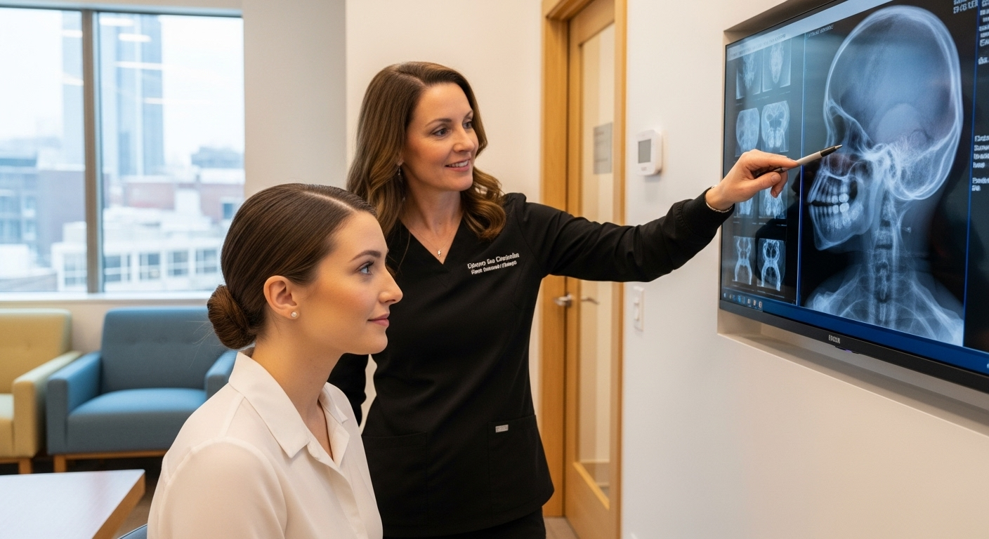
In revision FFS, accurate pre-operative assessment is paramount. Traditional 2D photographs and physical examinations provide valuable information, but **advanced 3D imaging, such as Computed Tomography (CT) scans with 3D reconstruction**, is indispensable. These scans offer an unprecedented view of the underlying bony architecture, allowing the surgeon to precisely map out existing bone reductions or augmentations, identify any residual prominences, and detect subtle asymmetries that may not be apparent externally. The review by Gursky et al. highlights that preoperative imaging and VSP are widely used, with CT scans with 3D reconstruction employed in 75% of included studies to enhance visualization and surgical planning (Gursky etal., 2024).
**Virtual Surgical Planning (VSP)**, a sophisticated digital tool, takes 3D imaging a step further. It allows surgeons to perform virtual osteotomies and contouring on a digital model of the patient’s face, simulating various surgical maneuvers and predicting their effects on both bone and soft tissue. This virtual rehearsal enables the surgeon to refine the surgical plan, optimize bone cuts, and design custom 3D-printed guides or plates for intraoperative precision. While VSP can be costly and time-consuming, it has been shown to improve accuracy, especially in complex cases like forehead reconstruction, and ensures that the revision addresses the patient’s precise aesthetic goals (Gursky et al., 2024). The Gender Confirmation Center advises considering facilities that use pre-surgery imaging like CT scans for planning (Allure Esthetic, n.d.). Facialteam emphasizes that their detailed assessment process with patient input and planning with 3D technology leads to predictable, safe, and precise results (Facialteam, n.d.).
2. Graft Selection (Bone/Cartilage) and Soft Tissue Management
In revision FFS, the need for **grafting materials** is often heightened due to potential over-resection in previous surgeries or the desire for augmentation.
**Bone grafts:** Autologous bone (harvested from the patient’s own body, such as rib or calvarial bone) is often preferred for large volume augmentation or structural support, particularly in cases of over-reduced brow bones or chins. Synthetic bone substitutes can also be considered.
**Cartilage grafts:** Autologous cartilage (typically from the nasal septum, ear, or rib) is invaluable for refining delicate structures like the nasal tip, reconstructing alar cartilages, or correcting minor contour irregularities in the brow or jaw.
The choice of graft material is critical, impacting long-term stability, integration, and overall aesthetic outcome.
**Soft tissue management** in revision FFS is particularly challenging. Prior surgery introduces scar tissue, which can alter tissue elasticity, vascularity, and the natural planes of dissection. This necessitates meticulous and careful surgical technique to avoid further scarring, nerve damage, or compromising blood supply. Techniques like targeted fat grafting (using the patient’s own fat) can be employed to camouflage irregularities, restore volume, and improve skin quality over previously operated areas (Dr. MFO FFS Revision 1, 2025). The Spiegel Center notes that soft tissue procedures are equally important and involve modifying skin, fat, and muscles (The Spiegel Center, n.d.).
3. Distinct Recovery Profile for Revision Cases
Patients undergoing revision FFS often experience a recovery that differs from their initial surgery.
**Increased swelling and bruising:** Due to the presence of scar tissue and altered lymphatic drainage, swelling and bruising can be more pronounced and prolonged in revision cases. Patients need to be prepared for an extended period of post-operative edema.
**Pain management:** While pain is manageable, the patient’s psychological state can influence their perception of discomfort.
**Emotional impact:** Patients seeking revision often carry a heavier emotional burden due to dissatisfaction with previous results. Comprehensive psychological support and realistic expectation setting are crucial. The Feminization Center emphasizes that redo FFS can be an emotionally charged process, and open communication with the surgeon and a trusted support system are crucial (Feminization Center, n.d.).
**Extended healing timeline:** Surgeons typically recommend waiting a minimum of 6-12 months, and often up to 18 months, after the initial FFS before considering revision. This allows for complete resolution of swelling and soft tissue settling, ensuring that any perceived irregularities are truly permanent and warrant further intervention (Dr. MFO FFS Revision 1, 2025; Allure Esthetic, n.d.).
In essence, key surgical considerations in revision FFS revolve around advanced diagnostic capabilities, strategic material selection, and a holistic understanding of the patient’s physical and emotional journey. The expertise of the surgeon in navigating these complexities is paramount to achieving transformative and enduring results.
Conclusion
The journey through Facial Feminization Surgery, and particularly the pursuit of revision FFS, underscores a profound commitment to achieving a true alignment between one’s inner self and outward presentation. While the initial FFS procedures represent a significant step toward gender affirmation for many, the reality of complex aesthetic interventions dictates that some individuals may require further refinement to fully realize their desired facial harmony. This detailed exploration into the methodologies and considerations of advanced revision FFS reveals not only the intricate technical demands of such surgeries but also the deeply personal and transformative impact they hold for patients.
We have traversed the nuanced landscape of revision across various facial zones, from the subtle yet impactful reshaping of the forehead and brow to the meticulous adjustments required for the jaw and chin. In the upper face, addressing under-reductions, irregularities, hairline concerns, and asymmetries necessitates precise bone work and often adjunctive soft tissue procedures, all aimed at fostering a smoother, more feminized contour. For the lower face, revision techniques delve into correcting insufficient jaw angle reductions, asymmetrical chins, and palpable bony irregularities, striving for a softer, tapered outline that complements the overall feminine aesthetic. Nasal revision, a particularly delicate undertaking, often corrects persistent dorsal humps, tip irregularities, and functional concerns, with advanced grafting techniques playing a critical role in structural and aesthetic refinement. The midface, with its emphasis on volume and contour, benefits from revisions that rectify discrepancies through targeted fat grafting or implant adjustments, ensuring a youthful and harmonious fullness. Finally, soft tissue revisions, including scar management and addressing skin laxity, are indispensable for perfecting the superficial elements and contributing to a seamless, natural-looking result.
The overarching theme in successful revision FFS is the integration of advanced diagnostic tools with exceptional surgical expertise and empathetic patient care. The indispensable role of 3D imaging and virtual surgical planning cannot be overstated, as these technologies provide surgeons with unparalleled insights into the underlying anatomy and the precision required for complex secondary alterations. Furthermore, the judicious selection and application of bone and cartilage grafts are fundamental to reconstructing and augmenting areas that may have been over-resected or require additional volume. However, the technical prowess must be balanced with a profound understanding of the distinct psychological and physiological recovery profile inherent in revision cases. Patients embarking on this second chapter of their surgical journey often bring with them a unique set of emotional considerations, necessitating comprehensive support and the setting of realistic expectations. Patience during the extended healing timeline, often 6 to 18 months, is crucial for allowing tissues to fully settle and for final results to emerge before definitive evaluation. The Feminization Center highlights the emotional journey of redo FFS, emphasizing the importance of open communication and a support system (Feminization Center, n.d.).
Ultimately, the decision to undergo revision FFS is a deeply personal one, driven by the unwavering pursuit of an authentic self. The critical importance of selecting a highly experienced and specialized surgeon—one with a proven track record in revision procedures and a comprehensive understanding of transgender facial aesthetics—cannot be overstated. Such a surgeon not only possesses the technical skill to navigate previously altered anatomy but also the artistic vision to sculpt features that are both naturally feminine and uniquely tailored to the individual. By embracing a holistic approach that prioritizes meticulous planning, advanced techniques, and compassionate post-operative care, individuals can navigate the complexities of revision FFS with greater confidence, ultimately achieving the profound sense of harmony and self-acceptance they seek. This iterative process in facial feminization is a testament to the continuous evolution of surgical science and the unwavering human spirit in pursuit of identity congruence, transforming not just faces, but lives.
What is revision FFS?
Revision Facial Feminization Surgery (FFS) is a specialized set of procedures performed to correct or refine suboptimal aesthetic or functional outcomes from a previous FFS. It aims to further harmonize facial features with a patient’s desired feminine appearance.
Why might someone need revision FFS?
Reasons can include under-correction or over-correction of features, persistent asymmetry, visible scarring, incomplete feminization, evolving aesthetic preferences, or, rarely, complications from the initial surgery.
How long should I wait before considering revision FFS?
Most surgeons recommend waiting a minimum of 6 to 18 months after the initial surgery. This allows for complete resolution of swelling and for soft tissues to fully settle, ensuring that any perceived irregularities are stable and genuinely warrant further intervention.
Is revision FFS more complex than initial FFS?
Yes, revision FFS can be more intricate due to the presence of scar tissue, altered anatomical planes, and potentially compromised tissue integrity from prior surgery. It requires a surgeon with advanced expertise and meticulous planning.
What role does advanced imaging play in revision FFS?
Advanced 3D imaging, such as CT scans with 3D reconstruction, and virtual surgical planning (VSP) are crucial. They provide detailed insights into underlying bone structure and allow surgeons to precisely plan corrective actions, design custom guides, and simulate outcomes.
Are soft tissue revisions common in revision FFS?
Yes, soft tissue revisions are often necessary to manage scars, address residual skin laxity, or refine volume through techniques like surgical scar revision, laser treatments, thread lifts, or targeted fat grafting.
Bibliography
Allure Esthetic. (n.d.). Revision Facial Feminization Seattle. Retrieved from https://www.allureesthetic.com/transgender/revision-facial-feminization/
Facialteam. (n.d.). Redoing Facial Feminization Surgery. Retrieved from https://facialteam.eu/redoing-facial-feminization-surgery/
Feminization Center. (n.d.). Achieve Your Vision: A Guide to Transformative Redo FFS (Revision Facial Feminization Surgery). Retrieved from https://feminizationcenter.com/redo-ffs-surgery/
Gender Confirmation Center. (n.d.). Facial Feminization Surgery (FFS) & Gender-Affirming Facial Surgery 101. Retrieved from https://www.genderconfirmation.com/what-is-ffs/
Gursky, A. K., Chinta, S. R., Wyatt, H. P., Belisario, M. N., Shah, A. R., Kantar, R. S., & Rodriguez, E. D. (2024). A Comprehensive Analysis of Genioplasty in Facial Feminization Surgery: A Systematic Review and Institutional Cohort Study. Journal of Clinical Medicine, 14(1), 182. https://pmc.ncbi.nlm.nih.gov/articles/PMC11721636/
The Spiegel Center. (n.d.). The Evolution of Facial Feminization Surgery. Retrieved from https://www.drspiegel.com/transgender/the-evolution-of-facial-feminization-surgery/
Facial Feminization Surgery (FFS) represents a transformative milestone for many transgender women, offering a path to align their physical appearance with their gender identity. However, like all complex surgical procedures, FFS is not without risks. Intraoperative challenges and immediate postoperative complications can arise unexpectedly, requiring swift, decisive action to ensure patient safety and optimal outcomes. This article explores the critical decision-making processes and immediate surgical interventions necessary when unforeseen challenges occur during or after FFS.
From managing unexpected bleeding and bone fractures to addressing nerve injuries and implant displacements, surgeons must be prepared to handle a range of complications. Additionally, early postoperative issues such as hematoma formation, seroma development, and infection management require precise protocols to mitigate risks and promote healing. This guide provides a surgeon’s perspective on maintaining patient safety and achieving the best possible results when faced with surgical deviations.
By understanding the nuances of these challenges, surgeons can refine their techniques, improve patient outcomes, and contribute to the broader field of gender-affirming care. This article serves as a resource for both experienced and emerging surgeons, offering practical strategies and evidence-based protocols to navigate the complexities of FFS.
Introduction
Facial Feminization Surgery (FFS) is a complex and highly individualized procedure designed to soften masculine facial features and create a more feminine appearance. While FFS can be life-changing for transgender women, it is not without risks. Intraoperative challenges such as unexpected bleeding, bone fractures, implant displacement, and nerve injuries can arise, requiring immediate and skilled intervention. Additionally, early postoperative complications like hematoma formation, seroma development, and infections must be managed promptly to ensure optimal healing and patient satisfaction.
This article delves into the critical decision-making processes and surgical protocols necessary to address these challenges. Drawing from clinical experience and evidence-based practices, we explore the strategies surgeons use to navigate intraoperative complications and immediate postoperative issues. By understanding these protocols, surgeons can enhance patient safety, improve outcomes, and contribute to the advancement of gender-affirming surgical care.
The importance of preparedness cannot be overstated. Surgeons must be equipped with the knowledge and skills to handle unexpected events, ensuring that patients receive the highest standard of care. This guide serves as a resource for both experienced and emerging surgeons, offering practical insights into managing the complexities of FFS.
Intraoperative Challenges in FFS
Intraoperative challenges in FFS require a surgeon’s expertise and quick decision-making. These challenges can range from unexpected bleeding to bone fractures, implant displacement, and nerve injuries. Each of these complications demands a tailored approach to ensure patient safety and optimal surgical outcomes.
Unexpected Bleeding Management
Unexpected bleeding is one of the most common intraoperative challenges in FFS. It can occur during bone contouring, soft tissue manipulation, or implant placement. Effective management of bleeding is crucial to maintain a clear surgical field and prevent complications such as hematoma formation or excessive blood loss.
Surgeons employ several strategies to manage bleeding, including:
Electrocautery: A widely used tool to coagulate blood vessels and control bleeding during surgery.
Bone Wax: Applied to bleeding bone surfaces to achieve hemostasis.
Topical Hemostatic Agents: Used to promote clotting and reduce bleeding in soft tissues.
Intravenous Fluids or Blood Transfusions: Administered in cases of significant blood loss to maintain hemodynamic stability.
Surgical Drains: Placed to prevent fluid accumulation and reduce the risk of hematoma formation.
In cases where bleeding is persistent, surgeons may need to revisit the surgical plan, ensuring that all bleeding sources are identified and addressed before proceeding. This proactive approach minimizes the risk of postoperative complications and ensures a smoother recovery for the patient.
Handling Unforeseen Bone Fracture Patterns
Bone fractures during FFS, particularly in procedures like forehead contouring or jaw reshaping, can occur due to the delicate nature of facial bones. These fractures may be linear or comminuted, requiring careful assessment and management to avoid long-term complications such as malunion or nonunion.
When a fracture occurs, surgeons must:
Assess Stability: Determine whether the fracture is stable or displaced. Stable fractures may not require intervention, while displaced fractures may need reduction and fixation.
Use Fixation Techniques: Apply plates, screws, or wires to stabilize the fracture and ensure proper healing.
Monitor for Complications: Watch for signs of infection, nerve damage, or improper healing, which may require additional intervention.
Surgeons must also consider the aesthetic implications of bone fractures. Improper alignment or healing can lead to asymmetry or dissatisfaction with the results. Therefore, meticulous planning and execution are essential to achieve both functional and aesthetic goals.
Managing Implant Displacement
Implant displacement is a potential complication in FFS, particularly in procedures involving cheek or chin augmentation. Displacement can occur due to improper placement, trauma, or inadequate fixation. Addressing this issue promptly is critical to prevent asymmetry, infection, or implant extrusion.
To manage implant displacement, surgeons should:
Assess Position: Use imaging or direct visualization to confirm the implant’s location and determine the extent of displacement.
Reposition the Implant: If displacement is significant, the implant may need to be repositioned or secured with additional sutures or fixation devices.
Monitor for Complications: Watch for signs of infection, seroma, or poor healing, which may indicate the need for further intervention.
Preventing implant displacement begins with precise surgical technique and secure fixation. Surgeons must also educate patients on postoperative care, including avoiding activities that could disrupt the implant’s position during the healing process.
Protocols for Accidental Nerve Injury
Nerve injuries during FFS can result from direct trauma, stretching, or compression. Commonly affected nerves include the facial nerve, trigeminal nerve, and sensory branches. Immediate recognition and management of nerve injuries are essential to minimize long-term functional deficits.
When a nerve injury is suspected, surgeons should:
Assess Function: Evaluate nerve function intraoperatively using nerve stimulators or direct visualization.
Repair if Necessary: If a nerve is severed or significantly damaged, primary repair or nerve grafting may be required.
Monitor for Recovery: Postoperative monitoring is crucial to assess nerve function and determine if further intervention is needed.
Preventing nerve injuries involves careful surgical technique, thorough knowledge of facial anatomy, and the use of nerve monitoring tools. Surgeons must balance the pursuit of aesthetic goals with the preservation of nerve function to ensure the best possible outcomes for their patients.
Immediate Postoperative Complications
Immediate postoperative complications in FFS can significantly impact patient recovery and outcomes. Hematoma formation, seroma development, and infections are among the most common issues that require prompt attention. Managing these complications effectively is essential to ensure a smooth recovery and achieve the desired aesthetic results.
Hematoma Formation
Hematomas are collections of blood that can form beneath the skin or within tissues following surgery. They are typically caused by inadequate hemostasis during surgery or postoperative bleeding. Hematomas can lead to swelling, pain, and potential infection if not managed promptly.
To manage hematomas, surgeons should:
Drainage: If a hematoma is significant, it may need to be drained to relieve pressure and prevent further complications.
Compression: Apply compression dressings to minimize swelling and promote reabsorption of the hematoma.
Monitor for Infection: Watch for signs of infection, such as increased pain, redness, or fever, which may require antibiotic treatment.
Preventing hematomas involves meticulous hemostasis during surgery, proper placement of surgical drains, and close postoperative monitoring. Patients should be educated on the signs of hematoma formation and instructed to seek medical attention if they experience unusual swelling or pain.
Seroma Development
Seromas are collections of serous fluid that can develop in the surgical site following FFS. They are typically caused by the body’s inflammatory response to surgery and can lead to swelling, discomfort, and potential infection if not managed properly.
To address seromas, surgeons should:
Aspiration: If a seroma is large or causing discomfort, it may need to be aspirated to relieve pressure and promote healing.
Compression Garments: Use compression garments to reduce fluid accumulation and support the healing process.
Monitor for Infection: Watch for signs of infection, such as redness, warmth, or fever, which may require antibiotic treatment.
Preventing seromas involves minimizing tissue trauma during surgery, using surgical drains, and educating patients on postoperative care. Patients should be advised to avoid strenuous activities that could exacerbate fluid accumulation and delay healing.
Initial Infection Management
Infections are a serious postoperative complication that can arise following FFS. They may result from contamination during surgery, poor wound care, or underlying health conditions. Early recognition and treatment of infections are critical to prevent systemic complications and ensure optimal healing.
To manage infections, surgeons should:
Administer Antibiotics: Prescribe broad-spectrum antibiotics to treat the infection and prevent its spread.
Drain Abscesses: If an abscess forms, it may need to be drained to remove infected material and promote healing.
Monitor for Systemic Signs: Watch for signs of systemic infection, such as fever, chills, or elevated white blood cell count, which may require hospitalization and intravenous antibiotics.
Preventing infections involves adhering to strict aseptic techniques during surgery, educating patients on proper wound care, and monitoring for early signs of infection. Patients should be instructed to keep the surgical site clean and dry and to follow their surgeon’s postoperative care instructions closely.
Surgical Approaches to Resolution
The surgical approaches to resolving intraoperative and postoperative complications in FFS require a combination of technical skill, clinical judgment, and patient-centered care. Surgeons must be prepared to adapt their strategies based on the specific challenges they encounter, ensuring that patient safety and optimal outcomes remain the top priorities.
For intraoperative challenges such as bleeding, bone fractures, implant displacement, and nerve injuries, surgeons must employ a systematic approach. This includes assessing the situation, implementing appropriate interventions, and monitoring the patient’s response. Postoperative complications like hematomas, seromas, and infections require vigilant monitoring and timely intervention to prevent further issues.
Collaboration with a multidisciplinary team, including anesthesiologists, nurses, and postoperative care specialists, is essential to address complications effectively. Surgeons must also communicate clearly with patients, ensuring they understand the steps being taken to manage any issues and the importance of following postoperative care instructions.
Conclusion
Navigating the unforeseen challenges of FFS requires a combination of surgical expertise, clinical judgment, and a patient-centered approach. By understanding the protocols for managing intraoperative complications and immediate postoperative issues, surgeons can enhance patient safety, improve outcomes, and contribute to the advancement of gender-affirming care.
This article has explored the critical decision-making processes and surgical interventions necessary to address unexpected challenges in FFS. From managing bleeding and bone fractures to addressing nerve injuries and infections, surgeons must be prepared to handle a range of complications with skill and precision. By adhering to evidence-based protocols and maintaining open communication with patients, surgeons can ensure that FFS remains a safe and effective option for transgender women seeking to align their physical appearance with their gender identity.
As the field of gender-affirming surgery continues to evolve, ongoing research and collaboration among surgeons will be essential to refine techniques, improve outcomes, and address the unique needs of transgender patients. By sharing knowledge and best practices, the surgical community can continue to advance the standard of care for FFS and support patients on their journey to authenticity and self-confidence.
Frequently Asked Questions
What are the most common intraoperative challenges in FFS?
The most common intraoperative challenges in FFS include unexpected bleeding, bone fractures, implant displacement, and nerve injuries. Each of these complications requires immediate attention to ensure patient safety and optimal outcomes.
How can surgeons manage unexpected bleeding during FFS?
Surgeons can manage unexpected bleeding by using electrocautery, bone wax, topical hemostatic agents, intravenous fluids or blood transfusions, and surgical drains. These tools help maintain a clear surgical field and prevent complications.
What steps should be taken if a bone fracture occurs during FFS?
If a bone fracture occurs, surgeons should assess its stability, use fixation techniques such as plates or screws, and monitor for complications like infection or improper healing. Proper alignment is crucial for both functional and aesthetic outcomes.
How is implant displacement addressed in FFS?
Implant displacement is addressed by assessing the implant’s position, repositioning it if necessary, and securing it with additional sutures or fixation devices. Patients should be educated on postoperative care to prevent displacement.
What protocols are in place for accidental nerve injury during FFS?
Protocols for accidental nerve injury include assessing nerve function, repairing the nerve if necessary, and monitoring for recovery. Preventing nerve injuries involves careful surgical technique and the use of nerve monitoring tools.
What are the immediate postoperative complications in FFS?
Immediate postoperative complications in FFS include hematoma formation, seroma development, and infections. These issues require prompt attention to ensure a smooth recovery and optimal healing.
How are hematomas managed following FFS?
Hematomas are managed through drainage, compression dressings, and monitoring for infection. Preventing hematomas involves meticulous hemostasis during surgery and proper placement of surgical drains.
What is the approach to managing seromas after FFS?
Seromas are managed through aspiration, compression garments, and monitoring for infection. Preventing seromas involves minimizing tissue trauma during surgery and educating patients on postoperative care.
How are infections treated following FFS?
Infections are treated with antibiotics, drainage of abscesses if necessary, and monitoring for systemic signs. Preventing infections involves adhering to strict aseptic techniques and educating patients on proper wound care.
Why is collaboration important in managing FFS complications?
Collaboration with a multidisciplinary team, including anesthesiologists, nurses, and postoperative care specialists, is essential to address complications effectively and ensure patient safety and optimal outcomes.
Bibliography
Here are the key sources referenced in this article:
Cacciamani, G. E., et al. (2024). Study Protocol for the Intraoperative Complications Assessment and Reporting with Universal Standards (ICARUS) Global Cross-Specialty Surveys and Consensus. PLoS ONE.
Mayo Clinic Proceedings. (2020). Intraoperative Complications and Management Strategies.
ScienceDirect. (2022). Management of Surgical Complications in Facial Feminization Surgery.
Facial Feminization Surgery (FFS) is a transformative and highly specialized field of gender-affirming care that aims to align the facial features of transgender women with their gender identity. While FFS can significantly enhance femininity and alleviate gender dysphoria, it also presents unique challenges, particularly in preserving the intricate network of facial nerves. The facial nerves, including the supraorbital, infraorbital, mental, and marginal mandibular nerves, are critical to both sensory and motor functions. Damage to these nerves can result in temporary or permanent numbness, weakness, or loss of function, which can profoundly impact a patient’s quality of life. This guide explores the anatomical relevance of these nerves, preoperative planning strategies, intraoperative protection techniques, and postoperative management protocols to ensure optimal outcomes while minimizing risks.
Key Facial Nerves in FFS
The facial nerves most commonly encountered during FFS procedures include:
Supraorbital Nerve: Located near the forehead, this nerve is particularly vulnerable during forehead contouring and hairline lowering procedures. It provides sensory innervation to the forehead and scalp.
Infraorbital Nerve: Found beneath the eyes, this nerve is at risk during procedures involving the midface, such as cheek augmentation and orbital rim contouring. It supplies sensation to the lower eyelids, cheeks, and upper lip.
Mental Nerve: This nerve is critical during chin and jawline procedures, such as genioplasty and mandibular angle reduction. It provides sensation to the lower lip and chin.
Marginal Mandibular Nerve: A branch of the facial nerve, it controls the muscles of the lower lip and chin. Damage to this nerve can result in asymmetry or drooping of the lower face.
Understanding the anatomical pathways of these nerves is essential for surgeons to avoid inadvertent injury during FFS procedures. For example, the supraorbital nerve runs along the superior orbital rim, making it susceptible to damage during forehead contouring. Similarly, the infraorbital nerve emerges from the infraorbital foramen, which must be carefully avoided during midface procedures.
Pre-Surgical Planning for Nerve Preservation
Preoperative planning is a cornerstone of successful FFS outcomes. Advanced imaging techniques, such as CT scans and 3D virtual planning, allow surgeons to visualize the patient’s unique anatomy and identify potential risks to facial nerves. These tools enable the creation of custom cutting guides and surgical templates, which enhance precision and reduce the likelihood of nerve injury. For instance, virtual surgical planning can simulate the exact placement of incisions and osteotomies, ensuring that critical nerves are avoided.
Additionally, AI-driven morphing software allows patients to preview potential outcomes and align their expectations with realistic surgical goals. This technology also aids surgeons in refining their approach to minimize nerve-related complications. For example, AI-based simulations can help predict the impact of bone reshaping on surrounding nerves, allowing for adjustments before the actual surgery.
Intraoperative Nerve Protection Techniques
During FFS procedures, surgeons employ a variety of techniques to protect facial nerves:
Subperiosteal Dissection: This technique involves carefully elevating the soft tissue from the bone to avoid damaging nerves. It is commonly used in forehead contouring and brow lift procedures.
Precision Burring: During bone reshaping, such as forehead or jaw contouring, surgeons use specialized burrs to avoid excessive pressure on nerves. The use of piezosurgery can further enhance precision by selectively cutting bone while sparing soft tissues.
Nerve Identification and Monitoring: In procedures involving the chin or jaw, such as genioplasty or mandibular angle reduction, surgeons may use nerve stimulators to identify and avoid the mental and marginal mandibular nerves.
Endoscopic Assistance: Endoscopic tools provide enhanced visualization of nerve pathways, reducing the risk of accidental injury during procedures like brow lifts or hairline advancement.
For example, during genioplasty, the mental nerve is meticulously identified and preserved to prevent postoperative numbness. Similarly, mandibular angle reduction requires careful dissection to avoid the marginal mandibular nerve, which controls lower lip movement.
Managing Unexpected Nerve Encounters
Despite meticulous planning, unexpected nerve encounters can occur during FFS. If a nerve is inadvertently exposed or injured, surgeons follow a structured protocol:
Immediate Assessment: The surgeon evaluates the extent of the injury and documents the nerve’s condition.
Intraoperative Repair: If the nerve is severed, microsurgical techniques, such as nerve grafting or neurorrhaphy, may be employed to restore function.
Postoperative Monitoring: Patients are closely monitored for signs of nerve dysfunction, such as numbness or weakness, and may require additional interventions, such as physical therapy or medication, to support recovery.
For instance, if the supraorbital nerve is injured during forehead contouring, the surgeon may use a nerve graft to bridge the gap and promote regeneration. Early intervention is critical to maximizing the chances of functional recovery.
Postoperative Nerve Recovery and Surgical Interventions
Postoperative nerve recovery varies depending on the extent of the injury and the specific nerve involved. Temporary nerve effects, such as numbness or tingling, typically resolve within 6 to 12 months. However, persistent nerve damage may require additional surgical interventions:
Nerve Grafting: This procedure involves transplanting a nerve segment from another part of the body to replace the damaged section.
Neurotization: A healthy nerve is connected to the damaged nerve to restore function.
Physical Therapy: Exercises and massages can help improve nerve function and reduce stiffness. For example, facial physiotherapy is often recommended to enhance recovery after FFS.
Patients are advised to monitor their nerve function closely and report any unusual symptoms, such as prolonged numbness or muscle weakness, to their surgical team. Early intervention can significantly improve outcomes and reduce the risk of long-term complications.
Patient Guidelines for Nerve Health
Patients play a crucial role in their nerve recovery after FFS. The following guidelines can help optimize healing and minimize complications:
Follow Postoperative Instructions: Adhere to all care guidelines provided by your surgeon, including wound care, medication use, and activity restrictions.
Monitor for Symptoms: Report any signs of nerve dysfunction, such as numbness, tingling, or weakness, to your surgical team promptly.
Engage in Physical Therapy: Participate in recommended exercises or massages to improve nerve function and reduce stiffness.
Avoid Smoking and Alcohol: Both can impair healing and increase the risk of complications. Smoking, in particular, reduces blood flow to nerves and tissues, delaying recovery.
Maintain a Healthy Diet: Proper nutrition supports nerve regeneration and overall healing. Focus on a balanced diet rich in vitamins and minerals.
For example, patients recovering from FFS procedures are often advised to avoid strenuous activities for at least six weeks to prevent strain on healing nerves. Additionally, regular follow-up appointments are essential to monitor progress and address any concerns.
Conclusion
Navigating facial nerves in FFS requires a combination of advanced surgical techniques, meticulous planning, and proactive postoperative care. By understanding the anatomical relevance of key facial nerves, employing intraoperative protection strategies, and following evidence-based recovery protocols, surgeons can minimize risks and achieve optimal outcomes. Patients, in turn, must actively participate in their recovery by adhering to guidelines and communicating openly with their surgical team. Choosing an experienced FFS surgeon who specializes in nerve-sparing techniques is paramount to ensuring both functional and aesthetic success.
For those considering FFS, it is essential to consult with a board-certified surgeon who prioritizes nerve preservation and has a proven track record in gender-affirming facial surgery. With the right approach, FFS can be a life-changing experience that aligns physical appearance with gender identity while maintaining facial function and expression.
Frequently Asked Questions
What are the most critical facial nerves in FFS?
The most critical facial nerves in FFS include the supraorbital, infraorbital, mental, and marginal mandibular nerves. These nerves are essential for sensory and motor functions in the face and must be carefully preserved during surgery.
How do surgeons protect facial nerves during FFS?
Surgeons use techniques such as subperiosteal dissection, precision burring, nerve identification, and endoscopic assistance to protect facial nerves during FFS. These methods minimize the risk of nerve injury while achieving the desired aesthetic outcomes.
What should I do if I experience numbness after FFS?
Numbness after FFS is common and typically resolves within 6 to 12 months. However, if numbness persists or is accompanied by other symptoms, such as weakness or pain, contact your surgeon immediately for further evaluation.
Can nerve damage from FFS be repaired?
Yes, nerve damage from FFS can often be repaired using techniques such as nerve grafting or neurotization. Early intervention is crucial for maximizing the chances of functional recovery.
How long does it take to recover from nerve-related effects after FFS?
Recovery from nerve-related effects after FFS varies but typically ranges from 6 to 12 months for temporary symptoms. Persistent issues may require additional interventions, such as physical therapy or surgical repair.
What role does preoperative planning play in nerve preservation?
Preoperative planning, including advanced imaging and virtual simulations, allows surgeons to visualize nerve pathways and avoid them during surgery. This reduces the risk of nerve injury and enhances surgical precision.
Are there exercises to help with nerve recovery after FFS?
Yes, facial physiotherapy and exercises can help improve nerve function and reduce stiffness after FFS. Your surgeon or a physical therapist can provide guidance on specific exercises tailored to your recovery needs.
How can I minimize the risk of nerve damage during FFS?
Choosing an experienced FFS surgeon who specializes in nerve-sparing techniques is the best way to minimize the risk of nerve damage. Additionally, following all preoperative and postoperative instructions can further reduce risks.
Bibliography:
Barnett, S. L., Choe, J., Aiello, C., & Bradley, J. P. (2023). Facial Feminization Surgery: Anatomical Differences, Preoperative Planning, Techniques, and Ethical Considerations. Medicina, 59(12), 2070. https://doi.org/10.3390/medicina59122070
Facialteam. (2018). Tightness, Numbness, and Stiffness After FFS Surgery. Retrieved from https://facialteam.eu/blog/stiffness-after-ffs-surgery/
Gender Confirmation Center. (2024). FFS Recovery Timeline. Retrieved from https://www.genderconfirmation.com/ffs-recovery-timeline/
Mayo Clinic. (2024). Facial Feminization Surgery. Retrieved from https://www.mayoclinic.org/tests-procedures/facial-feminization-surgery/about/pac-20467962
Facial Feminization Surgery (FFS) represents a profound and transformative journey for individuals seeking to harmonize their outward appearance with their authentic gender identity. Traditionally, patients considering such significant procedures have navigated concerns related to prolonged recovery periods and the visibility of surgical marks. These considerations, while understandable, have prompted continuous innovation within the surgical community. The modern era of facial feminization has witnessed the advent of cutting-edge, minimally invasive techniques specifically designed to address these core concerns, paving the way for faster healing and remarkably fainter scars. This evolutionary leap in surgical practice signifies a patient-centric approach, focusing not only on achieving precise aesthetic outcomes but also on optimizing the journey to affirmation.
The essence of facial feminization lies in subtly reshaping features to align with societal perceptions of femininity. This involves intricate modifications across various facial regions, from the brow to the jawline. For many, the face is the primary locus of social interaction and self-identification, making congruence between internal identity and external presentation exceptionally vital. As such, the efficacy of FFS is measured not just by anatomical alteration but by its profound impact on an individual’s psychological well-being and social integration. The historical landscape of FFS often involved more extensive interventions, necessitating longer downtimes and more conspicuous incisions. However, contemporary advancements have allowed for a paradigm shift, favoring approaches that minimize tissue trauma while maximizing aesthetic refinement.
Specialized centers now emphasize methodologies that integrate subtle yet impactful changes, reducing the overall invasiveness of procedures. This commitment to minimally disruptive techniques stems from a deep understanding of patient needs, aiming to streamline the recovery process and enhance comfort without compromising the desired feminization effects. The focus is on precision and finesse, leveraging sophisticated tools and refined surgical strategies to sculpt delicate features. By embracing these advancements, practitioners are able to offer pathways to facial feminization that are not only effective but also considerate of the patient’s desire for discretion and efficiency in their recovery. This holistic evolution in surgical philosophy underscores a broader understanding of the patient experience, moving beyond mere physical alteration to encompass emotional and practical considerations. The development of these advanced approaches reflects a continuous dedication to enhancing both the technical aspects of surgery and the overall quality of life for individuals undergoing facial feminization.
The advancements in this field are crucial because, as noted by Murphygendercenter.com, specialized centers are continually improving cosmetic and body shaping procedures, including FFS, with a focus on minimally invasive techniques (Murphy Gender Center, n.d.). Similarly, Ricktroy.com highlights that facial features are the first part of the body that catches the eye in human interactions, and for a transgender woman, aligning outward appearance with gender identity is paramount, leading to increasing popularity and continuous improvement in surgical techniques (Troy, 2019). The Spiegel Center also emphasizes that technological advancements in imaging, surgical tools, and materials have played a crucial role in refining FFS procedures, with a significant shift towards minimally invasive techniques that reduce scarring, minimize recovery time, and enhance the overall patient experience (The Spiegel Center, n.d.). Stanfordhealthcare.org further corroborates this by stating that many surgical procedures offered are minimally invasive, leading to a faster recovery time (Stanford Health Care, n.d.). These collective insights underscore the transformative impact of minimally invasive FFS, making it a cornerstone of modern gender-affirming care by addressing critical patient concerns while delivering exceptional results.
This detailed exploration will delve into the specific techniques that define minimally invasive FFS, highlighting their mechanisms, benefits, and the factors that determine ideal candidacy. It will also examine the delicate balance between achieving comprehensive feminization and minimizing surgical footprint, providing a robust overview for those considering this life-altering journey. The aim is to illuminate the intricate world of advanced facial feminization, showcasing how modern surgical artistry is transforming patient experiences and delivering outcomes that are both aesthetically pleasing and aligned with individual aspirations for faster healing and reduced visibility of surgical evidence.
Understanding Minimally Invasive Techniques in Facial Feminization
Minimally invasive surgery, within the realm of facial feminization, encompasses a suite of sophisticated surgical methodologies designed to accomplish aesthetic objectives with considerably less tissue disruption than conventional open approaches. The bedrock principles underpinning these techniques revolve around the precise application of small, often meticulously hidden incisions, the deployment of highly specialized instruments capable of delicate maneuvering in constrained anatomical spaces, and the pervasive integration of endoscopic visualization. Endoscopic assistance involves the strategic insertion of a slender, flexible tube, typically equipped with a high-definition camera and a potent light source, which transmits a magnified and brightly illuminated view of the intricate underlying anatomical structures to an external monitor. This unparalleled visual clarity empowers the surgeon to navigate the sensitive facial tissues and underlying bone with exceptional accuracy, even when working through incisions no larger than a few millimeters.
The fundamental distinction between minimally invasive FFS and its traditional open counterparts resides in the magnitude of tissue trauma inflicted during the procedure. Historically, open surgical methods necessitated longer, more extensive incisions to afford the surgeon a broad, direct visual field for manipulation. While undeniably effective for comprehensive reshaping, these larger access points invariably led to more widespread soft tissue detachment, increased intraoperative bleeding, pronounced post-operative swelling, and consequently, an extended recovery period, often compounded by more noticeable scarring. In stark contrast, minimally invasive techniques are predicated on the principle of tissue preservation. By meticulously limiting the size and number of incisions, and carefully dissecting along existing anatomical planes—often utilizing techniques like hydrodissection to create precise working spaces with minimal damage—surgeons can drastically reduce incidental trauma to microvasculature, peripheral nerves, and the delicate lymphatic drainage system. This targeted and respectful approach to tissue handling directly translates into a cascade of advantageous outcomes for the patient.
The inherent reduction in tissue manipulation characteristic of these refined methods cultivates a far more conducive environment for healing. Less trauma instigates a subdued inflammatory response, which in turn leads to a palpable decrease in post-operative edema and ecchymosis (bruising), allowing the body to dedicate its resources more efficiently to repair. Furthermore, the preservation of vital anatomical structures and the less disruptive nature of these procedures are instrumental in mitigating certain risks historically associated with more extensive open surgeries, such as widespread nerve damage, prolonged paresthesia, or larger areas rendered vulnerable to infection. The specialized, miniaturized instruments employed in minimally invasive FFS, including micro-saws, precision burrs, fine osteotomes, and endoscopic elevators, are engineered for exacting work. This allows for intricate bone reshaping, precise cartilage modification, and meticulous soft tissue repositioning with an extraordinary degree of control and accuracy, all while minimizing collateral tissue damage.
This transformative pivot towards less invasive interventions is not merely an ephemeral trend but a profound testament to the continuous evolution of surgical science. It is a paradigm shift fueled by relentless technological advancements, a deeper physiological understanding of healing, and an unwavering commitment to optimizing both the aesthetic and recovery aspects of the patient journey. The overarching objective is to deliver equally effective, and often superior, aesthetic outcomes, while dramatically improving the patient experience in terms of reduced discomfort, significantly faster healing times, and the eventual virtual disappearance of any surgical evidence. The Spiegel Center notes that minimally invasive techniques reduce scarring, minimize recovery time, and enhance the overall patient experience, often utilizing procedures like FaceTite or FaceTite to the neck with Morpheus8 to lift skin through small incisions, resulting in less trauma to tissues and quicker healing (The Spiegel Center, n.d.). Stanford Health Care further emphasizes that many of their surgical procedures are minimally invasive, leading to a faster recovery time (Stanford Health Care, n.d.). These advancements collectively highlight a dedicated pursuit of refining surgical methodologies, ensuring that patients can achieve their desired feminization goals with unprecedented ease, greater safety, and enhanced confidence in their transformative journey.
Key Minimally Invasive Procedures in Facial Feminization
The integration of minimally invasive techniques has profoundly reshaped the landscape of facial feminization, enabling surgeons to achieve refined aesthetic outcomes with a significantly reduced surgical footprint. These advanced methods are meticulously tailored to address specific facial features, contributing to a harmonious and natural feminine appearance while prioritizing patient comfort, efficient recovery, and minimal visible surgical evidence.
Endoscopic Brow Lift
The endoscopic brow lift stands as a foundational procedure within minimally invasive upper facial feminization. This technique directly addresses a key masculine characteristic: a typically lower and flatter brow line. The procedure is performed through a series of small, typically 3 to 5, incisions, each approximately 1 to 2 centimeters in length, meticulously concealed within the hairline in the temporal region. Through these minimal access points, an endoscope – a specialized optical instrument – is introduced, providing the surgeon with a magnified, high-definition visualization of the underlying subgaleal or subperiosteal tissue planes. This enhanced clarity enables precise dissection and mobilization of the forehead and brow tissues.
Utilizing specialized endoscopic instruments, the surgeon carefully releases the tissues and repositions the brow to a more elevated and arched contour, which is characteristic of feminine aesthetics. Various anchoring techniques, such as absorbable fixation devices (e.g., Endotine), specialized sutures, or even small titanium screws, are then employed to secure the repositioned brow in its new, feminized position, ensuring stable and long-lasting results. This controlled elevation specifically targets the lateral brow tail, contributing significantly to a more open, refreshed, and aesthetically pleasing eye appearance (Dr. MFO, 2025). The inherent advantages of this technique are substantial: significantly reduced scarring, as the incisions are small and expertly hidden within the hairline, and a markedly shorter recovery period characterized by less swelling and bruising compared to more extensive traditional open brow lift methods (Dr. MFO, 2025). Stanford Health Care also lists brow lifts as a procedure to elevate the eyebrows to a more feminine position and appearance (Stanford Health Care, n.d.). This approach not only achieves the desired aesthetic but also respects the patient’s desire for a discreet and efficient recovery.
Targeted Forehead Contouring with Limited Incisions
Forehead contouring is a cornerstone procedure in facial feminization, given that the forehead constitutes a significant portion of the facial surface and profoundly influences the perception of gender (Troy, 2019). While certain cases necessitating extensive remodeling of the frontal sinus may still require more comprehensive approaches, advancements now permit targeted contouring for specific modifications through limited, strategically placed incisions. This is particularly applicable for Type 1 forehead contouring, which primarily involves burring down a prominent brow bossing. Rather than a full coronal incision, surgeons may use several smaller incisions, often concealed within the hairline, to access the supraorbital rims and frontal bone.
Through these limited access points, specialized, miniaturized burrs and drills are utilized to precisely reduce bony prominences. The process involves meticulous and gradual bone removal to create a smoother, more rounded forehead contour that transitions seamlessly into the orbital rims. Advanced pre-operative planning, often incorporating 3D computed tomography (CT) scans, is crucial to guide these precise reductions and ensure symmetrical and harmonious results. This detailed planning allows the surgeon to anticipate bone thickness and safely remove material without compromising the underlying frontal sinus. The concept of “feathering” the edges of the bone reduction is essential to avoid any palpable or visible lines of demarcation, ensuring a smooth, natural transition. Ricktroy.com explains that forehead reshaping, often including grinding or reduction of frontal bossing, is a cranio-facial surgery often included in FFS due to its pronounced impact on facial aesthetics (Troy, 2019). The Spiegel Center also highlights forehead contouring as a key FFS procedure (The Spiegel Center, n.d.), while Stanford Health Care notes that it reduces the prominent, bony ridge of a masculine forehead for a smoother appearance, often combined with hairline repositioning (Stanford Health Care, n.d.). This minimally invasive approach transforms a typically more prominent male forehead into a softer, more rounded, and definitively feminine one, minimizing the overall incision length and associated surgical trauma.
Small Incision Jaw and Chin Contouring
The jaw and chin play a pivotal role in gender identification, with masculine jaws typically presenting as wider and more angular, and feminine jaws characterized by narrower, more tapered contours (Troy, 2019). Minimally invasive jaw and chin contouring techniques focus on achieving these feminizing modifications through smaller, well-hidden access points. The primary method involves intraoral incisions, specifically a vestibular incision made within the lower lip sulcus, completely avoiding visible external scarring. This allows direct access to the mandible and mentum (chin bone).
Through these precise intraoral openings, surgeons employ specialized tools such as oscillating saws for precise osteotomies (bone cuts) and miniaturized burrs for meticulous bone reduction and shaping. For chin contouring (genioplasty), the bone can be reshaped to achieve a more pointed, rounded, or less protruding profile. This might involve reducing the chin’s width or vertically shortening it, often through a horizontal osteotomy where a segment of bone is removed or repositioned. For jawline feminization, the focus is often on reducing the prominence of the mandibular angles and the lateral cortex of the mandible, softening the squareness characteristic of masculine jaws. This creates a more oval or heart-shaped lower face. Critical to these procedures is an intimate knowledge of the anatomy to avoid damage to vital structures, particularly the mental nerve, which provides sensation to the lower lip and chin (Troy, 2019). Ricktroy.com describes how surgeons access the mandibular angle through an intraoral incision to grind it down or perform an osteotomy for significant modification (Troy, 2019). Stanford Health Care further details genioplasty to recontour the chin to be shorter, narrower, and rounder, and jaw contouring to narrow and soften the lower jaw by sculpting or removing its outer layers (Stanford Health Care, n.d.). These methods enable the creation of a softer, more delicate lower facial contour with minimal external evidence of surgical intervention, aligning the jaw and chin with desired feminine proportions.
Advanced Rhinoplasty Approaches
Rhinoplasty, or nose reshaping, is a central and often transformative procedure in facial feminization, aiming to create a smaller, more delicate nasal structure with refined contours and a slightly upturned tip (Troy, 2019). Advanced minimally invasive approaches to rhinoplasty largely fall into the category of closed rhinoplasty, where all surgical incisions are made entirely inside the nostrils (endonasal), thereby completely avoiding any visible external scarring on the columella. This technique requires exceptional skill and a deep understanding of nasal anatomy, as the surgeon works through a more restricted visual field.
Within closed rhinoplasty, specialized micro-instruments are employed to meticulously reshape nasal cartilage and bone with minimal tissue disruption. These tools include fine osteotomes for precise bone cuts, micro-rasps for gentle smoothing of the nasal dorsum, and specialized scissors and forceps for intricate cartilage remodeling. The primary goals of feminizing rhinoplasty include reducing the overall size of the nose, refining the nasal tip to make it less bulbous and slightly more projected, narrowing the nasal bridge, and achieving a gentle concave profile of the dorsum rather than a straight or convex one. Furthermore, techniques may involve reducing the alar base width to create narrower nostrils. The emphasis is on preserving the integrity of nasal support structures while making subtle yet impactful alterations that enhance overall facial femininity. Ricktroy.com notes that feminizing rhinoplasty, while not different from a common rhinoplasty, requires the surgeon to consider all facial changes to create a balanced and harmonious nose (Troy, 2019). Stanford Health Care states that rhinoplasty can reduce the nose’s width and reshape masculine angles for a softer look (Stanford Health Care, n.d.). This advanced, minimally invasive approach allows for significant aesthetic improvements to the nose, contributing to overall facial harmony without leaving any external evidence of surgery.
Facial Liposuction with Micro-Cannulas
Facial liposuction, particularly when performed with micro-cannulas, is an inherently minimally invasive procedure utilized for targeted fat reduction and meticulous facial contouring. This technique involves making tiny, inconspicuous incisions, typically no more than 2-3 millimeters in length, through which very fine cannulas (often 1-3 mm in diameter) are gently inserted into the subcutaneous fat layers. Before liposuction, a tumescent solution—a diluted mixture of saline, epinephrine (to constrict blood vessels and minimize bleeding), and lidocaine (for local anesthesia)—is often infiltrated into the target areas. This tumescent infiltration numbs the area, makes fat removal easier, and reduces bruising.
The micro-cannulas are then moved back and forth in a fanning motion to gently dislodge and suction out excess fat deposits. In facial feminization, this procedure is invaluable for refining the jawline by removing jowl fat, reducing submental fullness (often referred to as a double chin), and meticulously sculpting the cheeks to create a more defined and aesthetically feminine appearance. Specific areas targeted may include the submental region, pre-jowl sulcus, and even the lateral cheek areas to enhance angularity if desired, or to complement other bone work by thinning the overlying soft tissue. The small diameter of the micro-cannulas results in significantly less trauma to surrounding tissues, including blood vessels and nerves, compared to larger cannulas used in traditional body liposuction. This minimized trauma translates directly into less bruising, reduced post-operative swelling, and a markedly quicker recovery period. While the Spiegel Center mentions facial fat grafting to add volume, the use of micro-cannulas for fat management, whether for removal or precise grafting, aligns with minimally invasive principles for contouring (The Spiegel Center, n.d.). This precise fat removal allows for subtle yet impactful contouring, enhancing the overall feminization of the face by refining its natural lines and proportions, often resulting in a sharper jawline and a more delicate lower face.
Primary Benefits of Minimally Invasive Approaches in Facial Feminization
The widespread adoption of minimally invasive techniques in facial feminization has ushered in a new era of patient care, marked by significant advantages that directly address the traditional concerns associated with surgical interventions. These benefits extend well beyond mere aesthetic outcomes, significantly enhancing the patient’s recovery experience, overall comfort, and long-term satisfaction.
Accelerated Recovery Time
One of the most compelling and patient-valued advantages of minimally invasive procedures is the significantly accelerated recovery time. By utilizing smaller, meticulously placed incisions and highly specialized instruments, surgeons minimize the degree of disruption to the surrounding tissues, blood vessels, and intricate nerve networks. This profound reduction in surgical trauma translates directly into a less intense inflammatory response, allowing the body to initiate its natural healing cascades far more rapidly and efficiently. Consequently, patients typically experience a faster resolution of post-operative swelling and bruising, often seeing noticeable improvements within days rather than weeks. This expedited healing timeline enables individuals to return to their routine daily activities, work, and social engagements much sooner than with conventional open surgical methods, where recovery can often extend for several weeks to months. For instance, an endoscopic temporal lift is specifically noted for typically involving a shorter recovery period with less swelling and bruising (Dr. MFO, 2025). Stanford Health Care explicitly corroborates this by stating that many of their minimally invasive procedures lead to a faster recovery time (Stanford Health Care, n.d.). This inherent efficiency in the recovery process is a cornerstone of the modern approach to facial feminization, empowering individuals to embrace their affirmed appearance with minimal interruption to their lives and significantly less reliance on prolonged pain management.
Minimized and Concealed Scarring
The ultimate cosmetic outcome of any facial procedure is intrinsically linked to the visibility and quality of scars. Minimally invasive techniques are meticulously conceptualized and executed to drastically reduce the scar burden by employing smaller incisions that are strategically placed in the least conspicuous anatomical regions. These include incisions artfully hidden within the natural hairline, behind the ears, or inside the mouth (intraorally), where they are virtually undetectable to the casual observer. The endoscopic temporal lift, for example, is performed through small, discreet incisions within the hairline, resulting in minimal and exceptionally well-concealed scarring, presenting a profound advantage over traditional techniques that may necessitate longer, more visible incisions (Dr. MFO, 2025). The Spiegel Center also explicitly emphasizes that minimally invasive techniques are designed to reduce scarring and minimize recovery time, highlighting their aesthetic superiority (The Spiegel Center, n.d.). This deliberate and artistic approach to incision planning and execution ensures that while significant and transformative facial changes are achieved, the physical evidence of surgical intervention remains virtually undetectable, contributing to a more natural, seamless, and aesthetically pleasing feminized appearance that boosts patient confidence.
Decreased Post-Operative Swelling and Bruising
The reduced tissue manipulation that is intrinsic to minimally invasive procedures directly translates into a notable and welcome decrease in both post-operative swelling (edema) and bruising (ecchymosis). Less extensive surgical dissection and the precise application of advanced hemostatic techniques (methods to control bleeding) meticulously minimize the accumulation of fluid and blood in the surgical site. This physiological advantage is twofold: it not only contributes significantly to a more comfortable and less painful recovery period for the patient, but also allows for a much quicker and clearer visualization of the initial results, often within the first few days. This early glimpse of the desired outcome can be immensely beneficial for patient morale, reducing anxiety and enhancing overall satisfaction during the critical early stages of healing. The endoscopic temporal lift is specifically noted for involving less swelling and bruising (Dr. MFO, 2025). Furthermore, The Spiegel Center’s emphasis on techniques that cause less trauma to tissues implicitly points to a direct correlation with reduced edema and ecchymosis (The Spiegel Center, n.d.), thereby fostering a more rapid and pleasant convalescence.
Potential Reduction in Certain Complication Risks
While it is imperative to acknowledge that no surgical procedure is entirely devoid of inherent risks, minimally invasive techniques can demonstrably reduce the incidence of specific complications commonly associated with more extensive open surgeries. By deliberately limiting the scope of surgical dissection and maintaining a precise working field, the risk of iatrogenic damage to delicate superficial nerves, such as sensory nerves leading to temporary or permanent numbness or paresthesia, and smaller blood vessels is significantly decreased. Moreover, the inherent nature of smaller incisions may present a reduced surface area exposed to the external environment, thereby potentially lowering the risk of post-operative infections. Ricktroy.com broadly discusses the intrinsic surgical risks of any surgery, including patient dissatisfaction, asymmetries, scarring, hemorrhage, swelling, pain, and nerve damage, and implies that the less invasive methods can mitigate some of these general risks by minimizing the extent of tissue disruption (Troy, 2019). However, it is paramount to underscore that the success and safety of these advanced, intricate procedures hinge critically on the profound expertise and meticulous skill of the operating surgeon, highlighting the non-negotiable importance of selecting a highly qualified specialist. The precision afforded by endoscopic visualization, for example, allows for direct identification and preservation of vital structures, thereby actively contributing to a reduction in certain adverse events.
Enhanced Patient Comfort
The synergistic effect of reduced tissue trauma, minimized post-operative swelling and bruising, and the typically faster healing trajectory culminates in a significantly enhanced patient comfort level throughout the entire post-operative period. Patients undergoing minimally invasive facial feminization procedures generally report experiencing noticeably less acute pain and overall discomfort compared to those undergoing more extensive open surgeries. This discomfort is often effectively managed with standard, over-the-counter or mild prescription analgesics, allowing for a far more serene and less physically challenging recovery experience. The endoscopic temporal lift is specifically mentioned for providing less discomfort (Dr. MFO, 2025), a benefit that extends to other minimally invasive techniques. This improved physical comfort is inextricably linked to psychological well-being, fostering a more positive overall experience, reducing pre- and post-operative anxiety, and facilitating a smoother and more confident transition back to daily life. Ultimately, the enhanced comfort significantly amplifies the profound psychological benefits associated with achieving successful facial feminization, allowing individuals to fully embrace and enjoy their affirmed appearance.
Ideal Candidates for Minimally Invasive Facial Feminization Surgery
The determination of an ideal candidate for minimally invasive facial feminization surgery necessitates a thorough and highly individualized assessment. It is crucial to recognize that not every individual will be optimally suited for every minimally invasive technique, as the most effective surgical approach is intricately dependent on a unique confluence of factors pertaining to the patient’s specific facial anatomy, their desired aesthetic outcomes, and their overall health status.
Key determinants that significantly influence a patient’s suitability for these advanced procedures include the precise degree of feminization desired and the existing underlying facial bone structure. Individuals seeking subtle to moderate feminization, where extensive bone removal, significant structural repositioning, or dramatic reshaping is not the primary objective, often derive the most substantial benefits from minimally invasive approaches. For instance, a patient presenting with a less prominent brow ridge, but desiring a more arched and elevated brow, would likely be an excellent candidate for an endoscopic brow lift to achieve brow feminization, rather than someone requiring a more substantial frontal sinus setback, which typically necessitates a more open approach (Dr. MFO, 2025). The inherent elasticity and overall quality of the patient’s skin also play a critical role; individuals possessing good skin elasticity are far more likely to achieve smooth, natural-looking contours without experiencing significant skin laxity or irregularities following less invasive lifting and contouring procedures.
Beyond anatomical considerations, the patient’s overall health is a paramount factor. As with any surgical intervention, candidates must be in robust general health, free from any uncontrolled chronic medical conditions that could potentially complicate the surgical procedure or impede the recovery process. The patient’s psychological readiness and the formation of realistic expectations are equally vital components of candidate selection. It is essential for individuals to possess a clear and nuanced understanding of both the capabilities and inherent limitations of minimally invasive techniques, thereby ensuring that their desired outcomes are genuinely achievable through these less extensive methods. The Spiegel Center emphasizes a holistic approach, where surgical plans are meticulously tailored to meet each patient’s unique goals and concerns, underscoring the personalized nature of this journey (The Spiegel Center, n.d.).
The critical importance of a thorough and detailed consultation with a highly experienced and board-certified facial feminization surgeon cannot be overstated. During this pivotal consultation, the surgeon will conduct a comprehensive facial analysis, often augmented by advanced imaging technologies such as high-resolution photographs and 3D computed tomography (CT) scans. This sophisticated evaluation allows for the meticulous assessment of angles, widths, lengths, projections, and the intricate proportional relationships between various facial features (Troy, 2019). This detailed data-driven assessment empowers the surgeon to precisely determine which specific minimally invasive techniques are most appropriate for the individual’s unique needs and to effectively establish realistic expectations regarding the potential post-operative outcomes. For example, Dr. MFO highlights that suitability for an endoscopic temporal lift, and the anticipated degree of brow elevation and temporal refinement, are contingent upon individual facial anatomy, brow position, temporal fullness (or hollowing), and overall feminization goals, all of which are thoroughly evaluated during consultation (Dr. MFO, 2025).
Furthermore, the consultation serves as an invaluable opportunity for the patient to openly articulate their aspirations, concerns, and any questions they may have in a supportive and informed environment. The surgeon’s profound expertise in both traditional and cutting-edge minimally invasive FFS techniques is indispensable for recommending the most suitable, effective, and safest treatment plan. Stanford Health Care strongly emphasizes individualized care and thorough consultation to develop a treatment plan that is meticulously tailored to the patient’s desired results (Stanford Health Care, n.d.). A truly skilled and ethical surgeon will provide a nuanced understanding of how various procedures will seamlessly integrate to create a harmonious and natural feminine appearance, always prioritizing patient safety, long-term stability, and ultimate satisfaction. The judicious selection of the right surgical approach, whether it is purely minimally invasive or a thoughtful combination of techniques, is ultimately a collaborative decision forged between the patient and an expert surgeon, guided by an unwavering commitment to profound anatomical knowledge and exquisite aesthetic principles.
Challenges and Important Considerations in Minimally Invasive Facial Feminization
While minimally invasive techniques offer a compelling array of advantages in facial feminization, it is imperative to approach their application with a thorough understanding of their inherent challenges and crucial considerations. Recognizing these aspects ensures that both surgical practitioners and patients engage with these advanced procedures from a position of comprehensive knowledge regarding their capabilities, limitations, and specific requirements.
A paramount challenge lies in the recognition that minimally invasive techniques are not universally applicable to all cases of facial feminization. They may not be suitable for individuals who require extensive bone work or fundamental, complex structural changes to achieve their desired degree of feminization. For patients presenting with profoundly prominent masculine skeletal features, such as a severely projecting brow ridge, a significantly robust and square jawline, or a prominent Adam’s apple that necessitates substantial bone reduction, osteotomy, or cartilage reshaping, traditional open surgical methods may often remain the most effective, and sometimes the only viable, pathway to accomplishing the desired transformative results. Ricktroy.com explicitly states that while an adept surgeon can identify procedures that yield significant aesthetic impact, extensive changes might still necessitate more comprehensive approaches, and that not all features can be effectively modified by purely non-surgical or minimally invasive treatments (Troy, 2019). The overarching objective is invariably to achieve the most impactful, harmonious, and enduring results, and in certain complex anatomical presentations, this objective genuinely necessitates a more direct and expansive surgical approach to the underlying skeletal framework to ensure adequate access and definitive modification.
Furthermore, these advanced and intricate techniques demand an exceptional confluence of skill, meticulous precision, and extensive specialized experience from the operating surgeon. Working through considerably smaller incisions with the aid of endoscopic visualization requires an extraordinarily refined anatomical understanding, a high degree of spatial awareness, and superlative surgical dexterity. The inherently limited field of view and the necessity to deftly maneuver specialized miniaturized instruments in confined anatomical spaces significantly elevate the technical complexity of these procedures. Consequently, the selection of a board-certified plastic surgeon or facial plastic surgeon who possesses not only a profound general expertise in facial feminization but also demonstrable, extensive specialized training and experience specifically in endoscopic and other minimally invasive techniques, is absolutely paramount for achieving safe and optimal outcomes. Dr. MFO explicitly underscores that surgeon expertise is critical for endoscopic temporal lift, demanding specialized skill and a deep understanding of facial anatomy and feminine aesthetics (Dr. MFO, 2025). The Spiegel Center similarly highlights that the fundamental success of facial feminization surgery is inextricably linked to the expertise and extensive experience of the performing surgeon (The Spiegel Center, n.d.).
There can also be inherent potential limitations in achieving certain ambitious aesthetic goals when compared to the capabilities of more extensive open techniques, particularly in highly complex or revisional scenarios. While minimally invasive methods excel at subtle sculpting, precise repositioning, and targeted volume modification, they may encounter constraints when confronted with the imperative for dramatic volume reduction of dense cortical bone, or the need for fundamental re-contouring of expansive skeletal structures. It is an ethical and professional imperative for the surgeon to possess the discerning judgment and candor to advise patients when a less invasive option might not realistically yield the most comprehensive, impactful, or fully desired aesthetic outcome. This ensures that realistic expectations are not only established but diligently maintained throughout the entire consultation, planning, and post-operative phases. The true art of surgical decision-making lies not merely in performing the least invasive procedure, but in executing the *most appropriate* procedure that safely and effectively achieves the patient’s feminization goals, which often entails a judicious balance between minimizing surgical impact and delivering truly transformative and enduring results. The significant investment required for specialized minimally invasive equipment and ongoing training also represents a practical consideration for surgical centers.
The Future Landscape of Minimally Invasive Facial Feminization Surgery
The trajectory of facial feminization surgery is unequivocally pointing towards a future characterized by relentless innovation and increasingly sophisticated refinement, with minimally invasive techniques firmly positioned at its vanguard. This field is inherently dynamic, propelled forward by ongoing rigorous research, the continuous development of exquisitely engineered surgical tools, and a perpetually deepening understanding of the intricate nuances of facial aesthetics, biomechanics, and physiological patient recovery.
One of the most prominent and rapidly evolving aspects of this future involves the further, seamless integration of advanced imaging and predictive simulation technologies. Three-dimensional (3D) modeling, derived from high-resolution computed tomography (CT) scans, and sophisticated virtual surgical planning software are already profoundly enhancing precision in FFS. These powerful tools empower surgeons to meticulously plan intricate bone alterations, precise cartilage modifications, and subtle soft tissue movements with unparalleled foresight, long before the first incision is ever made. The Spiegel Center specifically notes that technological advancements in imaging and surgical tools have played a crucial role in refining FFS procedures, indicating a trend towards more sophisticated planning (The Spiegel Center, n.d.). As these technologies continue their relentless march towards even greater sophistication, higher resolution, and broader accessibility, they will enable surgeons to simulate outcomes with even greater fidelity, generate patient-specific custom cutting guides and implants, and predict post-operative changes with unprecedented detail and accuracy. This hyper-personalized planning will further optimize individual patient treatment plans, leading to profoundly predictable, highly harmonious, and exquisitely tailored results that perfectly align with each individual’s unique anatomy and desired aesthetic vision.
The concurrent development of new generations of surgical instruments is also a pivotal area of ongoing innovation. The future promises even smaller, more agile, and more ergonomically designed instruments, equipped with enhanced articulation, capable of executing extraordinarily intricate maneuvers through even more minimal access points. This expansion of instrumental capability will broaden the spectrum of procedures that can be executed with a truly minimally invasive approach. This includes significant advancements in endoscopic visualization systems (e.g., 4K and 8K resolution, flexible endoscopes with multi-angle views), the burgeoning role of robotic assistance for enhanced dexterity and tremor reduction, and the emergence of specialized energy devices (e.g., ultrasonic bone scalpels, bipolar radiofrequency instruments) that can precisely cut, reshape, and coagulate tissues with minimal collateral thermal spread and reduced trauma. Such cutting-edge tools will facilitate even finer sculpting of delicate bone and soft tissue structures, relentlessly pushing the boundaries of what is safely and effectively achievable through less invasive means.
Moreover, groundbreaking research into tissue regeneration, scar modulation, and accelerated healing modalities is poised to play an increasingly significant role. The application of biologics, such as growth factors, platelet-rich plasma (PRP), and even advancements in stem cell therapies, along with highly sophisticated advanced wound care strategies, holds immense promise. These innovations could potentially further shorten recovery times, enhance the intrinsic quality and elasticity of healed tissues post-surgery, significantly diminish the visibility and texture of scars, and actively promote a more robust, resilient, and aesthetically superior healing environment. The Spiegel Center expresses profound excitement about the future possibilities and reaffirms its unwavering dedication to continuously pushing the boundaries of what is achievable, ensuring that patients attain their desired outcomes with both confidence and grace (The Spiegel Center, n.d.). This comprehensive and holistic approach to surgical innovation meticulously encompasses not only the operative techniques but also the entire perioperative journey, aiming to optimize every conceivable aspect of the patient’s experience from initial consultation through to long-term results.
The continuous and rapid evolution of minimally invasive techniques in facial feminization is fundamentally reshaping the landscape of gender-affirming care. It stands as a powerful testament to the unwavering dedication of surgical specialists to perpetually improve both the aesthetic and functional outcomes for patients, ultimately enabling them to achieve a more authentic, congruent, and fulfilling self-image with greater ease, enhanced safety, and unprecedented discretion. The forthcoming era promises even more refined, exquisitely personalized, and exceptionally efficient pathways to facial feminization, thereby firmly solidifying its indispensable role as a pivotal and ever-advancing component of comprehensive gender affirmation.
Conclusion: The Critical Role of Minimally Invasive Approaches
The profound emergence of minimally invasive techniques in facial feminization surgery marks a significant and enduring milestone in the evolution of gender-affirming care, fundamentally redefining and profoundly enhancing the patient experience. These avant-garde methods, precisely characterized by their utilization of meticulously smaller incisions, advanced specialized instrumentation, and augmented visualization technologies such as endoscopy, offer a powerful and often superior alternative to traditional open surgical approaches. By directly addressing long-standing patient concerns regarding prolonged recovery durations and the visibility of surgical evidence, these innovative techniques represent a true paradigm shift. This patient-centric revolution in surgical practice is driven by an unwavering commitment to not only achieving exquisitely precise and harmonious aesthetic outcomes but also to rigorously optimizing patient comfort, accelerating the delicate healing process, and meticulously preserving the natural integrity of facial tissues. This compassionate and scientific approach ensures that individuals embarking on their transformative feminization journey can do so with substantially greater confidence and significantly reduced apprehension about the intricate and critical post-operative period.
The multifaceted benefits derived from the judicious adoption of minimally invasive FFS are both compelling and far-reaching. Patients are now able to experience demonstrably accelerated recovery times, which translates into a remarkably quicker return to their personal daily routines, professional endeavors, and vital social engagements. The strategic placement and inherently smaller dimensions of the incisions utilized in these techniques result in minimized and, in many cases, virtually imperceptible scarring. This critical advantage ensures that the profound and transformative results achieved appear impeccably natural and seamlessly integrated into the overall facial aesthetic, bolstering patient self-esteem. Furthermore, the significantly reduced tissue trauma intrinsic to these procedures directly contributes to a notable decrease in post-operative swelling and bruising, thereby enhancing physical comfort and enabling patients to appreciate their newly feminized features much sooner in the recovery trajectory. While a nuanced understanding of potential limitations is crucial, particularly for highly complex cases necessitating extensive bone restructuring, the continuous advancements in these techniques unequivocally underscore an unyielding pursuit of surgical excellence and an unwavering dedication to patient well-being. The Spiegel Center emphatically highlights that minimally invasive techniques are specifically designed to reduce scarring, minimize recovery time, and profoundly enhance the overall patient experience, positioning them at the forefront of modern surgical practice (The Spiegel Center, n.d.).
The diverse array of procedures falling under the expansive umbrella of minimally invasive FFS, encompassing techniques such as endoscopic brow lifts for a more arched and elevated contour, targeted forehead contouring with meticulously limited incisions for subtle bone reshaping, small incision jaw and chin contouring for a softer lower facial outline, advanced rhinoplasty approaches for a refined nasal aesthetic, and precise facial liposuction with micro-cannulas for delicate soft tissue sculpting, each contribute uniquely and synergistically to creating a harmonious and authentically feminine appearance. These cutting-edge techniques empower highly skilled surgeons to sculpt and refine facial features with unparalleled precision and artistry, working in concert to achieve a balanced and natural presentation. The absolutely critical importance of a thorough and deeply individualized consultation with an exceptionally experienced and board-certified facial feminization surgeon cannot be overstated. This collaborative and comprehensive process is the cornerstone that ensures the meticulously chosen surgical plan aligns perfectly with the individual’s unique facial anatomy, their cherished aesthetic aspirations, and their overall health profile, thereby meticulously setting the stage for optimal, enduring, and ultimately profoundly satisfying results. Stanford Health Care powerfully emphasizes individualized care with a treatment plan meticulously tailored to the patient, developed through close and empathetic collaboration with expert providers (Stanford Health Care, n.d.).
Looking towards the horizon, the future of minimally invasive FFS is exceptionally promising and bright, fundamentally characterized by relentless ongoing research, the continuous development of even more sophisticated and intelligent surgical tools, and the perpetual evolution of refined surgical methodologies. The seamless integration of highly advanced imaging modalities, sophisticated 3D modeling and virtual planning software, and the burgeoning role of artificial intelligence and robotics assistance will undoubtedly further enhance surgical precision, predictability, and safety. These technological leaps will offer even more refined, exquisitely personalized, and exceptionally efficient pathways to facial feminization. Such innovations will continue to push the boundaries of what is achievable, solidifying minimally invasive FFS as an indispensable, pivotal, and ever-advancing component of comprehensive gender affirmation. The unwavering dedication to these continuous advancements reflects a profound and empathetic commitment to supporting individuals on their deeply personal journey to live authentically, confidently, and harmoniously in alignment with their true selves.
For any individual considering facial feminization, a profound understanding of the cutting-edge, minimally invasive options currently available is not merely beneficial but absolutely paramount. Minimally invasive FFS offers a highly effective and compassionate pathway to achieving natural-looking, genuinely feminine results, coupled with the invaluable advantages of a more efficient and comfortable recovery process and significantly reduced visible surgical impact. This represents the pinnacle of modern surgical artistry blended with rigorous scientific innovation. To confidently embark on this life-altering and transformative journey, the essential and most crucial next step is to proactively seek a comprehensive and empathetic consultation with a board-certified surgeon who specializes specifically in these advanced gender-affirming facial procedures. Such an expert professional possesses the unique capability to provide a meticulous, personalized anatomical assessment, gain a profound understanding of the most appropriate minimally invasive techniques for your singular needs, and ultimately guide you with profound care through a process that consistently prioritizes both your cherished aesthetic goals and your holistic well-being.
Key Takeaways
Minimally invasive Facial Feminization Surgery utilizes smaller incisions, specialized instruments, and often endoscopic visualization for reduced tissue trauma and enhanced precision.
These techniques significantly accelerate recovery times, allowing patients to return to daily activities and social engagements much sooner than with traditional methods.
A primary advantage is minimized and strategically concealed scarring, as incisions are placed in inconspicuous areas like the hairline.
Patients benefit from decreased post-operative swelling and bruising, contributing to a more comfortable recovery and quicker visualization of results.
Key minimally invasive procedures include endoscopic brow lifts, targeted forehead contouring with limited incisions, small incision jaw and chin contouring, advanced rhinoplasty approaches, and facial liposuction with micro-cannulas.
Ideal candidates typically seek subtle to moderate feminization, possess favorable existing facial bone structures, exhibit good skin quality, and maintain overall robust health.
A thorough and individualized consultation with a highly experienced and board-certified surgeon is unequivocally crucial for accurate assessment and the formulation of a tailored treatment plan.
The future landscape of minimally invasive FFS is poised for continuous advancement, with further integration of advanced imaging, 3D modeling, robotics, and regenerative medicine enhancing precision and outcomes.
These approaches prioritize patient comfort, safety, and discreet results, allowing for a more harmonious alignment of external appearance with gender identity.
Next Steps
If you are considering facial feminization, the most important and empowering step is to schedule a comprehensive consultation with a highly qualified, experienced, and board-certified surgeon who specializes specifically in these advanced gender-affirming facial procedures. This initial and crucial meeting will provide an invaluable opportunity for you to openly discuss your deeply personal aesthetic goals, gain a profound understanding of the various cutting-edge minimally invasive options currently available, and receive a meticulously tailored surgical plan that profoundly respects your individual facial anatomy and desired authentic outcomes.
Take the decisive initiative to thoroughly research and connect with specialists who consistently prioritize both innovative surgical techniques and compassionate, comprehensive patient care. Engage in an open, honest, and in-depth dialogue about the potential results you can realistically anticipate, the expected recovery trajectory, and any lingering concerns or questions you may have. By actively collaborating with a skilled and empathetic professional, you can confidently and serenely navigate your transformative path to achieving a more authentic, harmonized, and aesthetically pleasing appearance that truly reflects your inner self.
Frequently Asked Questions
What defines minimally invasive facial feminization surgery?
Minimally invasive facial feminization surgery is characterized by the use of smaller incisions, specialized instruments, and often endoscopic visualization to achieve aesthetic goals with reduced tissue disruption, leading to quicker healing and less visible scarring.
What are the main benefits of minimally invasive techniques in FFS?
The primary benefits include accelerated recovery times, minimized and concealed scarring, decreased post-operative swelling and bruising, and generally enhanced patient comfort due to less extensive tissue manipulation.
Which procedures are considered minimally invasive in FFS?
Key minimally invasive procedures include endoscopic brow lifts, targeted forehead contouring with limited incisions, small incision jaw and chin contouring, advanced rhinoplasty approaches, and facial liposuction using micro-cannulas.
Who is an ideal candidate for minimally invasive FFS?
Ideal candidates typically seek subtle to moderate feminization, have suitable existing facial bone structures, good skin quality, and are in overall good health. A thorough consultation with an experienced surgeon is essential for assessment.
Are there limitations to minimally invasive FFS?
Yes, minimally invasive techniques may not be suitable for all cases, especially those requiring extensive bone work or complex structural changes. They demand exceptional surgical skill and precision, and may have limitations compared to more extensive open techniques in highly complex scenarios.
How do minimally invasive techniques impact scarring?
Minimally invasive techniques significantly reduce scarring by utilizing smaller incisions that are strategically placed in less conspicuous areas, such as within the hairline, making them minimal and well-concealed.
What role does technology play in minimally invasive FFS?
Technological advancements in imaging, surgical tools, and materials are crucial. Advanced imaging and 3D modeling enhance precision and planning, while specialized instruments allow for intricate maneuvers through minimal access points, contributing to improved outcomes and reduced trauma.
Murphy Gender Center. (n.d.). Specializing in minimally invasive Female-to-Male Top Surgery, Male-To-Female cosmetic and body shaping procedures, and Facial Feminization Surgery (FFS). Retrieved from https://murphygendercenter.com/
Stanford Health Care. (n.d.). Gender-Affirming Facial Plastic Surgery. Retrieved from https://stanfordhealthcare.org/medical-treatments/t/transgender-facial-plastic-surgery.html
The Spiegel Center. (n.d.). The Evolution of Facial Feminization Surgery. Retrieved from https://www.drspiegel.com/transgender/the-evolution-of-facial-feminization-surgery/
Troy, R. (2019, May 22). Facial feminization surgery (FFS) for MtF transgender women. Retrieved from https://www.ricktroy.com/facial-feminization-surgery-ffs-for-mtf-transgender-women/
Facial asymmetry is a common yet often overlooked challenge in Facial Feminization Surgery (FFS). While perfect symmetry is rare in nature, pronounced asymmetry can significantly impact the perception of femininity and overall facial harmony. For transgender women undergoing FFS, addressing asymmetry is not merely an aesthetic concern—it is a critical component of achieving a balanced, feminine appearance that aligns with their gender identity. This comprehensive guide explores the advanced surgical strategies used to correct facial asymmetry in FFS, ensuring harmonious and natural results.
Facial asymmetry can arise from skeletal discrepancies, soft tissue irregularities, or a combination of both. These asymmetries may be congenital, developmental, or the result of trauma or previous surgeries. In FFS, the goal is not to achieve perfect symmetry—an unattainable and often unnatural ideal—but to create a harmonious balance that enhances femininity while preserving individuality. This requires a meticulous, multi-disciplinary approach that combines advanced diagnostic tools, surgical precision, and personalized treatment planning.
Understanding Facial Asymmetry in FFS
Types of Facial Asymmetry
Facial asymmetry can be categorized into three primary types:
Skeletal Asymmetry: Involves discrepancies in the underlying bone structure, such as the forehead, cheekbones, jaw, or chin. These asymmetries often require surgical intervention to reshape or reposition bone.
Soft Tissue Asymmetry: Refers to irregularities in the distribution of fat, muscle, or skin. These can often be addressed with fat grafting, fillers, or muscle modification techniques.
Combined Asymmetry: A mix of both skeletal and soft tissue discrepancies, requiring a multi-faceted approach to achieve balance.
Understanding the type and extent of asymmetry is crucial for developing an effective treatment plan. For example, skeletal asymmetry in the jawline may require mandibular angle reduction on one side, while soft tissue asymmetry in the cheeks might be corrected with strategic fat grafting or implants.
The Impact of Asymmetry on Gender Perception
Facial features play a significant role in gender perception. Masculine faces often exhibit more pronounced and angular contours, while feminine faces tend to be softer and more rounded. Asymmetry can accentuate masculine features, leading to increased gender dysphoria for transgender women. For instance, an asymmetric jawline with a more pronounced angle on one side can create a more masculine appearance. Addressing these asymmetries is essential for achieving a harmonious and feminine facial structure.
Pre-Operative Assessment for Asymmetry
A thorough pre-operative assessment is the foundation of successful asymmetry correction in FFS. This process involves:
3D Imaging and CT Scans: Advanced imaging tools like 3D CT scans provide detailed views of the skeletal structure, allowing surgeons to identify and quantify asymmetries. These scans are essential for planning precise surgical corrections.
Morphometric Analysis: This involves measuring facial proportions and angles to assess the degree of asymmetry. Digital tools can simulate potential outcomes, helping patients visualize the expected results.
Patient Consultation: Understanding the patient’s goals and concerns is critical. A detailed discussion ensures that expectations are realistic and aligned with what can be surgically achieved.
Virtual Surgical Planning: Using AI-driven software, surgeons can create a virtual model of the patient’s face and simulate the effects of different procedures. This technology enhances precision and allows for personalized treatment planning.
By combining these tools, surgeons can develop a tailored surgical plan that addresses the unique asymmetries of each patient, ensuring optimal outcomes.
Surgical Techniques for Skeletal Asymmetry Correction
Forehead Contouring and Brow Bone Reduction
The forehead is a key area for feminization, and asymmetry here can be particularly noticeable. Techniques such as brow bone reduction or forehead reconstruction can be performed asymmetrically to correct discrepancies. For example, if one side of the brow bone is more prominent, it can be reduced to match the other side. Bone cement or implants may also be used to augment a flatter side, ensuring a balanced and feminine contour.
Jawline Contouring (Mandibuloplasty)
Asymmetry in the jawline is common and can significantly impact facial harmony. Procedures like mandibular angle reduction or body reduction can be tailored to each side of the jaw. For instance, if one side of the jaw is more square or prominent, it can be contoured to match the other side, creating a softer, more feminine jawline. Custom implants or fat grafting may also be used to achieve symmetry.
Chin Contouring (Genioplasty)
Chin asymmetry can be addressed through genioplasty, which involves reshaping or repositioning the chin bone. If the chin is deviated to one side, it can be centered through osteotomy (cutting and repositioning the bone). Augmentation with implants or bone grafts can also be used to balance the chin’s width and projection, ensuring it complements the rest of the facial features.
Rhinoplasty for Nasal Asymmetry
The nose is a central facial feature, and even minor asymmetries can be noticeable. Rhinoplasty can correct nasal asymmetry by reshaping the nasal bones and cartilage. For example, a deviated septum can be straightened, and the nasal bridge can be narrowed or augmented to achieve symmetry. Cartilage grafts may be used to build up a flatter side, ensuring the nose blends harmoniously with the rest of the face.
Soft Tissue Asymmetry Management
Fat Grafting and Fillers
Soft tissue asymmetry can often be corrected with fat grafting or dermal fillers. Fat harvested from other parts of the body, such as the abdomen or thighs, can be strategically injected into areas of the face that lack volume. For example, if one cheek is flatter than the other, fat grafting can restore balance. Dermal fillers offer a temporary solution for minor asymmetries and can be used to refine results post-surgery.
Muscle Modification
Asymmetry in facial muscles, such as the masseter (jaw muscle), can be addressed with Botox or surgical reduction. For instance, if one masseter muscle is more developed, Botox can be used to relax it, creating a more symmetrical jawline. In cases of significant hypertrophy, surgical reduction may be necessary to achieve long-term balance.
Localized Lift Techniques
For patients with soft tissue laxity contributing to asymmetry, localized lift techniques can be employed. For example, a brow lift can correct asymmetry in the eyebrows, while a facelift can address unevenness in the lower face and neck. These procedures are tailored to the patient’s specific needs, ensuring a natural and balanced result.
Combined Approaches for Complex Asymmetry
Many patients present with complex asymmetries that require a combination of skeletal and soft tissue procedures. For example, a patient may have both a deviated chin and uneven cheek volume. In such cases, a genioplasty to correct the chin deviation may be combined with cheek augmentation to restore balance. The key to success lies in the strategic integration of multiple procedures, ensuring that each correction complements the others.
Virtual surgical planning plays a crucial role in these combined approaches. By simulating the effects of different procedures, surgeons can develop a comprehensive plan that addresses all aspects of asymmetry, ensuring a harmonious and feminine outcome.
Intra-Operative Techniques and Tools
Achieving symmetry during surgery requires precision and the use of advanced tools. Some of the techniques and tools employed include:
Surgical Navigation Systems: These systems provide real-time guidance during surgery, allowing surgeons to make precise adjustments to bone and soft tissue. They are particularly useful in complex cases where multiple asymmetries need to be addressed.
Intraoperative Measurements: Continuous measurements during surgery ensure that corrections are accurate and balanced. For example, during genioplasty, measurements can confirm that the chin is properly centered.
Custom Implants and Cutting Guides: 3D-printed implants and cutting guides, created from pre-operative scans, ensure that bone reshaping is precise and symmetrical. These tools are especially valuable in procedures like forehead contouring and jawline reduction.
By leveraging these technologies, surgeons can achieve a higher degree of accuracy, reducing the risk of post-operative asymmetry and enhancing overall outcomes.
Post-Operative Considerations
Recovery from asymmetry correction in FFS involves managing differential swelling and ensuring that the corrections heal properly. Some key considerations include:
Differential Swelling: Asymmetrical procedures may result in uneven swelling. Patients should be prepared for this and follow their surgeon’s instructions for managing swelling, such as using cold compresses and keeping the head elevated.
Physical Therapy: In some cases, physical therapy or lymphatic massage may be recommended to reduce swelling and promote healing. This is particularly important for procedures involving soft tissue adjustments.
Follow-Up Appointments: Regular follow-up appointments allow the surgeon to monitor healing and make any necessary adjustments. These visits are crucial for ensuring that the corrections remain balanced as swelling subsides.
Patients should also be aware that final results may take several months to become apparent, as swelling resolves and tissues settle into their new positions.
Potential Limitations and Expectations
While FFS can achieve remarkable improvements in facial symmetry, it is important to set realistic expectations. Perfect symmetry is neither attainable nor necessarily desirable, as some degree of asymmetry is natural and contributes to individuality. The goal of FFS is to create a harmonious balance that enhances femininity while preserving the patient’s unique features.
In some cases, residual asymmetry may persist after surgery. This can often be addressed with minor revisions or non-surgical treatments like fillers. Patients should discuss their expectations with their surgeon during the consultation to ensure they have a clear understanding of what can be achieved.
Conclusion: Achieving Facial Balance in FFS
Mastering facial asymmetry in FFS requires a combination of advanced diagnostic tools, surgical precision, and personalized treatment planning. By addressing both skeletal and soft tissue asymmetries, surgeons can create a harmonious and feminine facial structure that aligns with the patient’s gender identity. The use of virtual surgical planning, intraoperative navigation systems, and custom implants ensures that corrections are precise and tailored to the individual’s unique anatomy.
For transgender women, achieving facial balance through FFS is a transformative experience that enhances self-confidence and reduces gender dysphoria. By working with a skilled and experienced surgeon, patients can achieve results that are not only aesthetically pleasing but also deeply affirming of their identity.
Frequently Asked Questions
What causes facial asymmetry in FFS patients?
Facial asymmetry can be caused by a variety of factors, including genetic differences, developmental variations, trauma, or previous surgeries. In FFS, asymmetry is often addressed to create a more balanced and feminine appearance.
Can FFS completely eliminate facial asymmetry?
While FFS can significantly improve facial symmetry, perfect symmetry is neither attainable nor necessarily desirable. The goal is to achieve a harmonious balance that enhances femininity while preserving individuality.
What are the most common procedures for correcting asymmetry in FFS?
Common procedures include forehead contouring, jawline reduction, chin genioplasty, rhinoplasty, fat grafting, and muscle modification techniques like Botox or surgical reduction.
How long does it take to see the final results of asymmetry correction in FFS?
Final results may take several months to become apparent, as swelling resolves and tissues settle into their new positions. Patients often begin to see improvements within the first few weeks.
Are there non-surgical options for correcting facial asymmetry?
Yes, non-surgical options like dermal fillers and Botox can be used to address minor asymmetries. These treatments are often used in conjunction with surgical procedures for optimal results.
What should I expect during recovery from asymmetry correction in FFS?
Recovery involves managing swelling, following post-operative care instructions, and attending follow-up appointments. Differential swelling is common and should resolve over time.
Facial Feminization Surgery (FFS) has undergone a profound evolution, moving beyond standard aesthetic adjustments to encompass highly specialized reconstructive approaches. This advancement is particularly crucial for individuals presenting with severe facial skeletal deficiencies, a complex subset of cases demanding innovative and meticulous surgical intervention. These deficiencies can stem from various etiologies, including congenital deformities, such as syndromic craniofacial anomalies, which manifest as significant structural deviations from typical facial architecture. Additionally, severe facial trauma, resulting from accidents or injuries, can lead to devastating bone loss and distortion, necessitating extensive reconstruction. Extreme facial asymmetry, whether developmental or acquired, also falls within this category, often involving substantial skeletal disharmony that cannot be addressed by conventional feminization techniques alone. Unlike routine feminization procedures that primarily involve reshaping existing masculine features, reconstructive FFS is fundamentally about rebuilding and re-establishing a foundational feminine framework where significant structural deficits exist. This distinction highlights a shift from refinement to comprehensive restoration, aiming not just for aesthetic alignment but also, in many instances, for functional recovery and psychological well-being. The surgical strategies employed in these challenging scenarios require an unparalleled depth of anatomical knowledge, advanced surgical skill, and the integration of cutting-edge technologies to achieve predictable and harmonious outcomes. The goal transcends merely softening features; it involves creating a structurally sound and aesthetically pleasing face that harmonizes with the individual’s gender identity, often from a compromised or deficient starting point. This requires a multidisciplinary approach, drawing upon principles of both craniofacial reconstruction and gender-affirming care, to navigate the intricate interplay of bone, cartilage, and soft tissue. The subsequent sections will delve into the specific indications, advanced surgical techniques, planning methodologies, and considerations unique to performing reconstructive feminization surgery in the context of severe facial skeletal deficiencies, offering a detailed analysis for practitioners and patients alike.
The necessity for reconstructive facial feminization surgery extends beyond typical aesthetic enhancement, addressing profound structural challenges that impede both feminine presentation and, at times, facial function. This specialized intervention is indicated in cases where the underlying skeletal architecture exhibits significant deviations, making conventional feminization techniques insufficient or ineffective. The patient profiles requiring reconstructive FFS are diverse but share a common thread of severe skeletal deficiencies. A primary indication includes congenital deformities, such as various forms of craniofacial dysostosis or syndromic conditions that result in hypoplastic midface development, orbital dystopia, or severe mandibular abnormalities from birth. These conditions often present with pronounced facial asymmetry, underdevelopment of specific facial bones, or abnormal proportions that inherently masculinize the face or simply deviate significantly from typical human facial structures, let alone feminine ideals. The goal is to establish a more harmonious and gender-congruent appearance from a baseline of substantial anatomical divergence. Moreover, individuals who have experienced significant facial trauma, such as high-impact injuries, vehicular accidents, or ballistic trauma, may suffer extensive bone loss, fragmentation, or malunion. This can lead to severe skeletal defects, orbital collapse, jaw discrepancies, and overall facial disfigurement. In such scenarios, reconstructive FFS becomes essential not only for feminization but also for restoring structural integrity and often vital functions like mastication, vision, and breathing. The intricate nature of these cases demands a surgical approach that can rebuild the foundation of the face, often requiring bone grafting or custom implants to restore lost volume and contour. Furthermore, patients presenting with extreme facial asymmetry that impacts the skeletal framework, whether developmental or acquired through previous unsuccessful surgeries, may also require reconstructive FFS. This can manifest as discrepancies in orbital position, malar projection, or mandibular shape that are so severe they dictate a complex, multi-stage correction involving significant skeletal manipulation. The objective is to normalize and feminize the facial skeleton to achieve a balanced and harmonious appearance, significantly improving both aesthetic and psychological outcomes. Therefore, the decision to pursue reconstructive FFS is driven by the presence of severe underlying skeletal deficiencies that necessitate a fundamental restructuring of the face to achieve both form and function consistent with a feminized aesthetic.
Key Reconstructive Surgical Techniques
Addressing severe facial skeletal deficiencies in the context of feminization necessitates a repertoire of advanced reconstructive surgical techniques. These methodologies are designed to restore lost volume, correct structural malformations, and establish a harmonious feminine contour. The choice of technique is highly individualized, depending on the nature and extent of the deficiency.
Autologous Bone Grafting for Volume and Contour Restoration
Autologous bone grafting is a cornerstone of reconstructive facial surgery, involving the transplantation of bone tissue from one part of the patient’s body to the face. This technique is invaluable for cases requiring substantial volume augmentation or structural support, particularly when a significant portion of bone is missing or severely deficient. The primary advantage of autologous grafts is their biological compatibility; being the patient’s own tissue, the risk of rejection or immune response is virtually eliminated. Furthermore, autologous bone contains osteocytes, osteoblasts, and growth factors, which are vital for successful integration and remodeling into the recipient site, leading to stable, long-lasting results. Common donor sites include the cranial vault, ribs, and iliac crest. Cranial bone grafts are often favored for facial reconstruction due to their proximity, ease of harvest, and membranous origin, which makes them less prone to resorption compared to endochondral bone. They are particularly suitable for reconstructing areas like the forehead, orbital rims, and malar regions, where a thin, contoured piece of bone is required. Rib grafts, with their curved shape and ample volume, are useful for larger defects or for creating contours that require a specific curvature, such as in jawline or chin augmentation. The iliac crest offers a generous supply of corticocancellous bone, providing excellent structural support and osteogenic potential, making it ideal for substantial augmentations or complex reconstructions involving the jaw or midface. The harvest technique for each site is meticulously planned to minimize donor site morbidity, including pain, scarring, and functional impairment. Once harvested, the bone graft is meticulously shaped and secured to the recipient site using micro-screws and plates, ensuring precise placement and stable fixation. Successful integration of the graft relies on adequate vascularization of the recipient bed and close contact between the graft and the existing bone, facilitating osteoinduction and osteoconduction, ultimately leading to the restoration of feminine contours and skeletal integrity.
Custom Alloplastic Implants: Precision through Advanced Materials
In scenarios where autologous bone is limited, or precise, intricate contouring is paramount, custom alloplastic implants offer an excellent alternative. These implants are fabricated from biocompatible synthetic materials, circumventing the need for a donor site and its associated morbidity. Advanced materials such as Polyetheretherketone (PEEK) and porous polyethylene (Medpor) are commonly employed due to their inertness, strength, and ability to be custom-designed. PEEK implants are robust and can be precisely milled to match complex anatomical contours, offering predictable structural support. Porous polyethylene, on the other hand, allows for tissue ingrowth, fostering better integration with the surrounding soft tissues and reducing the risk of implant migration or extrusion. The process of designing these patient-specific implants is highly sophisticated, leveraging Computer-Aided Design (CAD) and Computer-Aided Manufacturing (CAM) technologies. High-resolution computed tomography (CT) scans or cone-beam CT (CBCT) data of the patient’s facial skeleton are used to create a detailed 3D digital model. Surgeons then use specialized software to virtually sculpt the desired feminine contours and design an implant that perfectly complements the existing bone structure while correcting deficiencies. This virtual surgical planning allows for unparalleled precision, ensuring an exact anatomical fit and optimal aesthetic outcome (Barnett et al., 2023). The digital design is then sent for fabrication, often via 3D printing, resulting in an implant tailored specifically for the individual. The use of custom implants is particularly advantageous in cases of severe asymmetry, large volume deficits, or when a very specific and intricate shape is required that would be challenging to achieve with freehand bone grafting. While these implants offer significant advantages in terms of predictability and reduced surgical time, careful consideration of potential long-term complications, such as infection or exposure, is essential, necessitating meticulous surgical technique and patient selection.
Complex Osteotomies and Skeletal Repositioning
Complex osteotomies involve precise surgical cuts to the facial bones, allowing for their repositioning, reduction, or augmentation. These procedures are fundamental in reconstructive FFS to correct significant skeletal discrepancies and achieve a more feminized facial framework. One of the most intricate and powerful techniques is the Le Fort osteotomies, traditionally used in orthognathic surgery for correcting severe jaw and midface malocclusions. In the context of reconstructive FFS, modified Le Fort osteotomies can be employed to advance a hypoplastic midface, reshape the maxilla, or correct significant vertical or transverse discrepancies that contribute to a masculine appearance or functional impairment. For instance, a Le Fort I osteotomy can be used to reposition the upper jaw, improving midface projection and harmonizing dental occlusion. In more complex cases, Le Fort II or III osteotomies may be necessary for comprehensive midface and orbital region reshaping. Similarly, sagittal split osteotomies of the mandible are crucial for repositioning the lower jaw. This technique allows for advancement or setback of the chin and jawline, as well as correction of open bites or prognathism, which can contribute to a masculine facial profile. By carefully sectioning the mandible, surgeons can alter its shape and projection, creating a softer, more tapered jawline and a delicate chin that aligns with feminine ideals. These osteotomies are often combined with bone grafting to fill gaps created by repositioning or to augment specific areas. Custom plates and screws are then used to rigidly fixate the repositioned bone segments, ensuring stability and predictable healing. The precision required for these procedures is immense, often guided by virtual surgical planning (Barnett et al., 2023) and intraoperative navigation systems to minimize risks and optimize outcomes. Such intricate bone work forms the structural bedrock upon which subsequent soft tissue modifications can build, contributing significantly to both the aesthetic and functional goals of reconstructive FFS.
In the most extreme cases of severe facial skeletal deficiencies, particularly those involving extensive bone loss and compromised vascularity of the recipient site, vascularized bone grafts may be considered. Unlike non-vascularized autologous grafts, which rely on diffusion for initial survival and eventual revascularization, vascularized grafts come with their own dedicated blood supply. This means a segment of bone, along with its nourishing artery and vein, is harvested from a distant site and then meticulously transplanted to the facial defect. The blood vessels of the graft are then surgically reconnected to recipient vessels in the face using microsurgical techniques. This approach significantly enhances graft survival, especially in large defects or previously irradiated tissues where the local blood supply is poor. While offering superior integration and viability, vascularized bone grafting is a highly complex procedure, demanding specialized microsurgical expertise and prolonged operative times. It is reserved for the most challenging reconstructive FFS cases where conventional grafting methods are deemed insufficient, representing the pinnacle of reconstructive artistry.
Pre-Surgical Planning and Technological Advancements
The success of advanced reconstructive feminization surgery for severe facial skeletal deficiencies hinges critically on comprehensive pre-surgical planning. This phase has been revolutionized by the integration of cutting-edge imaging modalities and virtual technologies, allowing for an unprecedented level of precision and predictability. High-resolution imaging, such as Cone-Beam Computed Tomography (CBCT) and standard Computed Tomography (CT) scans, provides detailed three-dimensional anatomical data of the patient’s skull and soft tissues (Barnett et al., 2023). This data is essential for accurate diagnosis of the skeletal deficiencies, including bone volume deficits, malposition of facial segments, and assessment of critical structures like nerve pathways and sinus cavities. The granularity of these images allows surgeons to meticulously map out the existing anatomy and identify all areas requiring correction or augmentation. This detailed understanding forms the bedrock of the surgical strategy. Building upon this imaging data, 3D virtual surgical planning (VSP) systems have become indispensable tools. VSP involves importing the patient’s CT or CBCT data into specialized software, where a precise 3D digital model of the face and skull is generated. Within this virtual environment, surgeons can meticulously plan every osteotomy, graft placement, and implant positioning. They can simulate different surgical scenarios, adjust bone segment movements, and visualize the potential aesthetic and functional outcomes before stepping into the operating room. This iterative process allows for precise measurement of bone reductions or augmentations, ensuring that the final contours align with feminizing principles while addressing the specific reconstructive needs. For instance, customized cutting guides and drilling templates can be virtually designed and then 3D printed, which are used intraoperatively to execute the planned osteotomies with remarkable accuracy, minimizing human error (Barnett et al., 2023). Intraoperative navigation systems further enhance precision during the actual surgery. These systems, often akin to a GPS for the surgeon, track the position of surgical instruments in real-time relative to the patient’s anatomy and the pre-planned virtual model. This allows for continuous validation of instrument placement and bone repositioning, ensuring that the surgery adheres precisely to the virtual plan, even in complex cases with distorted anatomy. The amalgamation of advanced imaging, virtual surgical planning, and intraoperative navigation systems not only significantly improves the accuracy and safety of reconstructive feminization surgery but also enhances the predictability of outcomes, leading to higher patient satisfaction. This technological synergy transforms complex cases from challenging guesswork into precisely engineered solutions.
Intraoperative Considerations and Challenges in Reconstructive Procedures
The execution of reconstructive feminization surgery for severe skeletal deficiencies presents a unique set of intraoperative considerations and challenges that demand exceptional surgical skill and adaptability. Unlike routine aesthetic procedures, these cases often involve highly altered anatomy due to congenital conditions, trauma, or previous interventions, which can obscure normal landmarks and increase the complexity of dissection and bone manipulation. One significant challenge lies in managing potential nerve damage, especially the facial nerve and trigeminal nerve branches. Precise knowledge of anatomical variations and meticulous surgical technique are crucial to preserve nerve function, minimizing risks of facial paralysis or sensory deficits. The use of intraoperative nerve monitoring can be invaluable in identifying and protecting these delicate structures. Vascular concerns are also heightened in reconstructive cases. Scarred or previously operated tissues may have compromised blood supply, increasing the risk of flap necrosis, graft failure, or excessive bleeding. Careful dissection, gentle tissue handling, and meticulous hemostasis are paramount. In situations requiring large bone grafts, ensuring adequate vascularity of the recipient bed is critical for graft survival, sometimes necessitating the use of vascularized grafts as previously discussed. Managing complex anatomical variations is another major hurdle. No two cases of severe skeletal deficiency are identical, requiring surgeons to adapt their techniques dynamically. While virtual surgical planning provides a robust roadmap, the reality of the operating field can present unexpected findings. This necessitates a surgeon with extensive experience in craniofacial and reconstructive surgery who can make informed decisions in real-time, deviating from the plan if necessary while maintaining the overarching feminization goals. For instance, unexpected bone density, presence of fibrous tissue, or abnormal sinus anatomy can influence osteotomy execution and plate fixation strategies. Furthermore, achieving precise symmetry and harmonious contours in a distorted face requires continuous assessment during the surgery. This often involves techniques like intraoperative cephalometry or repeated visual inspection and palpation to ensure that bone reductions, advancements, or augmentations are balanced and aligned with the feminizing objectives. The sheer volume and intricacy of bone work can also lead to prolonged operative times, increasing risks associated with anesthesia and patient recovery. Therefore, a highly coordinated surgical team, efficient instrumentation, and judicious patient selection are essential. Ultimately, navigating these intraoperative complexities successfully relies on the surgeon’s profound expertise, meticulous technique, and the ability to combine pre-planned strategies with flexible, adaptive execution.
The post-operative phase following reconstructive feminization surgery for severe skeletal deficiencies is a critical period demanding comprehensive care and a structured management plan. The recovery timeline can be more protracted and intense compared to standard aesthetic FFS, given the extensive nature of bone manipulation and tissue reconstruction. Patients typically experience significant swelling, bruising, and discomfort, which are managed with appropriate analgesia, anti-inflammatories, and diligent cold compression. Hospitalization may extend for several days, particularly if complex osteotomies or large grafts were performed, allowing for close monitoring of vital signs, wound healing, and early detection of complications. A soft or liquid diet is often prescribed for several weeks, especially after jaw or chin osteotomies, to prevent trauma to healing bone and intraoral incisions (Gender Confirmation Center). Meticulous oral hygiene is paramount to prevent infection, often involving antimicrobial mouth rinses. Activity restrictions are strict in the initial weeks to avoid any strain on the healing skeletal structures, gradually increasing as recovery progresses. Physical therapy or lymphatic drainage massages may be recommended to help reduce swelling and improve soft tissue recovery.
Reconstructive procedures carry potential complications unique to their complexity. Graft resorption is a concern with autologous bone grafts, where a portion of the transplanted bone may be reabsorbed by the body, potentially leading to a loss of contour or volume. While some degree of resorption is expected, excessive loss can necessitate revision. Implant exposure or infection is a serious risk with alloplastic implants. Infection can compromise implant integration and potentially require removal. Strict sterile technique during surgery and appropriate antibiotic prophylaxis are crucial for prevention. If exposure occurs, aggressive local wound care and sometimes surgical intervention are required. Non-union or malunion of osteotomies can occur if bone segments do not heal properly, leading to persistent asymmetry or functional issues, often requiring further surgical correction. Nerve damage, though carefully mitigated intraoperatively, can manifest post-operatively as persistent numbness, altered sensation, or, in rare cases, motor weakness (Barnett et al., 2023).
Long-term stability expectations are a crucial aspect of patient counseling. While the aim is for durable results, facial structures continue to undergo natural aging processes. The extensive bone reshaping in reconstructive FFS provides a more stable and lasting foundation for the feminized face compared to soft tissue procedures alone. However, ongoing soft tissue changes due to aging, weight fluctuations, or continued hormone therapy may necessitate minor revisions or non-surgical touch-ups years down the line. Regular follow-up appointments are essential for monitoring the long-term integrity of the reconstruction, addressing any emerging concerns, and ensuring sustained patient satisfaction. A commitment to ongoing care and realistic long-term expectations are vital for a successful reconstructive feminization journey.
Functional and Aesthetic Restoration: Comprehensive Outcomes
The overarching goal of advanced reconstructive feminization surgery, particularly in cases of severe facial skeletal deficiencies, extends beyond mere aesthetic transformation to encompass the restoration of optimal facial function. For individuals presenting with congenital deformities or post-traumatic defects, functional impairments can significantly impact quality of life. These may include difficulties with mastication due to jaw misalignment, compromised vision from orbital dystopia or malposition, impaired breathing due to nasal obstruction or midface hypoplasia, and speech impediments arising from abnormal oral or pharyngeal anatomy. Therefore, a successful reconstructive FFS outcome is defined by the synergistic achievement of both highly feminine aesthetics and robust functional recovery. The intricate bone work, including complex osteotomies and grafting, plays a direct role in re-establishing proper skeletal support for critical facial structures. For instance, correcting mandibular discrepancies not only feminizes the jawline but also restores proper dental occlusion and chewing efficiency. Reconstructing the orbital rims and midface can alleviate visual field defects and provide better protection for the eyes, while rhinoplasty, in addition to creating a more delicate nasal appearance, improves nasal airflow and respiratory function (Barnett et al., 2023). The integration of soft tissue management techniques further refines both outcomes. Accurate redraping of soft tissues over the newly contoured skeletal framework ensures natural-looking transitions and minimizes visible surgical evidence. Fat grafting, beyond its aesthetic benefits of adding feminine volume to cheeks and lips, can also improve tissue quality and camouflage minor irregularities. The meticulous planning involved, utilizing 3D virtual surgical planning and intraoperative navigation, contributes significantly to achieving this dual objective. By precisely defining bone movements and graft placements, surgeons can optimize both the feminizing aesthetic and the structural integrity necessary for function. Patient-reported outcome measures consistently demonstrate that individuals undergoing comprehensive facial feminization experience significant psychological benefits, including a reduction in gender dysphoria and improved self-esteem (Barnett et al., 2023). However, in reconstructive cases, these psychological gains are compounded by the profound impact of regaining lost or never-had functions, which further enhances overall quality of life and integration into society. The ability of reconstructive FFS to simultaneously sculpt a face that is aesthetically feminine and fully functional represents the pinnacle of modern craniofacial and gender-affirming surgery.
Selecting a Specialist for Complex Reconstructive FFS
The decision to undergo advanced reconstructive feminization surgery for severe facial skeletal deficiencies is monumental, necessitating the selection of a highly specialized and experienced surgeon. The complexity of these cases demands expertise far beyond that of a general plastic surgeon or even one specializing solely in aesthetic feminization. Therefore, the importance of choosing a surgeon with dual proficiency in both routine facial feminization procedures and complex maxillofacial reconstruction cannot be overstated. Such a specialist possesses a deep understanding of intricate craniofacial anatomy, the biomechanics of bone remodeling, and advanced reconstructive techniques, including microvascular surgery when vascularized grafts are indicated. They are adept at managing large skeletal defects, correcting severe asymmetries, and reconstructing compromised facial units. Crucially, this dual expertise means the surgeon not only understands how to create feminine contours but also how to rebuild a stable and functional facial framework from a significantly compromised starting point. This includes proficiency in complex osteotomies, advanced bone grafting techniques, and the use of custom alloplastic implants. Furthermore, a highly skilled reconstructive feminization surgeon will be well-versed in utilizing cutting-edge technologies such as 3D virtual surgical planning, intraoperative navigation, and custom guide fabrication (Barnett et al., 2023). Their ability to leverage these tools ensures optimal precision, minimizes risks, and maximizes the predictability of outcomes in the most challenging anatomical situations. Beyond technical skill, the ideal specialist will demonstrate a patient-centered approach, engaging in thorough consultations to understand the individual’s unique goals, concerns, and psychological needs. They will provide realistic expectations regarding the surgical process, recovery, and potential long-term outcomes, especially given the inherent complexities of reconstructive cases. Verification of the surgeon’s credentials, including board certification in relevant specialties (e.g., plastic surgery, oral and maxillofacial surgery with craniofacial fellowship training), and a portfolio demonstrating successful outcomes in complex reconstructive cases are paramount. Consulting with a surgeon who is part of a multidisciplinary team, including oral surgeons, orthodontists, and mental health professionals, further ensures a holistic and comprehensive approach to care. Ultimately, the selection of a highly experienced and uniquely qualified surgeon is the single most critical factor in achieving safe, functional, and aesthetically transformative results in advanced reconstructive facial feminization surgery.
Conclusion: The Art and Science of Reconstructive Feminization
Advanced reconstructive facial feminization surgery represents a pinnacle in the convergence of surgical artistry and scientific innovation, offering profound transformations for individuals confronting severe facial skeletal deficiencies. This specialized field extends far beyond conventional aesthetic alterations, delving into the intricate realm of rebuilding and restoring a foundational feminine facial structure where significant anatomical challenges exist. The journey of reconstructive feminization is a testament to the remarkable capabilities of modern surgery, addressing issues ranging from congenital deformities and traumatic injuries to extreme asymmetries with unparalleled precision and foresight. The core principle lies in establishing a harmonious and functional facial framework that authentically aligns with an individual’s gender identity. Key surgical techniques, including sophisticated autologous bone grafting, the strategic deployment of custom alloplastic implants meticulously designed through advanced imaging and manufacturing processes, and complex osteotomies that precisely reposition and reshape facial bones, are indispensable tools in this endeavor. Each approach is carefully selected and executed to address specific deficits, whether it involves augmenting missing bone volume or correcting profound structural malalignments. The emphasis on pre-surgical planning, driven by high-resolution 3D imaging and virtual surgical simulation, has fundamentally reshaped the landscape of reconstructive FFS. This technological integration allows for an extraordinarily detailed blueprint of the surgical intervention, minimizing uncertainties and enhancing the predictability of outcomes. Surgeons can virtually rehearse complex procedures, design patient-specific guides, and anticipate challenges, thereby optimizing patient safety and surgical efficacy. Intraoperative considerations, such as meticulous nerve protection, vascular management in compromised tissues, and real-time adaptation to anatomical variations, underscore the demanding nature of these procedures. The post-operative phase, while often more extensive, is managed with comprehensive protocols aimed at optimizing healing, preventing complications like graft resorption or infection, and ensuring long-term stability. The ultimate success of reconstructive FFS is measured not only by the achievement of aesthetically pleasing feminine contours but also by the restoration of crucial facial functions, such as mastication, vision, and breathing, which may have been severely impaired by the initial deficiency. This dual emphasis on form and function significantly enhances an individual’s physical comfort and psychological well-being, fostering a profound sense of authenticity and confidence. The selection of a surgeon who possesses specialized expertise in both facial feminization and complex maxillofacial reconstruction is paramount, as their unique skill set is crucial for navigating the intricacies of these advanced cases. This expertise, combined with a compassionate, patient-centered approach, defines the highest standard of care in reconstructive feminization. As the field continues to advance through ongoing research and technological innovations, the potential to further refine these transformative techniques offers immense promise. The ability to reconstruct and feminize a severely deficient face not only alleviates profound gender dysphoria but also reclaims a sense of wholeness and self-alignment for individuals who have long faced significant physical and emotional challenges. This intricate dance between art and science continues to push the boundaries of what is possible, fostering hope and delivering life-changing results for those in greatest need of advanced facial reconstruction.
To embark on this transformative journey, or to gain a deeper understanding of the possibilities in advanced reconstructive facial feminization surgery, it is imperative to seek consultation with a highly experienced and board-certified specialist. An initial consultation offers the opportunity to discuss individual needs, assess the complexity of your specific facial skeletal deficiencies, and collaboratively develop a personalized treatment plan that aligns with both reconstructive necessities and feminizing goals. This critical first step ensures that you receive expert guidance, understand the nuanced surgical pathways available, and can make informed decisions with confidence. Schedule a consultation today to explore how these advanced techniques can create a harmonious, functional, and authentically feminine facial appearance tailored to your unique circumstances.
Frequently Asked Questions
What defines severe facial skeletal deficiencies in the context of feminization surgery?
Severe facial skeletal deficiencies refer to significant structural deviations in the facial bones, often stemming from congenital deformities, extensive trauma, or extreme asymmetry. These conditions require advanced reconstructive techniques beyond standard aesthetic feminization to rebuild and re-establish a feminine facial framework.
How do autologous bone grafts contribute to reconstructive feminization?
Autologous bone grafts, harvested from the patient’s own body (e.g., cranium, ribs, iliac crest), are used to restore lost bone volume, correct structural malformations, and provide stable support. They are biologically compatible and integrate well with existing bone, offering durable and natural contours.
What role do custom alloplastic implants play in complex reconstructive cases?
Custom alloplastic implants, made from advanced biocompatible materials like PEEK or porous polyethylene, are patient-specific and designed using 3D virtual planning. They are crucial for precise contouring, especially in large defects or when autologous bone is insufficient, eliminating the need for a donor site.
What are complex osteotomies and why are they necessary in reconstructive feminization?
Complex osteotomies involve precise surgical cuts to facial bones, allowing for their repositioning, reduction, or augmentation. Procedures like modified Le Fort osteotomies or sagittal split osteotomies correct severe jaw and midface malalignments, crucial for achieving both feminine aesthetics and functional restoration.
How does pre-surgical planning utilizing 3D technology enhance outcomes?
Advanced pre-surgical planning leverages high-resolution CT/CBCT scans, 3D virtual surgical planning (VSP), and intraoperative navigation systems. This technology allows surgeons to meticulously plan osteotomies, graft placements, and implant positioning with exceptional precision, optimizing both aesthetic and functional results.
What are the unique challenges during recovery from reconstructive feminization surgery?
Recovery from reconstructive feminization is often more intensive, involving significant swelling, bruising, and discomfort. Potential complications include graft resorption, implant exposure or infection, non-union of osteotomies, and nerve issues. Adherence to post-operative care and realistic long-term expectations are vital.
Why is choosing a specialist with dual expertise important for these advanced procedures?
Selecting a surgeon with expertise in both facial feminization and complex maxillofacial reconstruction is critical. This dual proficiency ensures the surgeon can effectively rebuild a stable and functional facial framework while achieving feminine aesthetics, navigating intricate anatomical challenges with advanced surgical and technological skills.
Bibliography
Barnett, S. L., Choe, J., Aiello, C., & Bradley, J. P. (2023). Facial Feminization Surgery: Anatomical Differences, Preoperative Planning, Techniques, and Ethical Considerations. Medicina (Kaunas), 59(12), 2070. https://pmc.ncbi.nlm.nih.gov/articles/PMC10744788/
Capitán, L., Santamaría, J. G., Simon, D., Coon, D., Bailón, C., Bellinga, R. J., Tenório, T., & Capitán-Cañadas, F. (2020). Facial Gender Confirmation Surgery: A Protocol for Diagnosis, Surgical Planning, and Postoperative Management. Plastic and Reconstructive Surgery, 145(4), 818e–828e. https://www.plasticsurgery.org/news/press-releases/new-therapeutic-approach-to-facial-feminization-surgery-for-transgender-women
Choe, J., Parikh, S., Barnett, S. L., Sam, S., Chen, K., & Bradley, J. P. (2021). Facial feminization surgery review: Diagnosis, preoperative planning, surgical techniques, and outcomes. FACE, 2(4), 426–435. https://pmc.ncbi.nlm.nih.gov/articles/PMC10744788/ (cited content available through Barnett et al. (2023) link)
Gender Confirmation Center. (n.d.). FFS Surgery Options: Facial Feminization Surgery Procedures: Complete Guide. Retrieved October 24, 2025, from https://www.genderconfirmation.com/ffs-surgery-options/
The Spiegel Center. (n.d.). The Evolution of Facial Feminization Surgery. Retrieved October 24, 2025, from https://www.drspiegel.com/transgender/the-evolution-of-facial-feminization-surgery/
Facial Feminization Surgery (FFS) represents a transformative journey for transgender women, non-binary individuals, and gender non-conforming individuals seeking to align their external appearance with their gender identity. This process involves a multi-layered approach that integrates bone restructuring, cartilage refinement, and soft tissue management to achieve harmonious and natural results. The goal of FFS is not merely to feminize facial features but to create a cohesive and balanced appearance that reflects the individual’s true self. This guide explores the intricate techniques and considerations involved in each layer of FFS, emphasizing the importance of surgical artistry and personalized planning.
Introduction: The Multi-Layered Nature of Facial Feminization
Facial feminization is a complex process that requires a deep understanding of the anatomical differences between male and female facial structures. The male face typically exhibits more angular and pronounced features, such as a prominent brow ridge, square jawline, and larger nasal structures. In contrast, feminine facial features are characterized by softer contours, a rounded forehead, and delicate facial proportions. To achieve a seamless transition, FFS must address these differences through a combination of bone restructuring, cartilage refinement, and soft tissue augmentation.
The integration of these layers is critical to ensuring that the results are not only aesthetically pleasing but also functional and long-lasting. Bone restructuring provides the foundation, cartilage refinement adds subtle sculpting, and soft tissue management delivers the finishing touches. Each layer must be carefully planned and executed to create a harmonious and natural appearance that aligns with the individual’s gender identity.
This guide will delve into the specific techniques used in each layer of FFS, the importance of surgical sequencing, and the long-term considerations for maintaining the results. By understanding the interplay between bone, cartilage, and soft tissue, individuals can make informed decisions about their FFS journey and achieve the best possible outcomes.
Bone Restructuring: The Foundation of FFS
Bone restructuring is often the first and most foundational step in FFS. This process involves modifying the underlying skeletal structure to create a more feminine facial framework. Key procedures in this category include forehead contouring, jaw and chin reshaping, and mandibular angle reduction. These procedures are essential for addressing the most sexually dimorphic areas of the face, which are heavily influenced by testosterone during puberty.
The forehead is one of the most prominent areas of gender difference. Male foreheads tend to have a more pronounced brow ridge and a longer, flatter appearance, while female foreheads are shorter and more rounded. Forehead contouring involves reducing the prominence of the brow ridge through techniques such as burring, frontal sinus setback, or type 3 cranioplasty. Hairline lowering can also be performed to shorten the forehead and create a more feminine hairline shape.
According to Barnett et al. (2023), virtual surgical planning and 3D imaging are increasingly used to enhance the precision of forehead contouring. These tools allow surgeons to visualize the desired outcomes and plan the surgical approach with greater accuracy, reducing the risk of complications and improving patient satisfaction.
Jaw and Chin Contouring
The jaw and chin are critical areas for achieving a feminine facial appearance. Male jaws are typically wider and more angular, while female jaws are narrower and more tapered. Jaw contouring involves reducing the width of the jawline and softening the angles to create a more oval or heart-shaped appearance. Chin contouring may include genioplasty, which reshapes the chin to achieve a more delicate and feminine profile.
As noted by Gender Confirmation Center, these procedures are often performed through intraoral incisions to avoid visible scarring. The recovery process can be challenging, with swelling and discomfort peaking in the first few weeks, but the long-term results are typically transformative.
The Role of Bone Work in Setting the Stage
Bone restructuring sets the stage for subsequent cartilage and soft tissue modifications. By altering the underlying skeletal structure, surgeons create a framework that supports the feminine contours achieved through later procedures. This foundational work is essential for ensuring that the final results are balanced and harmonious.
For example, reducing the prominence of the brow ridge allows for a smoother transition to the softer features created through cartilage refinement and soft tissue augmentation. Similarly, reshaping the jaw and chin provides a more feminine base for the overlying soft tissues, enhancing the overall feminization effect.
Cartilage refinement focuses on the nasal and laryngeal structures, which play a significant role in facial femininity. The nose, in particular, is a central feature that can greatly influence the overall perception of gender. Rhinoplasty, or nose reshaping, is one of the most common procedures in FFS, aimed at creating a smaller, more delicate nasal structure with a slightly upturned tip.
Rhinoplasty in FFS typically involves reducing the size of the nasal bridge, refining the nasal tip, and narrowing the nostrils. The goal is to create a nose that is proportionate to the rest of the feminized face and enhances the overall feminine appearance. According to Dr. Weinfeld, this procedure can be combined with other FFS techniques to achieve a more comprehensive feminization effect.
The surgical approach may vary depending on the individual’s nasal structure and desired outcomes. Open rhinoplasty, which involves an incision across the columella, provides greater access to the nasal structures and allows for more precise modifications. Closed rhinoplasty, on the other hand, involves incisions inside the nostrils and is less invasive but may offer limited access for complex reshaping.
Tracheal Shave for Adam’s Apple Reduction
The tracheal shave, or chondrolaryngoplasty, is a procedure designed to reduce the prominence of the Adam’s apple, which is a masculine feature caused by the thyroid cartilage. This procedure involves making a small incision in the neck and shaving down the cartilage to create a smoother, more feminine neckline. As highlighted by Dr. MFO, the tracheal shave is a relatively simple procedure with minimal recovery time but can have a significant impact on the overall feminine appearance.
It is important to note that the tracheal shave does not alter the pitch of the voice, as the vocal cords remain unaffected. However, the procedure can greatly enhance the feminine contour of the neck, contributing to a more cohesive and natural feminization effect.
The Role of Cartilage in Blending Bone and Soft Tissue
Cartilage refinement plays a crucial role in blending the changes made through bone restructuring with the softer modifications achieved through soft tissue augmentation. By refining the nasal and laryngeal structures, surgeons create a more feminine and harmonious facial appearance that complements the underlying bone work.
For example, a feminized nose can enhance the overall facial balance by drawing attention to the eyes and cheekbones, which are often highlighted through soft tissue procedures. Similarly, a reduced Adam’s apple creates a smoother transition from the neck to the jawline, further enhancing the feminine contours achieved through bone and soft tissue modifications.
Soft Tissue Management: The Finishing Touches
Soft tissue management involves the redraping, repositioning, and augmentation of the facial soft tissues to create natural transitions and hide surgical evidence. Procedures in this category include brow lifts, facelifts, cheek augmentation, lip augmentation, and fat grafting. These techniques are essential for achieving a seamless and natural feminization effect that complements the underlying bone and cartilage work.
Brow Lift and Facelift
A brow lift elevates the eyebrows to create a more open and feminine appearance, while a facelift repositions the facial soft tissues to reduce sagging and create a more youthful contour. These procedures are often combined with other FFS techniques to achieve a comprehensive feminization effect. As noted by Dr. Henry Chen, brow lifts and facelifts can greatly enhance the overall feminine appearance by creating a more lifted and open facial expression.
The brow lift is particularly effective in feminizing the upper face, as it raises the eyebrows and reduces the heaviness of the brow ridge. This procedure can be performed through endoscopic techniques, which minimize scarring and reduce recovery time. Facelifts, on the other hand, are more extensive and involve repositioning the deeper layers of the facial tissues to create a more youthful and feminine contour.
Cheek and Lip Augmentation
Cheek augmentation involves adding volume to the cheeks to create a more feminine and youthful appearance. This can be achieved through the use of implants or fat grafting, which enhances the natural contours of the face. Lip augmentation, on the other hand, involves increasing the fullness of the lips to create a more feminine and sensual appearance. According to Gender Confirmation Center, these procedures are often combined with other FFS techniques to achieve a balanced and harmonious feminization effect.
Fat grafting is a popular technique for both cheek and lip augmentation, as it uses the patient’s own fat to create natural and long-lasting results. The fat is harvested from areas such as the abdomen or thighs, processed, and then injected into the cheeks and lips to enhance their volume and contour. This technique not only provides a more feminine appearance but also improves the overall facial harmony by blending the changes made through bone and cartilage work.
Fat Grafting for Natural Contours
Fat grafting is a versatile technique used in FFS to enhance the facial contours and create a more feminine appearance. This procedure involves harvesting fat from one area of the body, processing it, and then injecting it into the face to add volume and improve the overall facial harmony. Fat grafting can be used to augment the cheeks, lips, temples, and other areas of the face, creating a softer and more feminine contour.
As highlighted by Telang (2020), fat grafting is particularly effective in addressing the volume loss that occurs with aging, which can further enhance the feminine appearance achieved through FFS. The procedure is minimally invasive and can be combined with other FFS techniques to achieve a comprehensive and natural feminization effect.
Managing Skin Elasticity and Preventing Irregularities
Managing skin elasticity is a critical aspect of soft tissue management in FFS. As the skin ages, it loses elasticity, which can affect the overall feminization results. Techniques such as laser resurfacing, chemical peels, and radiofrequency treatments can be used to improve skin texture and elasticity, enhancing the feminine contours achieved through surgery.
Preventing irregularities, such as asymmetry or visible scarring, is also essential for achieving a seamless feminization effect. Surgeons must carefully plan the incisions and use advanced techniques to minimize scarring and ensure that the results are as natural and harmonious as possible.
Achieving Seamless Integration: The Surgical Artistry
The integration of bone, cartilage, and soft tissue techniques requires careful planning and surgical artistry. Surgeons must consider the sequence of procedures, the individual’s unique facial anatomy, and the desired outcomes to create a harmonious and natural feminization effect. The goal is to blend the changes made through each layer of FFS to achieve a cohesive and balanced appearance that aligns with the individual’s gender identity.
The sequence of procedures in FFS is critical for achieving optimal results. Typically, bone restructuring is performed first, followed by cartilage refinement and then soft tissue management. This sequence allows for a more controlled and precise feminization process, as each layer builds upon the foundation created by the previous procedures.
For example, forehead contouring and jaw reshaping are often performed first to create the underlying skeletal framework. Rhinoplasty and tracheal shave are then performed to refine the nasal and laryngeal structures. Finally, soft tissue procedures such as brow lifts, cheek augmentation, and lip augmentation are performed to add the finishing touches and create a seamless feminization effect.
Intraoperative strategies are essential for blending the different techniques used in FFS. Surgeons must use advanced imaging and virtual planning tools to visualize the desired outcomes and ensure that the procedures are performed with precision. Techniques such as 3D modeling and computer-assisted design allow for greater accuracy and control, reducing the risk of complications and improving the overall feminization effect.
For instance, virtual surgical planning can be used to create custom cutting guides for bone restructuring, ensuring that the modifications are precise and tailored to the individual’s unique facial anatomy. Similarly, advanced imaging techniques can be used to guide cartilage refinement and soft tissue augmentation, creating a more harmonious and natural feminization effect.
Considering Tissue Thickness, Elasticity, and Healing Properties
Tissue thickness, elasticity, and healing properties are important considerations in FFS. Surgeons must assess the individual’s skin type, age, and overall health to determine the most appropriate techniques for achieving the desired feminization effect. For example, individuals with thicker skin may require more extensive bone restructuring to achieve the same level of feminization as those with thinner skin.
Additionally, the healing process can vary greatly depending on the individual’s age, health, and the specific procedures performed. Surgeons must provide detailed post-operative care instructions to ensure that the healing process is as smooth and successful as possible, minimizing the risk of complications and achieving the best possible outcomes.
Long-Term Harmony and Stability
Long-term harmony and stability are critical considerations in FFS. The goal is to create results that are not only aesthetically pleasing but also functional and enduring. This requires a comprehensive approach that addresses the underlying skeletal structure, the overlying soft tissues, and the individual’s unique healing properties.
Contributing to Stable, Long-Lasting Results
The integration of bone, cartilage, and soft tissue techniques contributes to stable and long-lasting results in FFS. By addressing each layer of the face, surgeons create a harmonious and balanced appearance that aligns with the individual’s gender identity. The use of advanced imaging and virtual planning tools further enhances the precision and accuracy of the procedures, ensuring that the results are as natural and enduring as possible.
For example, bone restructuring provides the foundation for the feminized facial structure, while cartilage refinement and soft tissue augmentation add the subtle sculpting and finishing touches. This comprehensive approach ensures that the results are not only aesthetically pleasing but also functional and long-lasting.
Minimizing Visible Lines of Demarcation
Minimizing visible lines of demarcation between treated areas is essential for achieving a seamless feminization effect. Surgeons must carefully plan the incisions and use advanced techniques to ensure that the transitions between the different layers of FFS are as smooth and natural as possible. This requires a deep understanding of the individual’s unique facial anatomy and the desired outcomes.
Techniques such as fat grafting and soft tissue redraping can be used to blend the changes made through bone and cartilage work, creating a more natural and harmonious feminization effect. Additionally, the use of advanced imaging and virtual planning tools allows surgeons to visualize the desired outcomes and ensure that the procedures are performed with precision and accuracy.
Conclusion: The Critical Role of a Multi-Layered Approach
A multi-layered approach is critical for achieving natural, harmonious, and enduring FFS results. By integrating bone restructuring, cartilage refinement, and soft tissue management, surgeons create a comprehensive feminization effect that aligns with the individual’s gender identity. This approach requires careful planning, surgical artistry, and a deep understanding of the individual’s unique facial anatomy and healing properties.
Facial Feminization Surgery (FFS) involves a multi-layered approach that integrates bone restructuring, cartilage refinement, and soft tissue management to achieve harmonious and natural results.
Bone restructuring provides the foundation for FFS, addressing the most sexually dimorphic areas of the face, such as the forehead, jaw, and chin.
Cartilage refinement focuses on the nasal and laryngeal structures, creating a more feminine and delicate appearance that complements the underlying bone work.
Soft tissue management involves the redraping, repositioning, and augmentation of the facial soft tissues to create natural transitions and hide surgical evidence.
The sequence of procedures in FFS is critical for achieving optimal results, with bone restructuring typically performed first, followed by cartilage refinement and then soft tissue management.
Long-term harmony and stability are essential considerations in FFS, requiring a comprehensive approach that addresses the underlying skeletal structure, the overlying soft tissues, and the individual’s unique healing properties.
A multi-layered approach is critical for achieving natural, harmonious, and enduring FFS results that align with the individual’s gender identity.
If you are considering FFS, the next step is to consult with a qualified and experienced surgeon who specializes in gender-affirming procedures. During your consultation, you can discuss your goals, ask questions, and learn more about the specific techniques that will be used to achieve your desired feminization effect. Your surgeon will work with you to create a personalized surgical plan that addresses your unique facial anatomy and gender identity.
To embark on your FFS journey, schedule a consultation with a board-certified surgeon today. Whether you are seeking to feminize specific features or undergo a comprehensive transformation, FFS can help you achieve a more authentic and harmonious appearance that aligns with your true self.
Bone restructuring in FFS aims to modify the underlying skeletal structure to create a more feminine facial framework. This process addresses the most sexually dimorphic areas of the face, such as the forehead, jaw, and chin, which are heavily influenced by testosterone during puberty.
How does cartilage refinement contribute to facial feminization?
Cartilage refinement focuses on reshaping the nasal and laryngeal structures to create a more feminine and delicate appearance. Procedures such as rhinoplasty and tracheal shave are essential for achieving a harmonious feminization effect that complements the underlying bone work.
What are the key soft tissue procedures in FFS?
Soft tissue procedures in FFS include brow lifts, facelifts, cheek augmentation, lip augmentation, and fat grafting. These techniques are used to redrape, reposition, and augment the facial soft tissues, creating natural transitions and hiding surgical evidence.
Why is the sequence of procedures important in FFS?
The sequence of procedures in FFS is critical for achieving optimal results. Bone restructuring is typically performed first to create the underlying skeletal framework, followed by cartilage refinement and then soft tissue management. This sequence allows for a more controlled and precise feminization process.
What are the long-term considerations in FFS?
Long-term harmony and stability are essential considerations in FFS. The goal is to create results that are not only aesthetically pleasing but also functional and enduring. This requires a comprehensive approach that addresses the underlying skeletal structure, the overlying soft tissues, and the individual’s unique healing properties.
How can I prepare for FFS?
To prepare for FFS, consult with a qualified and experienced surgeon who specializes in gender-affirming procedures. During your consultation, discuss your goals, ask questions, and learn more about the specific techniques that will be used to achieve your desired feminization effect. Your surgeon will work with you to create a personalized surgical plan.
What is the recovery process like after FFS?
The recovery process after FFS varies depending on the specific procedures performed. Generally, patients can expect swelling, bruising, and discomfort in the first few weeks, with gradual improvement over several months. It is important to follow your surgeon’s post-operative care instructions to ensure a smooth and successful recovery.
Are the results of FFS permanent?
The results of FFS are designed to be long-lasting, but it is important to note that all faces undergo changes with aging. Techniques such as fat grafting and soft tissue augmentation can help maintain the feminization effect over time, but some touch-ups may be required as the individual ages.
For more information or to schedule a consultation, visit Dr. MFO’s website or explore resources from the Gender Confirmation Center.
Facial Feminization Surgery (FFS) has evolved significantly over the past decade, offering transformative results for transgender individuals seeking to align their facial features with their gender identity. However, despite advancements in surgical techniques and pre-operative planning, a subset of patients may require revision surgery due to undercorrection, complications, or evolving aesthetic goals. This comprehensive guide explores the intricate surgical methodologies employed in complex FFS revision scenarios, providing actionable insights for both patients and practitioners.
Understanding Revision FFS: Why Complex Revisions Are Necessary
Revision FFS addresses a range of anatomical and aesthetic challenges that may arise post-initial surgery. Common issues include:
Undercorrection: Insufficient feminization of key facial features, such as the forehead, nose, or jawline, which may not meet the patient’s expectations or align with feminine ideals.
Overcorrection: Excessive alterations that result in an unnatural or overly sculpted appearance, compromising facial harmony.
Persistent Asymmetry: Subtle or pronounced asymmetries that become more apparent during the healing process.
Implant Malposition: Displacement or improper placement of implants, such as those used in chin augmentation.
Soft Tissue Irregularities: Uneven fat distribution, scarring, or contour irregularities that detract from the desired outcome.
According to a study published in Plastic and Reconstructive Surgery Global Open, approximately 25% of FFS patients may require secondary surgery. The most common reasons for revision include undercorrection to feminine ideals, particularly in the nose, forehead, and chin areas. This underscores the importance of precise surgical planning and execution to minimize the need for revisions.
Pre-Operative Assessment & Planning
Effective revision FFS begins with a thorough pre-operative assessment. Advanced diagnostic tools, such as 3D CT scans and virtual surgical planning, are essential for mapping bone and soft tissue structures. These technologies allow surgeons to:
Identify areas requiring correction with precision.
Simulate potential outcomes to align with the patient’s aesthetic goals.
Develop a customized surgical plan that addresses both functional and aesthetic concerns.
As highlighted by Facialteam, a multidisciplinary approach involving surgeons, psychologists, and radiologists ensures comprehensive care. This collaborative effort minimizes risks and enhances the likelihood of achieving the desired results.
Forehead & Orbital Revision Techniques
The forehead and orbital region are critical to facial feminization. Revision techniques in this area often focus on refining contours and addressing specific concerns such as:
Under-Reduction of Brow Bone: Further burring or reshaping of the bone to achieve a smoother, more feminized contour.
Irregularities or Depressions: Smoothing out minor bumps or depressions using fat grafting or bone cement.
Hairline Adjustments: Correcting irregularities or recessions in the hairline to create a more natural and rounded feminine frame.
Brow Asymmetry: Targeted bone work or refined brow lifts to address persistent asymmetries.
Endoscopic methods and robotic assistance are increasingly used to minimize scarring and enhance precision. According to Dr. MFO, these techniques reduce recovery time and improve overall outcomes.
Midface & Nasal Revision Strategies
Rhinoplasty is one of the most frequently revised procedures in FFS. Common nasal revision strategies include:
Persistent Dorsal Hump or Width: Further reduction of bone and cartilage to create a smoother, more delicate profile.
Tip Irregularities: Addressing a bulbous, asymmetrical, or under-projected tip using cartilage grafting or suturing techniques.
Breathing Issues: Correcting functional impairments such as septal deviations or internal nasal valve collapse.
Asymmetry: Improving the symmetry of the nostrils, bridge, or tip.
Over-Reduction: Rebuilding structure with cartilage grafts to correct an overly scooped or pinched appearance.
Cheek and midface augmentations may also require revision to address volume loss or asymmetry. Fat grafting and custom implants are commonly used to restore harmony and balance. Dr. Spiegel emphasizes the importance of a tailored approach to ensure natural-looking results.
Jaw & Chin Revision Methodologies
Jaw and chin revisions are often necessary to achieve a softer, more feminine lower face. Key methodologies include:
Insufficient Jaw Angle Reduction: Further osteotomy to achieve a softer, more tapered jawline.
Chin Asymmetry or Undercorrection: Refining the chin’s shape and projection through additional bone reshaping or implant adjustments.
Palpable Irregularities: Smoothing bone irregularities or steps along the resected jawline or chin.
Custom implants and advanced osteotomy techniques are often employed to address these challenges. Allure Esthetic highlights the importance of selecting an experienced surgeon to minimize complications and optimize results.
Soft Tissue Management in Revision
Soft tissue revisions focus on refining the skin and soft tissue drape over corrected bone structures. Techniques include:
Scar Revision: Minimizing visible scarring through laser treatments, steroid injections, or surgical excision.
Fat Grafting: Restoring volume and contour in areas such as the cheeks, temples, and lips.
Non-surgical options, such as fillers and laser therapies, are often used for minor touch-ups. Feminization Center recommends a combination of surgical and non-surgical approaches to achieve optimal results.
Managing Complications & Risks
Revision FFS carries inherent risks, including nerve damage, vascular concerns, and wound healing issues. Strategies to mitigate these risks include:
Sterile Techniques: Adhering to rigorous sterilization protocols to minimize infection risks.
Precise Surgical Planning: Utilizing 3D imaging and virtual simulations to anticipate and avoid potential complications.
Experienced Surgical Teams: Ensuring that the surgical team has extensive experience in revision FFS to handle unexpected challenges.
Post-Operative Care: Providing comprehensive aftercare, including lymphatic drainage massages and scar management.
Patients are advised to follow all post-operative instructions diligently and attend follow-up appointments to monitor healing progress. Research indicates that adherence to these protocols significantly reduces the risk of complications.
Recent advancements in technology have revolutionized revision FFS, enhancing precision and outcomes. Key innovations include:
3D Printing: Custom implants and surgical guides tailored to the patient’s unique anatomy.
Surgical Navigation Systems: Real-time imaging to guide bone reshaping and implant placement.
Regenerative Techniques: Stem cell integration and platelet-rich plasma (PRP) therapy to enhance healing and tissue integration.
Minimally Invasive Techniques: Reducing recovery time and minimizing scarring through advanced endoscopic methods.
These technologies not only improve surgical outcomes but also enhance patient safety and satisfaction. Dr. Spiegel notes that the integration of these innovations has set new standards in FFS revision surgery.
The success of revision FFS hinges on the surgeon’s expertise. Key considerations when selecting a surgeon include:
Board Certification: Ensuring the surgeon is board-certified in plastic or maxillofacial surgery with a specialization in FFS.
Experience: Evaluating the surgeon’s track record in performing revision FFS, including before-and-after photos and patient testimonials.
Multidisciplinary Approach: Collaborating with psychologists, radiologists, and other specialists to provide holistic care.
Patient-Centered Communication: Ensuring open and transparent communication to align expectations and surgical goals.
Surgeons like Dr. MFO and Facialteam are recognized for their expertise in revision FFS, offering personalized care and predictable outcomes.
Conclusion: Key Takeaways and Future Directions
Revision FFS is a complex but highly rewarding process that enables patients to achieve their desired facial feminization outcomes. Key takeaways include:
Revision FFS addresses undercorrection, overcorrection, asymmetry, and soft tissue irregularities.
Advanced diagnostic tools and virtual surgical planning are essential for precise revisions.
Technological innovations, such as 3D printing and surgical navigation systems, enhance precision and outcomes.
Surgeon expertise and a multidisciplinary approach are critical for minimizing risks and achieving optimal results.
Comprehensive post-operative care and adherence to aftercare protocols significantly reduce complications.
As the field of FFS continues to evolve, ongoing research and innovation will further refine revision techniques, ensuring that patients can achieve their aesthetic goals with confidence and safety. For those considering revision FFS, consulting with an experienced surgeon and exploring the latest technological advancements are essential steps toward a successful outcome.
Frequently Asked Questions
What is revision FFS?
Revision FFS refers to corrective or additional surgery performed to address shortcomings or complications from a previous FFS procedure. It aims to refine facial features to better align with the patient’s aesthetic goals.
How common is revision FFS?
Approximately 25% of FFS patients may require revision surgery, according to studies published in Plastic and Reconstructive Surgery Global Open.
What are the most common reasons for revision FFS?
The most common reasons include undercorrection, asymmetry, persistent masculine features, and complications such as implant malposition or soft tissue irregularities.
How long should I wait before considering revision FFS?
It is generally recommended to wait at least 6-12 months after the initial surgery to allow for complete healing and swelling resolution before considering revision.
What technological advancements are used in revision FFS?
Advancements include 3D printing for custom implants, surgical navigation systems for real-time imaging, and regenerative techniques like stem cell integration and PRP therapy.
How do I choose the right surgeon for revision FFS?
Look for a board-certified surgeon with extensive experience in FFS and revision procedures. Review before-and-after photos, patient testimonials, and ensure they use advanced diagnostic tools and virtual planning.
What are the risks associated with revision FFS?
Risks include nerve damage, vascular concerns, wound healing issues, and infection. Choosing an experienced surgeon and adhering to post-operative care protocols can minimize these risks.
Can non-surgical options be used for FFS revisions?
Yes, non-surgical options such as fillers, laser therapies, and fat grafting can be used for minor touch-ups and soft tissue refinements.
Facial Feminization Surgery (FFS) represents a profound and life-altering journey for individuals seeking to align their outward appearance with their authentic gender identity. It is a highly specialized field of reconstructive surgery that demands not only artistic vision but also an intricate understanding of facial anatomy and surgical precision. While FFS is widely recognized for its transformative potential, leading to significant improvements in self-perception, social integration, and overall quality of life, it is imperative to acknowledge that, like any complex medical intervention, it carries inherent risks and potential for complications.
The decision to undergo FFS is deeply personal, often marking the culmination of extensive emotional, psychological, and physical preparation. It signifies a substantial investment in one’s well-being, underscoring the critical need for a comprehensive understanding of all aspects involved, including the rare but significant challenges that may arise. This advanced discourse is not intended to diminish the overwhelmingly positive outcomes associated with FFS, but rather to serve as an authoritative resource for expert audiences on the critical importance of surgical expertise, meticulous planning, and sophisticated intervention strategies in addressing and managing complex FFS complications.
For transgender individuals, FFS is often more than a cosmetic enhancement; it is a medically necessary component of gender-affirming care, directly impacting the alleviation of gender dysphoria. Therefore, navigating potential complications—ranging from general surgical concerns to FFS-specific anatomical challenges, prolonged post-operative realities, and psychological considerations—requires a nuanced, evidence-based approach. This article delves into the advanced surgical methodologies and comprehensive management protocols employed to mitigate and resolve intricate FFS complications, ensuring long-term patient well-being and satisfaction. By exploring the mechanisms of these challenges and the sophisticated solutions available, we aim to reinforce the critical role of specialized surgical knowledge and integrated care pathways in achieving optimal, lasting results in facial feminization.
The focus here is on empowering both patients and healthcare professionals with the in-depth knowledge necessary to anticipate, diagnose, and effectively manage the most intricate scenarios in FFS. From addressing nerve regeneration complexities to resolving bone healing anomalies and refining aesthetic outcomes, a proactive and expert-driven approach is paramount. This foundational understanding allows for more informed decision-making, setting realistic expectations, and fostering resilience throughout the entire transformative process. The following sections will dissect specific types of complications, advanced diagnostic approaches, the indispensable role of specialist surgeons, and realistic outlooks for revision interventions, providing a definitive framework for navigating the most challenging aspects of facial feminization surgery.
Understanding the physiological responses to FFS is fundamental in distinguishing expected recovery phenomena from actual complications. Immediately post-surgery, the body initiates a multi-step healing process. The initial inflammatory phase, characterized by redness, swelling, warmth, and mild pain, is a natural biological response aimed at forming a clot and protecting the surgical area. This is followed by the proliferative phase, where fibroblasts produce collagen to rebuild and strengthen damaged tissues, leading to new blood vessel formation. During this stage, the surgical site may appear reddish as scar tissue begins to form, and patients might experience gradual improvements in movement as tissues regain strength. The final maturative phase, extending over several months to up to two years, involves the remodeling of scar tissue, which gradually becomes less prominent and strengthens, with temporary collagen replaced by more resilient forms. This nuanced understanding helps frame expectations and guides early identification of deviations from normal healing, allowing for timely intervention (Facialteam, 2024).
Understanding Specific Complications and Advanced Interventions
Complex facial feminization surgery, while generally safe, can present unique challenges requiring advanced surgical interventions. A detailed understanding of these specific complications is crucial for effective management and optimal patient outcomes. This section delves into the nuances of various intricate issues that can arise and the sophisticated approaches employed to address them.
Nerve-Related Issues: Diagnosis and Microsurgical Repair
The intricate network of nerves in the face is highly susceptible to manipulation during FFS, leading to potential sensory or motor deficits. Sensory nerves, such as the supraorbital, supratrochlear, infraorbital, and mental nerves (branches of the trigeminal nerve), are frequently affected during forehead contouring, cheek augmentation, and chin/jaw reshaping. Post-operative numbness, tingling, or “electric shock” sensations are common as nerves regenerate, typically resolving over weeks to 18 months. However, significant or persistent numbness may indicate more extensive damage. Motor nerves, particularly branches of the facial nerve (Cranial Nerve VII), control facial expressions. Damage to these during procedures like hairline advancement, brow lift, or jaw contouring, though rare, can cause temporary or permanent weakness or paralysis, manifesting as eyebrow asymmetry or impaired smiling (Dr. MFO, 2025).
Advanced surgical interventions for nerve damage include nerve grafting, where a segment of a less critical nerve (e.g., sural nerve from the leg) is used to bridge a gap in the damaged facial nerve. Neurolysis, the surgical freeing of a nerve from scar tissue or compression, can alleviate symptoms and promote regeneration. Microsurgical repair techniques, utilizing specialized instruments and magnification, are employed for direct nerve repair, meticulously reconnecting severed nerve ends to restore function. The success of these interventions hinges on early diagnosis and the expertise of the surgical team. The great auricular nerve, responsible for sensation to the earlobe, can also be affected during jawline or neck procedures, leading to localized numbness (Facialteam, 2024).
Persistent Edema and Seroma: Advanced Drainage and Compression Strategies
While some post-operative swelling (edema) and bruising are expected after FFS, persistent or chronic fluid accumulation can impede healing and delay final aesthetic results. Swelling typically peaks within the first 1-2 weeks and largely subsides within 3-6 weeks, but residual edema, especially in areas of extensive bone work, can last 6-18 months or longer (Dr. MFO, 2025). A seroma, a collection of clear fluid, can also form under the skin. Unlike hematomas, which contain blood, seromas are usually clear or yellowish fluid and can persist if not managed.
Surgical drainage techniques are employed when seromas are large or persistent. This may involve aspiration with a needle and syringe or, in some cases, placement of a temporary surgical drain to continuously remove fluid. Specialized compression garments are vital, applying consistent pressure to reduce swelling and prevent fluid accumulation. For chronic fluid accumulation or lymphedema, which results from temporary disruption of delicate lymphatic vessels, manual lymphatic drainage (MLD) performed by a trained therapist can significantly stimulate lymphatic flow, reduce puffiness, and accelerate healing (Dr. MFO, 2025). Maintaining head elevation, applying cold compresses initially, and restricting sodium intake also contribute to managing edema.
Many FFS procedures involve reshaping or repositioning facial bones, often requiring the use of fixation hardware such as plates and screws to stabilize bone segments. While generally well-tolerated, these can lead to complications. Hardware-related issues include infection, extrusion (where hardware pushes through the skin), or palpable hardware that causes discomfort or an undesirable aesthetic contour. The risk of serious complications from hardware is minimal (Gender Confirmation Center, n.d.).
Management of these complications often necessitates the removal or replacement of the hardware in a secondary surgical procedure. If an infection is present, the hardware must typically be removed, and the infection treated with antibiotics and debridement before considering replacement. In cases of palpable hardware without infection, removal can alleviate discomfort and improve aesthetic results. Careful pre-operative planning, precise placement, and selection of biocompatible materials are crucial to minimize these issues (Dr. MFO, 2025).
Bone Healing Complications: Non-Union and Malunion Correction
Procedures involving osteotomies, such as forehead setback or jaw contouring, carry a rare risk of bone healing complications. Non-union occurs when bone segments fail to fuse properly, while malunion refers to healing in an incorrect position. These can lead to persistent pain, functional deficits, or noticeable asymmetry, affecting both the structural integrity and aesthetic outcome. Reabsorption of the anterior wall of the frontal sinus, often due to improper bone cuts or weak fixation, is another specific bone healing complication that can cause functional and aesthetic problems (Facialteam, 2024).
Corrective surgical strategies for non-union or malunion typically involve re-osteotomy, where the bone is recut and repositioned, often with enhanced fixation techniques. Bone grafts, either autologous (from the patient’s own body) or allogeneic (from a donor), may be used to promote healing and fill any bone defects. Advanced imaging, such as CBCT scans, is vital for precise pre-operative planning, enabling the surgeon to identify the exact nature of the healing anomaly and devise an individualized corrective plan. Revision surgeries for bone issues require significant expertise due to altered anatomy and scar tissue (Dr. MFO, 2025).
Infection Beyond Initial Antibiotics: Surgical Debridement and Reconstruction
While the risk of infection after FFS is low with proper hygiene protocols and prophylactic antibiotics, severe or persistent infections can occur. A study on multiprocedural FFS indicated that 6 out of 31 patients experienced post-operative infection, with 4 requiring a washout procedure (Hazkour et al., 2022). Factors like preoperative dental checks, intraoperative antibiotic irrigation, and postoperative antibiotics are crucial preventative measures (Hazkour et al., 2022).
When infections do not respond to initial antibiotic therapy, surgical debridement becomes necessary. This involves the meticulous removal of infected or necrotic tissue to eliminate bacteria and promote healing. In cases involving implants or hardware, removal of the foreign body may be required to clear the infection. Following infection resolution, reconstruction may be needed to address any tissue loss or contour deformities. This can involve local tissue flaps, skin grafts, or fat grafting to restore form and function. Severe infections in jaw/chin areas can stem from dental issues due to the proximity to teeth, requiring a multidisciplinary approach involving oral and maxillofacial surgeons (Facialteam, 2024; Dr. MFO, 2025).
Achieving a harmonious and natural feminine appearance is the primary goal of FFS. However, occasionally, aesthetic outcomes may not meet patient expectations, necessitating advanced revision surgery. These issues can include significant asymmetry, contour irregularities, or functional deficits not resolved by primary surgery. Minor asymmetries are natural and often resolve with swelling, but pronounced or persistent differences may require intervention (Facialteam, 2024).
Specific surgical techniques for complex aesthetic revisions are highly personalized. Re-osteotomy may be performed to correct residual brow bossing, uneven jawline, or chin discrepancies. Fat grafting is a valuable technique for addressing severe depressions, contour deficiencies, or hollowing, using the patient’s own fat cells to add subtle volume and improve overall facial harmony. Specialized soft tissue adjustments, such as targeted excisions or repositioning of facial musculature, can correct puckering, dimpling, or unevenness in the skin overlying reshaped bone. For scarring issues, silicone-based treatments, steroid injections, laser therapy, or scar revision surgery can improve texture and color (Dr. MFO, 2025). Dissatisfaction can also stem from unmet expectations or persistent dysphoria, highlighting the importance of thorough pre-operative counseling and psychological support (Dr. MFO, 2025).
Other Specific Complications
Beyond the primary categories, other complications can arise. In forehead feminization, risks include hematoma, suture dehiscence (wound pulling apart), temporary alopecia (hair loss), and sinus issues such as perforation of the posterior wall of the frontal sinus leading to cerebrospinal fluid (CSF) leakage. CSF leaks are rare but serious, requiring immediate sealing to prevent meningitis. Sinus dysfunction, like blocked frontonasal ducts, can cause pressure or infection. Reabsorption of the anterior wall of the frontal sinus is a later complication impacting both function and aesthetics (Facialteam, 2024; Dr. MFO, 2025).
Hairline lowering, though less common, can result in poor scarring at the hairline, which can be challenging to manage due to its visibility. Jaw and chin reduction procedures carry risks such as hematoma in the floor of the mouth, which requires immediate attention to prevent airway blockage. Infections in this region can arise from dental issues. Fractures of fragile jaw areas can occur during or after surgery, necessitating immediate realignment and fixation. Tracheal shave procedures, while common, have rare risks of vocal cord damage, potentially leading to voice issues, or airway impairment from over-burring the cricothyroid membrane. Damage to salivary glands or ducts, though very rare, can result in swelling, pain, infection (sialadenitis), or a salivary fistula (Facialteam, 2024; Dr. MFO, 2025).
Diagnostic Tools and Pre-Surgical Planning for Revisions
The successful management of FFS complications, particularly in revision scenarios, hinges on meticulous diagnostic evaluation and comprehensive pre-surgical planning. This phase is paramount in identifying the precise root cause of the complication, assessing the altered anatomy, and formulating a tailored surgical strategy. Standard two-dimensional imaging provides limited insight into complex bone and soft tissue relationships, making advanced three-dimensional technologies indispensable.
Cone Beam Computed Tomography (CBCT) is a cornerstone diagnostic tool, offering high-resolution volumetric images of facial bone structures with lower radiation exposure compared to traditional CT scans. CBCT allows for detailed visualization of bone irregularities, malunion, non-union, or issues related to previously placed hardware. It can precisely pinpoint areas of inadequate bone reduction, persistent masculine contours, or even the subtle reabsorption of bone segments that might be causing aesthetic or functional concerns. This level of detail is critical for planning precise corrective osteotomies or bone grafting procedures (Dr. MFO, 2025).
Beyond static imaging, advanced 3D planning software plays a transformative role. These platforms enable surgeons to create virtual surgical simulations, allowing for a precise “rehearsal” of the revision procedure. Surgeons can virtually reposition bone segments, design custom implants for contour augmentation, or plan intricate nerve repairs. This technology facilitates accurate measurements, predicts post-operative outcomes, and minimizes surprises during the actual surgery. For example, in cases of complex asymmetry, 3D planning can highlight discrepancies that are imperceptible in standard clinical examination, guiding symmetrical re-contouring of the forehead, jaw, or chin. It also aids in identifying the optimal placement for new hardware or the removal of problematic existing hardware with minimal collateral damage.
Pre-surgical planning for revisions often extends to soft tissue analysis. While 3D bone models are crucial, understanding the overlying soft tissue envelope, scar tissue formation, and vascularity is equally important. Advanced photographic analysis, sometimes coupled with AI-driven simulations, helps to visualize potential aesthetic outcomes and manage patient expectations. In cases of nerve injury, specialized electrodiagnostic studies, such as electromyography (EMG) and nerve conduction studies (NCS), can assess the extent of nerve damage and monitor regeneration progress, informing the decision for surgical intervention versus conservative management (Dr. MFO, 2025).
Furthermore, an exhaustive review of the patient’s initial surgical records, including operative notes and previous imaging, is indispensable. This historical context provides vital information about the original procedures performed, the techniques used, and any documented intraoperative findings. For patients who underwent multiprocedural FFS, a preoperative dental check is also a key preventative measure, as issues in the oral cavity can contribute to infection risks, particularly in jaw and chin surgeries (Hazkour et al., 2022). This multi-faceted diagnostic and planning approach ensures that revision surgery is performed with the highest degree of precision, safety, and predictability, enhancing the likelihood of a successful and satisfying outcome for the patient.
The Role of the Specialist FFS Surgeon in Complication Management
The management of complex FFS complications demands a level of expertise that extends far beyond that of a general plastic surgeon. It requires a specialist FFS surgeon with an unparalleled depth of knowledge in craniofacial surgery, a nuanced understanding of gender-affirming aesthetics, and extensive experience in revision procedures. This specialization is critical for accurately diagnosing the underlying issues, formulating effective corrective strategies, and executing intricate surgical interventions.
A specialist FFS surgeon possesses an intimate understanding of the unique anatomical variations between typically masculine and feminine facial structures, and how these are altered during feminization. This knowledge is paramount when addressing complications such as persistent masculine features, disproportionate feminization, or significant asymmetries. They are adept at differentiating between expected post-operative healing phenomena and genuine complications that require intervention. For instance, distinguishing between normal post-operative swelling and lymphedema, or between temporary nerve stunning and permanent nerve damage, requires significant clinical acumen gained through dedicated experience in FFS.
Their expertise in advanced surgical techniques is equally vital. Revision FFS often involves operating in scarred tissue, which presents anatomical distortions and increased technical challenges. A specialist surgeon is proficient in intricate procedures such as re-osteotomies in previously operated bone, complex nerve repairs using microsurgical techniques, and sophisticated soft tissue rearrangements. They understand how to safely navigate altered vascular and neural pathways, minimizing further risk during corrective surgery. Furthermore, their experience allows them to judiciously apply techniques like fat grafting for contour restoration or custom implant design for precise augmentation, ensuring a harmonious and natural aesthetic outcome (Dr. MFO, 2025).
Moreover, the specialist FFS surgeon plays a crucial role in preventative measures. Through meticulous pre-operative assessment, they identify patient-specific risk factors, optimize overall health, and engage in thorough planning using 3D imaging and virtual simulations. During the initial surgery, they employ pioneering surgical techniques and stringent health protocols, including meticulous bleeding control, appropriate drain placement, and prophylactic antibiotic regimens, to minimize the incidence of complications. For example, implementing intraoperative antibiotic irrigation and locking sutures has been shown to reduce post-operative infections in multiprocedural FFS (Hazkour et al., 2022).
Beyond technical skill, a specialist FFS surgeon embodies a patient-centered approach. They engage in transparent communication, setting realistic expectations for both primary and revision surgeries. They offer comprehensive post-operative care, including structured follow-up appointments, guidance on managing recovery symptoms, and access to a supportive team that includes nurses and psychological support. Their commitment extends to addressing patient concerns promptly and empathetically, recognizing the profound emotional impact of complications. This holistic approach, combining surgical mastery with compassionate care, is fundamental to achieving both physical restoration and psychological well-being for patients navigating complex FFS challenges (Facialteam, 2024; Dr. MFO, 2025).
Patient Expectations and Realistic Outcomes for Revision
Embarking on the journey of FFS revision surgery requires a deeply informed perspective on patient expectations and the realistic outcomes that can be achieved. While primary FFS is a transformative process, revision surgery, necessitated by complications or aesthetic dissatisfaction, adds layers of complexity and emotional consideration. It is paramount for patients to engage in candid discussions with their specialist surgeon to establish clear, achievable goals and navigate the psychological landscape of corrective procedures.
Realistic expectations for revision surgery differ significantly from those for primary procedures. The presence of scar tissue from previous operations alters the facial anatomy, affects tissue elasticity, and can make dissection more challenging. This means that while significant improvements are often possible, achieving “perfection” or a flawless outcome may not always be surgically feasible. Patients should understand that the goal of revision is to ameliorate specific concerns, correct deficiencies, and enhance overall harmony rather than to achieve an idealized image that might defy anatomical limitations or the realities of prior surgical alterations (Dr. MFO, 2025).
For individuals seeking revision for persistent asymmetry, contour irregularities, or palpable hardware, the outcomes can be highly satisfying, especially when performed by an experienced revision surgeon. Specialists in FFS revisions possess the refined skills to address these specific issues through targeted bone contouring, soft tissue adjustments, or the precise removal of problematic implants (Dr. MFO, 2025). However, the healing process for revisions can be protracted, with swelling and bruising potentially lasting longer due to disrupted lymphatic pathways and increased tissue manipulation. Scar maturation, too, will follow its own timeline, often requiring patience and diligent scar care over many months (Dr. MFO, 2025).
Psychological adjustment is a critical component of the revision journey. Patients who have experienced complications or dissatisfaction with their initial FFS may face heightened anxiety, frustration, or even renewed gender dysphoria. The emotional “post-op blues” can be more pronounced, and the process of accepting and integrating a revised self-image can be challenging. Comprehensive pre- and post-operative psychological support is therefore indispensable. Therapists specializing in gender-affirming care can help patients process previous surgical experiences, manage current anxieties, and cultivate realistic expectations for the revised outcome. Support groups with others who have undergone similar journeys can also provide invaluable peer support and a sense of shared experience (Dr. MFO, 2025).
Long-term satisfaction from revision surgery is often high, particularly when patients have a clear understanding of the possibilities and limitations. The ability to address a specific concern that has been a source of distress can lead to a profound improvement in self-confidence and quality of life. Even small, targeted revisions can make a significant psychological impact. However, patients must commit to rigorous post-operative care, including adherence to medication regimens, wound care, activity restrictions, and all follow-up appointments, as this compliance is as crucial for a successful revision as it is for primary surgery (Dr. MFO, 2025).
Ultimately, the revision journey is one of careful consideration, open dialogue, and a partnership between the patient and a highly skilled specialist surgeon. By fostering realistic expectations, prioritizing comprehensive care, and embracing patience throughout the healing process, patients can achieve meaningful and satisfying enhancements that further affirm their authentic selves, even in the face of complex surgical challenges.
Conclusion
Facial Feminization Surgery represents a profoundly impactful and transformative medical journey, offering individuals a critical pathway to aligning their physical presentation with their intrinsic gender identity. The success of FFS is measured not only by its aesthetic outcomes but also by its capacity to enhance psychological well-being, foster social congruence, and significantly improve overall quality of life. While the vast majority of FFS procedures lead to highly satisfying results, an exhaustive and informed understanding of potential complex challenges and their sophisticated management is an essential cornerstone of responsible and expert medical practice.
This comprehensive exploration has systematically dissected the intricate landscape of FFS complications, moving beyond general surgical risks to focus on specific, advanced scenarios. We have illuminated the multifaceted nature of nerve-related issues, detailing the spectrum from temporary sensory alterations to rare instances requiring microsurgical nerve repair. The persistent challenges of edema and seroma have been addressed with emphasis on advanced drainage techniques and specialized compression strategies. Furthermore, we have delved into the complexities of hardware-related complications, bone healing anomalies such as non-union and malunion, and the imperative for surgical debridement and reconstruction in the face of severe or recalcitrant infections. Crucially, the discourse extended to the critical arena of unsatisfactory aesthetic outcomes, underscoring the bespoke nature of revision surgery with advanced techniques like re-osteotomy and judicious fat grafting.
The imperative for advanced diagnostic tools, particularly Cone Beam Computed Tomography (CBCT) and sophisticated 3D planning software, cannot be overstated in this highly specialized field. These technologies enable unprecedented precision in identifying the precise etiology of complications and meticulously pre-planning corrective interventions, thereby mitigating inherent risks and optimizing outcomes. Such technological integration epitomizes the evolution of FFS into a domain characterized by scientific rigor and innovative application.
Central to navigating these complex challenges is the indispensable role of the specialist FFS surgeon. Their profound expertise, honed through extensive experience in both primary and revision gender-affirming facial surgeries, is the bedrock upon which successful complication management rests. This specialization encompasses not only superior technical skill but also a holistic, patient-centric approach that prioritizes comprehensive pre-operative assessment, stringent intraoperative protocols, and empathetic post-operative support. The surgeon’s ability to engage in transparent communication, manage patient expectations, and provide continuous psychological support is as vital as their surgical prowess.
For patients considering or undergoing FFS, particularly those facing the prospect of revision, understanding the journey and cultivating realistic expectations is paramount. The path to resolution, while often highly successful, demands patience, a commitment to rigorous post-operative care, and a robust support system. The psychological adjustment, often an emotional continuum, necessitates readily accessible mental health resources to navigate potential “post-op blues” or lingering dysphoria, ensuring that the transformative physical changes are harmonized with internal well-being.
In conclusion, while FFS offers immense benefits, the readiness to address and expertly manage its complex challenges is a hallmark of advanced surgical care. By prioritizing a multidisciplinary approach that integrates cutting-edge diagnostics, specialized surgical techniques, and comprehensive patient support, the long-term well-being and profound satisfaction of patients can be consistently achieved. This enduring commitment ensures that every individual’s journey toward authentic selfhood is supported with the highest standards of medical excellence and compassionate care, reinforcing FFS as a truly empowering and life-affirming intervention.
What are the most common complex complications in FFS?
Common complex complications can include persistent nerve damage leading to sensory changes, prolonged edema, hardware-related issues such as infection or exposure, bone healing problems like non-union or malunion, and unsatisfactory aesthetic outcomes requiring revision. Infections unresponsive to initial antibiotics may also be considered complex.
How are nerve injuries in FFS typically managed?
Nerve injuries can be managed through various advanced surgical techniques including neurolysis (freeing the nerve from scar tissue), direct microsurgical repair to reconnect severed ends, or nerve grafting where a segment of a less critical nerve is used to bridge a gap. The specific approach depends on the type and severity of the injury.
What diagnostic tools are essential for planning FFS revision surgery?
Essential diagnostic tools include Cone Beam Computed Tomography (CBCT) for high-resolution 3D imaging of bone structures, advanced 3D planning software for virtual surgical simulations, and detailed photographic analysis. For nerve issues, electrodiagnostic studies like EMG and NCS may also be utilized.
What should patients realistically expect from FFS revision surgery?
Patients should realistically expect significant improvement in specific concerns rather than absolute perfection. Revision surgery can correct asymmetries, refine contours, and address functional deficits. However, the presence of scar tissue from previous operations can make healing protracted, and outcomes are optimized with a highly specialized surgeon and diligent post-operative care.
Why is a specialist FFS surgeon crucial for managing complications?
A specialist FFS surgeon possesses an unparalleled understanding of craniofacial anatomy and gender-affirming aesthetics, with extensive experience in revision procedures. This expertise enables accurate diagnosis, sophisticated surgical planning, and precise execution of intricate interventions in altered tissues, minimizing further risks and optimizing complex outcomes.
Facialteam. (2024, October 25). Facial Feminization Surgery Complications: The Risks of FFS. Facialteam. https://facialteam.eu/blog/complications-in-facial-feminization-surgery/
Gender Confirmation Center. (n.d.). Tips for Reducing Risks and Complications Associated With Gender-Affirming Facial Surgery. Gender Confirmation Center. https://www.genderconfirmation.com/ffs-risks-complications/
Hazkour, N., Palacios, J., Lu, W., Goote, P., Rivera, R., & Bastidas, N. (2022). Multiprocedural Facial Feminization Surgery: A Review of Complications in a Cohort of 31 Patients. Journal of Craniofacial Surgery, 33(8), 2502-2506. https://pubmed.ncbi.nlm.nih.gov/36102911/