

Imagine looking in the mirror and seeing a reflection that finally aligns with who you truly are. For many transgender women, Facial Feminization Surgery (FFS) is the key to unlocking that harmony. But what if the procedures designed to bring you closer to your ideal self could also compromise something as fundamental as your ability to breathe? This isn’t just about aesthetics—it’s about balancing transformation with function. In this guide, we’ll explore the intricate world of FFS, focusing on how bone and soft tissue modifications structurally alter male facial morphology into feminine proportions—without sacrificing critical functions like nasal airway integrity.
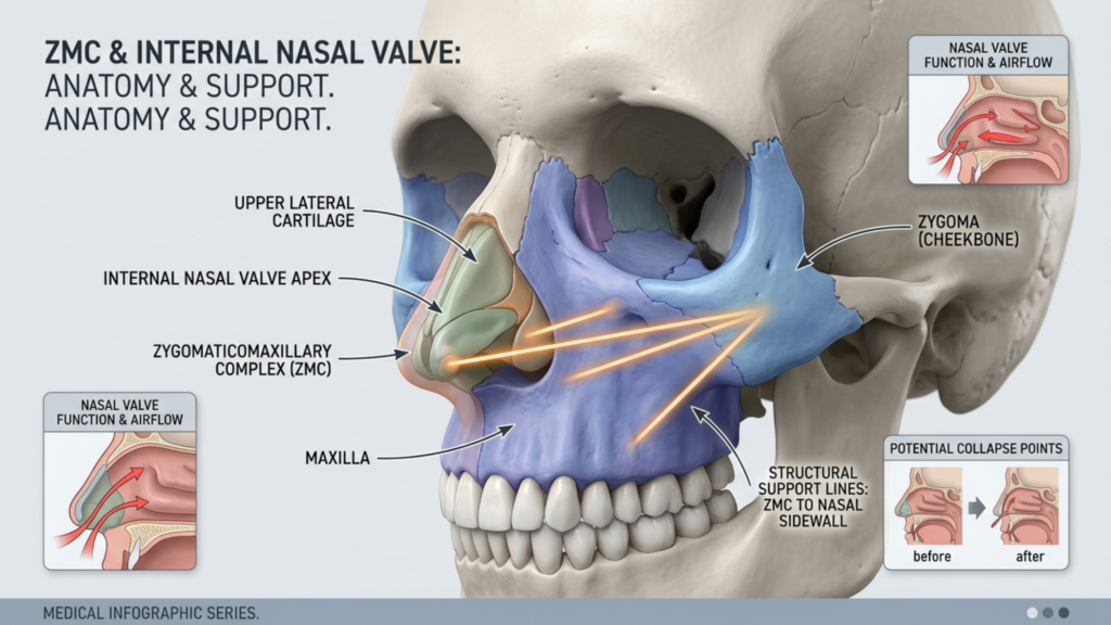
Table of Contents
Why FFS Is More Than Just Aesthetics: The Science of Facial Harmony
FFS is not merely about altering appearances—it’s about reconstructing identity through precision. The human face is a complex interplay of bone, cartilage, and soft tissue, each contributing to gender-specific proportions. For transgender women, the goal is to soften angular male features, such as a prominent brow ridge, wide jaw, or pronounced chin, and create a more delicate, feminine contour. However, achieving this requires a deep understanding of craniofacial anatomy and the potential functional trade-offs, such as nasal airway obstruction, that can arise from aggressive bone reduction.
Studies in Plastic and Reconstructive Surgery reveal that the most common FFS procedures—such as forehead contouring, cheekbone reduction, and jaw reshaping—can inadvertently alter the structural support of the nasal airway. For example, reducing the zygomatic arch (cheekbone) may weaken the lateral nasal wall, leading to nasal valve collapse and chronic breathing difficulties (Frontiers in Surgery, 2025). This guide will help you navigate these complexities, ensuring that your transformation is both beautiful and functional.
The Core Procedures of FFS: Bone and Soft Tissue Modifications
FFS is a collection of procedures tailored to feminize the face by addressing key structural differences between male and female facial anatomy. Below, we break down the most impactful modifications:
1. Forehead Contouring: Redefining the Brow Ridge
The forehead is one of the most gender-distinct areas of the face. Male foreheads tend to have a more pronounced brow ridge (frontal bossing) and a flatter slope, while female foreheads are smoother and more rounded. Forehead contouring involves:
- Bone Reduction: The frontal bone is reshaped to reduce the prominence of the brow ridge, creating a softer, more feminine contour.
- Hairline Advancement: For patients with a high hairline, the forehead can be reduced in height by advancing the hairline forward, which also helps to create a more youthful appearance.
- Fat Grafting: Soft tissue augmentation can further enhance the smoothness and femininity of the forehead.
This procedure is often combined with forehead reduction to achieve a harmonious result.
2. Cheekbone Reduction: The Delicate Balance of Midface Feminization
The zygomatic arch (cheekbone) is a defining feature of facial masculinity. In men, it tends to be wider and more prominent, while in women, it is narrower and less pronounced. Cheekbone reduction involves:
- Osteotomies: Surgical cuts are made to reposition or reduce the zygomatic arch, creating a more slender midface.
- Soft Tissue Adaptation: The overlying soft tissues are redraped to match the new bone structure, enhancing the feminine contour.
- Nasal Airway Considerations: As discussed earlier, reducing the zygomatic arch can compromise the lateral support of the nasal airway, leading to potential breathing issues. Surgeons must carefully plan to avoid this complication.
For those concerned about the risks of cheekbone reduction, alternatives like cheek augmentation with fat grafting or fillers can achieve a similar feminizing effect without altering bone structure.
3. Jaw Reduction: Softening the Lower Face
A strong, square jaw is a hallmark of male facial structure. Jaw reduction aims to create a softer, more oval or heart-shaped lower face by:
- Bone Shaving: The mandible (jawbone) is carefully reshaped to reduce its width and prominence.
- Angle Refinement: The jaw angle is softened to eliminate the sharp, masculine edge.
- Chin Reshaping: Often performed in conjunction with jaw reduction, chin reshaping can further enhance facial harmony. Procedures like genioplasty or chin reduction can refine the lower face.

4. Rhinoplasty: The Nose as the Centerpiece of Feminization
The nose plays a central role in facial gender perception. Male noses tend to be larger, with a more pronounced bridge and wider nostrils, while female noses are typically smaller, with a softer bridge and more refined tip. Feminization rhinoplasty focuses on:
- Bridge Refinement: Reducing the height and width of the nasal bridge to create a more delicate profile.
- Tip Definition: Enhancing the definition and projection of the nasal tip for a more feminine appearance.
- Nostril Reshaping: Narrowing the nostrils to achieve a more refined and balanced look.
- Airway Preservation: Ensuring that aesthetic changes do not compromise nasal function, particularly in patients undergoing concurrent cheekbone reduction.
Rhinoplasty is often combined with other FFS procedures to achieve a cohesive result. For more details, explore our rhinoplasty guide.
The Hidden Risks: How FFS Can Compromise Nasal Airway Function
While FFS can achieve remarkable aesthetic results, it is not without risks. One of the most critical yet overlooked complications is nasal airway obstruction. This occurs when procedures like cheekbone reduction or rhinoplasty alter the structural integrity of the nasal cavity, leading to breathing difficulties. Below, we explore the mechanisms behind this risk and how to mitigate it.
The Anatomical Connection: Zygomatic Arch and Nasal Airway
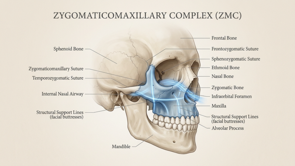
The zygomatic arch (cheekbone) is not an isolated structure—it is intricately connected to the midface and nasal cavity. The arch forms the lateral boundary of the midface, supporting the soft tissues of the cheek and contributing to the structural integrity of the nasal valve and airway. The zygomaticomaxillary complex, which includes the zygomatic bone, maxilla, and nasal bones, plays a pivotal role in maintaining nasal airway patency.
When the zygomatic arch is reduced or repositioned, the lateral support for the nasal sidewall may be compromised. This can lead to nasal valve collapse, a condition where the nasal passages narrow or collapse during inhalation, causing significant airway obstruction. The nasal valve is the narrowest segment of the nasal airway, and even minor anatomical changes can substantially increase nasal resistance and impair breathing (Frontiers in Surgery, 2025).
Clinical Evidence: The Link Between Cheekbone Reduction and Airway Obstruction
Emerging clinical evidence underscores the importance of considering nasal airway function during FFS planning. A 2025 study published in Frontiers in Surgery found that patients undergoing midface contouring procedures, including cheekbone reduction, experienced a 20% increase in nasal airway resistance postoperatively, with a subset developing symptomatic nasal valve collapse (Frontiers in Surgery, 2025). Similarly, research in Plastic and Reconstructive Surgery demonstrated that patients with pre-existing nasal valve insufficiency were at higher risk for postoperative airway obstruction following midface procedures.
These findings highlight the need for preoperative functional assessment and surgical techniques that preserve or enhance nasal airway patency. As FFS surgeons, we must recognize that aesthetic goals should not come at the expense of functional integrity.
Mitigating Risks: Strategies for Safe and Effective FFS
To minimize the risk of nasal airway obstruction during FFS, a multidisciplinary, anatomy-guided approach is essential. Below are evidence-based strategies for surgical planning and execution:
1. Comprehensive Preoperative Assessment
- Nasal Airway Evaluation: Conduct a thorough nasal airway assessment, including physical examination (e.g., Cottle maneuver) and, if indicated, rhinomanometry or acoustic rhinometry to quantify nasal resistance and identify pre-existing valve collapse.
- 3D Imaging and Virtual Surgical Planning: Utilize 3D CT scans and virtual surgical planning software to simulate the effects of cheekbone reduction on nasal airway dimensions. This allows for precise osteotomy planning and prediction of postoperative airway changes (ESMED, 2024).
- Patient-Specific Risk Stratification: Identify patients at higher risk for postoperative airway obstruction, such as those with pre-existing nasal valve weakness, septal deviations, or a history of nasal trauma.
2. Anatomy-Guided Surgical Techniques
- Preservation of Lateral Nasal Support: Avoid over-resection of the zygomatic arch, particularly in the region adjacent to the nasal sidewall. Consider segmental osteotomies that preserve the structural integrity of the midface while achieving aesthetic goals.
- Concurrent Nasal Valve Repair: In patients with pre-existing nasal valve collapse or high-risk anatomy, consider concurrent nasal valve repair techniques, such as spreader grafts, alar batten grafts, or lateral crural strut grafts, to reinforce the nasal sidewall and prevent collapse (Plastic Surgery Key, 2017).
- Minimally Invasive Approaches: Utilize endoscopic or intraoral approaches for cheekbone reduction to minimize soft tissue trauma and preserve critical support structures.
- Staged Procedures: In complex cases, consider staging cheekbone reduction and nasal airway procedures to allow for soft tissue readaptation and reduce the risk of compounded functional complications.
3. Postoperative Care and Monitoring
- Airway Monitoring: Closely monitor nasal airway function postoperatively, particularly in the first 48 hours, when swelling and soft tissue changes are most pronounced.
- Patient Education: Educate patients about the signs of nasal airway obstruction (e.g., difficulty breathing through the nose, increased snoring) and the importance of follow-up if symptoms arise.
- Functional Rehabilitation: Consider postoperative nasal dilators or breathing exercises to support nasal valve function during the healing process.

Alternative Approaches to Midface Feminization
For patients at high risk of nasal airway obstruction, alternative techniques for midface feminization may be considered:
- Soft Tissue Augmentation: Instead of bone reduction, consider fat grafting or filler placement to achieve midface contouring while preserving skeletal support for the nasal airway.
- Orthognathic Surgery: In patients with midface hypoplasia or malocclusion, orthognathic surgery (e.g., Le Fort I osteotomy) can advance the maxilla, improving both facial balance and nasal airway dimensions (Mayo Clinic, 2024).
- Combined Procedures: Combine cheekbone reduction with septoplasty or turbinate reduction to optimize nasal airway function and reduce the risk of postoperative obstruction.
The Future of FFS: Balancing Aesthetics and Function
The field of FFS is evolving rapidly, with advancements in virtual surgical planning and 3D imaging revolutionizing our ability to predict and prevent complications. By leveraging these tools, surgeons can simulate the effects of cheekbone reduction on nasal airway dimensions and adjust their approach accordingly, ensuring both aesthetic and functional success (Hospimedica, 2024).
As surgeons, our responsibility extends beyond achieving aesthetic goals. We must prioritize functional outcomes and patient safety, particularly in procedures as complex as FFS. The zygomatic arch dilemma underscores the importance of:
- Multidisciplinary Collaboration: Work closely with rhinoplasty and airway specialists to ensure comprehensive care for patients undergoing midface procedures.
- Informed Consent: Discuss the potential risks of nasal airway obstruction with patients during the consent process, ensuring they understand the balance between aesthetic and functional outcomes.
- Continuous Education: Stay abreast of the latest research and techniques in both FFS and rhinoplasty to refine surgical approaches and minimize complications.
- Patient-Centered Care: Tailor surgical plans to each patient’s unique anatomy and risk profile, prioritizing long-term health and well-being over short-term aesthetic results.
The integration of technology and a patient-centered approach will continue to shape the future of FFS, ensuring that every individual can achieve their desired transformation safely and effectively.

Frequently Asked Questions
How does FFS structurally alter male facial features into feminine proportions?
FFS modifies bone and soft tissue structures to create a more feminine appearance. Procedures like forehead contouring, cheekbone reduction, jaw reshaping, and rhinoplasty address gender-specific differences in facial anatomy, such as brow ridge prominence, midface width, and nasal proportions.
What are the risks of nasal airway obstruction during FFS?
Cheekbone reduction and rhinoplasty can compromise the structural support of the nasal airway, leading to nasal valve collapse and increased airway resistance. This risk is higher in patients with pre-existing nasal valve weakness or septal deviations.
How can virtual surgical planning improve FFS outcomes?
Virtual surgical planning uses 3D imaging to simulate the effects of FFS procedures on facial anatomy and nasal airway function. This allows surgeons to optimize surgical techniques, minimize risks, and achieve more predictable results.
What alternatives exist for patients at high risk of nasal airway obstruction?
Patients at high risk may consider soft tissue augmentation (e.g., fat grafting), orthognathic surgery (e.g., Le Fort I osteotomy), or combined procedures that address both aesthetic and functional goals without compromising airway integrity.
How do surgeons balance aesthetic goals with functional outcomes in FFS?
Surgeons prioritize functional outcomes by conducting comprehensive preoperative assessments, using anatomy-guided surgical techniques, and collaborating with airway specialists. Patient education and informed consent are also critical to achieving a balanced result.
What is the role of 3D imaging in FFS planning?
3D imaging allows surgeons to visualize and simulate the effects of FFS procedures on facial structures and nasal airway dimensions. This technology enhances precision, reduces risks, and improves overall surgical outcomes.
How can patients prepare for FFS to minimize risks?
Patients should undergo a thorough preoperative assessment, including nasal airway evaluation and 3D imaging. Discussing potential risks with their surgeon and following postoperative care instructions can further minimize complications.







