The pursuit of a refined, V-shaped jawline has led to increased demand for treatments targeting lower facial width. Two primary approaches exist: masseter Botox for temporary muscle reduction and mandibuloplasty (jaw surgery) for permanent structural changes. The choice between these methods depends on the underlying cause—masseter muscle hypertrophy or mandibular bone protrusion—as well as patient goals, budget, and tolerance for downtime.
This article explores the diagnostic criteria, clinical outcomes, risks, and cost comparisons of both treatments, supported by peer-reviewed studies and official medical sources. It also provides SEO-optimized insights for practitioners and patients seeking evidence-based guidance.
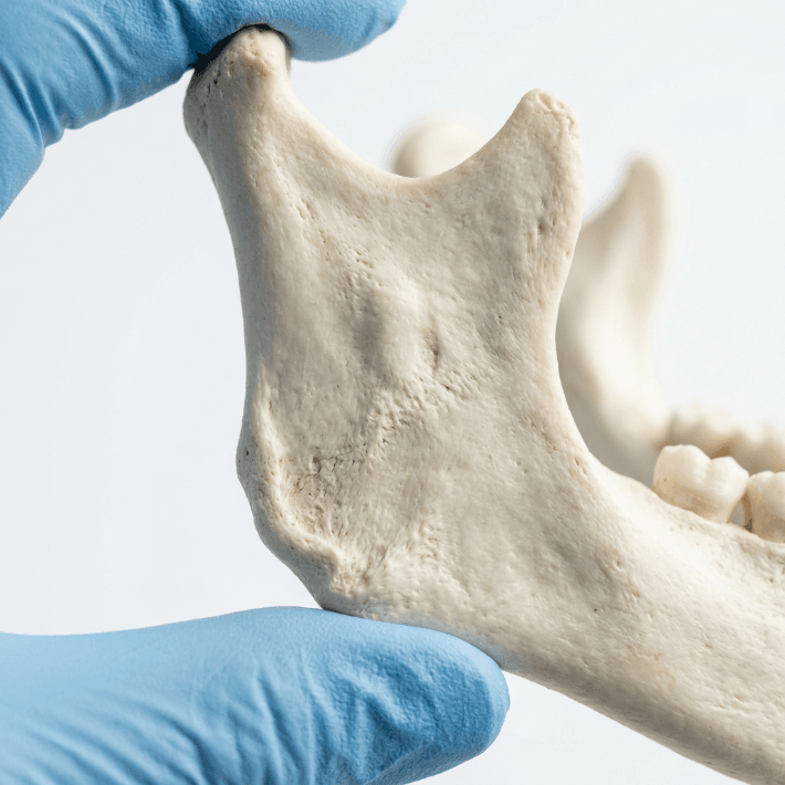
Diagnostic Differentiation: Masseter Hypertrophy vs. Mandibular Bone Protrusion
Clinical Examination and Imaging
Accurate diagnosis is critical to determining the appropriate treatment. Masseter muscle hypertrophy and mandibular bone protrusion present differently and require distinct approaches:
- Masseter Hypertrophy:
- Palpation: The masseter muscle is palpated during clenching, revealing a prominent, thickened muscle fiber at the angle of the mandible (20XJNEak, EelsQLvR).
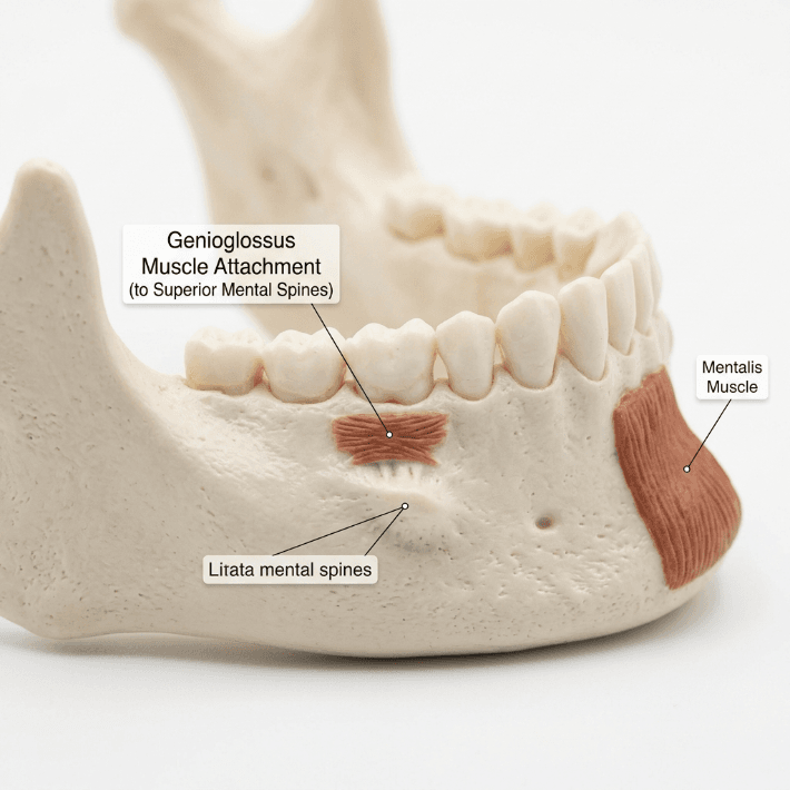
- Imaging: CT, MRI, and panoramic radiographs confirm muscle enlargement and rule out bone involvement. Bone spurs or hyperostosis at the mandibular angle may also be visible (xunUAhzf, qg4kRYCH, Yr7koQ28).
- Etiology: Often linked to bruxism, chronic clenching, or genetic factors (Lelwo1V2, eUBf6ryx).
- Mandibular Bone Protrusion:
- Radiographic Findings: OPG (orthopantomogram) and AP X-rays reveal broadening of the mandibular angle, bone spurs, or hyperostosis (SvjnOYoe, qg4kRYCH).
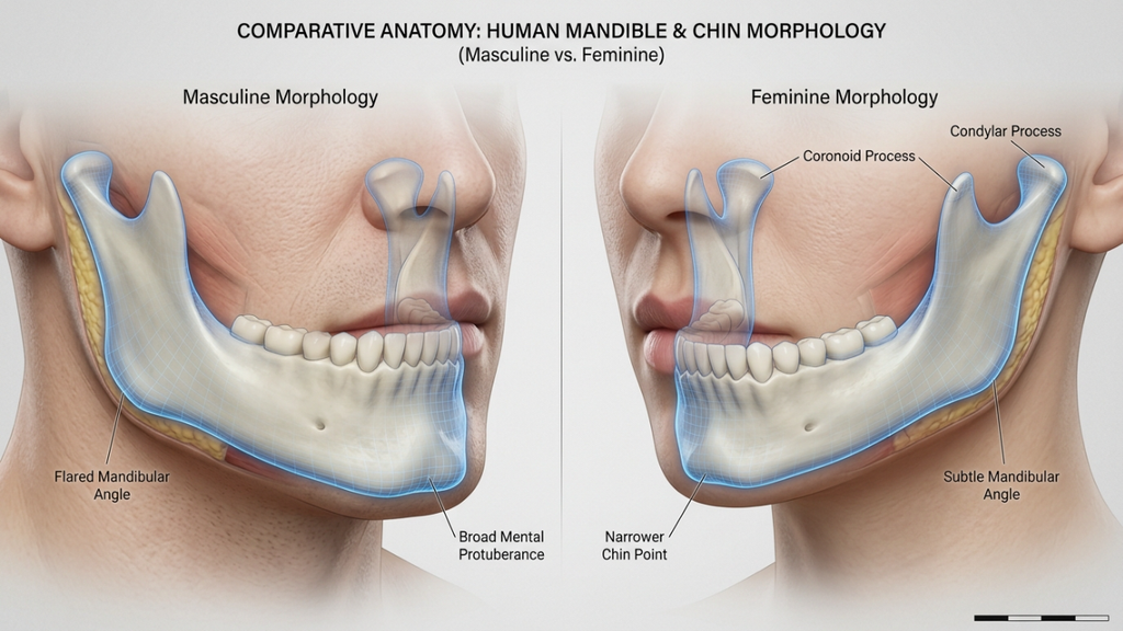
- Clinical Presentation: A square or wide jawline persists even when the masseter muscle is relaxed, indicating skeletal involvement (fonfKtxE).
Key Diagnostic Tools
| Tool | Masseter Hypertrophy | Mandibular Bone Protrusion |
|---|---|---|
| Palpation | Muscle bulge during clenching | Hard, fixed prominence |
| CT/MRI | Muscle enlargement | Bone spurs, angle hypertrophy |
| Panoramic X-ray | Normal bone structure | Broadened mandibular angle |
| CBCT | Muscle tension, slight bone changes | Prominent angle, bone spur development (TCLxTqnb) |
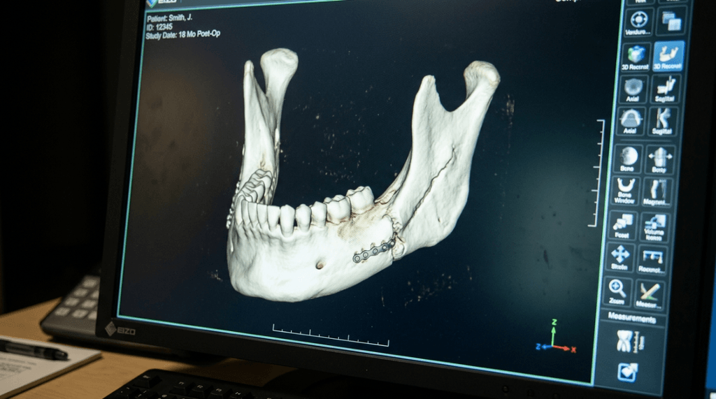
Treatment Modalities: Masseter Botox vs. Jaw Surgery
Masseter Botox: Temporary Muscle Reduction
Mechanism of Action
Masseter Botox involves botulinum toxin type A (BoNT-A) injections into the masseter muscle, causing temporary paralysis and atrophy. This reduces muscle bulk, slimming the lower face (555inSZk, 2zNx6q7S).
Clinical Efficacy
- Reduction in Muscle Thickness: Studies report a 20–30.9% reduction in masseter muscle thickness within 3 months, with 95% patient satisfaction for aesthetic narrowing (4qEf3FHe, 2zNx6q7S).
- Onset and Duration: Initial softening appears within 2 weeks, with peak effects at 6 weeks. Results last 3–6 months, requiring maintenance injections (xl0pxczi, 2zNx6q7S).
- Functional Benefits: Reduces bruxism, jaw pain, and TMJ symptoms (x0Xhooeo).
Limitations
- Temporary: Effects reverse as muscle regains strength (UGny6fZW).
- Muscle-Only: Cannot address bony prominence (fonfKtxE, C5VjcL52).
- Off-Label Use: In the U.S., masseter Botox for jaw slimming is off-label (KDTnAIvL).
Cost
- $500–$1,000 per session (4qEf3FHe).
Mandibuloplasty (Jaw Surgery): Permanent Structural Change
Mechanism of Action
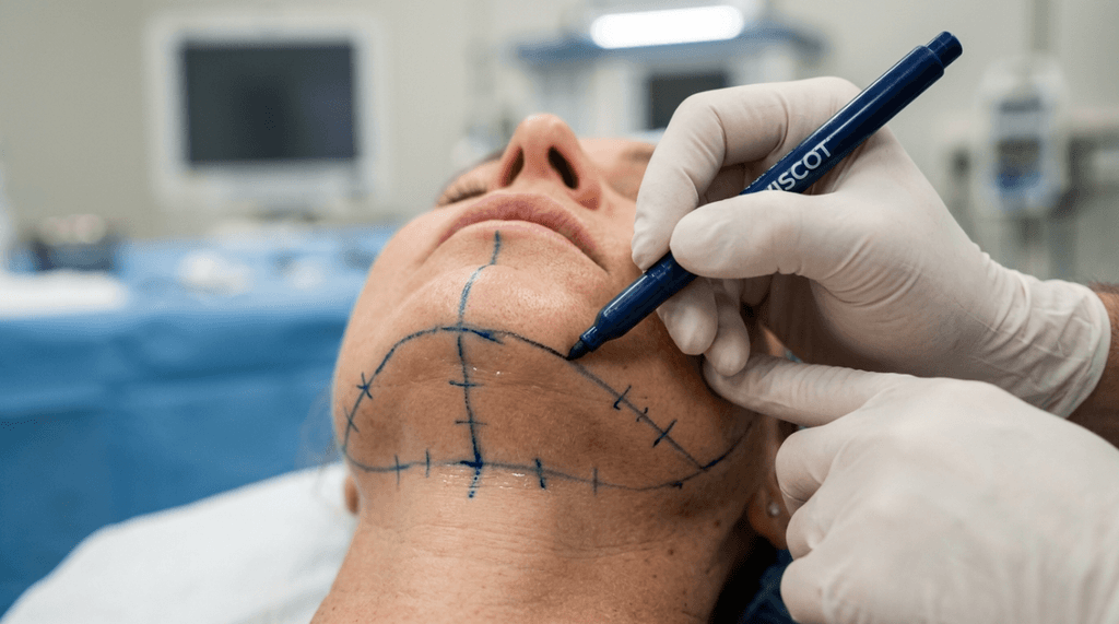
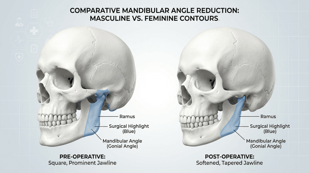
Mandibuloplasty involves surgical resection of the mandibular angle or bone shaving to permanently reduce lower facial width. It is the gold standard for skeletal causes of wide jaw (SvjnOYoe, pUJuYmuE).
Clinical Efficacy
- Permanent Results: Structural changes are irreversible, providing long-term contouring (4qEf3FHe).
- Dramatic Transformation: Ideal for patients with severe mandibular angle prominence or asymmetry (SvjnOYoe).
- Combined Approaches: Surgery may be paired with masseter muscle resection for optimal results (pUJuYmuE).
Risks and Recovery
- Surgical Risks: Infection, nerve damage, difficulty opening the mouth, and prolonged recovery (2–3 months) (FeZ46LCn).
- Cost: $3,000–$20,000, depending on complexity (9Wk8EEmR).
Comparative Analysis: Masseter Botox vs. Jaw Surgery
| Factor | Masseter Botox | Jaw Surgery (Mandibuloplasty) |
|---|---|---|
| Target | Masseter muscle | Mandibular bone |
| Permanence | Temporary (3–6 months) | Permanent |
| Onset of Results | 2–6 weeks | Months (full recovery) |
| Invasiveness | Non-surgical, minimal downtime | Surgical, extended recovery |
| Cost | $500–$1,000 per session | $3,000–$20,000 |
| Best For | Muscle hypertrophy, bruxism, non-surgical preference | Bone protrusion, severe skeletal issues |
| Risks | Bruising, swelling, temporary weakness | Infection, nerve damage, asymmetry |
| Maintenance | Requires repeat injections | None |
Clinical Decision-Making: Which Treatment Is Right for You?
Patient Selection Criteria
Masseter Botox Is Ideal For:
- Patients with masseter muscle hypertrophy confirmed via palpation and imaging.
- Those seeking non-surgical, temporary contouring.
- Individuals with bruxism or TMJ symptoms (x0Xhooeo).
- Patients unwilling or unfit for surgery.
Jaw Surgery Is Ideal For:
- Patients with mandibular bone protrusion or skeletal asymmetry.
- Those desiring permanent, dramatic changes.
- Cases where non-surgical methods fail to address structural concerns (fonfKtxE).
Hybrid Approaches
In some cases, Botox and surgery are combined:
- Pre-Surgery: Botox may be used to test aesthetic outcomes before committing to surgery.
- Post-Surgery: Botox can refine muscle balance during healing (f1VoJDdr).
Case Studies and Clinical Outcomes
Masseter Botox
- Study (2019): 20–30% reduction in muscle thickness in East Asian patients, with high satisfaction rates (555inSZk).
- MRI/Ultrasound Confirmation: Objective measurements validate muscle atrophy post-treatment (2zNx6q7S).
Jaw Surgery
- Case Report (2020): Bilateral mandibuloplasty for idiopathic masseter hypertrophy with retrognathic mandible, achieving symmetrical, aesthetic results (SvjnOYoe).
- Long-Term Stability: Surgical outcomes are permanent, with minimal recurrence (pUJuYmuE).
Risks and Complications
Masseter Botox
- Common: Bruising, swelling, mild headache (UGny6fZW).
- Rare: Asymmetry, over-relaxation of muscles, temporary chewing difficulty (zfsVLpf6).
Jaw Surgery
- Early: Pain, swelling, infection (FeZ46LCn).
- Late: Nerve damage (facial paralysis), asymmetry, bone over-resection (SvjnOYoe).

Cost Comparison and Accessibility
| Treatment | Cost Range | Insurance Coverage | Accessibility |
|---|---|---|---|
| Masseter Botox | $500–$1,000/session | Rare (cosmetic) | Widely available |
| Jaw Surgery | $3,000–$20,000 | Possible (if medically necessary) | Specialized clinics |
Conclusion
The choice between masseter Botox and jaw surgery hinges on the underlying cause of lower facial width—muscle or bone. Masseter Botox offers a non-surgical, temporary solution for muscle hypertrophy, while mandibuloplasty provides permanent, structural changes for skeletal issues. A thorough diagnostic workup, including palpation, CT/MRI, and X-rays, is essential to guide treatment selection.
For patients and practitioners alike, understanding the distinctions, benefits, and limitations of each approach ensures informed, evidence-based decision-making.
Sources
- La Belle Vie Med Spa: Masseter Botox vs Jaw Surgery (4qEf3FHe, xl0pxczi)
- PubMed: Efficacy of Botulinum Toxin in Masseter Muscle Hypertrophy (555inSZk)
- PMC: Idiopathic Masseter Muscle Hypertrophy (20XJNEak)
- PMC: Surgical Management of Masseteric Hypertrophy (SvjnOYoe)
- PMC: Masseter and Medial Pterygoid Muscle Hypertrophy (qg4kRYCH)
- Sage Journals: Management of Masseter Muscle Hypertrophy (6lWM5jpI)
- Wiley: Management of Unilateral Masseter Hypertrophy (EelsQLvR)
- Hindawi: Masseter Hypertrophy Case Report (Yr7koQ28)
- Dr. Troell: Masseter Botox vs Surgery (KDTnAIvL, zfsVLpf6)
- Greenwich MD: Masseter Botox for Jaw Slimming (2zNx6q7S)
- Dr. Kenneth Kim: Non-Invasive Jaw Reduction (Lelwo1V2, fonfKtxE)
- Qunomedical: Jaw Shaping (FeZ46LCn)
- The Bruxism Clinic: Botox vs Jaw Surgery for TMJ (x0Xhooeo)
- Korea Clinic Guide: Square Jaw Correction (9Wk8EEmR)
- Hugo Plastic Surgery: Wide Jawline Options (f1VoJDdr)
- JOMOS: Masticatory Muscle Hypertrophy (TCLxTqnb)