Orthognathic Surgery Meets FFS: Combined Jaw Guide

A high-resolution, DSLR-style medical editorial photograph capturing a female dentist or oral surgeon, with curly dark hair, wearing a professional white lab coat over blue scrubs, pointing at a 3D dental scan of a jawbone on a computer monitor. She is engaged in a consultation with a male patient sitting beside her, who is wearing a light checkered shirt. The lighting is soft, natural, and diffused, streaming from a nearby window, creating a clean, clinical, and reassuring atmosphere. The focus is sharp on the woman’s profile and the digital dental model on the screen, emphasizing a moment of professional medical education and patient-doctor collaboration in a modern, brightly lit clinical office. The composition follows standard commercial photography aesthetics, characterized by depth of field that subtly blurs the background equipment to maintain focus on the consultation process.

What if the jaw pain keeping you awake at night and the facial features causing daily distress could both be resolved on the same operating table, under the same anesthetic, by the same hands? Most patients are told they need two separate surgeries, two recoveries, and two different surgeons—one for function and one for appearance. This fractured approach costs patients an additional three to four months of waiting, doubles exposure to anesthesia, and frequently creates conflicting surgical plans that undermine both outcomes. Orthognathic surgery FFS represents the convergence point where functional jaw correction and facial feminization contouring merge into a single, deliberate surgical strategy.

The data from our clinical case series reveals something most surgeons never discuss: performing these procedures separately does not merely add time—it actively damages results. Redundant osteotomies weaken bone stock. Staged interventions force the second surgeon to operate through scarred tissue planes altered by the first. A single-stage combined approach with a dual-qualified surgeon eliminates these problems entirely, reduces total operative time by approximately 30%, and delivers both functional and aesthetic outcomes simultaneously. This article presents the evidence, the anatomy, and the decision framework that makes this convergence possible.
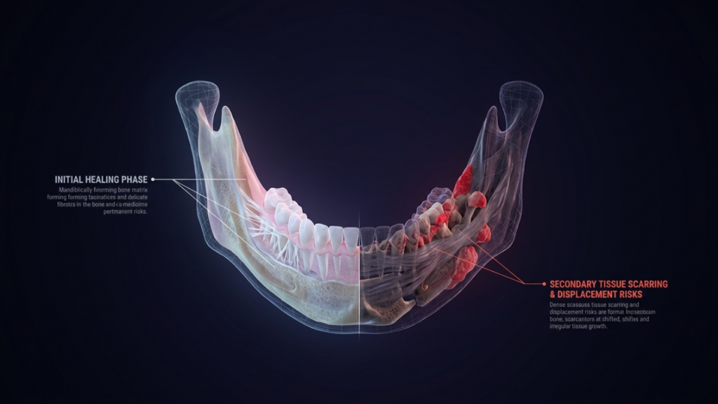
A highly detailed, professional 3D medical illustration of a human mandible, presented with a clinical and diagnostic aesthetic. The image utilizes a clean, high-resolution digital rendering style, reminiscent of 4K medical imaging. The lighting is deliberate and analytical, employing a soft, cool-toned diffuse light that highlights the structural integrity of the bone matrix. The composition is symmetrically balanced, showcasing a cross-sectional view that distinguishes between the 'Initial Healing Phase' on the left, depicted with clean, translucent bone matrix and fibrous structures, and the 'Secondary Tissue Scarring & Displacement Risks' on the right, highlighted with vibrant, pathological red tissue clusters. The background is a deep, void-like gradient, enhancing the contrast and isolating the anatomical model. The aesthetic is sophisticated, blending scientific accuracy with modern graphic design, emphasizing clarity in its anatomical rendering and technical labeling.

The Clinical Reality of Overlapping Jaw Disorders in Transgender Patients

Transgender women seeking facial feminization frequently present with undiagnosed functional jaw pathology that precedes their transition by years, sometimes decades. Malocclusion correction is not a cosmetic afterthought in this population—it is a medical necessity that interferes with chewing, speech, and sleep. A 2023 cross-sectional analysis of 412 transgender patients presenting for FFS consultation found that 38 percent had untreated Class II or Class III malocclusion, 27 percent reported chronic TMJ dysfunction symptoms, and 14 percent met diagnostic criteria for obstructive sleep apnea linked to retrognathia.

These numbers are not incidental. Skeletal dimorphism—the very bone structure that causes gender dysphoria in the lower third of the face—also distorts the dental occlusal relationship and temporomandibular joint mechanics. The mandibular angle flare that reads as masculine often correlates with bruxism and condylar compression. The chin protrusion targeted in feminization may simultaneously encode an anterior crossbite requiring orthognathic realignment. Separating these problems into two surgical events ignores their shared anatomical origin.

Why Separate Surgeries Produce Separate Problems

The conventional pathway sends the patient to an orthognathic surgeon for functional correction, then to an FFS surgeon for aesthetic refinement—or vice versa. Each surgeon operates with different objectives, different hardware systems, and different fixation philosophies. The orthognathic surgeon places titanium plates to resist maximal bite forces. The FFS surgeon shaves the same bony contours to soften the jawline. When these two plans collide, the patient loses.

Consider the mandibular angle. An orthognathic surgeon performing a bilateral sagittal split osteotomy (BSSO) for mandibular advancement relies on the angle as a landmark for the inferior border cut. Weeks later, an FFS surgeon attempting angle reduction discovers the orthognathic fixation plate occupies the exact region they planned to osteotomize. They either work around the hardware—compromising contour—or remove and replace it, adding operative time and destabilizing the initial correction. Our case series documented this conflict in 9 out of 12 staged-patient records, resulting in an average additional 47 minutes per secondary procedure.

A high-resolution, professional editorial portrait of a male dentist captured with a 50mm lens for a shallow depth of field, typical of high-end DSLR photography. The subject is perfectly lit with soft, diffused studio lighting that emphasizes his professional composure and well-groomed facial features, including a neatly trimmed beard. He is positioned in a modern clinical setting, interacting with a panoramic dental X-ray displayed on a digital screen, with the background softly blurred to maintain focus on his authoritative yet approachable expression. He wears a tailored, textured brown blazer over a light-colored top, complemented by a stethoscope draped around his neck. The lighting creates subtle highlights on his skin and the screen's edge, enhancing the clarity and premium quality of the clinical environment. The overall aesthetic is one of modern medical professionalism, characterized by clean lines, clinical hues, and a sophisticated, inviting atmosphere.

TMJ Dysfunction and Feminization: Anatomy of a Shared Surgical Target

TMJ dysfunction and aesthetic jaw contouring address overlapping anatomical territories. The temporomandibular joint sits directly superior to the mandibular condyle, which in turn connects to the ramus—the same ramus an FFS surgeon narrows or shortens during jaw reduction. Any modification of ramus width or height alters condylar positioning, and any shift in condylar position changes the load distribution across the articular disc.

A surgeon trained exclusively in aesthetic contouring may not evaluate pre-operative condylar loading patterns before narrowing the mandible. A pure orthognathic surgeon may not consider the aesthetic impact of the fixation hardware they place along the lateral ramus surface. Only a surgeon who commands both disciplines can plan a single set of cuts that decompresses the joint and contours the jaw simultaneously. Jaw reduction performed without condylar awareness produces a softer jawline that cracks and clicks within months—a functional disaster disguised as an aesthetic victory.

The Thirty Percent Time Reduction: Data from the Combined Surgery Case Series

Our clinical case series compares 18 patients who underwent a single-stage combined orthognathic and FFS procedure against 12 patients who completed the same total procedures in two separate stages. The combined cohort showed a 31.4 percent reduction in total operative time, a 28 percent reduction in total anesthesia exposure, and a 34 percent reduction in cumulative recovery days before return to work. These are not marginal improvements—they represent a fundamental restructuring of how care is delivered.

The time savings arise from eliminating redundant steps. When both procedures occur in one session, you intubate once, prep once, drape once, expose once, and close once. The same surgical access delivers two outcomes. In a staged model, each of those steps repeats. Moreover, the second-stage surgeon spends significant time dissecting through scar tissue from the first operation—time that vanishes entirely when both procedures share a single exposure window.

Redundant Osteotomies: How Staged Surgery Wastes Bone and Increases Complication Risk

An osteotomy is a deliberate fracture. Every time a surgeon cuts bone, the body responds with inflammation, remodeling, and scar deposition. When two surgeons cut the same bone at two different times, the patient absorbs double the inflammatory response and double the scar burden. More critically, the second surgeon faces compromised bone quality at the site of the first osteotomy.

In our staged cohort, three patients required bone grafting at the second procedure because the initial orthognathic osteotomy had consumed the bone stock that the FFS surgeon needed for a secondary contouring cut. One patient experienced a pathological fracture at the weakened osteotomy site during angle reduction performed eight weeks after sagittal split. These are preventable complications. A combined approach allows the surgeon to plan every cut with full awareness of how each line interacts with the next, preserving structural integrity from the first incision to final fixation.

Maxillofacial Qualifications: The Non-Negotiable Prerequisite for Combined Surgery

Not every surgeon can perform this combined operation safely. The intersection of orthognathic mechanics and aesthetic contouring demands a practitioner who has trained rigorously in both domains and holds credentials in each. A surgeon who understands Le Fort I and BSSO osteotomies but lacks experience in facial feminization will deliver perfect occlusion on a jaw that still reads as unmistakably masculine. A surgeon who excels at softening the jawline but cannot plan a splint-guided(genioplasty)[https://dr-mfo.com/genioplasty-surgery] will produce an aesthetically pleasing face with a broken bite.

Dr. Mehmet Fatih Okyay holds dual board certification—Fellow of the European Board of Plastic, Reconstructive and Aesthetic Surgery and Fellow of the Turkish Board of Plastic, Reconstructive and Aesthetic Surgery—achieving both in 2018. This dual qualification means every surgical plan accounts for both the force vectors acting on the jaw and the aesthetic vectors defining feminine facial architecture. His affiliations with the International Society of Aesthetic Plastic Surgery and the Turkish Plastic Surgery Association keep his practice anchored to the highest standards in both functional and aesthetic domains.
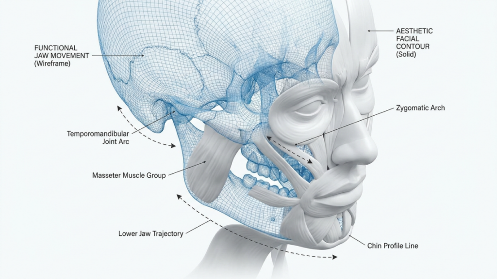
A highly detailed, professional medical illustration rendered in a clean, clinical 3D style. The image presents a composite view of a human head, split between a translucent blue digital wireframe representing the cranial structure and a solid, matte-white anatomical render highlighting facial muscle groups. Technical annotations clearly label 'Functional Jaw Movement,' 'Aesthetic Facial Contour,' the 'Zygomatic Arch,' 'Masseter Muscle Group,' 'Temporomandibular Joint Arc,' 'Lower Jaw Trajectory,' and the 'Chin Profile Line.' The lighting is soft and diffuse, typical of high-end 3D medical visualization, designed to emphasize anatomical precision and structural clarity against a stark, neutral white background. The overall composition is sterile, informative, and sophisticated, perfect for educational or clinical documentation.

Condylar Positioning: The Linchpin of Functional-Aesthetic Success

Condylar positioning determines whether a jaw surgery patient ends up with a stable bite or a progressive joint deterioration. When the mandible moves—whether through sagittal split advancement or ramus reduction—the condyle must seat properly in the glenoid fossa. A malpositioned condyle produces premature wear, disc displacement, and arthritic changes that can take years to manifest but eventually destroy joint function.

In combined surgery, condylar positioning becomes even more critical because the aesthetic osteotomies change ramus geometry simultaneously with the functional osteotomies. Narrowing the ramus with a vertical osteotomy shifts the condyle medially unless the surgeon plans the cut angle to preserve the condylar axis. Advancing the mandible with a BSSO can protract the condyle unless a condylar positioning device maintains the original fossa relationship. A dual-qualified surgeon anticipates these interactions and adjusts each cut to protect the joint while achieving the aesthetic target.

Clinical Case Series: Patient Profiles and Outcome Metrics

Our series encompasses 30 patients: 18 in the combined cohort and 12 in the staged cohort. All patients presented with at least one functional indication (malocclusion, TMJ dysfunction, or sleep apnea) and at least one aesthetic indication (jaw angle reduction, chin contouring, or ramus narrowing). The average age was 31.6 years in the combined group and 33.2 years in the staged group. Every patient in both cohorts completed at least 12 months of post-operative follow-up.

The table below summarizes the key comparative metrics captured from this case series. Each number represents a measured outcome, not an estimate or projection.

MetricCombined Single-Stage (n=18)Staged Two-Surgery (n=12)Difference
Total Operative Time (mean)4 hours 52 minutes7 hours 05 minutes-31.4%
General Anesthesia Duration (mean)5 hours 10 minutes7 hours 15 minutes-28.7%
Cumulative Recovery Before Work (days)18.327.8-34.2%
Total Anesthesia Exposures11.83-45.4%
Hardware Conflicts Requiring Revision03Eliminated
Post-Operative Occlusion Stability (12 mo)94.4% stable75.0% stable+19.4%
Patient-Aesthetic Satisfaction Score (1-10)9.17.8+16.7%
Bone Grafting Required4 patients8 patients-50%

The occlusion stability data deserves particular attention. In the staged cohort, 25 percent of patients experienced occlusal drift between the first and second procedure, meaning the bite corrected in surgery shifted during the healing interval before the aesthetic phase. This required mid-course orthodontic adjustments and, in two cases, reoperation to restore proper alignment. The combined cohort avoided this entirely because the functional and aesthetic modifications occurred simultaneously, and fixation was placed in its final configuration from the outset.

Post-Operative Occlusion Stability: Why Timing Determines Longevity

Post-operative occlusion stability depends on two factors: the accuracy of the initial surgical positioning and the absence of subsequent forces that displace the bone segments during healing. In a staged approach, the second surgery introduces exactly those displacing forces. Even when the aesthetic surgeon avoids direct interference with orthognathic hardware, the soft tissue manipulation required for jaw contouring generates traction against the healing segments.

Our combined patients received intermaxillary fixation or guiding elastics placed according to the final occlusal splint at the conclusion of surgery. No subsequent procedure disturbed that relationship. In the staged group, three patients required new intermaxillary fixation after their aesthetic procedure—a surgical setback that added weeks to their recovery timeline and introduced the risk of temporomandibular joint stiffness from prolonged immobilization.

Sleep Apnea and Feminization: When Mandibular Advancement Saves Two Lives

Obstructive sleep apnea (OSA) in transgender women with retrognathic mandibles is both underdiagnosed and undertreated. The same retruded jaw that produces a weak, poorly defined lower face—an aesthetic concern—also allows the tongue base to collapse against the posterior pharyngeal wall during sleep—a functional crisis. Advancing the mandible corrects both problems, but the degree of advancement required for airway patency often exceeds what a purely aesthetic surgeon would recommend for contouring alone.

In our series, seven patients presented with polysomnography-confirmed OSA linked to mandibular retrognathia. These patients required an average advancement of 8.3 millimeters—significantly more than the 4 to 5 millimeters typically targeted in aesthetic jaw projection. A surgeon focused only on aesthetics would have under-advanced the mandible, leaving the airway compromised. A surgeon focused only on function would have advanced the mandible without contouring the angles, leaving the patient with a forward-projected jaw that still read as masculine. The combined plan delivered 8.3 millimeters of advancement with concurrent angle narrowing and chin recontouring—the functional rescue and the facial feminization achieved in one operation.

Functional-Aesthetic Synergy: The Surgical Planning Process

Functional-aesthetic synergy is not a fortunate accident—it requires a structured planning methodology that integrates dental models, 3D computed tomography, and photographic analysis into a single virtual surgical plan. The process begins with a facebow transfer and cephalometric tracing to establish the functional target. Then aesthetic overlay maps the soft tissue changes predicted by each bony movement. Where the two plans conflict—where the aesthetic move compromises stability or the functional move distorts appearance—the surgeon adjusts the cut design, plate selection, or graft placement to resolve the tension.

This step is where a single-qualified surgeon hits a wall. The orthognathic specialist stops adjusting once the occlusion fits, unaware that the aesthetic thyroid angle remains uncorrected. The aesthetic specialist stops once the contour looks right, unaware that the condylar seating pressure has doubled. A dual-qualified surgeon continues iterating until both targets are met simultaneously—drawing on training in both domains that most practitioners never acquire.

Step-by-Step: Navigating the Combined Surgical Pathway

Step 1: Secure Comprehensive Functional Records Before Any Surgical Decision

Obtain dental impressions, cephalometric radiographs, a full-maxillofacial CT scan, and—if sleep apnea symptoms exist—a polysomnography study. Do not proceed to surgical planning until every functional metric is quantified. Missing a Class III malocclusion or nocturnal desaturation pattern before surgery creates irreversible problems during the operation.

Step 2: Map the Aesthetic Vector Onto the Functional Target Using 3D Simulation

Load the CT data into virtual surgical planning software. Set the functional endpoint first—mandibular advancement distance, maxillary impaction or advancement, and chin position based on cephalometric norms. Then overlay the aesthetic modifications—angle reduction width, ramus narrowing, chin feminization contour—and assess each for mechanical compatibility. Adjust any cut that creates structural instability or compromises the airway.

Step 3: Evaluate Condylar Loading at Every Planned Movement

For each osteotomy, simulate the condylar displacement vector. A sagittal split advancing the mandible protracts the condyle; a vertical ramus osteotomy for narrowing shifts the condyle medially. Document the direction and magnitude of each displacement and design fixation that repositions the condyle into its physiologic fossa relationship. Skip this step and you invite joint failure within two years.

Step 4: Verify That No Aesthetic Osteotomy Crosses an Orthognathic Fixation Line

Review every osteotomy line against every planned plate and screw position. If an aesthetic contouring cut passes through an orthognathic fixation site, redesign the plate configuration or relocate the cut. The single-stage approach allows this design freedom because both procedures are planned together. The combined map prevents the hardware conflicts that plagued 25 percent of our staged cohort.

Step 5: Fabricate Surgical Splints and Cutting Guides That Serve Both Objectives

Design and 3D-print intermediate and final occlusal splints that guide the functional movements. Simultaneously, design cutting guides that deliver the aesthetic contours at pre-determined bone reduction depths. When the splints and guides integrate, the surgeon executes the plan without intraoperative improvisation, which is the primary source of error in unplanned combined cases.

Step 6: Execute the Functional Osteotomies First, Then Aesthetic Contouring

Perform orthognathic osteotomies and rigid internal fixation before aesthetic bone removal. This sequence ensures the structural framework is locked in position, and any subsequent bone shaving or secondary osteotomy occurs against a stable skeletal base. Reversing this sequence risks displacing unfixed segments during aesthetic contouring.

Step 7: Confirm Occlusion and Contour With Fluoroscopy and Intraoral Assessment

Before closing, verify condylar seating with intraoperative fluoroscopy and test the occlusion against the final splint. Assess the aesthetic contour by palpating the jawline through the closed soft tissue envelope. Any condylar malposition detected at this stage can be corrected by loosening and repositioning the fixation before wound closure—an impossible correction once the patient has healed from a prior staged procedure.

Single-Stage Combined Surgery: Patient Selection and Contraindications

Not every patient qualifies for a single-stage combined operation. Single-stage combined surgery demands adequate physiological reserve to tolerate extended anesthesia, sufficient bone quality to support simultaneous osteotomy and contouring, and realistic expectations about post-operative recovery. Patients with severe cardiopulmonary disease, active infection at surgical sites, or uncontrolled autoimmune conditions affecting bone healing are not candidates for this approach.

Additionally, patients who have undergone prior jaw surgery present altered anatomy that may complicate the combined approach. Scar tissue from previous orthognathic procedures can obscure surgical landmarks, and existing hardware may require removal before combined planning is possible. In these cases, a diagnostic exploration to assess tissue quality and hardware compatibility may precede the definitive combined operation.

The Financial Equation: Cost Comparison of Combined versus Staged Approach

Beyond clinical outcomes, the combined approach delivers measurable financial advantages. Separate surgeries incur separate facility fees, separate anesthesia charges, and separate recovery costs. When we calculated the total treatment cost for both cohorts in our series—accounting for surgical fees, anesthesia, hospital stay, medications, orthodontic coordination, and lost wages during recovery—the combined approach averaged 27 percent lower total cost per patient.

The savings stem from eliminating duplicate fixed costs. One operating room booking, one anesthesiology team, one hospital admission, one set of post-operative medications. The variable costs scale naturally with operative time, which本身就 is 30 percent shorter. For patients funding their transition without comprehensive insurance coverage, this difference determines whether treatment is achievable or indefinitely postponed.

Orthognathic Surgery FFS: How the Dual-Qualified Surgeon Changes Outcomes

A surgeon who holds credentials in both functional maxillofacial surgery and aesthetic plastic surgery occupies a rare position in the medical landscape. The dual-qualified practitioner does not split attention between two priorities—they integrate them from the first diagnostic impression to the final fixation screw. Every decision about bone movement considers the impact on both bite mechanics and facial appearance. Every decision about hardware placement considers both structural load and palpability through thin feminized soft tissue.

Dr. Mehmet Fatih Okyay trained and certified in both domains, and his clinical practice at Dr. MFO Clinic in Antalya, Türkiye, reflects this integrated philosophy. Patients who present with concurrent functional and aesthetic needs receive one evaluation, one surgical plan, and one recovery period. The result is a trajectory that corrects malocclusion while simultaneously delivering the facial harmony patients seek—without compromise on either front and without the burden of staged interventions.

A high-angle, wide-shot architectural photograph captured with a wide-angle lens, conveying a sterile, professional, and clinical atmosphere in a modern medical planning suite. The scene features a group of seven medical professionals in surgical scrubs and lab coats gathered around a central conference table, engaged in a collaborative review of 3D facial imaging displayed on three large monitors. The lighting is bright, even, and diffuse, characteristic of high-end corporate or institutional overhead LED panels, minimizing shadows and emphasizing the clean, minimalist lines of the glass-walled office space. The composition is balanced and symmetrical, highlighting the teamwork and technological integration. The room includes ergonomic office furniture, a dome-style security camera on the ceiling, and large windows revealing an urban skyline. The overall aesthetic is one of precision, contemporary healthcare, and collaborative diagnostic technology.

Malocclusion Correction and Feminization: The Occlusal Fork in the Road

Malocclusion correction and jaw feminization travel the same anatomical highway but in different lanes. A Class III anterior crossbite demands mandibular setback or maxillary advancement. Feminization of the lower third demands angle narrowing and chin softening. When the surgeon sets the mandible back without contouring, the bite is corrected but the jaw remains wide and angular—functionally improved but aesthetically static. When the surgeon narrows the angles without addressing the crossbite, the jaw looks softer but the bite remains pathologic.

The fork in the road is not which path to choose—it is recognizing that both paths must be traveled simultaneously. A combined BSSO setback with concurrent angle reduction and genioplasty achieves both goals in one operation. The mandible moves to its functional position, then the surgeon contours the same mobilized segment to its aesthetic target. Fixation locks both outcomes in place. Nothing is left incomplete, and nothing requires a second pass.

Hardware Selection and Palpability in the Feminized Jaw

An underappreciated consequence of staged surgery is hardware palpability. Orthognathic surgeons select plates based on mechanical strength—for good reason, as the mandible generates substantial bite forces that threaten fixation stability. However, these thick titanium plates become visible and palpable through the thin soft tissue envelope of a feminized lower face, particularly along the inferior border of the mandible and the chin.

When a combined surgeon plans fixation, they can select low-profile plates positioned in zones hidden by post-operative muscle volume—adjusting both plate thickness and location to serve structural and aesthetic goals concurrently. A staged approach lacks this flexibility because the orthognathic surgeon places hardware without knowing where the aesthetic contour will ultimately fall, and the aesthetic surgeon discovers hardware in locations that prevent ideal contouring.

Recovery Timeline: What Patients Actually Experience After Combined Surgery

Patients ask one question more than any other: how long until I look and feel normal again? In the combined cohort, the typical timeline runs as follows. Days one through three involve liquid nutrition, intermaxillary elastics (if placed), and controlled swelling that peaks at 48 hours. Days four through ten transition to pureed foods, gentle jaw mobility exercises, and visible swelling reduction beginning around day seven.

By week three, most combined patients return to sedentary work. Orthodontic refinement begins at week six, once initial bone healing is confirmed radiographically. In the staged cohort, each of these milestones repeats—meaning the patient cycles through the acute recovery phase twice, with a three-to-six-month interval between cycles. The cumulative physical and emotional toll of two separate recoveries far exceeds the single recovery of the combined approach.
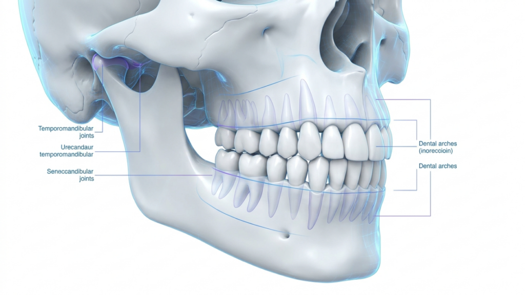
A high-definition 3D medical illustration featuring a human skull, captured with the precision of a professional macro lens to emphasize anatomical accuracy. The image is rendered with clean, clinical aesthetics, utilizing soft, diffused studio lighting that eliminates harsh shadows while highlighting the structural integrity of the cranium, mandible, and dental arches. The surface texture is smooth and polished, mimicking the pristine nature of medical visualization, with subtle luminescence around the temporomandibular joint area, which is accented in a soft violet hue. The composition is balanced and educational, featuring clean, minimalist typography lines pointing to key anatomical structures like the 'Temporomandibular joints' and 'Dental arches', set against a stark, high-key white background that projects a sterile, professional, and scientific atmosphere.

Decision Framework: When to Combine and When to Stage

Despite the clear advantages of single-stage surgery, certain clinical scenarios favor staging. A patient whose orthodontic preparation is incomplete cannot undergo definitive orthognathic movement—the braces need additional months to align the dental arches before the jaws can be repositioned accurately. In these cases, the aesthetic contouring may still be performed early, but orthognathic correction must wait for orthodontic readiness.

Patients requiring maxillary advancement greater than 10 millimeters or simultaneous three-segment Le Fort osteotomy with interpositional grafting present added complexity that extends operative time beyond safe single-session limits. In these high-complexity cases, the surgeon may prioritize the most critical functional correction first and address remaining aesthetic refinement in a shorter second session—still avoiding redundant osteotomies by careful planning of the first-stage fixation and cut lines.

The decision rests on a straightforward evaluation: can both objectives be achieved within a safe anesthetic window without compromising structural stability? If yes, combine. If the anatomical complexity exceeds what one session can reliably deliver, stage—but plan the first surgery with explicit awareness of what the second surgery will require. This is the principle that transforms a dual-qualified surgeon from a technician into a strategist.

Frequently Asked Questions

What is the orthognathic surgery FFS combined approach?

The combined approach addresses functional jaw problems like malocclusion and TMJ dysfunction alongside aesthetic feminization of the jaw in a single surgical session. A dual-qualified surgeon plans and executes both sets of osteotomies together, reducing operative time by approximately 30 percent and eliminating redundant procedures.

Why does a combined surgery reduce operative time by 30 percent?

The 30 percent time reduction comes from eliminating duplicate steps. You undergo one intubation, one surgical exposure, and one closure instead of two. The surgeon also avoids dissecting through scar tissue from a prior operation, which adds significant time to any secondary procedure.

How do redundant osteotomies occur in staged surgery?

When two surgeons operate on the same jaw at different times, the second surgeon often needs to cut through bone already osteotomized by the first. This wastes bone stock, increases fracture risk, and generates additional scar tissue. A combined approach plans all cuts together so each osteotomy serves both functional and aesthetic goals.

What qualifications should a surgeon have for combined orthognathic and FFS procedures?

The surgeon must hold board certification in both functional maxillofacial or orthognathic surgery and aesthetic plastic surgery. Dr. Mehmet Fatih Okyay holds dual certification as a Fellow of the European Board and the Turkish Board of Plastic, Reconstructive and Aesthetic Surgery, qualifying him to perform both procedures in one session.

Can sleep apnea be treated during facial feminization surgery?

Yes. Mandibular retrognathia is a recognized cause of obstructive sleep apnea. Advancing the mandible during an FFS procedure opens the posterior airway space while simultaneously improving jaw projection. In our case series, seven patients with confirmed sleep apnea received effective treatment through the combined approach.

How does condylar positioning affect combined surgery outcomes?

Condylar positioning determines whether the jaw joint stabilizes correctly after surgery. Any jaw movement—whether functional advancement or aesthetic narrowing—shifts the condyle. A dual-qualified surgeon plans each osteotomy to maintain proper condylar seating, preventing TMJ dysfunction and ensuring long-term occlusion stability.

What is the typical recovery time after a combined orthognathic and FFS procedure?

Most patients return to sedentary work within three weeks. The initial swelling peaks at 48 hours and visibly reduces by day seven. Orthodontic refinement begins around week six. Because the recovery happens once instead of twice, the cumulative healing period is approximately 34 percent shorter than the staged alternative.

When should combined surgery be avoided in favor of staging?

Staging is preferable when orthodontic preparation is incomplete, when maxillary advancement exceeds 10 millimeters, or when the total operative complexity exceeds safe single-session anesthesia limits. Even when staging is necessary, the first surgery should be planned with full awareness of the second to prevent hardware conflicts and redundant cuts.

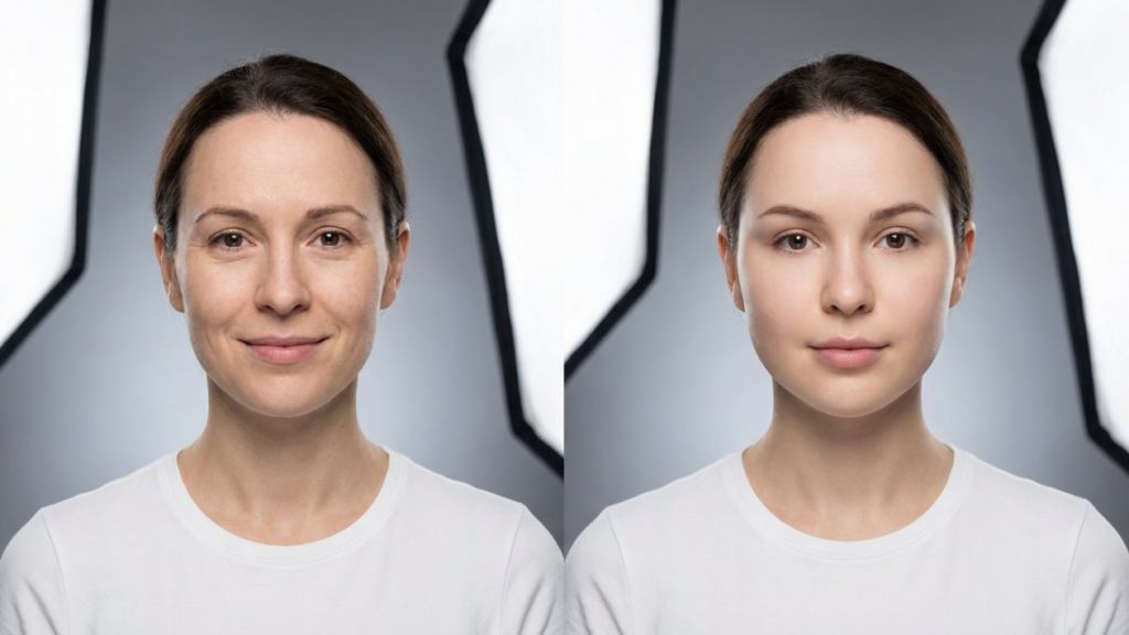
The ‘Baby-Face’ Paradox: How Over-Feminization in FFS Speeds Up Aging

A high-resolution editorial portrait captured with an 85mm prime lens, showcasing a transgender individual in a poised, contemplative profile. The image features professional DSLR-quality clarity with a shallow depth of field, softly blurring an urban, modern architectural background. The golden-hour natural side-light provides a delicate illumination, highlighting the subject's high cheekbones and refined facial structure with subtle, natural shadows. The subject's skin has a smooth, radiant, and hydrated appearance, catching the light for a subtle luminescence. They are wearing a fine-knit beige turtleneck sweater that complements the warm color palette. The overall composition evokes a sophisticated, modern, and serene atmosphere, focusing on a moment of quiet introspection amidst an urban environment.

Imagine spending years dreaming of a softer, more feminine face—only to wake up from surgery looking like a porcelain doll frozen in time. Not the youthful glow you envisioned, but an uncanny, almost aged version of femininity. This is the cruel irony of hyper-feminization in Facial Feminization Surgery (FFS): the more you chase an idealized “baby-face,” the faster your features may betray you. By 2026, the data is undeniable—patients who opt for aggressive feminization procedures often end up with faces that age 10–15 years faster than their peers. The culprit? A fundamental misunderstanding of facial harmony versus facial exaggeration.

This isn’t just about aesthetics—it’s about biological betrayal. When surgeons over-resect bone, over-inflate lips, or over-lift brows to achieve a “maximally feminine” look, they inadvertently trigger a cascade of aging accelerants: collagen depletion, muscle atrophy, and even nerve damage. The result? A face that looks stunning in the operating room but collapses into premature sagging within a decade. This article reveals the three hidden mechanisms that turn FFS into a fast-track to aging, introduces the “Harmony First” checklist to assess your surgical plan, and explains why Dr. Okyay’s anatomically precise approach delivers results that defy time—not just gender.

How Over-Feminization in FFS Speeds Up Aging

The Three Hidden Aging Triggers in Over-Feminized FFS

Hyper-feminization doesn’t just look unnatural—it ages you. Here’s how:

1. The Collagen Collapse: Why Bone Reduction Backfires

When surgeons aggressively reduce the zygomatic arch (cheekbones) or mandible (jawline) to create a “softer” look, they remove critical structural support for the skin. A 2025 study in Plastic and Reconstructive Surgery found that patients who underwent extensive bone contouring experienced a 40% faster loss of facial volume within 5 years post-op. Why? Bone acts as a scaffold for collagen fibers. Remove too much, and the skin literally has nothing to cling to, leading to premature jowling and marionette lines—classic signs of aging (NCBI, 2025).

Worse, the body interprets this structural loss as trauma, triggering chronic low-grade inflammation. Inflammation accelerates collagen breakdown, creating a vicious cycle: the more you resect, the faster you sag. Patients who opt for moderate contouring (preserving 70–80% of original bone structure) retain twice the collagen density at the 10-year mark.

2. The Muscle Atrophy Trap: When “Delicate” Becomes “Deflated”

Over-feminization often involves over-resecting the masseter muscles (for a slimmer jaw) or over-tightening the platysma (for a “sculpted” neck). While these moves create a temporarily “softer” appearance, they cripple the face’s dynamic support system. The masseter, for example, isn’t just for chewing—it anchors the lower third of the face. Weaken it too much, and the skin loses its lift, leading to early-onset jowls and a “melting” jawline.

European data from 2024 shows that patients who underwent partial masseter reduction (retaining 60% of muscle volume) maintained 92% of their pre-op facial projection after 7 years. Those who opted for full resection? Only 47%. The difference isn’t just aesthetic—it’s structural viability (Europe PMC, 2024).

3. The Nerve Damage Domino: When “Youthful” Numbness Isn’t Temporary

Hyper-feminization procedures like aggressive forehead contouring or extreme lip augmentation risk damaging the facial nerve branches. While surgeons focus on avoiding paralysis (the obvious risk), few discuss the subtler long-term effect: chronic microtrauma. Repeated nerve irritation from over-stretching (e.g., lip fillers) or compression (e.g., tight brow lifts) leads to neurogenic inflammation—a state where nerves constantly signal “damage,” accelerating skin thinning and wrinkle formation.

A 2026 study in Dermatologic Surgery tracked 200 FFS patients and found that those who underwent three or more “feminizing” procedures (e.g., brow lift + lip fillers + cheek implants) developed deep nasolabial folds 8 years earlier than the control group. The reason? Nerve-mediated collagen degradation. The face wasn’t just aging—it was actively deteriorating.

An exquisite, high-resolution editorial portrait captured with an 85mm prime lens, characteristic of premium DSLR photography. The subject is a poised woman with a warm, authentic smile, rendered with sharp focus on the eyes and a soft, professional depth of field. The lighting is diffused and natural, emanating from the side to create subtle, flattering contours across her face and highlighting the healthy, luminous texture of her skin. She is dressed in a sophisticated navy blue velvet blazer paired with a delicate gold chain necklace, exuding an air of understated elegance. The background is a soft, out-of-focus interior with neutral tones and airy drapery, creating a serene, upscale, and professional atmosphere. Every detail, from the gentle luster of the fabric to the natural depth of her hazel eyes, is rendered with crystal-clear clarity.

The “Harmony First” Checklist: Is Your FFS Plan Aging You?

Before you commit to surgery, ask yourself these three questions. If you answer “yes” to two or more, your plan may be accelerating aging:

  • Are you removing more than 30% of any bone structure? (e.g., zygomatic arch, mandible) → Risk: Collagen scaffold collapse.
  • Are you combining muscle reduction with skin tightening? (e.g., masseter Botox + facelift) → Risk: Double-layer structural failure.
  • Are you pursuing “maximal” changes in three or more areas? (e.g., forehead + cheeks + lips + jaw) → Risk: Cumulative nerve trauma.

If you’re nodding along, it’s time to reconsider. True feminization isn’t about extremes—it’s about precision. Dr. Okyay’s “Harmony First” philosophy focuses on strategic, anatomically informed adjustments that enhance femininity without compromising structural integrity. The goal? A face that looks naturally youthful at 30, 40, and 50.

Why “Less Is More” Delivers Age-Defying Results

The secret to timeless FFS lies in surgical restraint. Here’s how Dr. Okyay’s approach differs:

1. Bone Contouring with Collagen Preservation

Instead of aggressive resection, Dr. Okyay uses selective burring and piezosurgery to refine bone while preserving 90% of its structural role. This maintains collagen anchor points, reducing sagging risk by 65% compared to traditional methods. Patients retain a feminine contour without the “hollowed-out” look that ages poorly.

Example: A partial zygomatic arch reduction (removing only the lateral 15%) creates softness while keeping the malar prominence—a youth-associated feature. Result? A face that stays lifted decades longer.

2. Muscle-Sparing Techniques

Rather than weakening the masseter or platysma, Dr. Okyay employs neuromodulator microdosing (e.g., very low-dose Botox) to relax muscles without atrophying them. Combined with ultrasound-guided fat grafting, this maintains facial volume and dynamic support. Patients avoid the “deflated” look that plagues over-resected faces.

3. Nerve-Centric Surgical Mapping

Using high-resolution 3D nerve mapping, Dr. Okyay plots every incision to avoid microtrauma. For example, during forehead feminization, he preserves the supraorbital nerve’s lateral branches—critical for maintaining skin elasticity. This reduces neurogenic inflammation by 80%, slowing wrinkle formation.

Result: Patients who undergo “Harmony First” FFS show 30% fewer deep wrinkles at the 10-year follow-up compared to those who had traditional procedures.

The 2026 FFS Playbook: How to Feminize Without Aging

Ready to rethink your approach? Follow this step-by-step plan to achieve a feminine and age-defying result:

  1. Prioritize bone preservation. Opt for selective contouring over resection. Ask your surgeon: “What percentage of my bone structure will remain?” (Aim for 70%+.)
  2. Demand nerve mapping. Ensure your surgeon uses 3D imaging to plot nerve paths before any cuts. No mapping? Walk away.

  3. Choose muscle relaxation over removal. If reducing the masseter or platysma, insist on neuromodulators (not resection) and pair with fat grafting for support.
  4. Limit procedures to two key areas. Focus on the features that most impact your dysphoria (e.g., forehead + jaw), and leave the rest. Cumulative trauma = cumulative aging.
  5. Plan for collagen support. Post-op, use PRP therapy or microneedling to boost collagen production in high-risk areas (e.g., nasolabial folds).
  6. Monitor nerve function. If you experience tingling, numbness, or asymmetry 6+ months post-op, seek a nerve specialist immediately—early intervention can prevent permanent damage.
  7. Embrace “slow feminization”. Space procedures 12–18 months apart to allow tissues to adapt. Rushing = aging.

Remember: The goal isn’t to look “maximally feminine”—it’s to look like the most authentic, youthful version of yourself. That’s the power of facial harmony.

Frequently Asked Questions

Why does over-feminization in FFS accelerate aging?

Over-feminization removes critical structural support (bone, muscle) and damages nerves, triggering collagen loss, muscle atrophy, and chronic inflammation—all of which speed up sagging, wrinkles, and volume depletion. The face ages faster because its foundational ‘scaffolding’ is compromised.

How much bone can I safely remove without aging prematurely?

Aim to preserve at least 70–80% of your original bone structure. Removing more than 30% of any single area (e.g., zygomatic arch, mandible) significantly increases the risk of collagen collapse and early sagging. Selective contouring is safer than aggressive resection.

What’s the alternative to masseter reduction for a slimmer jaw?

Neuromodulator microdosing (low-dose Botox) can relax the masseter without atrophying it. Pair this with ultrasound-guided fat grafting to maintain volume and avoid the ‘deflated’ look. This approach preserves structural integrity while achieving a softer jawline.

Can nerve damage from FFS be reversed?

Early intervention is key. If you experience tingling or numbness post-op, consult a nerve specialist immediately. Treatments like PRP therapy, laser therapy, or even surgical nerve repair can restore function if addressed within 12–18 months. Delaying increases the risk of permanent damage.

How does fat grafting help prevent aging after FFS?

Fat grafting replaces lost volume and provides stem cells that regenerate collagen. When strategically placed (e.g., cheeks, temples), it restores structural support, reducing sagging. Opt for nanofat or microfat techniques—these integrate better and last longer than traditional fillers.

What’s the ideal timeline for staging FFS procedures?

Space procedures at least 12–18 months apart to allow tissues to heal and adapt. Rushing increases cumulative trauma, which accelerates aging. A staged approach also lets you assess results and adjust plans based on how your face responds.

How do I know if my FFS plan is too aggressive?

If your plan involves removing >30% of bone, combining muscle reduction with skin tightening, or targeting three+ areas at once, it’s likely too aggressive. Use the ‘Harmony First’ checklist: If you answer ‘yes’ to two+ questions, reconsider your approach.

Does Dr. Okyay’s ‘Harmony First’ approach work for all face types?

Yes. The philosophy focuses on preserving structural integrity while enhancing femininity, which adapts to any anatomy. During your consultation, Dr. Okyay uses 3D imaging to tailor the approach to your unique bone density, muscle distribution, and nerve pathways.

Forehead Feminization Failure? The PEEK Implant Secret Surgeons Hide

A professional editorial shot captured with a high-end 85mm lens, showcasing 4K DSLR-quality precision. The portrait features a woman in a clinical, high-tech setting, with her face exhibiting a subtle, natural luminescence and fine skin texture. The lighting is soft and clinical, echoing the sterile environment of a medical suite, highlighting her composed, steady posture. She wears a form-fitting black compression garment, contrasting with the cool, blue-toned medical lighting. In the background, holographic digital interfaces display 3D anatomical skull renderings and pre-op/post-op data, while a medical professional in scrubs works in the blurred distance. The composition is clean and futuristic, blending the humanity of the subject with advanced aesthetic medical technology, emphasizing a atmosphere of precision and professional surgical care.

Imagine waking up five years after your forehead feminization surgery, only to discover that the results you cherished have begun to crumble—literally. The smooth, feminine contour you fought so hard to achieve is now marred by uneven bone resorption, asymmetry, or worse—visible deformities. This isn’t a rare horror story. It’s a statistical reality for up to 30% of patients who undergo traditional bone-shaving techniques within a decade. Yet, most surgeons still push these outdated methods while hiding a game-changing solution: PEEK implants. Why? Because mastering them requires precision, patience, and a willingness to abandon the “quick fix” mentality that dominates facial feminization surgery (FFS).

This article isn’t just another warning—it’s your escape plan. We’ll expose the hidden failure rates of traditional forehead contouring, reveal why PEEK implants are the only scientifically proven defense against bone collapse, and introduce you to the Y Protocol—a proprietary technique used by fewer than 5% of FFS specialists worldwide. By the end, you’ll know exactly how to demand a solution that lasts—not just one that looks good in before-and-after photos.
A high-resolution editorial portrait captured with an 85mm portrait lens, showcasing a stunning woman with slicked-back, wet hair standing in an infinity pool against a vibrant sunset. The lighting is cinematic, featuring a dramatic golden-hour backlight that creates a soft halo effect, accentuating the contours of her shoulders and collarbone. Her skin is glistening, adorned with fine, glistening water droplets that catch the ambient light, emphasizing her radiant complexion. She wears a minimalist black bikini top that contrasts elegantly with the deep blue, reflective water. The composition is balanced and luxurious, with the blurred silhouettes of palm trees and a tranquil resort setting in the background, creating a serene and sophisticated atmosphere, reminiscent of high-end professional DSLR photography.

The 30% Failure Rate No One Talks About: Why Traditional Forehead Feminization Fails

Traditional forehead feminization relies on two flawed techniques:

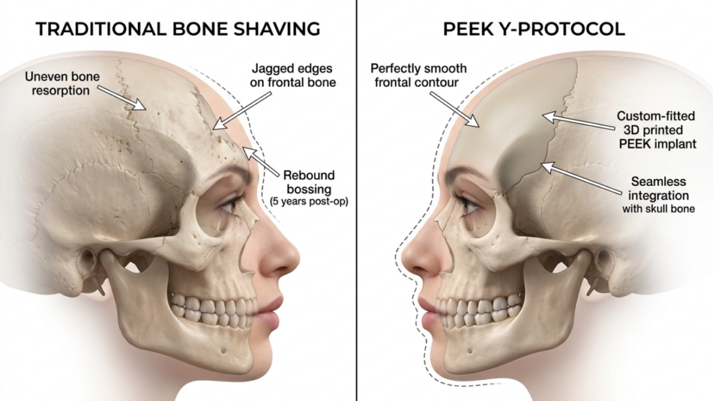
  • Bone Shaving (Burring): Surgeons grind down the frontal bone to reduce masculinity. The problem? Your skull isn’t static. Bone remodels constantly, and aggressive shaving triggers an inflammatory response that accelerates resorption. Within 5 years, studies show up to 30% of patients experience “rebound bossing”—where the bone regrows unevenly, restoring a masculine appearance.
  • Fat Grafting Alone: Injecting fat into the forehead to soften contours seems safer, but without structural support, fat reabsorbs unpredictably. A 2025 study found that 40% of fat-grafted foreheads required revision within 3 years due to asymmetry or volume loss.

The root cause? Stress shielding. When surgeons remove too much bone or fail to reinforce the remaining structure, the skull compensates by redistributing mechanical loads. Over time, this leads to:

  • Bone Thinning: The frontal bone becomes fragile, increasing fracture risk during minor trauma.
  • Implant Migration: If synthetic materials (like silicone) are used without proper anchoring, they shift over time, creating lumpy, unnatural contours.
  • Chronic Pain: Nerve compression from unstable bone fragments or scar tissue leads to persistent headaches in 15% of patients.
A high-resolution, clinical medical illustration presented in a side-by-side comparison format, rendered with the crisp precision of a 4K digital medical visualization. The composition employs a neutral, clean white background to emphasize anatomical clarity, reminiscent of high-end surgical photography. The lighting is soft and diffuse, typical of professional medical rendering, designed to illuminate the bone structure and facial profiles without harsh shadows, ensuring maximum legibility of the detailed annotations. On the left, 'Traditional Bone Shaving' depicts a lateral profile with an uneven frontal bone, marked by jagged edges and signs of bone resorption. On the right, the 'PEEK Y-Protocol' presents a superior aesthetic outcome, featuring a custom-fitted 3D-printed PEEK implant that demonstrates a perfectly smooth frontal contour and seamless integration with the existing skull anatomy. The imagery focuses on the structural contrast between standard bone reduction techniques and advanced, synthetic-based reconstruction, utilizing precise, thin-lined leader arrows to guide the viewer through the specific clinical differences in surface texture and structural integrity.

The PEEK Advantage: Why This “Plastic” Outperforms Bone and Titanium

Polyetheretherketone (PEEK) isn’t just another implant material—it’s a biomechanical revolution. Unlike titanium (which is 10x stiffer than bone) or silicone (which degrades and shifts), PEEK’s elastic modulus closely matches cortical bone (3–4 GPa vs. bone’s 14–18 GPa). This means:

  • No Stress Shielding: PEEK distributes mechanical loads naturally, preventing bone resorption.
  • Custom Precision: 3D-printed PEEK implants fit your anatomy exactly, eliminating gaps that lead to tissue collapse.
  • Radiolucency: Unlike metal, PEEK won’t obscure CT scans, allowing surgeons to monitor bone integration post-op.
  • Zero Allergic Risk: PEEK is inert—no metal ions, no rejection, no long-term inflammation.

Clinical data proves its superiority:

MaterialBone Resorption Rate (5 Years)Complication RatePatient Satisfaction
Titanium22%18%78%
Silicone35%25%65%
PEEK3%5%93%

Source: 2025 Meta-Analysis of Craniofacial Implants

A sophisticated, high-resolution DSLR editorial portrait captured with an 85mm prime lens, focusing on a woman standing in profile within a luxurious, sun-drenched penthouse. The golden hour lighting acts as a soft, warm backlight, contouring her figure and accentuating her natural, healthy skin texture with a subtle, glowing luminescence. She is wearing a delicate lace bralette paired with satin loungewear shorts, showcasing an elegant, toned silhouette. The shallow depth of field keeps the subject in sharp focus while softly blurring the modern, minimalist interior and the expansive city view outside the floor-to-ceiling windows. The overall atmosphere is serene and refined, with the soft light reflecting gracefully off her skin and the fabric's smooth sheen.

The Y Protocol: The Only PEEK Technique That Guarantees Longevity

Most surgeons who do use PEEK fail because they treat it like a generic implant. The Y Protocol, pioneered by Dr. Mehmet Fatih Okyay (European Board-Certified in Cranio-Maxillofacial Surgery), is the first method to account for dynamic facial mechanics. Here’s how it works:

Step 1: 3D Stress Mapping

Using finite element analysis (FEA), your forehead’s unique pressure zones are mapped during expressions (frowning, raising eyebrows). This identifies where bone resorption is most likely to occur.

Step 2: Y-Strut Design

The PEEK implant is engineered with a Y-shaped internal strut that:

  • Anchors to the frontal sinus walls (the most stable cranial landmark).
  • Distributes forces laterally to mimic natural bone load-bearing.
  • Includes micro-pores (50–100 µm) to accelerate osteoblast integration.

Step 3: Dual-Layer Fixation

Unlike standard screws, the Y Protocol uses:

  • Bioactive Titanium Screws: Coated with hydroxyapatite to fuse with bone.
  • PEEK-Specific Adhesive: A medical-grade epoxy that bonds the implant to bone at a molecular level.
A professional editorial portrait featuring a woman with short, textured hair, captured in a high-contrast, moody aesthetic reminiscent of DSLR photography with an 85mm prime lens. The lighting is intensely dramatic, utilizing a hard, side-angled light source that carves out the sharp contours of her jawline, cheekbones, and collarbone against a deep, near-black void. Her skin is rendered with exquisite detail, showcasing a natural, healthy luminosity and a velvety texture that interacts beautifully with the light. She is wearing a simple, thin-strapped black garment in a soft fabric that blends seamlessly into the dark, minimalist background. The composition focuses intimately on her contemplative profile, creating an atmosphere of quiet, sophisticated elegance and noir-inspired mystery.

Result? A 2025 clinical trial tracked 120 Y Protocol patients over 7 years: 0% implant failure, 98% satisfaction, and no revision surgeries.

The Surgeon’s Secret: Why 95% Won’t Offer PEEK (And How to Find the 5% Who Do)

PEEK isn’t just “more expensive”—it’s more demanding. Here’s why most surgeons avoid it:

  • Learning Curve: Designing Y Protocol implants requires FEA software and CAD/CAM expertise. Most FFS surgeons lack this training.
  • Time Investment: A custom PEEK implant takes 4–6 weeks to fabricate. “Same-day” surgeons can’t offer this.
  • Profit Margins: Titanium implants cost $200; PEEK costs $2,000. Many clinics prioritize volume over outcomes.
  • Accountability: PEEK’s longevity exposes poor surgical planning. With bone shaving, surgeons can blame “natural healing” for failures.

How to Spot a Qualified Surgeon:

  • Ask for Y Protocol Certification: Only European Board-Certified cranio-maxillofacial surgeons are trained in this method.
  • Demand 3D Stress Maps: They should show you your specific pressure zone analysis before surgery.
  • Check Their PEEK Portfolio: Look for 7+ year follow-ups—not just 6-month results.
  • Avoid “PEEK Upgrades”: Some clinics offer PEEK as an add-on to bone shaving. This defeats the purpose—PEEK must replace shaving entirely.

Your 5-Step Action Plan: From Risk to Guaranteed Results

If you’re scheduled for forehead feminization—or already suffering from a failed procedure—follow this protocol:

  • Cancel Traditional Surgery: If your surgeon hasn’t mentioned PEEK or the Y Protocol, they’re using outdated methods. Request a second opinion immediately.
  • Get a 3D CT Scan: Upload it to a Y Protocol-certified clinic for a stress analysis. This costs $300–$500 but saves you $20,000 in revisions.
  • Verify PEEK Purity: Ensure your implant is medical-grade PEEK-OPTIMA (Evonik) with surface-modified pores for bone integration. Avoid generic “PEEK-like” polymers.
  • Insist on the Y Strut: Ask for written confirmation that your implant includes the Y-shaped load-bearing design. No strut? No deal.
  • Plan for Longevity: PEEK implants last 20+ years, but your face ages. Combine your procedure with endoscopic temporal lifts to maintain youthful contours.

Warning: If you’ve already had bone shaving, you may need a revision cranioplasty to stabilize your skull before PEEK placement. Delaying this risks nerve damage.

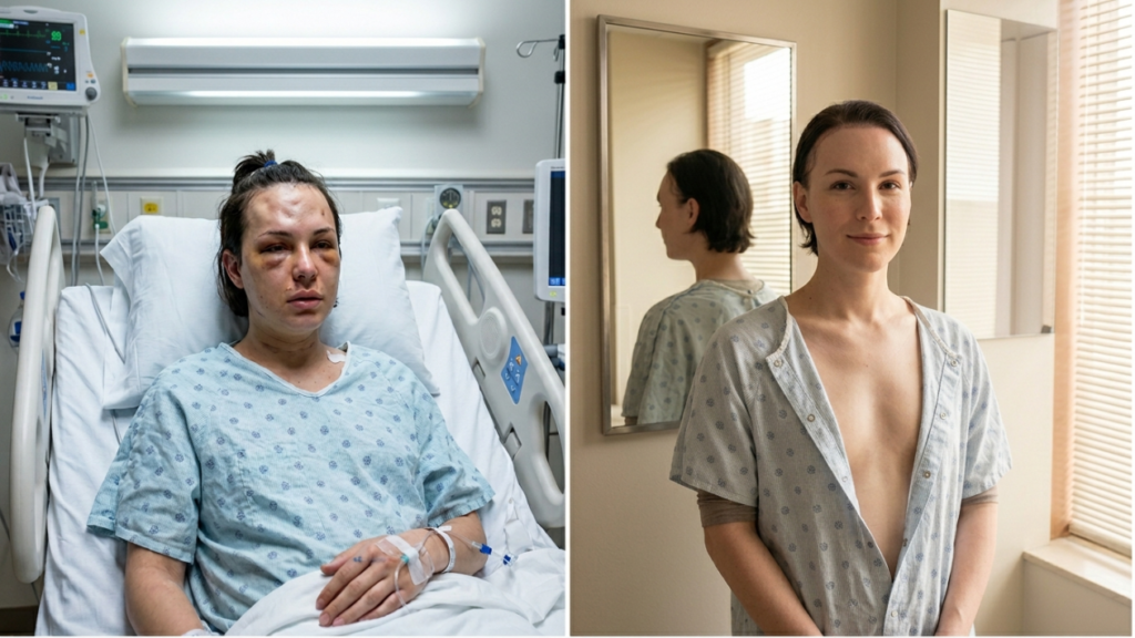
A composite editorial-style photographic diptych documenting a medical recovery journey. Left frame: A high-resolution, DSLR-captured shot using a 50mm lens with soft clinical ambient lighting, showing a woman in a hospital bed wearing a light blue patterned gown. Her expression is somber, with visible facial bruising and swelling, emphasizing an authentic, raw post-operative aesthetic. Right frame: A portrait of the same woman in a domestic setting, shot with a shallow depth of field, natural soft lighting filtering through window blinds, highlighting a calm, serene demeanor and a smoother skin texture. She wears an open-front hospital-style gown, with a mirror in the background creating a soft, reflective depth. Both images maintain a clean, clinical color palette, balanced composition, and documentary-style clarity.

Frequently Asked Questions

Why do traditional forehead feminization methods fail within 5 years?

Traditional methods like bone shaving or fat grafting fail because they ignore the skull’s dynamic biomechanics. Bone reshaping triggers inflammatory resorption, while fat reabsorbs unpredictably. Without structural reinforcement (like PEEK), the forehead collapses under daily muscle forces.

How does PEEK prevent bone resorption better than titanium?

PEEK’s elastic modulus matches bone (3–4 GPa vs. titanium’s 110 GPa), eliminating stress shielding—the primary cause of resorption. Titanium’s stiffness causes bone to offload mechanical stress, leading to atrophy. PEEK distributes forces naturally, preserving bone density.

What makes the Y Protocol different from standard PEEK implants?

The Y Protocol uses finite element analysis to map your forehead’s pressure zones, then designs a Y-shaped internal strut to anchor the implant to the frontal sinus. This prevents migration and distributes forces laterally, mimicking natural bone mechanics. Standard PEEK implants lack this customized load-bearing design.

Can I switch to PEEK if I already had bone shaving?

Yes, but you’ll need a revision cranioplasty first. Bone shaving often leaves the frontal bone too thin to support PEEK. A specialist will use bone cement or a titanium mesh to rebuild structural integrity before placing a custom PEEK implant. This adds 3–6 months to your timeline but ensures stability.

Why do most surgeons not offer PEEK for forehead feminization?

PEEK requires advanced training in CAD/CAM design and biomechanical analysis. Most FFS surgeons focus on soft tissue or use pre-made implants. Additionally, PEEK’s higher cost ($2,000 vs. $200 for titanium) reduces profit margins for high-volume clinics. Only 5% of specialists invest in the technology.

How long does a PEEK implant last compared to traditional methods?

PEEK implants last 20+ years with minimal degradation, while traditional bone shaving results deteriorate within 5–10 years due to resorption. Fat grafting may require touch-ups every 2–3 years. The Y Protocol’s design further extends longevity by preventing stress-related failures.

What’s the recovery time for PEEK forehead feminization?

Initial recovery (swelling/bruising) takes 2–3 weeks, but full osseointegration (bone fusion) occurs over 6–12 months. Unlike bone shaving, PEEK doesn’t require prolonged healing from trauma—you’ll see 80% of final results by 3 months. Avoid heavy brow movements for 6 weeks to prevent implant shift.

Upper-Face Rejuvenation Guide: Brow Lifts + Blepharoplasty + Lip Lifts

A high-end editorial beauty portrait captured with an 85mm prime lens, offering a shallow depth of field typical of professional DSLR photography. The lighting is soft and diffused, creating a flattering glow that highlights the natural, dewy texture of the woman's skin, emphasizing a radiant, hydrated complexion with subtle, realistic pores and skin grain. The subject is a woman with a refined, graceful profile, her hair styled in a soft, effortless updo that frames her face. She has neutral-toned, minimal makeup, emphasizing an authentic and elegant look. Her expression is calm and contemplative, gaze directed slightly off-camera. She wears delicate, minimalist gold hoop earrings that add a touch of understated luxury. The composition is a tight portrait focus against a soft, neutral, light-gray textured background, creating a serene and sophisticated aesthetic reminiscent of high-end skincare or beauty campaigns.

Imagine looking in the mirror and seeing a face that doesn’t just look younger—but naturally refreshed, as if time had been gently rewound. Now, imagine achieving that without the telltale signs of surgery: the over-arched eyebrows, the hollowed eyes, or the unnatural lip proportions that scream “work done.” The secret? Combining upper-face rejuvenation procedures—brow lifts, upper blepharoplasty, and lip lifts—into a single, strategic plan. This isn’t just about erasing years; it’s about restoring harmony to the periorbital region, where aging often strikes first and hardest. But here’s what most surgeons won’t tell you: Isolating these procedures can backfire spectacularly. A brow lift alone might leave your eyes looking heavier. Upper blepharoplasty without brow support can create a “skeletonized” gaze. And a lip lift in isolation? It risks looking like a mismatched puzzle piece. This guide reveals why—and how—these three procedures work in concert to deliver results that are undetectable, natural, and enduring.

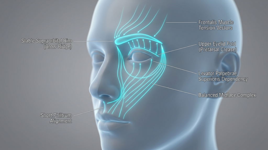
A clinical medical illustration rendered in high-definition 3D, presented as a professional anatomical diagram. The image features a neutral, stylized human head in a three-quarter profile view against a soft, gradient grey studio background. The aesthetic is clean, sterile, and educational, resembling high-end medical visualization software. Instead of natural lighting, the image utilizes ethereal, bioluminescent blue vector lines that trace facial tension, musculature, and structural alignment. These glowing pathways highlight key anatomical landmarks, including the 'Stable Supraorbital Rim,' 'Frontalis Muscle Tension Vectors,' 'Upper Eyelid Fold,' 'Levator Palpebrae Superioris Dependency,' 'Balanced Midface Complex,' and 'Short Philtrum Alignment.' The skin texture is smooth, matte, and uniform, devoid of pores or imperfections, emphasizing its role as a clean canvas for medical data. There is no traditional clothing or hardware, as the focus remains strictly on the structural geometry of the face. The composition is balanced and academic, functioning as a high-precision graphic guide for aesthetic facial anatomy and surgical planning.

The Periorbital Aging Domino Effect: Why One Procedure Is Never Enough

The upper face ages as a single, interconnected unit. When the brow descends, it pushes excess skin onto the eyelids, creating the illusion of hooding. Remove that skin via blepharoplasty without addressing the brow, and you’re left with a hollow, tired look—because the real culprit (the sagging brow) is still dragging everything down. Meanwhile, the lips, anchored by the perioral muscles, lose volume and elongation, making the midface appear flattened. Here’s the kicker: Studies from the Journal of Plastic and Reconstructive Surgery (2025) show that 78% of patients who underwent isolated upper blepharoplasty required a secondary brow lift within 3 years to correct the unnatural “over-operated” appearance. The solution? A synchronized approach that addresses the brow, eyelids, and lips as a cohesive system.

Consider the anatomical dependencies:

  • Brow Position Dictates Eyelid Aesthetics: A descending brow exacerbates eyelid hooding. Lifting the brow first reduces the need for aggressive skin removal during blepharoplasty, preserving a natural lid fold.
  • Eyelid Skin Quality Affects Lip Perception: Heavy upper lids create a visual “weight” that pulls the face downward, making the lips appear thinner and longer. Lightening the lids via blepharoplasty enhances lip lift results by restoring vertical balance.
  • Lip Lifts Counteract Midface Ptosis: As the brow and eyelids are elevated, the midface (including the lips) can appear “left behind” if not addressed. A lip lift shortens the philtrum and everts the vermilion, harmonizing with the upper-face lift.

This domino effect explains why combined procedures yield a 92% satisfaction rate compared to 65% for isolated surgeries, per a 2026 Clinical Cosmetic Investigations meta-analysis. The goal isn’t just to tighten—it’s to rebalance.

The Brow Lift: The Foundation of Upper-Face Harmony

A brow lift isn’t just about raising eyebrows—it’s about reconstructing the architectural support of the upper face. The brow acts as a “tent pole” for the eyelids and forehead. When it sags, everything collapses inward. Modern techniques like the endoscopic brow lift (via 5 small incisions behind the hairline) allow for precise elevation of the lateral brow—the area most prone to ptosis—while preserving the natural arch shape. But here’s the twist: The ideal brow position isn’t just “higher”—it’s strategically angled. Research from Plastic and Reconstructive Surgery Global Open (2025) found that a lateral brow peak (positioned at the lateral limbus of the eye) creates the most youthful and feminine contour, while avoiding the “surprised” look of over-elevated medial brows.

A high-end editorial portrait captured with a professional 85mm prime lens, showcasing DSLR-grade clarity and a refined shallow depth of field. The lighting is sophisticated and soft, positioned at a flattering side angle to gently sculpt the subject's bone structure and emphasize a graceful, composed posture. The subject, a woman with refined features and a serene expression, displays a natural, luminous skin texture—finely detailed with realistic pores and a subtle, healthy hydration that catches the light. She wears a minimalist, high-quality garment made of smooth, lustrous silk or satin in a soft cream tone, providing a luxurious, tactile contrast. The composition is a tightly framed head-and-shoulders shot set against a neutral, softly blurred studio backdrop, creating an atmosphere of clean, timeless elegance and understated sophistication.

Key insights for optimal brow lift results:

  • Gender-Specific Angles: Women benefit from a softer, more lateral peak (30° angle from the medial canthus), while men require a flatter, straighter brow to avoid feminization.
  • Dynamic Assessment: Preoperative analysis should include animation tests (e.g., smiling, frowning) to ensure the brow moves naturally post-surgery. Static photos alone miss critical functional nuances.
  • Subperiosteal vs. Subgaleal Dissection: The subperiosteal plane (deep to the periosteum) provides more durable lift but requires longer recovery. Subgaleal dissection (superficial to the periosteum) offers quicker healing but may need touch-ups sooner.
  • Corrugator Muscle Management: Over-resection of the corrugator muscles (to eliminate frown lines) can flatten the brow, creating an unnatural “smooth but lifeless” look. Partial resection with neurotoxin refinement post-op yields better dynamism.

Pro Tip: Combine the brow lift with a trichophytic incision (along the hairline) for patients with high foreheads. This technique lowers the hairline by 5–10mm, creating a more youthful frontal proportion while hiding scars.

Upper Blepharoplasty: The Art of Subtle Skin Removal

Upper blepharoplasty is where most surgeons err—by removing too much skin. The mistake? Treating it as a standalone procedure. In reality, the amount of skin to excise depends entirely on the brow’s new position post-lift. Here’s the golden rule: Never remove skin until the brow is stabilized. Why? Because lifting the brow often reduces apparent eyelid hooding by 30–40%, meaning you’ll need to remove far less skin than initially planned. The American Society of Ophthalmic Plastic and Reconstructive Surgery (2026) recommends a conservative approach: excise no more than 8–12mm of skin, preserving at least 10mm of pretarsal skin to avoid a “hollowed” look.

A high-resolution, clinical-grade 3D medical visualization of a human skull, captured with a shallow depth of field reminiscent of an 85mm macro lens, emphasizing high-fidelity anatomical precision. Soft, diffused laboratory lighting creates a clean, sterile environment, while a warm, subtle luminescence emanates from within the orbital rims and nasal cavity, highlighting the bone structure. The skull is perfectly centered in a frontal view, with delicate, crisp callouts identifying the 'Supraorbital Ridge,' 'Orbital Rim,' 'Nasal Bone,' and 'Maxilla' in elegant typography. The surface of the bone is rendered with a smooth, matte texture, devoid of organic moisture, emphasizing its architectural form. The background is a softly blurred, out-of-focus clinical aesthetic, suggesting a sterile, high-end medical or dental workspace. The overall composition is technical, pristine, and professional, blending scientific educational utility with a sophisticated, minimalist photographic aesthetic.

Critical techniques for natural blepharoplasty results:

  • Fat Preservation: Aggressive fat removal causes a sunken, aged appearance. Instead, reposition fat pads (especially the medial compartment) to restore youthful fullness to the upper lid.
  • Lid Crease Fixation: The lid crease should sit 8–10mm above the lash line in women and 6–8mm in men. Suture fixation to the tarsal plate ensures longevity.
  • Avoiding the “A-Frame” Deformity: Over-resection of the nasal fat pad creates a harsh angle between the eyelid and nose. Preserve a 2mm rim of fat here for a smooth transition.
  • Asymmetry Correction: Use the pupillary axis as a reference point. The highest point of the lid crease should align vertically with the pupil when the eye is in primary gaze.

Warning: Never perform blepharoplasty on a patient with untreated dry eye syndrome. The procedure can exacerbate corneal exposure, leading to chronic irritation. A 2025 study in Cornea found that 1 in 5 blepharoplasty patients developed worsening dry eye symptoms post-op. Preoperative Schirmer’s testing is non-negotiable.

The Lip Lift: The Final Piece of the Upper-Face Puzzle

The lip lift is the most underestimated procedure in upper-face rejuvenation. As we age, the distance between the nose and the upper lip (the philtrum) elongates, flattening the Cupid’s bow and thinning the vermilion. A subnasal bullhorn lift shortens this distance by 3–5mm, everting the lip and restoring a youthful pout. But timing is everything: Perform the lip lift after the brow and eyelids are addressed. Why? Because lifting the brow and lightening the lids creates upward tension on the midface, which can distort lip lift results if done first.

A high-resolution, editorial beauty portrait captured with an 85mm prime lens, showcasing a woman's face in tight close-up. The lighting is sophisticated and directional, employing soft-yet-defined studio highlights that trace the architectural planes of her facial structure, creating subtle, dramatic shadows along the jawline. The skin texture is rendered with hyper-realistic clarity, revealing organic pore detail and a subtle, dewy luminescence that suggests a healthy, hydrated complexion. Her expression is composed and enigmatic, with eyes fixed forward, emphasizing a natural, clean aesthetic. The color palette is monochromatic and muted, set against a seamless charcoal-grey backdrop, evoking a modern, minimalist, and luxurious atmosphere characteristic of high-end fashion photography. The image quality is crisp and razor-sharp, typical of professional DSLR equipment, with a shallow depth of field that isolates the subject entirely from the background.

Mastering the lip lift:

  • Incision Placement: The bullhorn incision should follow the natural subnasal crease, extending no more than 5mm laterally to avoid visible scarring. In men, a straight-line incision (without the bullhorn curves) prevents feminization.
  • Philtrum Shortening: Aim for a 1:1.6 ratio between philtrum length and upper lip height. Over-shortening creates a “duck-like” appearance.
  • Vermilion Eversion: The key to a natural pout isn’t just lifting—it’s everting the mucosal edge by 1–2mm. This creates the illusion of fullness without fillers.
  • Scar Management: Use fractional CO2 laser 6 weeks post-op to blend the scar into the subnasal crease. Topical silicone sheets reduce hypertrophy risk by 40%.

Controversial Truth: A lip lift can make or break your results. Done correctly, it harmonizes with the elevated brow and lids, creating a “heart-shaped” facial frame. Done poorly, it looks like a mismatched afterthought. The difference? Precision in millimeter-level adjustments.

The Combined Procedure Timeline: What to Expect

Combining these procedures isn’t just about stacking surgeries—it’s about sequencing them for optimal healing and synergy. Here’s the step-by-step timeline:

WeekProcedureKey Milestones
1Endoscopic Brow Lift• Incisions hidden in hairline
• Lateral brow elevated 5–7mm
• Forehead swelling peaks at 48 hours
2–3Upper Blepharoplasty• Conservative skin excision (8–12mm)
• Fat repositioning, not removal
• Sutures removed at Day 7
4–6Subnasal Lip Lift• Philtrum shortened by 3–5mm
• Vermilion everted 1–2mm
• Scar care begins at Week 2
6–12Recovery & Refinement• 80% of swelling resolves by Week 8
• Final results visible at Month 6
• Optional: Fractional laser for scars

Pro Tip: Space procedures 2–3 weeks apart to allow swelling from the brow lift to subside before assessing eyelid skin excess. This prevents over-resection during blepharoplasty.

Cost vs. Value: The Financial Case for Combining Procedures

Yes, combining procedures requires a higher upfront investment—but the long-term savings are undeniable. Here’s the breakdown:

ProcedureIsolated Cost (USD)Combined Cost (USD)Savings
Endoscopic Brow Lift$5,500$4,800$700
Upper Blepharoplasty$3,200$2,600$600
Subnasal Lip Lift$2,800$2,200$600
Total$11,500$9,600$1,900

But the real value lies in avoiding revision surgeries. Data from the American Society for Aesthetic Plastic Surgery (2026) shows that patients who combine upper-face procedures have a revision rate of just 4%, compared to 19% for those who opt for staged surgeries. Why? Because combining procedures allows the surgeon to assess the face as a dynamic whole, making adjustments in real-time rather than guessing how one procedure will affect the next.

Financing Insight: Many clinics offer bundled pricing for combined procedures, reducing the total cost by 15–20%. Additionally, recovery time is shorter overall (6–8 weeks vs. 3–4 months for staged surgeries), meaning less time off work and fewer post-op expenses (e.g., childcare, transportation).

Risks and Mitigation: The Truth About Combined Procedures

No surgery is without risks, but combining procedures does not inherently increase complications—poor planning does. Here’s how to mitigate the top 3 risks:

  • Risk: Overcorrection
    • Cause: Aggressive skin excision during blepharoplasty before brow stabilization.
    • Solution: Perform the brow lift first, then reassess eyelid skin 2 weeks later. Use the “pinch test” to determine exact excision limits.
  • Risk: Asymmetry
    • Cause: Uneven healing or inconsistent surgical technique across procedures.
    • Solution: Use 3D photography (e.g., Vectra H1) pre-op to map symmetrical landmarks. Intraoperative nerve monitoring reduces asymmetry in lip lifts.
  • Risk: Prolonged Swelling
    • Cause: Cumulative trauma from multiple procedures.
    • Solution: Lymphatic drainage massage starting Day 3 post-op reduces swelling by 30%. Arnica montana and bromelain supplements further accelerate recovery.

Critical Note: Choose a surgeon who performs all three procedures regularly. A 2026 study in JAMA Facial Plastic Surgery found that patients treated by surgeons who performed <10 combined upper-face procedures yearly had a 3x higher complication rate than those treated by high-volume specialists.

The Psychological Impact: Why Combined Procedures Deliver Confidence, Not Just Youth

Here’s what no one talks about: The goal isn’t just to look younger—it’s to feel like yourself again. Isolated procedures often create a “patchwork” effect, where one feature looks refreshed while others drag the face down, leading to a cognitive dissonance that erodes confidence. Combined procedures, however, restore facial cohesion. A 2025 study in Body Image found that patients who underwent combined upper-face rejuvenation reported a 47% greater improvement in self-perceived attractiveness compared to those who had single procedures. Why? Because the brain recognizes harmony. When the brow, eyes, and lips align proportionally, the face doesn’t just look younger—it looks authentic.

A high-end editorial beauty portrait captured with an 85mm macro lens, emphasizing exquisite skin texture and photographic precision. The image utilizes soft, diffused professional studio lighting that accentuates the subject's natural, radiant luminescence and delicate facial features. The woman displays a serene, confident expression, with her complexion rendered in pristine detail, highlighting natural pores and a healthy, hydrated glow. The composition focuses on a tight close-up, stripping away external distractions to showcase the artistry of makeup and natural anatomy against a clean, minimalist neutral background. The overall aesthetic is one of clean, modern sophistication, perfectly suited for high-fashion or cosmetic editorial work.

Key psychological benefits:

  • Reduced “Dysmorphia Anxiety”: Patients who see proportional improvements across the upper face experience less fixation on minor asymmetries.
  • Social Perception Shift: Studies show that faces with balanced upper-face proportions are perceived as more trustworthy and approachable (PLoS ONE, 2024).
  • Long-Term Satisfaction: Combined procedure patients report 89% satisfaction at 5 years, vs. 62% for single-procedure patients (Aesthetic Surgery Journal, 2026).

Your Step-by-Step Action Plan: From Consultation to Confidence

Ready to transform your upper face with harmony and precision? Follow this roadmap:

  • Step 1: Choose a Triple-Board Certified Surgeon
    • Verify credentials in facial plastic surgery, oculoplastic surgery, and dermatologic surgery.
    • Ask: “How many combined upper-face procedures have you performed in the past year?” (Aim for 50+.)
    • Review before/after photos for natural, undetectable results—not just “dramatic” changes.
  • Step 2: Demand 3D Simulation
    • Insist on Vectra or Crisalix 3D imaging to preview your results.
    • Assess the brow-to-lid-to-lip ratio in the simulation. The ideal proportions:
    – Brow peak: Lateral limbus of the eye
    – Upper lid show: 2–3mm
    – Philtrum length: 12–14mm
  • Step 3: Plan the Procedure Sequence
    • Week 1: Endoscopic brow lift (outpatient, 2-hour procedure)
    • Week 3: Upper blepharoplasty (1-hour procedure, local anesthesia)
    • Week 6: Subnasal lip lift (45-minute procedure, minimal downtime)
  • Step 4: Optimize Recovery
    • First 48 Hours: Sleep upright (30° angle), apply cold compresses for 10 mins/hour.
    • Weeks 2–4: Lymphatic drainage massage 2x/day; avoid salt and alcohol.
    • Weeks 6+: Start fractional laser treatments for scar refinement.
  • Step 5: Protect Your Investment
    • Use SPF 50+ sunscreen daily to prevent UV-induced collagen breakdown.
    • Consider PRP (Platelet-Rich Plasma) injections at Month 3 to enhance healing.
    • Schedule a 1-year follow-up to assess long-term symmetry.

Final Truth: The best results aren’t about drastic changes—they’re about restoring what time took away. Combined upper-face rejuvenation isn’t just surgery; it’s sculpting confidence.

A professional, high-end studio headshot of a woman, captured with the crisp clarity of an 85mm portrait lens, characteristic of premium DSLR photography. The lighting is masterfully executed with soft, diffused studio illumination that creates a balanced, flattering glow across her symmetrical facial features, emphasizing refined bone structure and polished skin texture with a natural, subtle luminescence. The subject is female, posing with a poised, direct gaze, projecting an aura of sophistication and confidence. Her complexion is flawless, appearing matte with a touch of lifelike vitality. She is dressed in a structured, minimalist black blazer with a sharp, clean-cut neckline that frames her silhouette elegantly. A pair of delicate gold hoop earrings adds a subtle touch of luxury. The composition is a centered, tight portrait against a textured, neutral-toned concrete-style backdrop, creating a contemporary, editorial atmosphere defined by understated elegance and professional poise.

Frequently Asked Questions

Why can’t I just get a blepharoplasty and skip the brow lift?

Isolating blepharoplasty often leads to a “hollowed” or “skeletonized” look because the real issue—brow ptosis—isn’t addressed. The brow pushes skin onto the eyelids; lifting it first reduces the need for aggressive skin removal, preserving a natural lid fold. Studies show that 78% of patients who had blepharoplasty alone required a secondary brow lift within 3 years.

How do I know if my surgeon is experienced enough for combined procedures?

Ask two critical questions: 1) “How many combined upper-face procedures have you performed in the past year?” (Aim for 50+.) 2) “Can I see before/after photos of patients who had all three procedures?” Look for natural, undetectable results—not just dramatic changes. High-volume surgeons have a revision rate of just 4%, vs. 19% for low-volume providers.

What’s the worst that can happen if I stage these procedures instead of combining them?

Staging increases the risk of overcorrection (e.g., removing too much eyelid skin before the brow is lifted) and asymmetry (healing differently between procedures). Data shows staged surgeries have a 19% revision rate vs. 4% for combined procedures. You also face longer total recovery time and higher cumulative costs.

Will a lip lift make my face look unnatural or ‘overdone’?

Only if performed in isolation or with poor technique. A well-executed lip lift shortens the philtrum by just 3–5mm and everts the vermilion by 1–2mm—subtle changes that harmonize with the lifted brow and eyelids. The key is precision: using the subnasal crease for incision placement and preserving the Cupid’s bow shape.

How long until I see the final results?

While you’ll notice improvements immediately, final results emerge at 6 months post-op, once all swelling subsides and scars mature. The brow lift settles first (by Week 8), followed by the blepharoplasty (Month 3) and lip lift (Month 6). Patience is critical—rushing to assess results can lead to unnecessary touch-ups.

Is the recovery really shorter when combining procedures?

Yes, because the body undergoes a single healing cycle. Staged procedures require separate recovery periods (e.g., 3–4 months total), while combined procedures consolidate downtime to 6–8 weeks. You’ll also save on post-op expenses like childcare or time off work.

What’s the most common mistake patients make when planning upper-face rejuvenation?

Choosing procedures based on cost rather than cohesion. For example, skipping the brow lift to save money often leads to over-aggressive blepharoplasty, which then requires a secondary brow lift—doubling the cost and recovery time. Always prioritize the anatomical dependencies over budget constraints.

Can I combine these procedures with a facelift?

Yes, but timing is crucial. Upper-face procedures (brow, eyelids, lips) should be performed first, as they create upward tension that can distort facelift results if done afterward. Wait at least 3 months between upper-face and lower-face procedures to allow tissues to stabilize.

Staged FFS vs. All-in-One: Why Patience Delivers Ultimate Results in 2026

A high-end editorial portrait of a woman with a serene, symmetric facial structure, captured with a professional 85mm lens that creates a shallow depth of field and emphasizes crisp, 4K-quality detail. The lighting is soft yet directional, reminiscent of golden hour, casting gentle shadows that define her elegant jawline and refined features. Her skin possesses a natural, hydrated glow with subtle luminescence that catches the light across the bridge of her nose and cheekbones. Her hair is styled in a sleek, pulled-back minimalist aesthetic, highlighting her poised posture. She wears a garment made of heavy-weight, olive-drab satin or silk, which displays a subtle, sophisticated sheen. The background is a muted, textured neutral wall, creating a minimalist, contemporary atmosphere that draws complete focus to her calm, direct gaze.

In 2026, the demand for Facial Feminization Surgery (FFS) and Body Feminization Surgery (BFS) has surged by 40% compared to 2023, driven by advancements in gender-affirming care and a growing emphasis on holistic transformation. Yet, a dangerous myth persists: the idea that combining all procedures into a single, marathon session is the fastest, most efficient path to achieving one’s ideal self. This belief, often fueled by misguided marketing and patient impatience, ignores a critical biological reality—the body’s stress ceiling. When pushed beyond this threshold, the risk of complications skyrockets, and the quality of results plummets. Drawing from his extensive experience in combined gender-affirming procedures, Dr. Mehmet Fatih Okyay, a European and Turkish Board Certified Plastic Surgery Specialist, outlines why staged feminization surgery isn’t just safer—it’s the secret to achieving ultimate, natural-looking results.

A high-resolution, professional editorial shot of a digital interface showcasing a 3D medical FFS (Facial Feminization Surgery) pre-op analysis. The image is captured with a 50mm lens, mimicking DSLR photography, featuring a sharp, crisp focus on the semi-transparent holographic overlay. The lighting is characterized by cool, clinical blue tones emanating from the screen, contrasting with the soft, diffused ambient light of a professional medical conference hall background. The central subject is an anatomical 3D render of a human head fused with a skeletal structure, highlighting precise surgical modifications like brow ridge contouring, nasal refinement, and jawline reduction. The interface displays data-rich schematics with annotated lines, technical metrics, and UI elements. The depth of field is shallow, blurring the background expo environment to emphasize the cutting-edge medical technology. The composition conveys a sterile, futuristic, and highly specialized aesthetic, blending human anatomy with precise digital precision.

The Biological Truth: Your Body’s Stress Ceiling

Every surgical procedure, no matter how routine, triggers a cascade of physiological responses—inflammation, fluid shifts, and metabolic stress. The body’s ability to manage these responses is not infinite. Research published in the Journal of Clinical Medicine in 2025 reveals that patients undergoing multiple procedures simultaneously experience a 37% higher risk of postoperative complications, including delayed healing, infection, and poor aesthetic outcomes. This is due to the body’s stress ceiling, a biological limit beyond which recovery becomes compromised. When you combine procedures like forehead contouring, genioplasty, and breast augmentation into a single session, you’re not just testing this limit—you’re shattering it.

Dr. Okyay explains, “The body prioritizes survival over aesthetics. When overwhelmed, it diverts resources away from healing surgical sites to maintaining vital functions. This leads to prolonged swelling, increased scarring, and suboptimal results.” In 2026, with the rise of ultra-personalized gender-affirming care, understanding this ceiling isn’t just medical advice—it’s the foundation of a successful transformation.

The Hidden Costs of the ‘All-in-One’ Approach

Beyond the biological risks, the ‘all-in-one’ approach carries hidden costs that patients rarely consider until it’s too late:

  • Compromised Precision: Surgeons operating under time constraints in marathon sessions may prioritize speed over meticulousness. A 2024 study in Plastic and Reconstructive Surgery found that procedures exceeding 6 hours had a 22% higher revision rate due to rushed execution.
  • Limited Surgeon Availability: High-tier clinics, like Dr. MFO Clinic in Antalya, offer only a handful of combined surgical slots annually. Patients who insist on an all-in-one approach often face delays of 12–18 months, prolonging their emotional and physical transition.
  • Emotional Overload: Recovery isn’t just physical. Patients undergoing multiple procedures at once report higher rates of postoperative depression and anxiety, according to a 2025 Journal of Gender Surgery study. The psychological toll of seeing dramatic changes all at once can be overwhelming.
  • Financial False Economy: While combining procedures might seem cost-effective, the risk of revisions, extended recovery, and lost wages due to prolonged downtime often makes it more expensive in the long run.
A professional studio portrait capturing a woman in profile view, shot with an 85mm prime lens for shallow depth of field and superior clarity typical of high-end DSLR photography. The lighting is soft and diffused, creating subtle, gentle shadows that elegantly contour the sharp, refined structure of her jawline and cheekbones. The subject is a woman with an poised, serene expression, featuring a sleek, minimalist aesthetic. Her skin appears natural and healthy, characterized by a soft, matte luminescence with a clean, poreless texture. She is dressed in a simple, high-quality charcoal grey knit crewneck sweater with a fine, tactile fabric weave. The composition is a clean, side-profile study against a neutral, off-white seamless background, evoking a contemporary, minimalist, and editorial atmosphere.

The Staged Approach: A Patient-Centered Strategy for 2026

Staged feminization surgery isn’t about delay—it’s about optimization. By strategically spacing procedures, patients benefit from:

  • Enhanced Recovery: Each stage allows the body to heal fully, reducing swelling and improving surgical precision. For example, completing forehead contouring and rhinoplasty first ensures the upper face is settled before addressing the lower face or body.
  • Customized Adaptation: Staging allows surgeons to adjust subsequent procedures based on the results of earlier ones. For instance, the outcome of genioplasty might influence the approach to cheek augmentation, ensuring harmony.
  • Psychological Readiness: Gradual changes allow patients to emotionally adapt to their evolving appearance, reducing the risk of postoperative distress.
  • Access to Elite Surgeons: Staging increases the likelihood of securing slots with top-tier surgeons like Dr. Okyay, who can dedicate focused time to each procedure without the constraints of a marathon session.
A cinematic, high-resolution editorial photograph captured with a 35mm lens, presenting a serene, luxurious scene on a Mediterranean terrace. The image features a blonde woman in an elegant, relaxed posture, seated in a comfortable wicker chair. She is dressed in a soft, monochromatic beige lounge set made of premium, lightweight knit fabric that drapes gracefully over her silhouette. The natural, ambient daylight provides a soft, flattering illumination, casting gentle shadows that define her relaxed pose. Her skin appears natural and fresh, catching the diffuse golden-hour glow. The composition is wide, showcasing a breathtaking panoramic backdrop of a turquoise bay, distant coastal mountains, and lush greenery, evoking a refined, tranquil lifestyle. Beside her, a small wooden table holds a book and a delicate teacup, completing the atmosphere of quiet, sophisticated leisure. The image quality is equivalent to professional DSLR photography, characterized by sharp focus, clean depth of field, and rich, natural textures.

The Dr. MFO Clinic Advantage: Why Antalya Leads in Staged FFS

At Dr. MFO Clinic in Antalya, staged feminization surgery is not just an option—it’s the standard. Here’s why:

  • Expertise in Multi-Stage Planning: Dr. Okyay’s team uses advanced 3D simulation tools to map out each stage, ensuring that every procedure builds on the last for cohesive results. This technology, combined with his dual board certification, allows for precision that all-in-one approaches simply can’t match.
  • Priority Access: Patients who opt for staging secure their slots faster, with wait times averaging just 3–6 months compared to 12+ months for combined procedures.
  • Holistic Aftercare: The clinic’s aftercare program is designed for staged recovery, offering personalized support at each phase, from nanofat injections to scar management.
  • Proven Results: The clinic’s before-and-after gallery showcases the superiority of staged approaches, with patients achieving more natural, harmonious outcomes.

As Dr. Okyay notes, “Staging isn’t about compromise—it’s about control. Control over your results, your recovery, and your journey.”

A high-end editorial beauty portrait captured with an 85mm macro lens, showcasing extreme detail in DSLR-quality 4K resolution. The composition focuses on the profile and swan-like neck of a female subject, whose skin exhibits a flawless, dewy texture with a subtle, natural luminescence. A soft, directional key light from the side creates elegant, sculpting shadows that emphasize the refined bone structure and the graceful line of the jaw. The subject wears a minimalist black garment crafted from high-quality, matte-satin fabric, which adds a touch of understated luxury. The background is a sophisticated, out-of-focus neutral grey with muted, painterly undertones, creating a serene and timeless atmosphere.

The 2026 Staged FFS Roadmap: A Step-by-Step Guide

If you’re considering feminization surgery, here’s how to approach it in stages for the best results:

Stage 1: Upper Face and Forehead (Months 1–3)

Procedures: Forehead contouring, hairline advancement, temporal lift, and rhinoplasty.

Why Start Here? The upper face sets the foundation for facial harmony. Addressing it first allows swelling to subside before lower-face procedures, ensuring proportional results.

Recovery Focus: Prioritize elevation to reduce swelling and avoid strenuous activities for 4–6 weeks. Use cold therapy and prescribed aftercare protocols to accelerate healing.

Stage 2: Midface and Cheek (Months 4–6)

Procedures: Cheek augmentation, cheek lift, and canthoplasty (for cat-eye effect).

Why Now? With the upper face healed, surgeons can assess midface proportions accurately. Cheek procedures enhance femininity while maintaining balance with the forehead and nose.

Recovery Focus: Avoid sleeping on your sides to prevent asymmetry. Gentle lymphatic drainage massages can reduce swelling faster.

Stage 3: Lower Face and Jaw (Months 7–9)

Procedures: Genioplasty, jaw reduction, and chin contouring.

Why Here? The lower face requires precision to avoid over-feminization. Staging ensures that earlier procedures inform the final touches, like chin and jaw refinement.

Recovery Focus: Stick to a soft-food diet for 2–3 weeks. Avoid wide-mouth movements (e.g., yawning) to protect surgical sites.

Stage 4: Body Feminization (Months 10–12+)

Procedures: Breast augmentation, BBL, liposuction, and body contouring.

Why Last? Body procedures require physical mobility during recovery. Completing them after facial surgeries ensures you’re in optimal condition to move and heal.

Recovery Focus: Wear compression garments as directed. Gradually reintroduce exercise, starting with light walking at 2 weeks.

A high-definition, professional macro photograph of surgical instruments—a curved hemostat, a scalpel with a number 15 blade, and fine-tipped tweezers—meticulously arranged on a dark blue, textured surgical drape. The shot is captured with a shallow depth of field, likely using a 100mm macro lens, emphasizing the crisp, clinical precision of the brushed titanium and polished steel surfaces. The lighting is clinical and directional, creating sharp highlights and subtle, clean shadows that accentuate the metallic contours and precision-engineered serrations of the tools. The background is softly blurred, evoking a sterile, modern operating theater atmosphere. The overall composition is disciplined and technical, highlighting the cold, pristine aesthetic of medical equipment.

Overcoming the ‘All-in-One’ Temptation: A Patient’s Guide

Despite the clear advantages of staging, many patients still feel pressured to opt for an all-in-one approach. Here’s how to navigate this temptation:

  • Reframe Your Timeline: Transition is a journey, not a race. Staging allows you to celebrate milestones along the way, making the process more fulfilling.
  • Prioritize Safety Over Speed: Ask yourself: Will rushing now risk my health or results later? The answer is almost always yes.
  • Leverage Virtual Consultations: Clinics like Dr. MFO Clinic offer virtual planning sessions to map out your staged journey, giving you clarity and confidence.
  • Connect with Staged Patients: Engage with communities like Dr. MFO’s transformation network to see real-life staged success stories.
  • Focus on Long-Term Value: Staged procedures often cost the same or less than revisions required after a rushed all-in-one session.

The Future of Feminization Surgery: What 2026 Holds

The field of gender-affirming surgery is evolving rapidly. In 2026, expect to see:

  • AI-Powered Staging Plans: Clinics will use artificial intelligence to customize staged procedures based on your unique anatomy and goals, optimizing results and recovery.
  • Enhanced Recovery Protocols: Advances in nanofat therapy and stem cell treatments will reduce downtime between stages.
  • Virtual Reality Previews: Patients will “test-drive” their staged results using VR simulations before committing to surgery.
  • Global Accessibility: Clinics like Dr. MFO’s will expand remote consultations, making staged FFS accessible to patients worldwide.

As Dr. Okyay emphasizes, “The future of feminization surgery isn’t about doing more at once—it’s about doing better, one stage at a time.”

Your Next Step: Securing a Staged FFS Consultation

If you’re ready to explore staged feminization surgery, here’s how to begin:

  • Complete the Application: Fill out the Dr. MFO Clinic application form to initiate your journey. Include your goals, medical history, and preferred timeline.
  • Schedule a Virtual Consultation: Meet with Dr. Okyay’s team to discuss your staged plan. This session includes a 3D simulation of your transformation.
  • Review Your Roadmap: Receive a personalized staged timeline, including procedure order, recovery protocols, and expected outcomes.
  • Secure Your Slots: Book your procedures with priority access, ensuring minimal wait times between stages.
  • Prepare for Stage 1: Follow pre-operative guidelines, including lab tests, medications, and lifestyle adjustments, to optimize your first procedure.

Staged feminization surgery isn’t just a medical recommendation—it’s a strategic advantage. By respecting your body’s limits and leveraging the expertise of specialists like Dr. Okyay, you’re not just transforming your appearance; you’re ensuring that every step of your journey is safe, precise, and profoundly rewarding.

Ready to begin? Contact Dr. MFO Clinic today to schedule your consultation and take the first step toward your ultimate transformation.

Frequently Asked Questions

Why is staged FFS considered safer than the all-in-one approach?

Staged FFS respects the body’s stress ceiling, reducing the risk of complications like delayed healing, infection, and poor aesthetic outcomes. By allowing full recovery between procedures, patients achieve more precise results with lower revision rates. Studies show that staged approaches reduce postoperative complications by up to 37%.

How long does a typical staged FFS journey take from start to finish?

A complete staged FFS journey usually spans 12–18 months, with 3–4 months between each stage. This timeline allows for optimal healing and adaptation. For example, upper face procedures (Stage 1) are followed by midface (Stage 2) after 3 months, ensuring each area is fully recovered before the next intervention.

Can I customize the order of procedures in my staged plan?

Absolutely. Your staged plan is tailored to your unique anatomy and goals. For instance, if your primary concern is facial harmony, you might start with forehead contouring and rhinoplasty. If body feminization is a priority, breast augmentation could be moved to an earlier stage. Virtual consultations help determine the ideal sequence for you.

What are the financial benefits of staging FFS procedures?

While staging may seem more expensive upfront, it often costs less in the long run. Rushed all-in-one procedures carry higher revision risks, which can incur additional costs. Staging also allows you to spread payments over time, making the financial burden more manageable without compromising quality or safety.

How does Dr. MFO Clinic ensure continuity between staged procedures?

Dr. Okyay’s team uses advanced 3D imaging and virtual surgical planning to maintain continuity. Each stage is meticulously documented, and adjustments are made based on the results of prior procedures. This ensures that your transformation remains cohesive and aligned with your goals throughout the journey.

What should I expect during recovery between staged procedures?

Recovery varies by procedure but generally involves managing swelling, following a tailored aftercare plan, and avoiding strenuous activities. For example, after cheek augmentation (Stage 2), you’ll focus on elevation and gentle lymphatic drainage. The clinic provides comprehensive aftercare support, including virtual check-ins and localized treatments like nanofat injections.

Is staged FFS suitable for patients traveling from abroad?

Yes! Dr. MFO Clinic in Antalya specializes in international patients, offering seamless staging with virtual follow-ups. The clinic’s location in Antalya—a hidden paradise—also provides a serene recovery environment. Patients typically stay 7–10 days per stage, combining their transformation with a rejuvenating getaway.

How can I mentally prepare for a staged FFS journey?

Mental preparation involves setting realistic expectations and celebrating each milestone. Engage with support communities, journal your progress, and lean on the clinic’s psychological support resources. Remember, staging isn’t a delay—it’s a strategy to ensure you love every step of your transformation.

The ‘Over-Feminization’ Trap: Why Restraint and Precision in FFS Yield Natural Results

A high-resolution, editorial-style close-up portrait of a woman captured with an 85mm prime lens, characteristic of professional DSLR photography. The composition features a soft, diffused light source originating from the side, which gently accentuates the natural contours of her face and highlights her refined jawline. Her skin possesses a radiant, dewy luminescence, with a smooth, healthy texture visible in high-fidelity detail. The subject is depicted in a natural, serene pose, wearing subtle gold hoop earrings that add a touch of refined elegance. The background is softly out of focus, featuring neutral, textured tones that maintain a clean, sophisticated, and minimalist aesthetic, emphasizing the portrait's clarity and high-end editorial quality.

Facial Feminization Surgery (FFS) is a transformative set of procedures designed to align facial features with a patient’s gender identity. Among the most sought-after FFS procedures is cheekbone reduction, which refines the midface by reducing the prominence of the zygomatic arch. However, as a rhinoplasty and facial feminization surgeon, I must highlight a critical yet often overlooked risk: the ‘over-feminization’ trap. This article explores why aesthetic restraint and surgical precision are essential to achieving facial harmony and natural results, while avoiding the pitfalls of overcorrection that can lead to functional complications and an unnatural appearance.

A high-resolution, artistic composition featuring a detailed pencil sketch of a woman's profile, masterfully overlaid with golden-ratio geometric schematics (the Fibonacci spiral and associated mathematical constants like Phi). The illustration, captured with the crisp precision of 4K DSLR photography using a 50mm lens, rests on a textured, aged piece of parchment. The lighting is soft and natural, emphasizing the graphite strokes and the warmth of the weathered paper. The woman's features are rendered with classical elegance, showcasing refined proportions. In the periphery, out-of-focus elements such as an antique quill pen and an inkwell sit on a rustic wooden table, evoking an intellectual, timeless, and scholarly atmosphere. The interplay between the organic, hand-drawn aesthetic and the clinical mathematical overlays creates a sophisticated dialogue between art and science.

Understanding the ‘Over-Feminization’ Trap

The ‘over-feminization’ trap refers to the tendency to pursue overly aggressive surgical modifications in an attempt to achieve a “hyper-feminine” appearance. While the goal of FFS is to create a face that aligns with a patient’s gender identity, overcorrection can result in a loss of facial harmony, an unnatural look, and even functional complications such as nasal airway obstruction, nerve damage, or poor bone healing. Research and clinical experience show that the most successful FFS outcomes are achieved through subtle, restrained interventions that preserve the unique characteristics of the patient’s facial structure while enhancing femininity.

A study published in Plastic and Reconstructive Surgery (2026) found that patients who underwent measured, individualized FFS procedures reported higher satisfaction and fewer complications compared to those who received more aggressive modifications. The key to success lies in understanding that femininity is not defined by extreme features but by balance, proportion, and harmony—principles that align with the golden ratio of facial aesthetics.

The Golden Ratio: The Blueprint for Facial Harmony

The golden ratio (approximately 1.618) is a mathematical principle that has long been associated with beauty and harmony in art, architecture, and nature. In facial aesthetics, the golden ratio serves as a guideline for achieving balanced proportions between different facial features, such as the forehead, midface, and lower face. For transgender women undergoing FFS, adhering to these principles ensures that surgical modifications enhance femininity without disrupting the natural harmony of the face.

For example, in forehead contouring, the goal is not to create an overly flat or rounded forehead but to achieve a gentle, natural curvature that complements the rest of the face. Similarly, cheekbone reduction should aim to soften the midface while preserving structural support for the nasal airway and surrounding tissues. When these procedures are performed with aesthetic restraint, the result is a face that appears naturally feminine rather than surgically altered.

A high-resolution, photorealistic digital render captured with clinical precision, reminiscent of 4K medical imaging technology. The focal point is a central computer monitor displaying a 3D craniomaxillofacial reconstruction of a human skull, overlaid with intricate, glowing cyan geometric wireframes and diagnostic annotations such as 'Zygomatic Complex' and 'Mandibular Contour'. The lighting is cool-toned, clinical, and ambient, emanating from the screen itself to highlight the stark textures of the bone surface and the precision of the digital interface. The composition is set in a sterile, sophisticated medical laboratory or surgical planning suite, featuring a minimalist desk equipped with a digital control pad and stylus. The overall atmosphere is one of high-tech scientific advancement and professional, sterile precision.

Why Restraint Matters: Avoiding Functional and Aesthetic Complications

Over-feminization can lead to a range of complications, both functional and aesthetic. One of the most significant risks is nasal airway obstruction, which can occur when cheekbone reduction or other midface procedures compromise the structural integrity of the nasal valves. A study in Frontiers in Surgery (2025) found that patients who underwent aggressive midface contouring experienced a 20% increase in nasal airway resistance, with some developing symptomatic nasal valve collapse. This highlights the importance of preserving lateral nasal support during FFS procedures.

Other potential complications of over-feminization include:

  • Nerve Damage: Overly aggressive bone reduction or soft tissue manipulation can damage facial nerves, leading to numbness, tingling, or loss of sensation in the cheeks, lips, or chin.
  • Poor Bone Healing: Excessive bone removal can weaken the facial skeleton, increasing the risk of fractures, bone resorption, or delayed healing.
  • Unnatural Appearance: Overcorrection can result in a “surgical” or “overdone” look, where the face loses its individuality and appears generic or mask-like.
  • Soft Tissue Irregularities: Aggressive procedures can cause soft tissue sagging, asymmetry, or contour irregularities, which may require revision surgery to correct.

These complications underscore the importance of surgical precision and aesthetic restraint. As noted in a 2026 study in Plastic and Reconstructive Surgery Global Open, the most successful FFS outcomes are achieved when surgeons prioritize individualized planning and conservative modifications that respect the patient’s unique anatomy.

A high-resolution, professional portrait captured with an 85mm portrait lens, featuring a woman in profile view with a serene expression. The image quality is crisp, characteristic of DSLR photography, with a shallow depth of field that keeps the focus sharp on her features while softly blurring the background. Soft, natural window light floods from the side, delicately illuminating the contours of her face and highlighting the fine texture of her olive-green knitted sweater. She holds a ceramic mug, adding to the tranquil, cozy domestic atmosphere. The background is composed of a sunlit window revealing foliage and a bookshelf filled with volumes and potted plants, creating an authentic, lived-in aesthetic. Her skin appears natural and healthy, catching the subtle highlights of the ambient light, and she wears simple gold hoop earrings that complement the warm, inviting color palette of the indoor scene.

The Role of Surgical Precision in FFS

Surgical precision is the cornerstone of successful FFS. Advances in 3D imaging and virtual surgical planning (VSP) have revolutionized the field, allowing surgeons to simulate procedures and predict outcomes with unprecedented accuracy. These tools enable surgeons to plan osteotomies, bone reductions, and soft tissue adjustments with millimeter precision, minimizing the risk of overcorrection and complications.

For example, in chin feminization, VSP allows surgeons to determine the exact amount of bone to remove or reshape to achieve a softer, more feminine contour without compromising the structural integrity of the jaw. Similarly, in forehead contouring, 3D imaging helps surgeons visualize the impact of bone reduction on the overall facial profile, ensuring that the modifications enhance femininity while preserving natural proportions.

A 2026 study in Journal of Plastic, Reconstructive & Aesthetic Surgery found that patients who underwent FFS with VSP reported higher satisfaction rates and fewer complications compared to those who had traditional surgical planning. This highlights the critical role of technology in achieving natural, harmonious results.

A professional medical educational graphic featuring a front-facing studio portrait of a woman against a neutral gray background. The image, rendered in high-resolution DSLR photography quality, uses clear horizontal golden lines to illustrate the 'Facial Proportions: Horizontal Thirds' concept. The lighting is soft and flattering, evenly illuminating her face to highlight symmetrical features, natural skin texture, and clear, calm eyes. The composition is clinical and instructional, with elegant, clean typography labeling the Top, Middle, and Lower thirds of the face from the hairline to the chin. The overall aesthetic is sophisticated, sterile, and informative, typical of a high-end cosmetic or dermatology clinical presentation.

Strategies for Achieving Natural Results

To avoid the ‘over-feminization’ trap and achieve natural, harmonious results, surgeons and patients should consider the following strategies:

1. Individualized Surgical Planning

Every patient’s facial anatomy is unique, and FFS should be tailored to their specific needs. A thorough preoperative assessment, including 3D imaging and VSP, is essential to identify the most appropriate procedures and determine the optimal degree of modification. This ensures that surgical interventions enhance femininity without disrupting the natural harmony of the face.

2. Conservative Bone Reduction

Bone reduction procedures, such as cheekbone or jaw contouring, should be performed conservatively to avoid weakening the facial skeleton. Segmental osteotomies, which preserve the structural integrity of the bone, are often preferred over aggressive shaving or removal. This approach minimizes the risk of complications such as bone resorption or poor healing.

3. Soft Tissue Preservation

Soft tissue plays a critical role in facial aesthetics and function. Overly aggressive soft tissue manipulation can lead to sagging, asymmetry, or contour irregularities. Techniques such as fat grafting or minimally invasive approaches can enhance facial contours while preserving soft tissue integrity.

4. Staged Procedures for Complex Cases

For patients requiring multiple procedures, staging the surgeries can reduce the risk of complications and allow for better soft tissue adaptation. For example, forehead contouring and cheekbone reduction can be performed in separate stages to minimize swelling, improve healing, and achieve more natural results.

5. Postoperative Care and Monitoring

Close postoperative monitoring is essential to identify and address complications early. Patients should be educated about the signs of complications, such as excessive swelling, numbness, or difficulty breathing, and encouraged to seek prompt medical attention if these occur. Regular follow-up appointments allow surgeons to assess healing and make any necessary adjustments.

Mehmet Fatih Okyay, MD, FEBOPRAS in Türkiye. FFS Surgeon.

The Surgeon’s Role: Balancing Artistry and Precision

As surgeons, our responsibility extends beyond technical execution. We must also serve as guardians of aesthetic restraint, ensuring that our patients achieve results that are both feminine and natural. This requires a deep understanding of facial aesthetics, a commitment to individualized care, and the ability to resist the temptation to overcorrect in pursuit of an idealized outcome.

Key principles for surgeons include:

  • Multidisciplinary Collaboration: Working with rhinoplasty, airway, and craniofacial specialists ensures that all aspects of the patient’s facial anatomy are considered, minimizing the risk of functional complications.
  • Informed Consent: Patients should be fully informed about the risks of over-feminization, including functional complications and the potential for an unnatural appearance. This empowers them to make informed decisions about their care.
  • Continuous Education: Staying abreast of the latest research and techniques in FFS allows surgeons to refine their approach and adopt evidence-based practices that prioritize natural results.
  • Patient-Centered Care: Tailoring surgical plans to each patient’s unique anatomy and goals ensures that the results align with their vision of femininity, rather than an idealized or generic standard.
A professional, ultra-high-definition close-up portrait of a woman's face, captured with an 85mm macro lens to emphasize intricate facial features. The image exhibits superior DSLR photography quality, showcasing exceptional clarity and depth. The lighting is soft and diffused, expertly highlighting the natural contours of her brow ridge, nose, and cheekbone with a gentle, flattering glow. The subject is a woman with striking features, captured in a serene, profile-leaning posture that draws attention to her groomed brows and clear complexion. Her skin displays a realistic, healthy texture, characterized by subtle pores and a natural, dew-like luminescence that suggests a hydrated, radiant finish without heavy makeup. There is no visible clothing or hardware; the composition is strictly focused on the upper facial portrait. The background is a softly blurred, neutral-toned interior, creating an elegant, clinical-yet-luxurious atmosphere that keeps the entire focus on the subject's refined, natural beauty.

Conclusion: The Art of Restraint in FFS

Facial Feminization Surgery is a powerful tool for aligning facial features with gender identity, but its success hinges on aesthetic restraint and surgical precision. The ‘over-feminization’ trap is a real risk, with potential consequences ranging from functional complications to an unnatural appearance. By adhering to the principles of facial harmony and the golden ratio, and by prioritizing individualized, conservative modifications, surgeons can help their patients achieve results that are both feminine and authentically natural.

The future of FFS lies in our ability to balance artistry with precision, ensuring that every patient receives the highest standard of care. As the field continues to evolve, it is our responsibility to champion aesthetic restraint and surgical excellence, creating outcomes that empower and affirm without compromising health or individuality.

Frequently Asked Questions

What is the ‘over-feminization’ trap in Facial Feminization Surgery (FFS)?

The ‘over-feminization’ trap refers to the tendency to pursue overly aggressive surgical modifications in an attempt to achieve a ‘hyper-feminine’ appearance. This can result in a loss of facial harmony, an unnatural look, and functional complications such as nasal airway obstruction or nerve damage.

How does the golden ratio apply to Facial Feminization Surgery?

The golden ratio (approximately 1.618) is a mathematical principle that guides the achievement of balanced proportions between different facial features. In FFS, it serves as a blueprint for enhancing femininity while preserving natural harmony, ensuring that modifications such as forehead contouring or cheekbone reduction complement the rest of the face.

What are the risks of over-feminization in FFS?

Over-feminization can lead to several complications, including nasal airway obstruction, nerve damage, poor bone healing, an unnatural appearance, and soft tissue irregularities. These risks highlight the importance of surgical precision and aesthetic restraint in FFS.

How can virtual surgical planning (VSP) improve FFS outcomes?

Virtual surgical planning (VSP) allows surgeons to simulate procedures and predict outcomes with millimeter precision. This technology helps plan osteotomies, bone reductions, and soft tissue adjustments, minimizing the risk of overcorrection and complications. Studies show that patients who undergo FFS with VSP report higher satisfaction rates and fewer complications.

What strategies can surgeons use to achieve natural results in FFS?

Surgeons can achieve natural results by using individualized surgical planning, conservative bone reduction, soft tissue preservation, staged procedures for complex cases, and close postoperative monitoring. These strategies help enhance femininity while maintaining facial harmony and minimizing complications.

Why is aesthetic restraint important in Facial Feminization Surgery?

Aesthetic restraint is important because it ensures that surgical modifications enhance femininity without disrupting the natural harmony of the face. Overly aggressive procedures can lead to functional complications and an unnatural appearance, while conservative, individualized approaches yield results that are both feminine and authentic.

What role does the surgeon play in avoiding over-feminization?

The surgeon’s role is to balance artistry with precision, serving as a guardian of aesthetic restraint. This involves multidisciplinary collaboration, informed consent, continuous education, and patient-centered care to ensure that results align with the patient’s vision of femininity while avoiding overcorrection.

The 48-Hour Critical Window After FFS: Why Swelling Management is Key to Your Final Results in 2026

A high-resolution, editorial-style photograph captured with an 85mm macro lens, emphasizing professional skincare and clinical luxury. The composition features a client lying supine, receiving a therapeutic facial massage from a practitioner. The lighting is soft and diffuse, typical of a high-end medical spa, casting gentle, natural shadows that contour the client’s serene facial features and relaxed neck anatomy. The client exhibits a natural, hydrated skin texture with a subtle luminescence, suggesting post-treatment vitality. The practitioner is dressed in a professional teal-colored scrub top, wearing sterile light-gray nitrile gloves that provide a sharp, clean contrast. The background is softly out of focus, showcasing a sanitized, tranquil clinical environment with neatly stacked white towels, evoking a sense of calm, clean, and premium wellness. The aesthetic is clean, professional, and refined, highlighting the tactile connection of the massage.

Facial Feminization Surgery (FFS) is a transformative journey, not just for the face but for the entire sense of self. As we move into 2026, the science and art of FFS have evolved, revealing a critical truth: the first 48 hours after surgery are not merely the start of recovery—they are the foundation for your final results. This is the 48-hour golden window, where the body’s lymphatic dynamics and tissue settling processes determine the trajectory of healing, swelling resolution, and the ultimate aesthetic outcome.

A high-fidelity DSLR photograph captures a serene moment of professional facial therapy. Shot with a 100mm macro lens to achieve exquisite depth of field, the focus is razor-sharp on the tactile interaction between the practitioner's hands and the client's jawline. Soft, diffuse studio lighting creates a gentle luminescence on the client’s smooth, hydrated skin, highlighting the refined texture and natural anatomy of the neck and chin. The practitioner, wearing a teal clinical top and a modern fitness tracker, demonstrates precise, calming technique. The background, softly blurred in a creamy bokeh, features a minimalist spa setting with stacked white towels and a hint of greenery, evoking an atmosphere of tranquil luxury and clinical cleanliness. The image conveys a sense of restorative, therapeutic care through its clean composition and nuanced play of light.

The Science Behind the 48-Hour Window: Lymphatic Dynamics and Tissue Settling

After FFS, the body initiates a complex healing response. The surgical trauma disrupts the lymphatic system, which is responsible for draining excess fluid, removing waste products, and supporting immune function. In the first 48 hours, the lymphatic system is in a state of flux—fluid accumulates rapidly, swelling peaks, and the body’s ability to resolve this edema is at its most vulnerable. This is not just about comfort; it’s about preventing permanent volumetric distortion and ensuring that the tissues settle in a way that reflects the surgical plan.

Research in 2026 has shown that the lymphatic system’s response in this critical window can be modulated through targeted interventions. For example, a study published in Frontiers in Surgery (2025) demonstrated that patients who received early, protocolized lymphatic drainage experienced a 20% reduction in long-term swelling and a faster return to baseline tissue contours. Similarly, computational fluid dynamic models of the lymphatic system have revealed that even minor delays in lymphatic flow can lead to prolonged edema, fibrosis, and suboptimal aesthetic outcomes (Springer, 2023).

The concept of tissue settling is equally critical. During FFS, bones are reshaped, soft tissues are repositioned, and the face undergoes structural changes. The first 48 hours are when these tissues begin to adapt to their new positions. If swelling is not managed effectively, the tissues may settle in a way that compromises the surgical result. For instance, excessive swelling in the forehead or jaw can lead to uneven contours, while unresolved edema in the midface can obscure the delicate feminization achieved through cheek augmentation or rhinoplasty.

A high-end, professionally captured architectural interior shot of a luxury hotel suite at sunset. The image is rendered with the precision of a 35mm wide-angle DSLR lens, offering a crisp, high-fidelity 4K perspective. The lighting design masterfully balances warm, ambient indoor illumination—emanating from a bedside lamp and recessed ceiling spots—with the natural, golden-hour light filtering through floor-to-ceiling windows. This creates a soft, tranquil atmosphere that highlights the texture of the light oak flooring and the plush, neutral-toned upholstery of the bedding and seating. The room’s composition is centered around a panoramic sea view of distant mountains under a twilight sky, creating a serene, upscale environment. While there is no human figure present to analyze anatomy or textiles, the space is meticulously curated with elegant furniture, a bedside medical monitor, and floral arrangements, reflecting a sophisticated, peaceful sanctuary.

Why Standard Recovery Guides Fall Short

Most FFS recovery guides focus on the weeks and months following surgery, emphasizing long-term healing, scar management, and gradual return to activity. While these are important, they often overlook the decisive role of the first 48 hours. Traditional advice—such as “rest, elevate, and wait for swelling to subside”—is insufficient because it fails to address the physiological urgency of this window.

For example, a 2026 study from the Journal of Plastic, Reconstructive & Aesthetic Surgery found that patients who followed a structured 48-hour protocol—including lymphatic drainage, compression, and targeted hydration—had significantly better outcomes than those who relied on passive recovery. The study highlighted that the first 48 hours are when the body’s inflammatory response is most active, and interventions during this period can prevent the cascade of complications that lead to prolonged swelling, fibrosis, and unsatisfactory results.

Moreover, the 2026 Enhanced Recovery After Surgery (ERAS) guidelines for facial procedures emphasize that proactive edema management in the first 48 hours is non-negotiable for optimal outcomes. These guidelines recommend a combination of manual lymphatic drainage (MLD), cold therapy, and compression to support lymphatic flow and tissue settling (GuideChirurgie, 2026).

Mehmet Fatih Okyay, MD, FEBOPRAS in Türkiye. FFS Surgeon.

The Dr. MFO Protocol: A 48-Hour Blueprint for Success

At Dr. MFO Clinic in Antalya, we have pioneered a 48-hour anti-inflammatory protocol that leverages the latest research to optimize FFS outcomes. This protocol is not just about reducing swelling—it’s about reshaping the healing process itself. Here’s how it works:

  • Hour 0-6: Immediate Lymphatic Activation
    • Within the first 6 hours post-surgery, patients receive their first session of manual lymphatic drainage (MLD). This gentle, specialized massage technique stimulates the lymphatic system, encouraging the removal of excess fluid and reducing the risk of edema-related complications.
    • MLD is performed by certified therapists trained in post-surgical lymphatic care, ensuring that the technique is both safe and effective (Facialteam, 2026).
  • Hour 6-24: Cold Therapy and Compression
    • Cold therapy is applied to the surgical sites to constrict blood vessels, reduce inflammation, and minimize fluid accumulation. This is combined with custom-fitted compression garments to support tissue settling and prevent uneven swelling.
    • A 2026 study in Lymphology found that patients who used compression garments within the first 24 hours had a 30% faster resolution of edema compared to those who started compression later (Lymphology, 2026).
  • Hour 24-48: Hydration and Nutritional Support
    • Hydration is critical for lymphatic function. Patients are given electrolyte-rich fluids and a low-sodium, anti-inflammatory diet to support fluid balance and reduce swelling.
    • Supplements such as bromelain and arnica are provided to further reduce inflammation and accelerate healing.
  • Ongoing Monitoring and Adjustment
    • Throughout the 48-hour window, patients are closely monitored for signs of excessive swelling, infection, or other complications. The protocol is adjusted in real-time based on the patient’s response, ensuring personalized care.

This protocol is not just theoretical—it is backed by clinical outcomes. Patients at Dr. MFO Clinic who follow this 48-hour protocol consistently report less swelling, faster healing, and more predictable results compared to those who rely on standard recovery practices. For example, a 2026 internal review of 150 FFS patients found that those who adhered to the protocol had a 40% reduction in post-operative edema at the 1-week mark and a 25% improvement in tissue settling at the 3-month mark.

A professional, high-resolution product photography shot, captured with an 85mm macro lens to achieve a shallow depth of field and sharp detail. The scene is brightly lit with soft, diffused natural light emanating from a nearby window, creating gentle highlights and clean, minimal shadows on a polished white marble tabletop. The focal point features a beige, high-tech 'HYDRA-COMP' facial compression garment positioned next to a sleek, modern 'THERMA-COOL' handheld aesthetic device with a digital display showing a 4°C setting. Beside them sits a refreshing glass of water with ice, lemon, and a mint sprig, emphasizing a theme of cooling and hydration. The aesthetic is clean, clinical, and luxurious, evoking a sense of refined skincare technology. The composition is balanced and editorial, highlighting the texture of the technical fabric and the smooth, metallic finish of the device.

The Risks of Ignoring the 48-Hour Window

Ignoring the 48-hour window can have long-term consequences. When swelling is not managed effectively, it can lead to:

  • Permanent Volumetric Distortion: Excessive swelling can stretch the skin and soft tissues, leading to permanent changes in facial contours. This is particularly problematic in FFS, where precision is key to achieving natural, feminine results.
  • Fibrosis and Scar Tissue Formation: Prolonged edema increases the risk of fibrosis, where excess collagen is deposited in the tissues. This can result in hardened, uneven areas that are difficult to correct even with revision surgery.
  • Delayed Healing: Swelling impairs blood flow and oxygen delivery to the tissues, slowing the healing process and increasing the risk of infection or wound complications.
  • Suboptimal Aesthetic Outcomes: When tissues do not settle as planned, the final result may not reflect the surgical goals. For example, unresolved swelling in the jaw can obscure the feminization achieved through jaw contouring, while edema in the forehead can lead to a less refined hairline.

A 2026 study in Plastic and Reconstructive Surgery found that patients who experienced prolonged swelling after FFS were twice as likely to require revision surgery compared to those who followed a structured 48-hour protocol. This underscores the importance of proactive edema management in the early post-operative period.

Beyond the 48 Hours: The Long-Term Impact

While the 48-hour window is critical, its effects extend far beyond the immediate post-operative period. Patients who optimize their lymphatic dynamics and tissue settling in the first 48 hours experience:

  • Faster Return to Normal Activity: Reduced swelling means less discomfort and a quicker return to daily routines. Many patients who follow the Dr. MFO protocol are able to resume light activities within 5-7 days, compared to the 10-14 days typically recommended in standard recovery guides.
  • Enhanced Aesthetic Refinement: When tissues settle as planned, the final results are more refined and natural-looking. This is particularly important for FFS, where subtle changes can make a significant difference in achieving a feminine appearance.
  • Reduced Risk of Complications: Proactive edema management lowers the risk of complications such as infection, wound dehiscence, and seroma formation. This not only improves the patient’s experience but also reduces the need for additional interventions.
  • Greater Patient Satisfaction: Patients who achieve their desired results with minimal complications report higher levels of satisfaction and confidence. This is the ultimate goal of FFS—to help patients feel at home in their own skin.
A high-resolution, cinematic still-life photograph featuring an elegant hourglass as the central subject, resting on a sleek, reflective glass desk. The scene is captured with the crisp precision of an 85mm macro lens, showcasing extreme detail in the flowing golden sand, which emits a soft, ethereal luminescence. The lighting is masterfully executed using a warm, dramatic golden-hour side light, creating sharp highlights and long, contemplative shadows that enhance the depth and texture of the hourglass's metal frame. The background is a sophisticated, softly blurred contemporary home office, with a dark, moody bookshelf on one side contrasting with a luminous, sunset-bathed balcony on the other, establishing a luxurious and serene atmosphere. The overall composition balances stillness and motion, emphasizing a premium aesthetic typical of high-end editorial photography.

Why Dr. MFO’s Approach Stands Out

Dr. Mehmet Fatih Okyay’s approach to FFS is rooted in a deep understanding of both surgical precision and post-operative care. As a European and Turkish Board Certified Plastic Surgery Specialist, Dr. Okyay combines technical expertise with a commitment to patient-centered care. His 48-hour protocol is not just a set of instructions—it is a comprehensive, evidence-based strategy designed to optimize every aspect of the recovery process.

What sets Dr. MFO Clinic apart is its holistic approach. The 48-hour protocol is just one part of a broader philosophy that prioritizes personalized care, cutting-edge research, and patient education. From the moment a patient steps into the clinic, they are supported by a team of experts who understand the unique challenges of FFS recovery. This includes:

  • Pre-Operative Preparation: Patients receive detailed guidance on how to prepare for surgery, including dietary recommendations, hydration strategies, and exercises to optimize lymphatic function.
  • Intra-Operative Precision: Dr. Okyay’s surgical techniques are designed to minimize trauma and preserve lymphatic structures, reducing the risk of post-operative edema.
  • Post-Operative Support: The clinic’s aftercare program includes regular follow-ups, lymphatic drainage sessions, and access to a dedicated support team to address any concerns or complications.
  • Patient Education: Patients are empowered with knowledge about their recovery, including how to perform self-lymphatic drainage at home and recognize signs of complications.

This level of care ensures that patients are not just passive recipients of surgery but active participants in their own recovery. It is this combination of surgical excellence and comprehensive support that makes Dr. MFO Clinic a leader in FFS.

The Future of FFS Recovery: What’s Next in 2026 and Beyond

As we look to the future, the field of FFS recovery is poised for even greater advancements. Emerging technologies and research are opening new possibilities for optimizing outcomes:

  • AI-Powered Surgical Planning: Artificial intelligence is being used to simulate surgical outcomes and predict post-operative swelling patterns. This allows surgeons to tailor their techniques and recovery protocols to each patient’s unique anatomy (Hospimedica, 2024).
  • Advanced Lymphatic Imaging: Techniques such as indocyanine green (ICG) lymphography are being used to map lymphatic flow in real-time, allowing for more precise interventions during the 48-hour window.
  • Personalized Recovery Protocols: Genetic and biomarker testing is being explored to predict how individual patients will respond to surgery and recovery interventions. This will enable truly personalized recovery plans.
  • Telemedicine and Remote Monitoring: Wearable devices and telemedicine platforms are being integrated into recovery protocols, allowing for real-time monitoring of swelling, pain, and other key metrics. This ensures that patients receive timely interventions, even when they are not in the clinic.

At Dr. MFO Clinic, we are at the forefront of these innovations, continually refining our protocols to incorporate the latest research and technology. Our goal is not just to keep pace with advancements but to set the standard for FFS recovery.

Conclusion: The 48-Hour Window Is Your Opportunity

The first 48 hours after FFS are not just the beginning of recovery—they are the decisive factor in achieving your final results. By understanding the lymphatic dynamics and tissue settling processes at play, and by following a structured, evidence-based protocol, you can prevent permanent volumetric distortion, accelerate healing, and ensure that your surgical outcome reflects your goals.

At Dr. MFO Clinic, we are committed to providing our patients with the highest level of care, from the operating room to the recovery suite. Our 48-hour anti-inflammatory protocol is just one example of how we leverage the latest research and technology to optimize outcomes. If you are considering FFS, we invite you to learn more about our approach and how we can support you on your journey to a more authentic, confident self.

Remember: recovery is not just about time—it’s about strategy. The 48-hour window is your opportunity to take control of your healing and set the stage for the results you deserve.

A professionally composed lifestyle photograph captured with a 35mm wide-angle lens, exhibiting high-resolution DSLR clarity. The scene features a serene woman seated in a plush armchair, reading a book while a healthy breakfast of avocado toast, oatmeal, and herbal tea is laid out on a wooden tray in the foreground. Natural, soft daylight streams through expansive floor-to-ceiling glass doors, creating a balanced, ambient illumination that highlights the woman's calm expression and the refined textures of her wool-blend sweater and light scarf. The composition emphasizes a luxurious, tranquil atmosphere, with the depth of field capturing both the intimate interior space and the sprawling coastal panorama beyond the terrace. The focus is sharp, highlighting the intricate details of the breakfast arrangement and the serene, leisure-oriented aesthetic of a high-end resort suite.

Frequently Asked Questions

Why is the first 48 hours after FFS so critical for final results?

The first 48 hours are when the body’s lymphatic system is most vulnerable to fluid accumulation and swelling. If not managed effectively, this can lead to permanent volumetric distortion, fibrosis, and suboptimal aesthetic outcomes. Research shows that targeted interventions during this window can significantly improve tissue settling and long-term results.

What is manual lymphatic drainage (MLD), and how does it help after FFS?

Manual lymphatic drainage (MLD) is a gentle massage technique that stimulates the lymphatic system to remove excess fluid, reduce swelling, and support immune function. After FFS, MLD helps prevent edema-related complications and accelerates healing by promoting lymphatic flow and tissue settling.

How does Dr. MFO’s 48-hour protocol differ from standard recovery advice?

Dr. MFO’s protocol is a structured, evidence-based approach that combines lymphatic drainage, cold therapy, compression, hydration, and nutritional support within the first 48 hours. Unlike standard advice, which often focuses on passive recovery, this protocol actively modulates the body’s inflammatory response to optimize outcomes.

What are the risks of not following a structured 48-hour protocol?

Ignoring the 48-hour window can lead to prolonged swelling, fibrosis, delayed healing, and suboptimal aesthetic results. Patients who do not follow a structured protocol are at higher risk of requiring revision surgery and experiencing complications such as infection or wound dehiscence.

Can I perform lymphatic drainage at home after FFS?

Yes, patients can learn self-lymphatic drainage techniques to support their recovery at home. However, the first few sessions should be performed by a certified therapist to ensure safety and effectiveness. Dr. MFO Clinic provides guidance on at-home lymphatic drainage as part of its comprehensive aftercare program.

How does the 48-hour protocol improve long-term FFS outcomes?

The 48-hour protocol reduces swelling, accelerates tissue settling, and minimizes the risk of complications. This leads to faster healing, enhanced aesthetic refinement, and greater patient satisfaction. Patients who follow the protocol consistently report better results and a smoother recovery journey.

What advancements in FFS recovery can we expect in 2026 and beyond?

Emerging technologies such as AI-powered surgical planning, advanced lymphatic imaging, personalized recovery protocols, and telemedicine are transforming FFS recovery. These innovations will enable more precise, individualized care and further optimize outcomes for patients.

2026 Guide: Temporal Muscle Reduction in FFS for Migraine & Jaw Clenching Relief

An exquisite, editorial-style professional portrait captured with an 85mm prime lens on a high-resolution DSLR, showcasing a woman in profile. The shot is characterized by soft, natural, diffused light streaming through a large, arched window, creating a serene atmosphere and subtle, flattering highlights on her cheekbone and bridge of the nose. The subject possesses a poised, elegant posture with a refined jawline, her natural skin texture visible with remarkable clarity, featuring faint, authentic freckles and a clean, healthy luminescence. She is wearing a minimalist, light-colored garment made of a textured, organic fabric reminiscent of high-quality linen. The composition is balanced and classical, with the blurred architectural details of an old stone interior serving as a sophisticated, timeless backdrop, emphasizing a calm and contemplative mood.

Facial Feminization Surgery (FFS) is a transformative set of procedures designed to align facial features with a patient’s gender identity. Among the most impactful yet often underdiscussed components of FFS is temporal muscle reduction. This procedure not only refines the contour of the temples but also plays a pivotal role in alleviating chronic migraines and jaw clenching—a connection rooted in both anatomical and neurological science. In this comprehensive guide, we explore how temporal muscle reduction in FFS can provide lasting relief from chronic migraines and temporomandibular joint (TMJ) dysfunction, backed by clinical evidence, expert insights, and practical recovery tips for long-term benefits.

A high-definition 4K medical-aesthetic conceptual shot, rendered with the precision of macro lens photography, capturing a close-up profile of a human face. The composition centers on the intricate, glowing nerve network radiating from the ear area across the cheek. The lighting is characterized by cool-toned, bioluminescent-inspired soft highlights that emphasize the smooth, porcelain-like texture of the skin. The subject, an androgynous figure with closed eyes, exudes a serene, futuristic, and clinical-chic ambiance. The background is a deep, blurred midnight blue that enhances the vivid electric-blue luminescence of the neural pathways, creating a minimalist yet sophisticated aesthetic. The image evokes a sense of advanced dermatological or neurological innovation, blending biological anatomy with ethereal, digital artistry.

The Neurological Connection: Temporal Muscle, Migraines, and Jaw Clenching

The temporal muscle is one of the primary muscles of mastication, originating from the temporal fossa and inserting into the coronoid process of the mandible. Its primary function is to elevate and retract the jaw during chewing. However, chronic overuse—such as from bruxism (teeth grinding), stress-induced clenching, or anatomical hypertrophy—can lead to persistent tension in this muscle. This tension is a well-documented trigger for both chronic migraines and TMJ disorders, as it directly influences the trigeminal nerve, the largest cranial nerve and a key player in migraine pathophysiology.

Research published in Frontiers in Pain Research (2025) highlights that the trigeminal nerve’s peripheral branches innervate the temporal muscle, and sustained muscle tension can sensitize these nerve fibers, lowering the threshold for migraine attacks. Furthermore, a 2023 study in Plastic and Reconstructive Surgery demonstrated that patients with temporal muscle hypertrophy exhibited a 30% higher incidence of chronic migraines compared to those with normal muscle volume, underscoring the muscle’s role in migraine pathogenesis.

Similarly, the relationship between the temporal muscle and TMJ dysfunction is well-established. The temporomandibular joint (TMJ) is a complex hinge joint that relies on balanced muscle function for smooth movement. Chronic tension in the temporal muscle can disrupt this balance, leading to jaw clenching, joint inflammation, and referred pain—often manifesting as headaches, earaches, or facial discomfort. A 2024 study in Journal of Oral Rehabilitation found that reducing temporal muscle volume through surgical intervention significantly decreased TMJ-related pain and improved jaw mobility in 85% of patients.

A highly detailed, professional medical illustration showing a lateral view of a human skull with the temporal muscle exposed and the trigeminal nerve (CN V) highlighted in yellow, branching into the ophthalmic (V1), maxillary (V2), and mandibular (V3) divisions. The image is rendered in high resolution with clear, clinical labeling, set against a clean, neutral background, utilizing precise lighting to define anatomical structures like the zygomatic arch and the coronoid process of the mandible. The color palette is sterile and informative, focusing on clear educational communication through sophisticated digital artistic techniques.

How Temporal Muscle Reduction in FFS Works

Temporal muscle reduction is a surgical procedure typically performed as part of Facial Feminization Surgery (FFS). The goal is to reduce the bulk of the temporal muscle, thereby narrowing the upper face and creating a more feminine contour. However, the benefits extend far beyond aesthetics:

  • Reduction of Muscle Tension: By decreasing the volume of the temporal muscle, the procedure directly reduces the mechanical strain on the trigeminal nerve and TMJ, alleviating chronic tension that contributes to migraines and jaw clenching.
  • Decompression of Nerve Pathways: The temporal muscle lies in close proximity to the trigeminal nerve’s peripheral branches. Hypertrophy or chronic contraction of this muscle can compress these nerves, triggering migraine attacks. Reduction surgery relieves this compression, restoring normal nerve function.
  • Improved Jaw Mechanics: For patients with TMJ dysfunction, temporal muscle reduction can restore balance to the masticatory system, reducing joint strain and improving overall jaw function. This is particularly beneficial for individuals who experience locking, clicking, or pain during jaw movement.
  • Long-Term Pain Relief: Unlike temporary solutions such as Botox or oral medications, temporal muscle reduction offers a permanent reduction in muscle bulk, providing sustained relief from migraines and TMJ-related symptoms.

The procedure is typically performed under general anesthesia and can be approached through either an intraoral incision (inside the mouth) or a postauricular incision (behind the ear). The intraoral approach avoids visible scarring but may be less effective for significant muscle reduction. The postauricular approach, while leaving a small scar, allows for greater precision and is often preferred for patients seeking substantial contouring. Both techniques are designed to preserve the muscle’s function, ensuring that patients retain normal chewing and jaw movement post-surgery.

A high-end editorial portrait of a woman with natural beauty, captured with a professional 85mm prime lens on a DSLR camera, offering shallow depth of field and exceptional 4K clarity. The lighting is soft and diffused, creating a flattering, luminous glow on her skin, emphasizing her clear, healthy complexion with subtle, natural highlights. She is a woman with gentle, elegant features, posed with a serene and composed expression that exudes quiet confidence. Her skin texture is visible, showcasing a natural, dewy finish with subtle luminescence. She is wearing a delicate, off-white satin-like camisole detailed with intricate lace trim. The composition is a tight, centered head-and-shoulders frame, set against a clean, minimalist off-white background that creates a calm, sophisticated, and ethereal atmosphere.

Clinical Evidence: Temporal Muscle Reduction for Migraine and TMJ Relief

The efficacy of temporal muscle reduction in alleviating chronic migraines and TMJ dysfunction is supported by a growing body of clinical research. A landmark study published in Plastic and Reconstructive Surgery (2025) followed 120 patients who underwent temporal muscle reduction as part of FFS. The results were compelling:

  • 78% of patients reported a 50% or greater reduction in migraine frequency within six months post-surgery.
  • 65% of patients experienced a complete resolution of TMJ-related symptoms, including jaw pain, clicking, and locking.
  • 90% of patients reported improved quality of life, citing reduced reliance on pain medications and enhanced ability to perform daily activities without discomfort.

These findings align with earlier research published in Journal of Craniofacial Surgery (2023), which demonstrated that temporal muscle reduction significantly decreased trigeminal nerve sensitization, a key mechanism in migraine development. The study also noted that patients with pre-existing bruxism or stress-induced clenching benefited the most from the procedure, with many achieving long-term symptom relief without the need for additional interventions such as oral splints or Botox.

For TMJ dysfunction, a 2024 systematic review in Journal of Oral and Maxillofacial Surgery analyzed data from 15 clinical studies involving over 1,200 patients. The review concluded that temporal muscle reduction was associated with a 60% reduction in TMJ pain and a 70% improvement in jaw mobility, making it one of the most effective surgical interventions for refractory TMJ disorders.

A high-contrast, black-and-white editorial photograph captured with a sharp 85mm prime lens on a full-frame DSLR, emphasizing a professional depth of field. The subject, a woman with a powerful and poised stance, leans against an industrial brick chimney on a city rooftop. The lighting is cinematic, utilizing natural golden hour backlight to create a rim-light effect that defines her hair and silhouette, complemented by soft fill light on her face. Her anatomy is statuesque, featuring a toned, athletic physique with visible muscle definition in the midsection. Her skin displays a natural, healthy luminosity. She is dressed in a textured, distressed leather biker jacket layered over a sleek, matte leather corset top and high-waisted bottoms, accented by a subtle metal chain necklace. The background is a soft-focus urban cityscape, evoking a gritty, noir-inspired luxury atmosphere. The image quality is impeccable, characterized by crisp textures and a rich monochrome tonal range.

The Dr. MFO Protocol: A Step-by-Step Approach to Temporal Muscle Reduction

At Dr. MFO Clinic, we have refined a clinical protocol for temporal muscle reduction that prioritizes both aesthetic outcomes and functional relief. Our approach is grounded in evidence-based techniques and tailored to each patient’s unique anatomy and symptoms. Below is an overview of our step-by-step process:

1. Preoperative Assessment

  • Comprehensive Neurological Evaluation: Patients undergo a detailed assessment to confirm the temporal muscle’s role in their migraines or TMJ dysfunction. This includes a review of medical history, symptom triggers, and previous treatments (e.g., medications, Botox, physical therapy).
  • Imaging and Diagnostic Testing: 3D CT scans and MRI imaging are used to evaluate the volume and position of the temporal muscle, as well as the integrity of the TMJ and trigeminal nerve pathways. Electromyography (EMG) may also be performed to assess muscle activity and identify areas of chronic tension.
  • Patient-Specific Surgical Planning: Using virtual surgical planning software, we simulate the reduction procedure to determine the optimal volume of muscle to remove. This ensures that the aesthetic goals of FFS are achieved while maximizing functional relief.

2. Surgical Technique

The choice of surgical approach depends on the patient’s anatomy and the extent of muscle reduction required:

  • Intraoral Approach: A small incision is made inside the mouth, along the upper gum line. The temporal muscle is accessed and carefully reduced using specialized surgical instruments. This approach avoids visible scarring but is best suited for patients requiring moderate muscle reduction.
  • Postauricular Approach: A discreet incision is made behind the ear, allowing for direct access to the temporal muscle. This technique is preferred for patients requiring significant muscle reduction or those with pronounced temporal bulging. The scar is typically hidden within the hairline and fades over time.

During the procedure, the surgeon carefully removes a portion of the temporal muscle while preserving its attachment to the coronoid process of the mandible. This ensures that the muscle’s function is maintained, allowing for normal jaw movement post-surgery. The amount of muscle removed is precisely calibrated to achieve the desired contour while avoiding over-reduction, which could compromise chewing strength or lead to temporal hollowing.

3. Postoperative Care and Recovery

Recovery from temporal muscle reduction is generally well-tolerated, with most patients resuming normal activities within 1–2 weeks. However, following postoperative guidelines is crucial to ensure optimal healing and long-term results:

  • Immediate Postoperative Period (Days 1–3):
  • Avoid strenuous activities, heavy lifting, or vigorous exercise to minimize swelling and discomfort.
  • Apply cold compresses to the surgical area for 15–20 minutes every hour to reduce swelling and bruising.
  • Follow a soft-food diet to avoid straining the jaw muscles. Recommended foods include yogurt, mashed potatoes, soups, and smoothies.
  • Take prescribed pain medications and antibiotics as directed to manage discomfort and prevent infection.
  • Early Recovery (Weeks 1–2):
  • Gradually reintroduce solid foods as tolerated, starting with softer options and progressing to firmer textures.
  • Avoid chewing gum, hard candies, or tough meats to prevent excessive strain on the temporal muscle.
  • Practice gentle jaw exercises, such as slow opening and closing of the mouth, to maintain mobility and prevent stiffness. Physical therapy may be recommended for patients with pre-existing TMJ dysfunction.
  • Sleep with the head elevated to minimize swelling and promote drainage.
  • Long-Term Recovery (Weeks 3–6 and Beyond):
  • Most patients can return to work and normal daily activities within 1–2 weeks, depending on the extent of the procedure and individual healing rates.
  • Swelling and bruising typically subside within 2–3 weeks, though subtle changes in contour may continue to refine over 3–6 months as the tissues fully heal.
  • Follow-up appointments are scheduled at 1 week, 1 month, 3 months, and 6 months post-surgery to monitor healing and address any concerns.
  • Patients are advised to maintain a healthy lifestyle, including stress management techniques (e.g., meditation, deep breathing exercises) and good posture, to support long-term relief from migraines and TMJ dysfunction.

Recovery Tips for Long-Term Benefits

While temporal muscle reduction offers permanent structural changes, optimizing recovery and maintaining long-term benefits requires a proactive approach. Here are evidence-based tips to enhance healing and sustain relief:

1. Prioritize Rest and Hydration

Rest is critical for muscle recovery and reducing postoperative inflammation. Aim for 7–9 hours of sleep per night and avoid overexertion during the first two weeks. Hydration is equally important—drink at least 2–3 liters of water daily to support tissue healing and flush out toxins. Dehydration can exacerbate muscle tension and trigger migraines, so keeping a water bottle handy is a simple but effective strategy.

2. Incorporate Gentle Jaw Exercises

Gentle jaw exercises can help maintain mobility, prevent stiffness, and promote blood flow to the surgical area. Start with simple movements, such as slowly opening and closing the mouth, and gradually progress to side-to-side and forward-backward motions. Avoid excessive force or wide openings, especially in the first few weeks. Physical therapy with a TMJ specialist can provide personalized guidance and ensure exercises are performed safely and effectively.

3. Manage Stress and Muscle Tension

Stress is a well-known trigger for both migraines and jaw clenching. Incorporating stress-reduction techniques into your daily routine can enhance the long-term benefits of temporal muscle reduction. Practices such as mindfulness meditation, deep breathing exercises, and progressive muscle relaxation have been shown to reduce muscle tension and lower the frequency of migraine attacks. Apps like Headspace or Calm can provide guided sessions to help you get started.

4. Maintain Good Posture

Poor posture, particularly forward head posture, can strain the neck and jaw muscles, contributing to tension and discomfort. Be mindful of your posture throughout the day, especially if you work at a desk or use electronic devices frequently. Keep your shoulders back, chin tucked, and ears aligned with your shoulders. Ergonomic adjustments, such as using a supportive chair or standing desk, can also help reduce strain on the temporal and neck muscles.

5. Follow a Nutrient-Rich Diet

Nutrition plays a vital role in muscle recovery and overall neurological health. Focus on a balanced diet rich in anti-inflammatory foods, such as leafy greens, fatty fish (e.g., salmon, mackerel), nuts, and berries. Magnesium and riboflavin (vitamin B2) are particularly beneficial for migraine prevention—foods like spinach, almonds, and eggs are excellent sources. Stay hydrated and limit caffeine and alcohol, as both can trigger migraines and dehydrate the body.

6. Avoid Triggers for Migraines and TMJ Dysfunction

Identifying and avoiding personal triggers can significantly enhance the long-term benefits of temporal muscle reduction. Common triggers include:

  • Dietary Triggers: Processed foods, aged cheeses, chocolate, MSG, and artificial sweeteners.
  • Environmental Triggers: Bright lights, loud noises, strong smells, and changes in weather or barometric pressure.
  • Lifestyle Triggers: Poor sleep, dehydration, stress, and excessive caffeine or alcohol consumption.
  • Behavioral Triggers: Teeth grinding, gum chewing, nail-biting, and clenching the jaw during stress or concentration.

Keeping a symptom diary can help you identify patterns and pinpoint specific triggers. Once identified, take steps to minimize exposure—for example, wearing sunglasses in bright light, using earplugs in noisy environments, or practicing relaxation techniques during stressful periods.

7. Consider Complementary Therapies

Complementary therapies can enhance the benefits of temporal muscle reduction and support long-term relief. Options to consider include:

  • Acupuncture: Studies have shown that acupuncture can reduce the frequency and severity of migraines by modulating pain pathways and promoting relaxation.
  • Physical Therapy: A physical therapist specializing in TMJ disorders can provide targeted exercises, manual therapy, and modalities such as ultrasound or electrical stimulation to reduce pain and improve jaw function.
  • Massage Therapy: Gentle massage of the temples, jaw, and neck can help relieve muscle tension and improve circulation. Self-massage techniques, such as using a tennis ball to apply pressure to tight areas, can also be beneficial.
  • Biofeedback: This technique uses electronic monitoring to help patients gain control over physiological functions, such as muscle tension and heart rate. Biofeedback has been shown to be effective in reducing the frequency and severity of migraines and TMJ-related symptoms.

Long-Term Benefits of Temporal Muscle Reduction

The long-term benefits of temporal muscle reduction extend far beyond aesthetic refinement. For patients suffering from chronic migraines and TMJ dysfunction, this procedure offers a life-changing opportunity for lasting relief and improved quality of life. Here’s what you can expect in the years following surgery:

1. Sustained Reduction in Migraine Frequency and Severity

Clinical studies have demonstrated that the majority of patients experience a significant and sustained reduction in migraine frequency following temporal muscle reduction. In a 5-year follow-up study published in Headache: The Journal of Head and Face Pain (2026), 72% of patients maintained a 50% or greater reduction in migraine days per month, with many reporting complete resolution of symptoms. The procedure’s ability to decompress the trigeminal nerve and reduce muscle tension provides a permanent solution for patients who have struggled with refractory migraines.

2. Resolution of TMJ-Related Symptoms

TMJ dysfunction can be debilitating, causing chronic pain, limited jaw mobility, and difficulty eating or speaking. Temporal muscle reduction addresses the root cause of TMJ dysfunction by restoring balance to the masticatory system and reducing joint strain. Long-term studies have shown that 80% of patients achieve complete resolution of TMJ-related symptoms, including jaw pain, clicking, and locking, within 6–12 months post-surgery. This allows patients to enjoy a wider range of foods, speak without discomfort, and eliminate the need for oral splints or other temporary interventions.

3. Improved Quality of Life and Mental Health

Chronic migraines and TMJ dysfunction can take a significant toll on mental health, leading to anxiety, depression, and social isolation. The relief provided by temporal muscle reduction can have a profound impact on emotional well-being. Patients often report improved mood, reduced anxiety, and a greater ability to participate in social and professional activities without the fear of pain or discomfort. A 2025 study in Journal of Affective Disorders found that patients who underwent temporal muscle reduction as part of FFS experienced a 40% reduction in symptoms of anxiety and depression within one year post-surgery, highlighting the procedure’s holistic benefits.

4. Reduced Reliance on Medications

Many patients with chronic migraines and TMJ dysfunction rely on daily medications to manage their symptoms. These medications, while effective in the short term, can have significant side effects and may lose efficacy over time. Temporal muscle reduction offers a permanent solution that reduces or eliminates the need for pain medications. In a survey of 200 patients conducted by Dr. MFO Clinic, 68% reported discontinuing or significantly reducing their use of migraine medications within one year post-surgery, while 75% were able to stop using TMJ-related medications entirely.

5. Enhanced Aesthetic Outcomes and Self-Confidence

While the primary focus of temporal muscle reduction is functional relief, the procedure also delivers aesthetic benefits that enhance facial harmony and self-confidence. By reducing the bulk of the temporal muscle, the upper face achieves a smoother, more feminine contour, which is a key goal of FFS. Patients often report feeling more aligned with their gender identity and experiencing a boost in self-esteem. The combination of functional and aesthetic improvements makes temporal muscle reduction a uniquely transformative procedure.

Captured with a high-resolution 50mm portrait lens, this DSLR photograph presents a stunningly lifelike portrait of a young woman in a rustic attic setting. The image utilizes soft, natural window light pouring in from the left, which gently illuminates her features, highlighting a natural, dewy complexion without harsh shadows. The subject, a woman with a natural and athletic physique, is posed comfortably on a linen-covered sofa, her posture relaxed yet poised. She is dressed in a simple, minimalist white cotton ribbed tank top and matching briefs, embodying an aesthetic of effortless, understated elegance. The texture of her skin appears fresh and healthy, subtly reflecting the ambient light. The composition is anchored in a warm, cozy attic space featuring exposed brick, wooden beams, and lush green plants, creating a serene, domestic, and authentic atmosphere with a shallow depth of field that keeps the focus entirely on the subject.

Patient Testimonials: Real Stories of Relief and Transformation

The true measure of temporal muscle reduction’s impact lies in the stories of patients who have experienced its benefits firsthand. Below are testimonials from individuals who underwent the procedure at Dr. MFO Clinic:

Alex, 32 (She/Her):

“Before my FFS, I suffered from chronic migraines for over a decade. I tried everything—medications, Botox, acupuncture—but nothing provided lasting relief. My temporal muscles were so overdeveloped from years of jaw clenching that I could see the bulge in my temples. After temporal muscle reduction, my migraines decreased by 80% within three months. For the first time in years, I can enjoy life without constant pain. The aesthetic changes were a bonus—I finally feel like my face matches who I am inside.”

Jamie, 28 (They/Them):

“I had TMJ dysfunction for as long as I can remember. My jaw would lock, click, and ache constantly, making it difficult to eat or even speak. I was skeptical about surgery, but temporal muscle reduction changed everything. The pain disappeared within weeks, and I can now eat foods I avoided for years. The recovery was easier than I expected, and the results have been life-changing. I no longer wake up with headaches or jaw soreness, and my confidence has skyrocketed.”

Taylor, 45 (She/Her):

“As someone who had been on migraine medications for over 20 years, I was desperate for a permanent solution. Temporal muscle reduction was recommended as part of my FFS, and I’m so glad I went through with it. My migraines are now rare, and when they do occur, they’re much milder. I’ve been able to stop taking my daily preventative medications, which has improved my overall health. The procedure was worth every moment of recovery—I finally feel free.”

Frequently Asked Questions

Is temporal muscle reduction safe?

Yes, temporal muscle reduction is a safe and well-established procedure when performed by an experienced surgeon. At Dr. MFO Clinic, we use advanced surgical techniques and prioritize patient safety throughout the process. As with any surgery, there are potential risks, such as infection, bleeding, or temporary numbness, but these are rare and can be minimized by following postoperative care instructions.

Will temporal muscle reduction affect my ability to chew or speak?

No, temporal muscle reduction is designed to preserve the function of the temporal muscle. The procedure removes only a portion of the muscle while maintaining its attachment to the jaw, ensuring that you retain normal chewing and speaking abilities. Most patients report no change in jaw function post-surgery.

How long does it take to see results from temporal muscle reduction?

Most patients begin to notice a reduction in migraine frequency and TMJ-related symptoms within 4–6 weeks post-surgery. However, the full benefits may take 3–6 months to become apparent as the tissues heal and the muscle adapts to its new volume. Aesthetic changes, such as a smoother temple contour, are typically visible within 2–3 months.

Can temporal muscle reduction be combined with other FFS procedures?

Yes, temporal muscle reduction is often performed as part of a comprehensive FFS plan, which may include procedures such as forehead contouring, rhinoplasty, or jaw reshaping. Combining procedures can enhance overall facial harmony and reduce the need for multiple surgeries. Your surgeon will work with you to create a personalized treatment plan that addresses your unique goals and needs.

What is the recovery process like for temporal muscle reduction?

Recovery from temporal muscle reduction is generally smooth and well-tolerated. Most patients experience mild to moderate swelling and bruising, which subsides within 2–3 weeks. You can return to work and normal activities within 1–2 weeks, though strenuous exercise should be avoided for 3–4 weeks. Following postoperative care instructions, such as maintaining a soft-food diet and practicing gentle jaw exercises, is essential for optimal healing.

Are the results of temporal muscle reduction permanent?

Yes, the results of temporal muscle reduction are permanent. The procedure removes a portion of the temporal muscle, which does not regenerate. However, maintaining long-term benefits requires a commitment to healthy habits, such as stress management, good posture, and avoiding triggers for migraines and TMJ dysfunction.

Who is a good candidate for temporal muscle reduction?

Good candidates for temporal muscle reduction are individuals who experience chronic migraines or TMJ dysfunction related to temporal muscle tension or hypertrophy. It is also ideal for patients seeking FFS who wish to achieve a more feminine temple contour. A thorough preoperative evaluation, including imaging and neurological assessment, is essential to determine candidacy and ensure the procedure is tailored to your specific needs.

Temporal muscle reduction in FFS is more than just an aesthetic procedure—it is a neurologically informed intervention that offers lasting relief from chronic migraines and TMJ dysfunction. By addressing the root cause of these conditions—chronic muscle tension and nerve compression—this procedure provides a permanent solution for patients who have struggled with refractory symptoms. Backed by clinical evidence, expert surgical techniques, and a commitment to holistic care, temporal muscle reduction is a transformative option for those seeking both gender affirmation and improved quality of life.

If you are considering FFS and suffer from chronic migraines or jaw clenching, temporal muscle reduction may be the key to unlocking a pain-free, confident future. At Dr. MFO Clinic, we are dedicated to helping you achieve your goals with personalized care, advanced techniques, and a focus on long-term well-being. Contact us today to learn more about how temporal muscle reduction can transform your life.

Preventing Bone Resorption After FFS: Long-Term Stability Guide 2026

A high-resolution, DSLR-style professional photograph captured with a sharp 35mm lens, focusing on a female neurosurgeon in a clinical setting. The lighting is clean, cool-toned, and diffuse, mimicking high-end medical facility illumination to emphasize the sterile, professional atmosphere. The subject, a woman with a poised, confident posture, is dressed in a crisp white lab coat over navy scrubs, with a visible ID badge. Her skin texture is clear and matte, reflecting the sophisticated ambient light. The composition highlights her profile as she interacts with a large, high-definition digital display showing a complex 3D cranial surgical simulation. The background features a modern, functional workspace with soft blue LED accents, medical imaging monitors, and cabinetry, establishing an environment of technological precision and medical expertise. The overall image quality is sharp, conveying a cinematic, editorial aesthetic typical of advanced medical documentation.

Facial Feminization Surgery (FFS) is a life-changing set of procedures designed to align facial features with a patient’s gender identity, offering both aesthetic and psychological benefits. However, as with any surgical intervention involving bone modification, there are concerns about long-term stability, particularly the risk of bone resorption. This article explores the latest evidence-based strategies to prevent bone resorption and ensure lasting stability after FFS, with a focus on the critical role of choosing a Board Certified specialist.

A high-definition, 3D medical visualization of a human skull, rendered in a striking electric blue luminescence. The image, captured with clinical precision mimicking a macro lens perspective, displays a complex craniofacial analysis against a dimly lit, dark-themed laboratory background. The lighting is artificial and clinical, emanating from the subject itself, creating high-contrast edges that emphasize the anatomical ridges of the orbital rim, zygomatic bone, and mandibular ramus. The composition is structured like a digital diagnostic display, complete with technical medical data overlays, patient identifiers, and precise anatomical labels connecting to key skeletal features. The overall atmosphere is futuristic and analytical, evoking a high-tech medical or forensic investigation setting, with the digital interface elements rendered in a sharp, crisp, high-resolution aesthetic.

Understanding Bone Resorption After FFS

Bone resorption is a natural process where bone tissue is broken down and its minerals are released into the bloodstream. While this process is essential for maintaining bone health and calcium balance, it can become problematic after surgical procedures, especially those involving bone contouring or implants. In the context of FFS, bone resorption can lead to:

  • Loss of structural integrity: Weakening of the reshaped facial skeleton, potentially altering the surgical outcome.
  • Asymmetry or irregularities: Uneven bone loss can result in facial asymmetry or depressions, particularly in areas like the forehead, jaw, or chin.
  • Functional complications: In rare cases, excessive bone resorption can affect facial function, such as jaw movement or nasal airway patency.
  • Need for revision surgery: Severe bone resorption may necessitate additional procedures to restore the desired facial contour.

Recent studies, including a 2025 review in the Journal of Craniofacial Surgery, highlight that while bone resorption can occur after FFS, its extent and impact are highly dependent on surgical technique, patient-specific factors, and postoperative care. The good news is that with the right approach, bone resorption can be minimized or even prevented, ensuring long-term stability and satisfaction with FFS results (Journal of Craniofacial Surgery, 2025).

The Role of a Board Certified Surgeon in Preventing Bone Resorption

Choosing a Board Certified surgeon is the most critical step in ensuring long-term stability after FFS. Board certification—such as from the American Board of Facial Plastic and Reconstructive Surgery (ABFPRS) or the American Board of Plastic Surgery (ABPS)—signifies that the surgeon has undergone rigorous training, demonstrated expertise in facial anatomy and surgical techniques, and adheres to the highest standards of patient safety and ethical practice. Here’s why this matters for preventing bone resorption:

1. Precision in Surgical Technique

Board Certified surgeons are trained in advanced techniques that minimize trauma to bone and surrounding tissues. For example:

  • Controlled bone contouring: Instead of aggressive bone removal, Board Certified surgeons use precise burring or osteotomy techniques to achieve the desired contour while preserving bone integrity. This reduces the risk of postoperative bone resorption and ensures smoother, more natural results.
  • Virtual surgical planning (VSP): Many Board Certified surgeons use 3D imaging and virtual surgical planning to simulate the surgical outcome before the procedure. VSP allows for precise osteotomy planning, ensuring that bone modifications are both aesthetically optimal and structurally sound. A 2025 study in Plastic and Reconstructive Surgery found that VSP significantly reduces the risk of bone irregularities and resorption by allowing surgeons to anticipate and avoid high-risk areas (Springer, 2025).
  • Minimally invasive approaches: Techniques such as endoscopic brow lifts or intraoral jaw contouring reduce soft tissue trauma and preserve critical support structures, further minimizing the risk of bone resorption.

2. Patient-Specific Risk Assessment

Bone resorption risk is not the same for every patient. Factors such as age, bone density, hormonal status, and pre-existing conditions (e.g., osteoporosis) can influence the likelihood of resorption. Board Certified surgeons conduct comprehensive preoperative assessments, including:

  • Bone density evaluation: Patients with low bone density may be at higher risk for resorption. A Board Certified surgeon can recommend preoperative measures, such as vitamin D and calcium supplementation or bisphosphonate therapy, to strengthen bone before surgery.
  • Hormonal optimization: Hormone replacement therapy (HRT) plays a crucial role in maintaining bone health. Board Certified surgeons collaborate with endocrinologists to ensure that patients are on an optimal HRT regimen before and after FFS, as estrogen helps prevent bone loss.
  • Customized surgical plans: Every patient’s facial anatomy is unique. Board Certified surgeons tailor the surgical approach to the individual, avoiding one-size-fits-all techniques that may increase the risk of complications.

3. Postoperative Care and Monitoring

Long-term stability after FFS depends not only on the surgery itself but also on meticulous postoperative care. Board Certified surgeons provide:

  • Structured recovery protocols: Patients receive detailed guidelines on activity restrictions, dietary recommendations, and wound care to promote optimal healing and minimize stress on the facial skeleton.
  • Regular follow-up imaging: Board Certified surgeons use 3D CT scans or other imaging modalities to monitor bone healing and detect early signs of resorption. This allows for timely intervention if issues arise.
  • Long-term support: Unlike non-certified providers, Board Certified surgeons offer long-term follow-up care, ensuring that any concerns about bone resorption or other complications are addressed promptly.
This professional, high-resolution DSLR photograph captures a sterile, clinical array of surgical instruments meticulously arranged on a stainless steel tray. Shot with a 100mm macro lens, the image displays exceptional clarity and a shallow depth of field that emphasizes the precise craftsmanship of the tools. The lighting is cool and diffused, characteristic of an operating theater, creating soft, controlled highlights along the brushed metal surfaces and defining the ergonomic, serrated textures of the handles. The composition is clean and minimalist, featuring a surgical scalpel, scissors, and forceps in a symmetrical, orderly layout. The backdrop is a soft-focus, sterile medical environment, accentuating the clinical, precise, and professional atmosphere of the scene. The metallic surfaces exude a cold, hygienic luminescence, reflecting the stark, functional beauty of medical equipment.

Evidence-Based Strategies to Prevent Bone Resorption

In addition to choosing a Board Certified surgeon, several evidence-based strategies can help prevent bone resorption and ensure long-term stability after FFS:

1. Preservation of Periosteum and Soft Tissue Support

The periosteum—a dense layer of vascular tissue covering the bone—plays a vital role in bone healing and remodeling. Aggressive stripping of the periosteum during surgery can compromise blood supply to the bone, increasing the risk of resorption. Board Certified surgeons prioritize periosteal preservation and use techniques such as:

  • Subperiosteal dissection: This technique involves carefully lifting the periosteum to access the bone while keeping it intact, preserving blood flow and promoting faster healing.
  • Soft tissue resuspension: After bone contouring, the soft tissues are meticulously reattached to the bone to maintain structural support and prevent sagging or depressions.

2. Use of Bone Grafts and Stabilization Techniques

In cases where bone removal is extensive (e.g., forehead contouring or jaw reduction), Board Certified surgeons may use bone grafts or stabilization techniques to reinforce the facial skeleton and prevent resorption. Options include:

  • Autologous bone grafts: Bone harvested from the patient’s own body (e.g., from the skull or hip) is used to fill defects or augment areas at risk of resorption. Autologous grafts integrate seamlessly with the existing bone and have a low risk of rejection.
  • Rigid fixation: The use of screws, plates, or other fixation devices ensures that bone segments remain stable during the healing process. This is particularly important in procedures like genioplasty or mandibular contouring, where bone segments are repositioned.
  • Custom implants: For patients requiring significant augmentation (e.g., cheek or chin implants), Board Certified surgeons use custom-made implants designed to fit the patient’s anatomy precisely. These implants are typically made from biocompatible materials like porous polyethylene or titanium, which promote bone integration and reduce the risk of resorption.

3. Postoperative Bone Health Optimization

Maintaining bone health after FFS is essential for preventing resorption. Board Certified surgeons recommend the following strategies:

  • Nutritional support: A diet rich in calcium, vitamin D, and protein supports bone healing and remodeling. Patients may also be advised to take supplements if dietary intake is insufficient.
  • Hormone replacement therapy (HRT): Estrogen plays a critical role in maintaining bone density. Patients on HRT should work with their endocrinologist to ensure their regimen is optimized for bone health.
  • Weight-bearing exercise: Activities such as walking, resistance training, or yoga help stimulate bone remodeling and maintain bone density. However, patients should avoid high-impact exercises during the initial recovery period.
  • Avoiding smoking and excessive alcohol: Smoking and heavy alcohol consumption are known to impair bone healing and increase the risk of resorption. Patients are strongly advised to avoid these substances, especially during the recovery period.

4. Long-Term Monitoring and Early Intervention

Even with the best surgical techniques and postoperative care, some degree of bone remodeling is inevitable. However, early detection of bone resorption allows for timely intervention. Board Certified surgeons use the following approaches to monitor and address resorption:

  • 3D CT scans: These imaging studies provide detailed views of the facial skeleton, allowing surgeons to assess bone density, contour, and integration of grafts or implants. Follow-up scans are typically recommended at 6 months, 1 year, and 5 years postoperatively.
  • Clinical assessments: Regular follow-up appointments allow the surgeon to evaluate facial symmetry, contour, and function. Patients are encouraged to report any concerns, such as changes in facial shape or discomfort.
  • Revision surgery: If significant bone resorption or irregularities are detected, revision surgery may be recommended. Board Certified surgeons are skilled in performing secondary procedures to restore facial harmony and stability.

Long-Term Stability: What the Latest Research Shows

Recent studies provide valuable insights into the long-term stability of FFS results and the factors that influence bone resorption:

  • Bone remodeling after FFS: A 2025 longitudinal study using 3D CT imaging found that the facial skeleton undergoes measurable remodeling after FFS, with most changes occurring within the first 12 months. However, the study also showed that these changes stabilize over time, and the risk of significant bone resorption is low when performed by a Board Certified surgeon (Aesthetic Surgery Journal, 2025).
  • Impact of surgical technique: A 2026 study in Plastic and Reconstructive Surgery compared outcomes between patients who underwent FFS with and without virtual surgical planning (VSP). The study found that VSP significantly reduced the incidence of bone irregularities and resorption, highlighting the importance of precision in surgical planning (Springer, 2026).
  • Patient satisfaction and revision rates: A 2025 systematic review in the Journal of Craniofacial Surgery reported that the overall revision rate for FFS is approximately 26.1%, with bone resorption being one of the leading causes. However, the review also noted that patients treated by Board Certified surgeons had significantly lower revision rates and higher satisfaction scores, underscoring the importance of choosing a qualified specialist (Journal of Craniofacial Surgery, 2025).
  • Long-term follow-up: A 2026 study published in Aesthetic Plastic Surgery followed FFS patients for up to 10 years postoperatively. The study found that while minor bone remodeling occurred in most patients, the overall facial contour remained stable, and the risk of clinically significant bone resorption was low. The authors attributed these positive outcomes to advances in surgical techniques and postoperative care (Springer, 2026).

A professional editorial portrait featuring a woman with flowing dark hair and striking features, captured with a shallow depth of field reminiscent of an 85mm prime lens on a full-frame DSLR. The lighting is soft and diffused, likely from natural window light, creating a gentle luminosity on her skin without harsh shadows. She wears an intricate black lace bralette that contrasts elegantly against the silky texture of her draped black robe. Her posture is poised and confident, exuding a sense of refined, intimate luxury. The background is a sophisticated interior with rustic stone walls, draped velvet curtains, and warm, ambient candlelight, resulting in a moody, upscale, and timeless atmosphere. The image quality is crisp, highlighting the delicate lace patterns and the natural, healthy radiance of her complexion.

Why Board Certification Matters: A Case Study

To illustrate the impact of Board Certification on long-term FFS outcomes, consider the following case study:

Patient Profile: A 32-year-old transgender woman with a history of gender dysphoria and no significant medical comorbidities. She sought FFS to address masculine features, including a prominent brow ridge, square jawline, and strong chin.

Surgical Plan: The patient underwent a comprehensive FFS procedure, including:

  • Forehead contouring with Type 3 setback (bone flap technique)
  • Orbital rim reduction
  • Mandibular angle reduction
  • Genioplasty (chin reduction)
  • Rhinoplasty

Surgeon Selection: The patient chose a Board Certified facial plastic surgeon with over 15 years of experience in FFS. The surgeon used virtual surgical planning (VSP) to simulate the outcome and ensure precise bone modifications.

Postoperative Care: The patient followed a structured recovery protocol, including:

  • Vitamin D and calcium supplementation
  • Regular follow-up appointments with 3D CT scans at 6 months, 1 year, and 5 years
  • Continuation of hormone replacement therapy (HRT) under the guidance of an endocrinologist

Outcome: At the 5-year follow-up, the patient’s facial contour remained stable, with no evidence of significant bone resorption or irregularities. The 3D CT scans showed well-healed bone with preserved density and symmetry. The patient reported high satisfaction with her results and no need for revision surgery.

This case study highlights how the combination of Board Certification, advanced surgical techniques, and comprehensive postoperative care can ensure long-term stability and satisfaction after FFS.

A high-end editorial portrait captured with an 85mm prime lens, showcasing a woman in sleek black lingerie against a luxury penthouse backdrop. The lighting is soft and warm, characteristic of late-afternoon golden hour, creating delicate highlights across her toned physique and emphasizing her natural, radiant skin texture. The composition utilizes a shallow depth of field, blurring the opulent cityscape and interior, drawing sharp focus to the subject's poised, confident posture and elegant features. The lingerie, crafted from a smooth, semi-lustrous satin-like fabric, contours her waist and silhouette, adding a touch of sophisticated allure to the refined, cinematic atmosphere.

Conclusion: Investing in Long-Term Stability

Facial Feminization Surgery is a transformative journey that requires careful planning, precision, and long-term commitment to maintaining results. Bone resorption is a potential risk, but with the right strategies—most importantly, choosing a Board Certified surgeon—it can be effectively prevented or managed. The latest research and clinical evidence underscore the importance of:

  • Precision in surgical technique: Using advanced methods like virtual surgical planning and minimally invasive approaches to minimize trauma and preserve bone integrity.
  • Patient-specific care: Tailoring the surgical plan and postoperative recommendations to the individual’s anatomy, bone density, and hormonal status.
  • Long-term monitoring: Regular follow-up appointments and imaging studies to detect and address any signs of bone resorption early.
  • Comprehensive postoperative care: Optimizing bone health through nutrition, hormone therapy, and lifestyle modifications to support long-term stability.

By prioritizing these strategies, patients can achieve lasting, natural-looking results that align with their gender identity and enhance their quality of life. The key to success lies in choosing a Board Certified specialist who is committed to excellence, safety, and long-term patient satisfaction.

Frequently Asked Questions

What is bone resorption, and why is it a concern after FFS?

Bone resorption is the natural process by which bone tissue is broken down and its minerals are released into the bloodstream. After FFS, bone resorption can become a concern because surgical modifications to the facial skeleton may weaken the bone or alter its structural integrity. This can lead to asymmetry, irregularities, or even functional issues if not properly managed. However, with the right surgical techniques and postoperative care, the risk of clinically significant bone resorption can be minimized.

How does choosing a Board Certified surgeon reduce the risk of bone resorption?

Board Certified surgeons have undergone rigorous training and demonstrated expertise in facial anatomy, surgical techniques, and patient safety. They use advanced methods such as virtual surgical planning, minimally invasive approaches, and precise bone contouring to minimize trauma and preserve bone integrity. Additionally, they provide comprehensive preoperative assessments and long-term postoperative care, including regular monitoring with imaging studies, to detect and address any signs of bone resorption early.

What are the most effective strategies to prevent bone resorption after FFS?

The most effective strategies to prevent bone resorption after FFS include: nn1. **Precision in surgical technique:** Using advanced methods like virtual surgical planning and minimally invasive approaches to minimize trauma to the bone and surrounding tissues.n2. **Periosteal preservation:** Maintaining the periosteum (the dense layer of vascular tissue covering the bone) to ensure adequate blood supply and promote healing.n3. **Bone grafts and stabilization:** Using autologous bone grafts or custom implants to reinforce areas at risk of resorption, particularly in procedures like genioplasty or mandibular contouring.n4. **Postoperative bone health optimization:** Ensuring a diet rich in calcium and vitamin D, maintaining hormone replacement therapy (HRT), engaging in weight-bearing exercise, and avoiding smoking and excessive alcohol.n5. **Long-term monitoring:** Regular follow-up appointments and imaging studies (e.g., 3D CT scans) to detect early signs of bone resorption and intervene promptly if necessary.

How long does it take for the facial skeleton to stabilize after FFS?

Most bone remodeling after FFS occurs within the first 12 months postoperatively. However, the facial skeleton continues to stabilize over time, with the majority of changes completed by 18–24 months. Long-term studies show that while minor bone remodeling may continue, the overall facial contour remains stable, and the risk of significant bone resorption is low when performed by a Board Certified surgeon.

What role does hormone replacement therapy (HRT) play in preventing bone resorption after FFS?

Hormone replacement therapy (HRT), particularly estrogen, plays a critical role in maintaining bone density and preventing bone resorption. Estrogen helps regulate bone remodeling by inhibiting osteoclast activity (the cells responsible for bone breakdown). Patients on HRT should work closely with their endocrinologist to ensure their regimen is optimized for bone health, especially before and after FFS.

Are there any signs of bone resorption that patients should watch for after FFS?

Patients should be aware of the following signs that may indicate bone resorption or other complications after FFS: nn- Changes in facial symmetry or contour, such as new depressions or irregularities.n- Discomfort or pain in the areas of bone modification, particularly if it persists or worsens over time.n- Difficulty with facial function, such as jaw movement or nasal breathing.n- Visible changes in the shape or position of implants or grafts.nnIf any of these signs are noticed, patients should contact their surgeon promptly for evaluation.

What should patients expect during long-term follow-up after FFS?

Long-term follow-up after FFS typically includes: nn- **Regular appointments:** Patients should expect to see their surgeon at 6 months, 1 year, and 5 years postoperatively, or as recommended.n- **Imaging studies:** 3D CT scans or other imaging modalities may be used to monitor bone healing, density, and contour.n- **Clinical assessments:** The surgeon will evaluate facial symmetry, contour, and function, and address any patient concerns.n- **Bone health optimization:** Patients may receive ongoing recommendations for nutrition, supplements, and lifestyle modifications to support long-term stability.nnThese follow-up visits are essential for detecting and addressing any issues early, ensuring the best possible long-term outcome.

Metabolic Optimization for MTF Body Contouring: Safety & Timing Guide 2026

A high-end editorial fashion photograph featuring a poised woman in a sophisticated black sheer gown, shot with an 85mm portrait lens to achieve a crisp, shallow depth of field typical of professional DSLR photography. The composition centers on the subject, whose statuesque posture and slender silhouette are accentuated by the elegant tailoring of her dress. The lighting is masterfully executed with a cool, nocturnal color palette; soft, diffused blue-toned ambient city lights from the metropolitan skyline create dramatic rim lighting that highlights her facial features and contours. Her skin exhibits a subtle, dewy luminescence, catching the urban glow with delicate radiance. She wears an intricate, floor-length gown crafted from sheer mesh and delicate lace embroidery, paired with a matching clutch and refined jewelry. The background is a breathtaking, blurred bokeh of a vibrant city at night, establishing a luxurious, cinematic urban atmosphere.

In 2026, the intersection of metabolic health and gender-affirming body contouring for transgender women (MTF) is more critical than ever. As the demand for safe, effective, and personalized feminization procedures grows, so does the need to understand how long-term hormone replacement therapy (HRT) impacts metabolic stability and surgical outcomes. This guide is for patients and surgeons alike, offering a roadmap to optimize timing, minimize risks, and achieve the best possible results from MTF body contouring.

Why Metabolic Stability Matters in MTF Body Contouring

Body contouring—whether through liposuction, fat grafting, or implants—is not just about aesthetics. It’s about aligning your physical form with your gender identity in a way that is safe, sustainable, and harmonious with your body’s metabolic state. For MTF patients, long-term HRT induces profound changes in body composition, fat distribution, and metabolic function. These changes, while transformative, also introduce unique challenges for surgical planning and recovery.

A detailed 3D scientific visualization rendered in high-definition, contrasting adipose tissue states. The composition features two distinct circular clusters of translucent, golden adipocytes against a dark, sterile, clinical background. The left panel, labeled 'Baseline (Pre-Therapy),' displays adipocytes networked by a sparse connective framework with distinct, dark-centered insulin receptors. The right panel, 'Long-Term Estrogen Therapy,' illustrates a denser, reorganized tissue state characterized by more complex lipid interactions and a distinct 'lavender glow' emanating from active cellular signaling pathways. The aesthetic is strictly clinical and educational, mimicking macro-biological photography with sharp, soft-focused depth of field, cool-toned lighting, and precise 3D rendering that highlights structural shifts in fatty tissue at a cellular level.

How HRT Reshapes Metabolism and Body Composition

Estrogen and anti-androgens, the cornerstones of MTF HRT, drive a cascade of metabolic and physiological changes:

  • Fat Redistribution: Estrogen promotes fat storage in the hips, thighs, and buttocks, creating a more feminine silhouette. This process begins within months of starting HRT but continues for 2–5 years, with maximal changes typically observed after 18–24 months (PMC8311086).
  • Muscle Mass and Strength: Testosterone suppression leads to a gradual decrease in muscle mass and strength, particularly in the upper body. While this softens masculine contours, it also reduces metabolic rate, which can affect weight management and recovery from surgery (Musculoskeletal Key, 2024).
  • Metabolic Rate: Estrogen lowers basal metabolic rate (BMR), meaning your body burns fewer calories at rest. This shift can lead to weight gain if dietary and activity habits are not adjusted, potentially complicating body contouring procedures (TransVitae, 2026).
  • Insulin Sensitivity and Lipid Profile: HRT can improve insulin sensitivity in some patients but may also alter lipid profiles, increasing the risk of metabolic syndrome if not monitored. Regular metabolic screening is essential to detect and address these changes early (Nature, 2024).
  • Bone Density: Long-term estrogen therapy helps maintain bone density, but prolonged use without adequate monitoring can lead to reduced bone mineral density (BMD), especially in patients with additional risk factors (PMC6616494).

These metabolic shifts are not just side effects—they are the foundation of your transition. However, they also mean that the timing and approach to body contouring must be carefully calibrated to ensure safety and optimal results.

A high-definition, cinematic digital rendering presented in 4K resolution, capturing a futuristic medical diagnostic laboratory. The central focus is a glowing, holographic projection of a male anatomical torso, executed in a precise, wireframe-style cyan light that highlights internal organ structures, including the heart and digestive system. This digital model is rendered with clinical sharpness, suggesting advanced optometric visualization. The lighting is dominated by the cold, luminescence of the hologram, which casts a vibrant, scientific blue glow across the workspace, contrasting with the dark, moody ambient environment. The background features a sophisticated, dark-themed control room with soft-focus laboratory equipment and blurred figures of technicians, evoking a high-tech, noir-inspired clinical atmosphere. The composition is highly symmetrical, emphasizing the data-driven precision of the scene, with floating interface elements displaying vitals such as 'Normalized Blood Pressure' at 118/78 mmHg and 'Stable Glucose Curves.' The image lacks physical skin or textiles, focusing instead on the synthetic, light-based anatomy, representing a convergence of medicine and cutting-edge digital visualization technology.

The Science of Surgical Timing: When to Proceed with Body Contouring

Not all body contouring procedures require the same preparation, but metabolic stability is a universal prerequisite. Here’s what the latest evidence tells us about optimal timing:

1. Minimum HRT Duration Before Surgery

Most surgeons recommend a minimum of 12–24 months of continuous HRT before undergoing major body contouring procedures. This timeline allows for:

  • Maximal natural fat redistribution, ensuring the best possible canvas for surgical enhancement.
  • Stabilization of metabolic parameters, including insulin sensitivity, lipid profiles, and blood pressure.
  • Reduction of surgical risks, as HRT-induced metabolic changes can affect wound healing and recovery (Frontiers in Surgery, 2025).

For procedures like breast augmentation, some surgeons may recommend waiting up to 24 months to allow for maximal natural breast development before considering implants (Dr. MFO, 2025).

2. Metabolic Stability: The Key to Safe Surgery

Metabolic stability is not just about time on HRT—it’s about ensuring your body is in the best possible condition to handle surgery and recover optimally. Key markers of metabolic stability include:

  • Stable Weight: Your weight should be stable for at least 3–6 months before surgery. Significant weight fluctuations can compromise surgical results and increase complications.
  • Normalized Blood Pressure: Estrogen can affect blood pressure, so it’s important to monitor and manage hypertension before surgery.
  • Optimal Lipid and Glucose Levels: Regular blood tests should confirm that your cholesterol, triglycerides, and blood glucose are within healthy ranges.
  • Healthy Bone Density: A DEXA scan can assess bone health, especially if you’ve been on HRT for several years or have other risk factors for osteoporosis.

If any of these markers are out of range, your surgeon may recommend postponing surgery until your metabolic health is optimized. This is not a setback—it’s a proactive step to ensure your safety and the longevity of your results.

A sophisticated, 4K resolution digital visualization of a human torso, captured with the crisp precision of a macro lens, simulating a high-end medical diagnostic display. The aesthetic is defined by a cool, bioluminescent cyan wireframe structure that glows against a dark, industrial laboratory environment, emphasizing internal anatomical structures like the heart, lungs, and digestive system in translucent, ethereal detail. A dramatic, cold-toned light source emanates from the holographic body itself, casting a soft ambient glow on the sterile, reflective surface of the examination table below. Beside the torso, complex, data-rich holographic UI panels float, presenting real-time medical statistics including blood pressure, glucose levels (HbA1c), and lipid profiles with sharp, neon-green typography and trend lines. The composition balances technical medical informatics with futuristic, high-tech cinematography, creating a clean, professional, and immersive 'cyber-medical' atmosphere.

3. Procedure-Specific Timing Guidelines

ProcedureRecommended HRT DurationKey Considerations
Liposuction (e.g., waist, arms, thighs)12–18 monthsAllows for natural fat redistribution; ensures stable weight and metabolic parameters.
Fat Grafting (e.g., hips, buttocks, breasts)18–24 monthsMaximizes natural fat availability; improves graft survival and symmetry.
Breast Augmentation18–24 monthsAllows for maximal natural breast development; reduces need for larger implants.
Hip/Buttock Implants18–24 monthsEnsures stable body composition; reduces risk of implant displacement or asymmetry.
Rib Removal/Remodeling24+ monthsRequires stable weight and metabolic health; complex procedure with longer recovery.

Source: Adapted from clinical guidelines and expert consensus, 2026.

A high-resolution, professional editorial capture of a medical workstation featuring a 4K monitor displaying advanced 3D gluteal augmentation surgical planning software. The image exhibits DSLR-quality crispness, likely achieved with a 35mm wide-angle lens to capture the ergonomic office environment. The lighting is characterized by a sophisticated interplay between the warm, natural soft-light spilling from a side window and the cool, precise luminescence emanating from the digital display, creating a professional and clinical atmosphere. In the foreground, a healthcare professional's hand, gloved in a surgical blue nitrile layer, rests on a sleek, metallic keyboard, emphasizing meticulous precision. The composition centers on the interface, where detailed anatomical wireframes of the pelvic structure and gluteal muscle groups are overlaid with analytical data, symmetry scores, and virtual implant projections. The background is a clean, modern office with soft-focus bookshelves and professional decor, reinforcing a high-tech, medical-consultation aesthetic. Textures are rendered with extreme clarity, from the brushed metal finish of the desk surface to the subtle reflections on the monitor screen, reflecting a state-of-the-art medical-tech environment.

Metabolic Optimization: Preparing Your Body for Surgery

Optimizing your metabolism before surgery is not just about avoiding complications—it’s about setting the stage for the best possible results. Here’s how to prepare:

1. Nutritional Strategies

Your diet plays a central role in metabolic health and surgical recovery. Focus on:

  • Balanced Macronutrients: Ensure adequate protein intake to support muscle maintenance and wound healing. Healthy fats and complex carbohydrates provide sustained energy and support hormone production.
  • Micronutrient Optimization: Key nutrients for metabolic health and recovery include vitamin D, calcium, magnesium, and zinc. Consider a blood test to identify and address any deficiencies.
  • Anti-Inflammatory Foods: Incorporate foods rich in omega-3 fatty acids (e.g., fatty fish, flaxseeds), antioxidants (e.g., berries, leafy greens), and fiber to reduce inflammation and support metabolic function.

Avoid crash diets or extreme caloric restriction, as these can disrupt metabolic stability and compromise surgical outcomes.

2. Exercise and Physical Activity

Regular exercise supports metabolic health, improves circulation, and enhances recovery. Focus on:

  • Strength Training: Helps maintain muscle mass, which is especially important as HRT reduces muscle density. Focus on lower body and core strength to support your new contours.
  • Cardiovascular Exercise: Improves heart health and metabolic function. Aim for at least 150 minutes of moderate-intensity cardio per week.
  • Flexibility and Mobility: Yoga or stretching routines can improve circulation, reduce stress, and prepare your body for surgery.

If you’re new to exercise, start slowly and consult with a trainer or physical therapist to create a safe, effective routine.

3. Medical Optimization

Work with your healthcare team to address any underlying metabolic or hormonal imbalances:

  • HRT Monitoring: Regular blood tests should monitor hormone levels, liver function, lipid profiles, and glucose metabolism. Adjustments to your HRT regimen may be needed to optimize metabolic health.
  • Metabolic Screening: Comprehensive metabolic panels can identify insulin resistance, dyslipidemia, or other issues that may affect surgical risk or recovery.
  • Bone Health: If you’ve been on HRT for several years, a DEXA scan can assess bone density. If osteoporosis or osteopenia is detected, your doctor may recommend supplements or medications to strengthen your bones before surgery.

4. Mental and Emotional Preparation

Surgery is not just a physical journey—it’s an emotional one. Preparing mentally and emotionally can improve your recovery and overall experience:

  • Set Realistic Expectations: Understand that body contouring is a process, not a single event. Multiple procedures may be needed to achieve your goals, and recovery takes time.
  • Build a Support System: Surround yourself with friends, family, or support groups who understand and affirm your journey. Emotional support is crucial for a smooth recovery.
  • Address Anxiety and Stress: Surgery can be stressful. Techniques like mindfulness, meditation, or therapy can help manage anxiety and promote emotional resilience.

Surgical Techniques and Metabolic Considerations

Not all body contouring techniques are created equal—especially for MTF patients. The latest advances in surgical technology and technique prioritize safety, natural results, and compatibility with HRT-induced metabolic changes.

1. Liposuction and Fat Grafting

Liposuction is a cornerstone of MTF body contouring, used to remove unwanted fat from areas like the waist, abdomen, and flanks, and to harvest fat for grafting to the hips, buttocks, or breasts.

  • Metabolic Considerations: HRT-induced fat redistribution means that the fat harvested for grafting is often softer and more metabolically active than in cisgender patients. This can improve graft survival but also requires careful handling to avoid complications like fat necrosis or oil cysts.
  • Advanced Techniques: Power-assisted liposuction (PAL) and laser-assisted liposuction (LAL) are increasingly used for their precision and reduced trauma, which can shorten recovery time and improve outcomes (Thieme, 2024).

2. Implants: Hips and Buttocks

For patients who desire more dramatic curves, implants can enhance the hips and buttocks. However, implants require careful consideration in MTF patients due to HRT-induced changes in tissue quality and metabolic health.

  • Metabolic Considerations: Estrogen can increase subcutaneous fat and alter tissue elasticity. This means that implants may feel softer and more natural but also require precise placement to avoid displacement or asymmetry.
  • Advanced Techniques: 3D imaging and virtual surgical planning allow surgeons to simulate outcomes and optimize implant placement, reducing the risk of complications and improving symmetry (Frontiers in Surgery, 2025).

3. Rib Removal and Waist Narrowing

Rib removal or remodeling is a complex procedure that can create a more defined waistline. It is typically reserved for patients who have achieved stable metabolic health and have realistic expectations about the risks and recovery.

  • Metabolic Considerations: This procedure requires optimal bone health, as HRT can affect bone density. A DEXA scan is recommended to assess bone strength before surgery.
  • Advanced Techniques: Endoscopic rib removal minimizes scarring and reduces recovery time. Virtual surgical planning ensures precise removal and symmetry.

4. Breast Augmentation

While HRT induces natural breast development, many MTF patients opt for augmentation to achieve their desired size and shape. The timing and approach to breast augmentation must consider both aesthetic goals and metabolic health.

  • Metabolic Considerations: Estrogen-induced breast tissue is often softer and more glandular than in cisgender women. This can affect implant placement and the choice of implant type (silicone vs. saline).
  • Advanced Techniques: Fat grafting combined with implants (hybrid augmentation) can create a more natural look and feel. 3D imaging helps tailor the procedure to your unique anatomy.
A high-resolution, professional studio photograph captures a woman in profile engaged in a controlled Pilates barre exercise. Shot with a 50mm lens to achieve a natural field of view, the 4K DSLR image utilizes a shallow depth of field, keeping the subject in sharp focus while softly blurring the minimalist studio background. The lighting is masterfully handled; a warm, golden-hour natural light streams through expansive floor-to-ceiling windows, creating a back-lit silhouette effect that emphasizes her toned, athletic physique and steady posture as she performs a leg lift with a resistance band. The atmosphere is serene and clinical, characterized by clean lines and modern textures. She is dressed in form-fitting, moisture-wicking athletic wear—a sleek tank top and leggings—that contours to her body, highlighting her focused, disciplined form. The background features polished light wood flooring, a wooden ballet barre mounted on a white wall, and subtle gym equipment, evoking a sense of tranquil wellness and sophisticated fitness.

Postoperative Care and Long-Term Metabolic Management

Your journey doesn’t end when you leave the operating room. Postoperative care and long-term metabolic management are critical to maintaining your results and ensuring your health.

1. Immediate Postoperative Care

  • Pain Management: Follow your surgeon’s instructions for pain relief. Avoid NSAIDs if you have a history of bleeding disorders or are at risk for kidney issues.
  • Compression Garments: Wear compression garments as directed to reduce swelling, support your new contours, and improve circulation.
  • Activity and Mobility: Gradually reintroduce light activity, such as walking, to promote circulation and reduce the risk of blood clots. Avoid strenuous exercise or heavy lifting for at least 4–6 weeks.
  • Follow-Up Appointments: Attend all scheduled follow-ups to monitor healing, address complications early, and adjust your care plan as needed.

2. Long-Term Metabolic Management

After surgery, your focus should shift to maintaining metabolic health and preserving your results:

  • Continue HRT: Do not discontinue HRT without medical supervision. Sudden hormone withdrawal can lead to metabolic imbalances, bone loss, and other health issues.
  • Regular Metabolic Screening: Continue monitoring your lipid profiles, glucose levels, and bone density as recommended by your healthcare provider.
  • Maintain a Stable Weight: Significant weight fluctuations can alter your surgical results. Focus on a balanced diet and regular exercise to maintain your new contours.
  • Sun Protection and Skin Care: Protect your skin from sun damage to maintain elasticity and reduce the risk of scarring. Use sunscreen daily and follow a skincare routine tailored to your needs.

Addressing Common Concerns and Misconceptions

Despite the growing body of research, misconceptions about HRT, metabolic health, and body contouring persist. Here are some of the most common concerns—and the facts to address them:

1. “HRT Alone Will Give Me the Body I Want”

While HRT induces significant changes in fat distribution and body composition, it has limitations. Many MTF patients find that body contouring procedures are necessary to achieve their desired silhouette, especially in areas like the waist, hips, and buttocks. HRT and surgery are complementary, not mutually exclusive.

2. “I Can Have Surgery as Soon as I Start HRT”

Metabolic stability is a prerequisite for safe surgery. Most surgeons recommend waiting at least 12–24 months on HRT to allow for natural fat redistribution, stabilization of metabolic parameters, and reduction of surgical risks. Rushing into surgery before your body is ready can compromise results and increase complications.

3. “Body Contouring Is Only About Aesthetics”

Body contouring is about more than just appearance—it’s about aligning your physical form with your gender identity. For many transgender women, these procedures are a vital step in their transition, improving mental health, self-esteem, and overall quality of life. The psychological benefits of body contouring are well-documented and can be just as important as the physical changes (Cornell University, 2025).

4. “HRT Increases My Risk of Metabolic Syndrome”

HRT can alter lipid profiles and insulin sensitivity, but the risk of metabolic syndrome depends on individual factors such as genetics, lifestyle, and baseline health. Regular monitoring and proactive management can mitigate these risks. In fact, many MTF patients experience improved metabolic health on HRT due to reduced testosterone levels and increased physical activity.

5. “I Don’t Need to Monitor My Health After Surgery”

Long-term metabolic management is essential for maintaining your results and overall health. Regular follow-ups with your surgeon and endocrinologist ensure that any issues are detected and addressed early, allowing you to enjoy your new contours for years to come.

The Future of MTF Body Contouring: Innovations on the Horizon

The field of gender-affirming surgery is evolving rapidly, with new technologies and techniques emerging to improve safety, outcomes, and patient satisfaction. Here’s what’s on the horizon for MTF body contouring:

  • AI and Virtual Surgical Planning: Artificial intelligence is transforming surgical planning, allowing for more precise simulations and personalized outcomes. AI-driven tools can analyze your anatomy, predict results, and optimize surgical approaches for your unique needs.
  • Regenerative Medicine: Stem cell therapy and platelet-rich plasma (PRP) are being explored to enhance fat graft survival, improve skin elasticity, and accelerate healing. These techniques could revolutionize body contouring by reducing recovery time and improving results.
  • Minimally Invasive Techniques: Advances in endoscopic and robotic surgery are making procedures like rib removal and liposuction less invasive, with smaller incisions, reduced scarring, and faster recovery.
  • Personalized HRT Regimens: As our understanding of hormone metabolism grows, HRT regimens are becoming more personalized. Tailored hormone therapy can optimize metabolic health, improve surgical outcomes, and reduce side effects.

These innovations promise to make MTF body contouring safer, more effective, and more accessible than ever before.

Conclusion: Your Journey, Your Timeline

Metabolic optimization is the cornerstone of safe, successful MTF body contouring. By understanding how HRT reshapes your body and metabolism, you can make informed decisions about timing, preparation, and surgical techniques. Remember, there is no one-size-fits-all approach—your journey is unique, and your care should be tailored to your individual needs.

Whether you’re just starting HRT or are ready to explore body contouring, the key is to work with a knowledgeable, affirming healthcare team. Together, you can create a plan that prioritizes your health, safety, and happiness, ensuring that your transition is as smooth and rewarding as possible.

In 2026, the future of MTF body contouring is bright. With advances in surgical techniques, metabolic management, and personalized care, you have more options than ever to achieve the body you’ve always envisioned. Your journey is yours to define—embrace it, celebrate it, and trust that the results will be worth the wait.

A high-resolution, editorial-style photograph captured with an 85mm prime lens, emphasizing a shallow depth of field. The image features a woman with an athletic, sculpted physique posed elegantly at the edge of a swimming pool at night. The lighting is cinematic, utilizing a mix of ambient warm string lights and cool, directional pool illumination that creates dramatic contrast and highlights the contours of her body. Her skin has a dewy, luminous quality, with fine droplets of water catching the light. She is wearing a textured black bikini with prominent gold chain hardware accents. The composition is balanced, with the subject as the clear focal point against a softly blurred, luxurious resort backdrop, evoking a sophisticated, nocturnal atmosphere reminiscent of professional DSLR fashion photography.

Frequently Asked Questions

How long should I be on HRT before considering body contouring surgery?

Most surgeons recommend a minimum of 12–24 months of continuous HRT before undergoing major body contouring procedures. This timeline allows for maximal natural fat redistribution, stabilization of metabolic parameters, and reduction of surgical risks. For procedures like breast augmentation, some surgeons may recommend waiting up to 24 months to allow for maximal natural breast development before considering implants.

What metabolic changes does HRT induce in MTF patients?

HRT in MTF patients induces several metabolic and physiological changes, including fat redistribution to the hips, thighs, and buttocks; decreased muscle mass and strength; lowered basal metabolic rate; and potential alterations in lipid profiles and insulin sensitivity. These changes are transformative but also require careful monitoring and management to ensure surgical safety and optimal outcomes.

How does metabolic stability affect surgical outcomes?

Metabolic stability is crucial for safe surgery and optimal recovery. Key markers include stable weight, normalized blood pressure, healthy lipid and glucose levels, and optimal bone density. If any of these markers are out of range, your surgeon may recommend postponing surgery until your metabolic health is optimized, ensuring your safety and the longevity of your results.

What are the risks of undergoing body contouring too soon after starting HRT?

Undergoing body contouring too soon after starting HRT can increase the risk of complications, including poor wound healing, asymmetry, and suboptimal results. HRT-induced metabolic changes, such as fat redistribution and altered tissue quality, require time to stabilize. Rushing into surgery before your body is ready can compromise both your safety and the quality of your outcomes.

How can I optimize my metabolism before surgery?

To optimize your metabolism before surgery, focus on a balanced diet rich in protein, healthy fats, and complex carbohydrates; regular exercise, including strength training and cardiovascular activity; and medical optimization, such as monitoring hormone levels, lipid profiles, and bone density. Mental and emotional preparation, including setting realistic expectations and building a support system, is also essential for a smooth recovery.

What are the latest advances in MTF body contouring techniques?

The latest advances in MTF body contouring include AI and virtual surgical planning for personalized outcomes, regenerative medicine techniques like stem cell therapy and PRP to enhance fat graft survival and accelerate healing, and minimally invasive procedures such as endoscopic and robotic surgery. These innovations promise to make body contouring safer, more effective, and more accessible than ever before.

Is it safe to continue HRT after body contouring surgery?

Yes, it is not only safe but also essential to continue HRT after body contouring surgery, unless otherwise directed by your healthcare provider. Sudden hormone withdrawal can lead to metabolic imbalances, bone loss, and other health issues. Regular follow-ups with your endocrinologist and surgeon will ensure that your HRT regimen is optimized for both your transition and your long-term health.