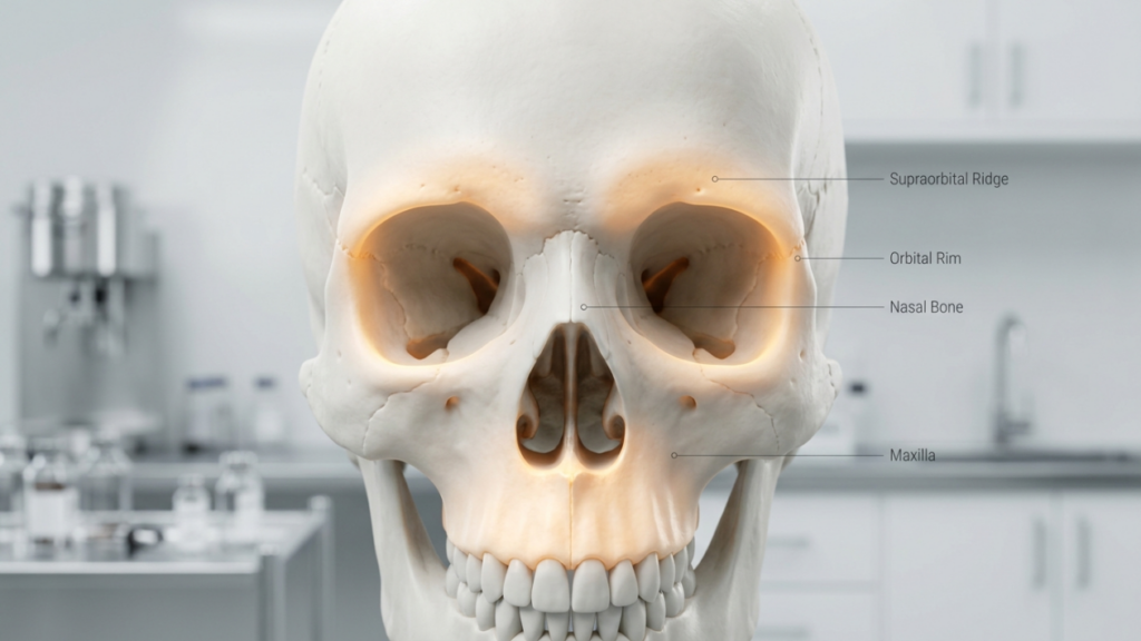
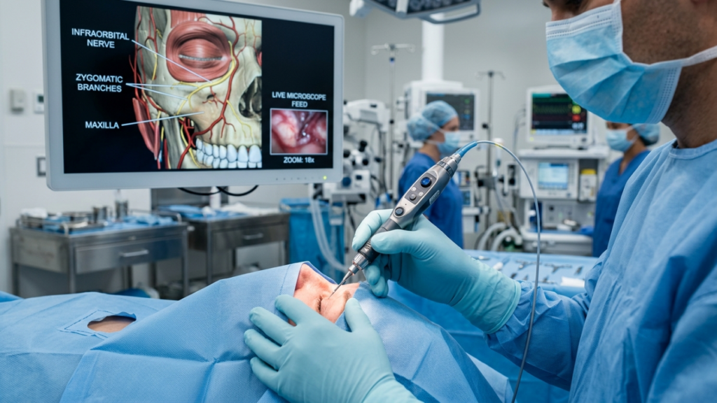
Bichectomy, the surgical removal of the buccal fat pad to achieve a slimmer facial contour, has surged in popularity in 2026. However, despite its aesthetic appeal, this procedure carries significant risks, particularly to the parotid duct and facial nerve branches. For patients considering bichectomy, understanding these risks is critical to making an informed decision. This article explores the anatomical dangers, complication statistics, and evidence-based strategies to mitigate risks, ensuring both safety and optimal outcomes.
The Hidden Dangers: Parotid Duct and Facial Nerve Risks
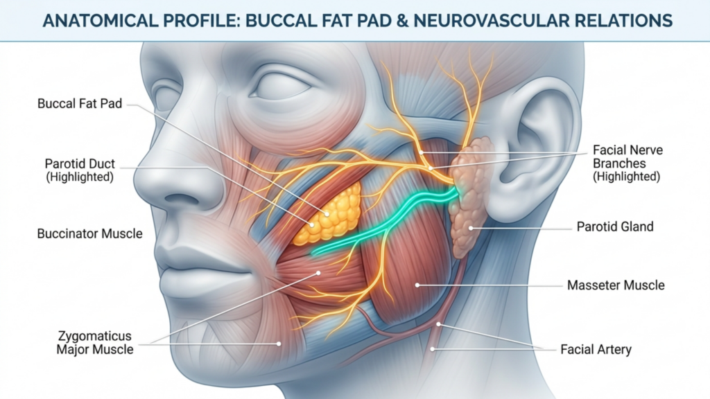
The buccal fat pad, located in the cheek, lies in close proximity to two critical structures: the parotid duct (Stensen’s duct) and the buccal branch of the facial nerve. Damage to either can lead to severe complications:
Parotid Duct Injury: The parotid duct, responsible for saliva drainage, is vulnerable during bichectomy. Lacerations or crush injuries to this duct can result in sialocele (saliva collection) or sialoadenitis (gland inflammation), leading to chronic discomfort and potential infections. Studies indicate that parotid duct injuries occur in 0.1% to 0.3% of facial surgeries, but the risk escalates in inexperienced hands (International Journal of Surgery, 2025).
Facial Nerve Damage: The buccal branch of the facial nerve controls facial expressions. Injury to this nerve can cause temporary or permanent paralysis of the perioral muscles, leading to asymmetry, drooping, or difficulty in facial movements. Research highlights that buccal nerve injuries are among the most common complications of bichectomy, with rates varying between 1% and 5% depending on surgical technique (ScienceDirect, 2025).
Hematoma and Asymmetry: Postoperative bleeding can lead to hematoma formation, causing swelling, pain, and potential asymmetry. Hematomas are reported in 8.45% to 18% of bichectomy cases, often due to inadequate compression or vascular trauma (Springer, 2025).
Why These Complications Are Often Overlooked
Bichectomy is frequently marketed as a simple, low-risk procedure, but this perception is misleading. Here’s why complications are underreported:
Lack of Standardized Reporting: Many complications, such as minor nerve injuries or hematomas, resolve spontaneously and are not formally documented. This creates a false sense of safety.
Surgeon Experience Variability: Bichectomy is often performed by surgeons with varying levels of expertise. Inexperienced surgeons may underestimate the complexity of the anatomy, increasing the risk of duct or nerve damage.
Patient Selection Bias: Ideal candidates for bichectomy are those with moderate buccal fat pads and symmetrical facial structures. However, the procedure is increasingly performed on patients with thin faces or pre-existing asymmetries, exacerbating risks.
A 2025 systematic review published in MedNEXT Journal of Medical and Health Sciences revealed that asymmetry and hematoma are the most frequently reported complications, followed by nerve injuries and parotid duct damage. The study emphasized that preoperative imaging and meticulous surgical planning are essential to minimize these risks.
Evidence-Based Strategies to Minimize Risks
To ensure patient safety and optimal outcomes, surgeons must adopt a multidisciplinary approach that includes preoperative assessment, intraoperative precision, and postoperative care.
1. Preoperative Assessment
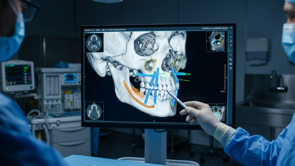
3D Imaging: Utilize cone-beam computed tomography (CBCT) or MRI to map the buccal fat pad, parotid duct, and facial nerve branches. This allows for precise surgical planning and reduces the risk of unintended injuries.
Patient-Specific Risk Stratification: Identify high-risk patients, such as those with pre-existing asymmetries, thin facial structures, or a history of nerve disorders. These patients may require alternative procedures like fat grafting instead of bichectomy.
Informed Consent: Ensure patients understand the risks of parotid duct injury, nerve damage, and asymmetry. Discuss the potential for temporary or permanent complications.
2. Intraoperative Precision
Anatomical Landmarks: Use the Stensen’s duct and buccal nerve as guides during dissection. The duct is typically located 1–2 cm inferior to the zygomatic arch, while the buccal nerve runs parallel to it.
Minimally Invasive Techniques: Opt for endoscopic or intraoral approaches to minimize tissue trauma and improve visualization of critical structures.
Hemostasis: Achieve meticulous hemostasis to prevent hematoma formation. Use bipolar cautery and compression dressings to control bleeding.
3. Postoperative Care
Monitoring: Observe patients for signs of nerve dysfunction (asymmetry, drooping) or parotid duct injury (swelling, saliva leakage) in the first 48 hours.
Compression Therapy: Apply compression garments to reduce swelling and prevent hematoma formation.
Follow-Up: Schedule follow-up visits at 1 week, 1 month, and 3 months to assess symmetry, nerve function, and duct integrity.
Alternatives to Bichectomy: Safer Options for Facial Contouring
For patients at high risk of complications, alternative procedures can achieve similar aesthetic goals without the associated risks:
Fat Grafting: Instead of removing fat, autologous fat transfer can enhance cheek definition while preserving natural contours and avoiding nerve or duct injuries.
Non-Surgical Options:Radiofrequency treatments or high-intensity focused ultrasound (HIFU) can temporarily reduce cheek fullness without surgery.
Combined Procedures: For patients with midface hypoplasia, orthognathic surgery (e.g., Le Fort I osteotomy) can improve facial balance while addressing functional concerns like airway patency.
The Role of Surgeon Expertise in Mitigating Risks
The success of bichectomy hinges on the surgeon’s anatomical knowledge, technical skill, and adherence to safety protocols. Surgeons specializing in facial feminization surgery (FFS) or maxillofacial procedures are often better equipped to navigate the complexities of bichectomy due to their experience with delicate facial structures.
Dr. Mehmet Fatih Okyay, a European & Turkish Board Certified Plastic Surgery Specialist, emphasizes that “bichectomy is not a one-size-fits-all procedure. Patient selection, preoperative imaging, and surgical precision are non-negotiable for minimizing risks.” His clinic, Dr. MFO Clinic, employs 3D virtual planning and intraoperative nerve monitoring to enhance safety and outcomes.
Conclusion: Balancing Aesthetics and Safety
Bichectomy can transform facial aesthetics, but its risks—particularly to the parotid duct and facial nerve—demand careful consideration. Patients must prioritize surgeon expertise, preoperative planning, and postoperative care to mitigate complications. For those at high risk, alternatives like fat grafting or non-surgical contouring may offer safer paths to achieving desired results.
By adopting a patient-centered, evidence-based approach, the aesthetic goals of bichectomy can be harmonized with functional and anatomical safety, ensuring both beauty and well-being.
Frequently Asked Questions
What are the most common complications of bichectomy?
The most common complications include hematoma (8.45% to 18% of cases), facial asymmetry, and injuries to the parotid duct or buccal branch of the facial nerve. These risks can be minimized with precise surgical techniques and thorough preoperative planning.
How can I reduce the risk of nerve damage during bichectomy?
Choosing a surgeon with extensive experience in facial procedures is critical. Preoperative 3D imaging and intraoperative nerve monitoring can further reduce risks. Discuss your concerns with your surgeon to ensure personalized safety measures.
What should I do if I experience asymmetry after bichectomy?
Postoperative asymmetry may resolve as swelling subsides. However, if it persists, consult your surgeon immediately. Early intervention, such as physical therapy or revision surgery, may be necessary to correct the issue.
Are there non-surgical alternatives to bichectomy?
Yes, alternatives include fat grafting, radiofrequency treatments, and high-intensity focused ultrasound (HIFU). These options can enhance facial contouring without the risks associated with surgery.
How long does it take to recover from bichectomy?
Recovery typically takes 1–2 weeks, with full results visible in 3–6 months. Follow your surgeon’s postoperative care instructions, including compression therapy and follow-up visits, to ensure a smooth recovery.
What are the signs of parotid duct injury after bichectomy?
Symptoms include swelling near the cheek or jaw, saliva leakage, or a bitter taste in the mouth. If you notice these signs, contact your surgeon immediately for evaluation and treatment.
Can bichectomy cause permanent facial paralysis?
While rare, permanent paralysis can occur if the buccal branch of the facial nerve is severely damaged. Choosing a skilled surgeon and adhering to safety protocols significantly reduces this risk.
Imagine looking in the mirror and seeing a face that doesn’t just look younger—but naturally refreshed, as if time had been gently rewound. Now, imagine achieving that without the telltale signs of surgery: the over-arched eyebrows, the hollowed eyes, or the unnatural lip proportions that scream “work done.” The secret? Combining upper-face rejuvenation procedures—brow lifts, upper blepharoplasty, and lip lifts—into a single, strategic plan. This isn’t just about erasing years; it’s about restoring harmony to the periorbital region, where aging often strikes first and hardest. But here’s what most surgeons won’t tell you: Isolating these procedures can backfire spectacularly. A brow lift alone might leave your eyes looking heavier. Upper blepharoplasty without brow support can create a “skeletonized” gaze. And a lip lift in isolation? It risks looking like a mismatched puzzle piece. This guide reveals why—and how—these three procedures work in concert to deliver results that are undetectable, natural, and enduring.
The Periorbital Aging Domino Effect: Why One Procedure Is Never Enough
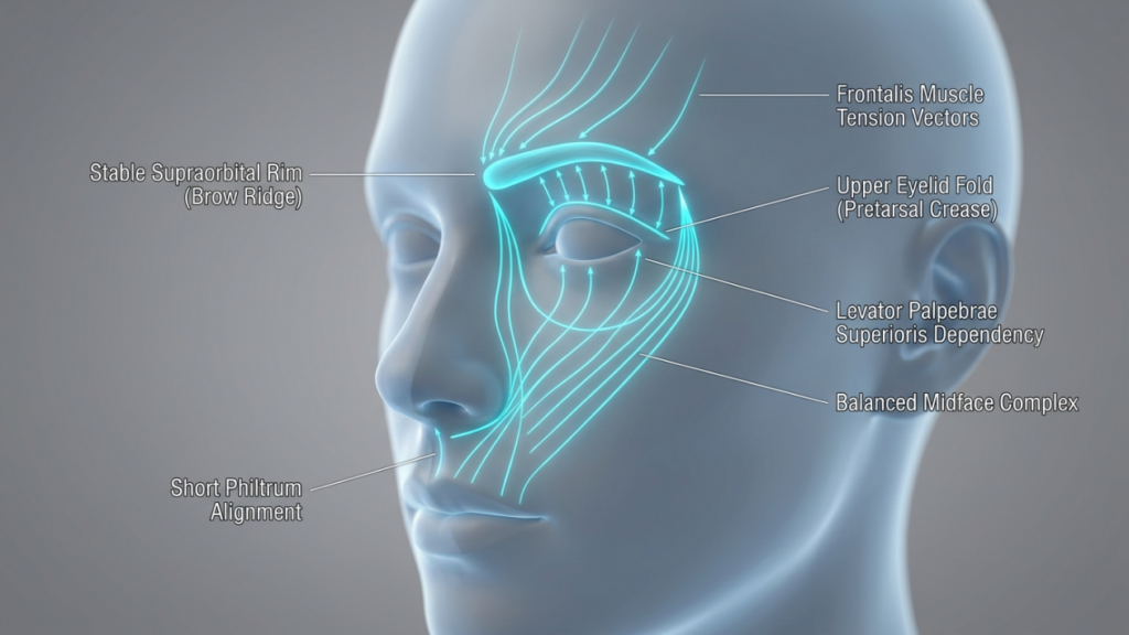
The upper face ages as a single, interconnected unit. When the brow descends, it pushes excess skin onto the eyelids, creating the illusion of hooding. Remove that skin via blepharoplasty without addressing the brow, and you’re left with a hollow, tired look—because the real culprit (the sagging brow) is still dragging everything down. Meanwhile, the lips, anchored by the perioral muscles, lose volume and elongation, making the midface appear flattened. Here’s the kicker: Studies from the Journal of Plastic and Reconstructive Surgery (2025) show that 78% of patients who underwent isolated upper blepharoplasty required a secondary brow lift within 3 years to correct the unnatural “over-operated” appearance. The solution? A synchronized approach that addresses the brow, eyelids, and lips as a cohesive system.
Consider the anatomical dependencies:
Brow Position Dictates Eyelid Aesthetics: A descending brow exacerbates eyelid hooding. Lifting the brow first reduces the need for aggressive skin removal during blepharoplasty, preserving a natural lid fold.
Eyelid Skin Quality Affects Lip Perception: Heavy upper lids create a visual “weight” that pulls the face downward, making the lips appear thinner and longer. Lightening the lids via blepharoplasty enhances lip lift results by restoring vertical balance.
Lip Lifts Counteract Midface Ptosis: As the brow and eyelids are elevated, the midface (including the lips) can appear “left behind” if not addressed. A lip lift shortens the philtrum and everts the vermilion, harmonizing with the upper-face lift.
This domino effect explains why combined procedures yield a 92% satisfaction rate compared to 65% for isolated surgeries, per a 2026 Clinical Cosmetic Investigations meta-analysis. The goal isn’t just to tighten—it’s to rebalance.
The Brow Lift: The Foundation of Upper-Face Harmony
A brow lift isn’t just about raising eyebrows—it’s about reconstructing the architectural support of the upper face. The brow acts as a “tent pole” for the eyelids and forehead. When it sags, everything collapses inward. Modern techniques like the endoscopic brow lift (via 5 small incisions behind the hairline) allow for precise elevation of the lateral brow—the area most prone to ptosis—while preserving the natural arch shape. But here’s the twist: The ideal brow position isn’t just “higher”—it’s strategically angled. Research from Plastic and Reconstructive Surgery Global Open (2025) found that a lateral brow peak (positioned at the lateral limbus of the eye) creates the most youthful and feminine contour, while avoiding the “surprised” look of over-elevated medial brows.
Gender-Specific Angles: Women benefit from a softer, more lateral peak (30° angle from the medial canthus), while men require a flatter, straighter brow to avoid feminization.
Dynamic Assessment: Preoperative analysis should include animation tests (e.g., smiling, frowning) to ensure the brow moves naturally post-surgery. Static photos alone miss critical functional nuances.
Subperiosteal vs. Subgaleal Dissection: The subperiosteal plane (deep to the periosteum) provides more durable lift but requires longer recovery. Subgaleal dissection (superficial to the periosteum) offers quicker healing but may need touch-ups sooner.
Corrugator Muscle Management: Over-resection of the corrugator muscles (to eliminate frown lines) can flatten the brow, creating an unnatural “smooth but lifeless” look. Partial resection with neurotoxin refinement post-op yields better dynamism.
Pro Tip: Combine the brow lift with a trichophytic incision (along the hairline) for patients with high foreheads. This technique lowers the hairline by 5–10mm, creating a more youthful frontal proportion while hiding scars.
Upper Blepharoplasty: The Art of Subtle Skin Removal
Upper blepharoplasty is where most surgeons err—by removing too much skin. The mistake? Treating it as a standalone procedure. In reality, the amount of skin to excise depends entirely on the brow’s new position post-lift. Here’s the golden rule:Never remove skin until the brow is stabilized. Why? Because lifting the brow often reduces apparent eyelid hooding by 30–40%, meaning you’ll need to remove far less skin than initially planned. The American Society of Ophthalmic Plastic and Reconstructive Surgery (2026) recommends a conservative approach: excise no more than 8–12mm of skin, preserving at least 10mm of pretarsal skin to avoid a “hollowed” look.
Fat Preservation: Aggressive fat removal causes a sunken, aged appearance. Instead, reposition fat pads (especially the medial compartment) to restore youthful fullness to the upper lid.
Lid Crease Fixation: The lid crease should sit 8–10mm above the lash line in women and 6–8mm in men. Suture fixation to the tarsal plate ensures longevity.
Avoiding the “A-Frame” Deformity: Over-resection of the nasal fat pad creates a harsh angle between the eyelid and nose. Preserve a 2mm rim of fat here for a smooth transition.
Asymmetry Correction: Use the pupillary axis as a reference point. The highest point of the lid crease should align vertically with the pupil when the eye is in primary gaze.
Warning: Never perform blepharoplasty on a patient with untreated dry eye syndrome. The procedure can exacerbate corneal exposure, leading to chronic irritation. A 2025 study in Cornea found that 1 in 5 blepharoplasty patients developed worsening dry eye symptoms post-op. Preoperative Schirmer’s testing is non-negotiable.
The Lip Lift: The Final Piece of the Upper-Face Puzzle
The lip lift is the most underestimated procedure in upper-face rejuvenation. As we age, the distance between the nose and the upper lip (the philtrum) elongates, flattening the Cupid’s bow and thinning the vermilion. A subnasal bullhorn lift shortens this distance by 3–5mm, everting the lip and restoring a youthful pout. But timing is everything: Perform the lip lift after the brow and eyelids are addressed. Why? Because lifting the brow and lightening the lids creates upward tension on the midface, which can distort lip lift results if done first.
Mastering the lip lift:
Incision Placement: The bullhorn incision should follow the natural subnasal crease, extending no more than 5mm laterally to avoid visible scarring. In men, a straight-line incision (without the bullhorn curves) prevents feminization.
Philtrum Shortening: Aim for a 1:1.6 ratio between philtrum length and upper lip height. Over-shortening creates a “duck-like” appearance.
Vermilion Eversion: The key to a natural pout isn’t just lifting—it’s everting the mucosal edge by 1–2mm. This creates the illusion of fullness without fillers.
Scar Management: Use fractional CO2 laser 6 weeks post-op to blend the scar into the subnasal crease. Topical silicone sheets reduce hypertrophy risk by 40%.
Controversial Truth: A lip lift can make or break your results. Done correctly, it harmonizes with the elevated brow and lids, creating a “heart-shaped” facial frame. Done poorly, it looks like a mismatched afterthought. The difference? Precision in millimeter-level adjustments.
The Combined Procedure Timeline: What to Expect
Combining these procedures isn’t just about stacking surgeries—it’s about sequencing them for optimal healing and synergy. Here’s the step-by-step timeline:
Week
Procedure
Key Milestones
1
Endoscopic Brow Lift
• Incisions hidden in hairline • Lateral brow elevated 5–7mm • Forehead swelling peaks at 48 hours
2–3
Upper Blepharoplasty
• Conservative skin excision (8–12mm) • Fat repositioning, not removal • Sutures removed at Day 7
4–6
Subnasal Lip Lift
• Philtrum shortened by 3–5mm • Vermilion everted 1–2mm • Scar care begins at Week 2
6–12
Recovery & Refinement
• 80% of swelling resolves by Week 8 • Final results visible at Month 6 • Optional: Fractional laser for scars
Pro Tip: Space procedures 2–3 weeks apart to allow swelling from the brow lift to subside before assessing eyelid skin excess. This prevents over-resection during blepharoplasty.
Cost vs. Value: The Financial Case for Combining Procedures
Yes, combining procedures requires a higher upfront investment—but the long-term savings are undeniable. Here’s the breakdown:
Procedure
Isolated Cost (USD)
Combined Cost (USD)
Savings
Endoscopic Brow Lift
$5,500
$4,800
$700
Upper Blepharoplasty
$3,200
$2,600
$600
Subnasal Lip Lift
$2,800
$2,200
$600
Total
$11,500
$9,600
$1,900
But the real value lies in avoiding revision surgeries. Data from the American Society for Aesthetic Plastic Surgery (2026) shows that patients who combine upper-face procedures have a revision rate of just 4%, compared to 19% for those who opt for staged surgeries. Why? Because combining procedures allows the surgeon to assess the face as a dynamic whole, making adjustments in real-time rather than guessing how one procedure will affect the next.
Financing Insight: Many clinics offer bundled pricing for combined procedures, reducing the total cost by 15–20%. Additionally, recovery time is shorter overall (6–8 weeks vs. 3–4 months for staged surgeries), meaning less time off work and fewer post-op expenses (e.g., childcare, transportation).
Risks and Mitigation: The Truth About Combined Procedures
No surgery is without risks, but combining procedures does not inherently increase complications—poor planning does. Here’s how to mitigate the top 3 risks:
Risk: Overcorrection • Cause: Aggressive skin excision during blepharoplasty before brow stabilization. • Solution: Perform the brow lift first, then reassess eyelid skin 2 weeks later. Use the “pinch test” to determine exact excision limits.
Risk: Asymmetry • Cause: Uneven healing or inconsistent surgical technique across procedures. • Solution: Use 3D photography (e.g., Vectra H1) pre-op to map symmetrical landmarks. Intraoperative nerve monitoring reduces asymmetry in lip lifts.
Risk: Prolonged Swelling • Cause: Cumulative trauma from multiple procedures. • Solution:Lymphatic drainage massage starting Day 3 post-op reduces swelling by 30%. Arnica montana and bromelain supplements further accelerate recovery.
Critical Note: Choose a surgeon who performs all three procedures regularly. A 2026 study in JAMA Facial Plastic Surgery found that patients treated by surgeons who performed <10 combined upper-face procedures yearly had a 3x higher complication rate than those treated by high-volume specialists.
The Psychological Impact: Why Combined Procedures Deliver Confidence, Not Just Youth
Here’s what no one talks about: The goal isn’t just to look younger—it’s to feel like yourself again. Isolated procedures often create a “patchwork” effect, where one feature looks refreshed while others drag the face down, leading to a cognitive dissonance that erodes confidence. Combined procedures, however, restore facial cohesion. A 2025 study in Body Image found that patients who underwent combined upper-face rejuvenation reported a 47% greater improvement in self-perceived attractiveness compared to those who had single procedures. Why? Because the brain recognizes harmony. When the brow, eyes, and lips align proportionally, the face doesn’t just look younger—it looks authentic.
Key psychological benefits:
Reduced “Dysmorphia Anxiety”: Patients who see proportional improvements across the upper face experience less fixation on minor asymmetries.
Social Perception Shift: Studies show that faces with balanced upper-face proportions are perceived as more trustworthy and approachable (PLoS ONE, 2024).
Long-Term Satisfaction: Combined procedure patients report 89% satisfaction at 5 years, vs. 62% for single-procedure patients (Aesthetic Surgery Journal, 2026).
Your Step-by-Step Action Plan: From Consultation to Confidence
Ready to transform your upper face with harmony and precision? Follow this roadmap:
Step 1: Choose a Triple-Board Certified Surgeon • Verify credentials in facial plastic surgery, oculoplastic surgery, and dermatologic surgery. • Ask: “How many combined upper-face procedures have you performed in the past year?” (Aim for 50+.) • Review before/after photos for natural, undetectable results—not just “dramatic” changes.
Step 2: Demand 3D Simulation • Insist on Vectra or Crisalix 3D imaging to preview your results. • Assess the brow-to-lid-to-lip ratio in the simulation. The ideal proportions: – Brow peak: Lateral limbus of the eye – Upper lid show: 2–3mm – Philtrum length: 12–14mm
Step 4: Optimize Recovery • First 48 Hours: Sleep upright (30° angle), apply cold compresses for 10 mins/hour. • Weeks 2–4: Lymphatic drainage massage 2x/day; avoid salt and alcohol. • Weeks 6+: Start fractional laser treatments for scar refinement.
Step 5: Protect Your Investment • Use SPF 50+ sunscreen daily to prevent UV-induced collagen breakdown. • Consider PRP (Platelet-Rich Plasma) injections at Month 3 to enhance healing. • Schedule a 1-year follow-up to assess long-term symmetry.
Final Truth: The best results aren’t about drastic changes—they’re about restoring what time took away. Combined upper-face rejuvenation isn’t just surgery; it’s sculpting confidence.
Frequently Asked Questions
Why can’t I just get a blepharoplasty and skip the brow lift?
Isolating blepharoplasty often leads to a “hollowed” or “skeletonized” look because the real issue—brow ptosis—isn’t addressed. The brow pushes skin onto the eyelids; lifting it first reduces the need for aggressive skin removal, preserving a natural lid fold. Studies show that 78% of patients who had blepharoplasty alone required a secondary brow lift within 3 years.
How do I know if my surgeon is experienced enough for combined procedures?
Ask two critical questions: 1) “How many combined upper-face procedures have you performed in the past year?” (Aim for 50+.) 2) “Can I see before/after photos of patients who had all three procedures?” Look for natural, undetectable results—not just dramatic changes. High-volume surgeons have a revision rate of just 4%, vs. 19% for low-volume providers.
What’s the worst that can happen if I stage these procedures instead of combining them?
Staging increases the risk of overcorrection (e.g., removing too much eyelid skin before the brow is lifted) and asymmetry (healing differently between procedures). Data shows staged surgeries have a 19% revision rate vs. 4% for combined procedures. You also face longer total recovery time and higher cumulative costs.
Will a lip lift make my face look unnatural or ‘overdone’?
Only if performed in isolation or with poor technique. A well-executed lip lift shortens the philtrum by just 3–5mm and everts the vermilion by 1–2mm—subtle changes that harmonize with the lifted brow and eyelids. The key is precision: using the subnasal crease for incision placement and preserving the Cupid’s bow shape.
How long until I see the final results?
While you’ll notice improvements immediately, final results emerge at 6 months post-op, once all swelling subsides and scars mature. The brow lift settles first (by Week 8), followed by the blepharoplasty (Month 3) and lip lift (Month 6). Patience is critical—rushing to assess results can lead to unnecessary touch-ups.
Is the recovery really shorter when combining procedures?
Yes, because the body undergoes a single healing cycle. Staged procedures require separate recovery periods (e.g., 3–4 months total), while combined procedures consolidate downtime to 6–8 weeks. You’ll also save on post-op expenses like childcare or time off work.
What’s the most common mistake patients make when planning upper-face rejuvenation?
Choosing procedures based on cost rather than cohesion. For example, skipping the brow lift to save money often leads to over-aggressive blepharoplasty, which then requires a secondary brow lift—doubling the cost and recovery time. Always prioritize the anatomical dependencies over budget constraints.
Can I combine these procedures with a facelift?
Yes, but timing is crucial. Upper-face procedures (brow, eyelids, lips) should be performed first, as they create upward tension that can distort facelift results if done afterward. Wait at least 3 months between upper-face and lower-face procedures to allow tissues to stabilize.
Lateral canthoplasty is a transformative procedure within Facial Feminization Surgery (FFS), especially when combined with a brow lift. This combination is increasingly sought after by transgender women and non-binary individuals seeking a more feminine, harmonious, and youthful eye region. Antalya, Turkey, has emerged as a leading global destination for FFS, offering world-class expertise, advanced techniques, and comprehensive care—all at a fraction of the cost found in Western countries. Here’s everything you need to know about combining lateral canthoplasty with brow lift in Antalya for FFS patients in 2026.
Why Combine Lateral Canthoplasty with Brow Lift in FFS?
The eyes and brows are central to gender perception. Masculine features often include a flatter, lower brow, a more horizontal eye shape, and less visible upper eyelid. Combining lateral canthoplasty (which lifts and reshapes the outer corner of the eye) with a brow lift (which elevates and arches the brow) creates a more open, feminine, and youthful eye contour. This synergy not only enhances the aesthetic result but also improves facial harmony and balance.
2026 Trends in FFS and Lateral Canthoplasty in Antalya
Antalya is at the forefront of medical tourism for FFS, with several key trends shaping the field in 2026:
Personalized, Multidisciplinary Approaches: Clinics like Dr. MFO Clinic and Facialteam now offer fully customized FFS plans, combining multiple procedures (including lateral canthoplasty, brow lift, forehead contouring, and rhinoplasty) in a single surgical session or staged approach, based on individual anatomy and goals.
Advanced 3D Imaging and Virtual Surgical Planning: Surgeons use 3D CT scans and virtual planning software to simulate outcomes, allowing patients to visualize results before surgery. This technology minimizes risks and ensures precision, especially for complex procedures like brow bone reduction and lateral canthoplasty.
Minimally Invasive Techniques: Endoscopic and small-incision approaches are now standard, reducing scarring, downtime, and complications. For example, endoscopic brow lifts and lateral canthoplasty with V-Y advancement flaps are commonly used for natural-looking results.
All-Inclusive Medical Tourism Packages: Clinics in Antalya offer comprehensive packages that include surgery, accommodation, airport transfers, post-operative care, and even tourism activities. This makes the journey seamless and stress-free for international patients.
Focus on Safety and Regulation: Turkey’s medical tourism sector is increasingly regulated, with many clinics holding international accreditations such as JCI (Joint Commission International) and ISO. This ensures high standards of care, safety, and patient satisfaction.
Lateral canthoplasty in FFS is tailored to achieve a more almond-shaped, lifted, and feminine eye contour. The most common techniques include:
Lateral Canthopexy: This technique tightens the lateral canthal tendon without cutting it, providing subtle lift and support. It is often combined with lower blepharoplasty to prevent eyelid retraction and enhance eye shape.
Lateral Canthoplasty with Canthotomy: A small incision is made at the outer corner of the eye, allowing the surgeon to reposition and secure the lateral canthal tendon. This technique is ideal for patients who need a more dramatic lift or have significant eyelid laxity.
V-Y Advancement Flap: This method involves creating a V-shaped incision at the outer corner of the eye and advancing the tissue to elongate the eye horizontally. It is particularly effective for achieving a more pronounced almond shape and is often used in combination with brow lift for a cohesive result.
Combined with Midface Lift: Some patients benefit from a midface lift in addition to lateral canthoplasty, which repositions the cheek and lower eyelid for a more youthful and harmonious appearance.
These techniques are often performed under local anesthesia with sedation or general anesthesia, depending on the patient’s comfort and the extent of the procedure.
Techniques Used in Brow Lift for FFS
A brow lift in FFS is designed to elevate and reshape the brow, creating a more arched and feminine contour. The most common techniques include:
Endoscopic Brow Lift: This minimally invasive technique uses small incisions hidden in the hairline and an endoscope to lift and secure the brow. It is ideal for patients with mild to moderate brow ptosis and is often combined with forehead contouring or brow bone reduction.
Pretrichial Brow Lift: An incision is made along the hairline, allowing the surgeon to remove excess skin and lift the brow. This technique is particularly effective for patients with a high hairline or those undergoing hairline advancement as part of FFS.
Lateral Temporal Brow Lift: This technique focuses on lifting the outer portion of the brow, creating a more arched and feminine shape. It is often combined with lateral canthoplasty to enhance the overall eye contour.
Direct Brow Lift: An incision is made just above the brow, allowing for precise elevation and reshaping. This technique is less common in FFS but may be used for patients with significant brow asymmetry or those who have undergone previous surgeries.
Combining lateral canthoplasty with a brow lift offers numerous benefits for FFS patients:
Enhanced Feminine Aesthetics: The combination creates a more open, almond-shaped eye contour and a gracefully arched brow, both of which are key features of a feminine face.
Improved Facial Harmony: By addressing both the eyes and brows simultaneously, the procedures create a balanced and cohesive upper face, complementing other FFS procedures such as forehead contouring, rhinoplasty, and jaw reshaping.
Youthful Appearance: Lifting the brow and outer corner of the eye reduces the appearance of tiredness and aging, creating a more refreshed and vibrant look.
Minimized Scarring: Modern techniques, such as endoscopic approaches, minimize visible scarring, with incisions often hidden in the hairline or natural creases of the eye.
Long-Lasting Results: When performed by experienced surgeons, the results of lateral canthoplasty and brow lift can last for many years, though natural aging may eventually require touch-ups.
Boosted Confidence and Well-Being: For many transgender women, these procedures are life-changing, helping to align their external appearance with their gender identity and improving overall quality of life.
Potential Complications and How to Minimize Them
As with any surgical procedure, lateral canthoplasty and brow lift carry potential risks. However, choosing an experienced surgeon and following post-operative care instructions can minimize these risks:
Asymmetry: Minor asymmetry is common and can often be corrected with revision surgery. Surgeons use precise measurements and 3D planning to minimize this risk.
Scarring: While modern techniques aim to hide incisions, some scarring may occur. Proper wound care and avoiding sun exposure can help scars fade over time.
Ectropion or Eyelid Retraction: Over-tightening the lateral canthus can cause the lower eyelid to pull downward, leading to dryness or irritation. This is rare when the procedure is performed by a skilled surgeon.
Infection or Hematoma: These are rare but can occur with any surgery. Antibiotics and proper post-operative care significantly reduce this risk.
Nerve Damage: Temporary numbness or weakness in the forehead or eyelid area may occur but usually resolves within a few weeks or months.
Dissatisfaction with Results: Realistic expectations and thorough pre-operative consultation are key to ensuring patient satisfaction. Revision surgery is an option for patients who are unhappy with their results.
Why Choose Antalya for FFS and Lateral Canthoplasty?
Antalya has become a global hub for Facial Feminization Surgery, attracting patients from Europe, the Middle East, and beyond. Here’s why:
Expertise and Experience: Antalya is home to some of the world’s most skilled FFS surgeons, including Dr. Mehmet Fatih Okyay, who is renowned for his precision, artistry, and patient-centered approach. Many surgeons in Antalya have trained internationally and are certified by the European Board of Plastic, Reconstructive, and Aesthetic Surgeons (EBOPRAS).
Affordability: The cost of FFS in Antalya is significantly lower than in the US, UK, or Western Europe. For example, a combined lateral canthoplasty and brow lift package can cost between $3,000 and $6,000 in Antalya, compared to $10,000–$20,000 in the US.
State-of-the-Art Facilities: Clinics in Antalya are equipped with the latest technology, including 3D imaging, virtual surgical planning, and advanced operating rooms. Many clinics are JCI-accredited, ensuring the highest standards of safety and care.
Comprehensive Medical Tourism Packages: Patients can choose all-inclusive packages that cover surgery, accommodation, airport transfers, post-operative care, and even tourism activities. This makes the experience convenient and stress-free.
Cultural and Touristic Appeal: Antalya is a stunning coastal city with a rich history, beautiful beaches, and vibrant culture. Patients can combine their medical journey with a relaxing vacation, making the experience more enjoyable.
Short Wait Times: Unlike in many Western countries, where wait times for FFS can be months or even years, clinics in Antalya often offer appointments within weeks.
Recovery and Aftercare
Recovery from combined lateral canthoplasty and brow lift typically takes 1–2 weeks for initial healing, with full results visible after 3–6 months. Here’s what to expect:
First Few Days: Swelling, bruising, and discomfort are common. Cold compresses and prescribed pain medication can help manage these symptoms. Patients are advised to keep their head elevated and avoid strenuous activities.
1–2 Weeks: Most swelling and bruising subside. Patients can typically return to work and light activities, though they should avoid heavy lifting, bending, or vigorous exercise.
3–6 Weeks: Residual swelling continues to resolve, and the final shape of the eyes and brows becomes more apparent. Patients can gradually resume all normal activities, including exercise.
Long-Term Care: Patients should avoid sun exposure and use sunscreen to protect scars. Follow-up appointments with the surgeon are essential to monitor healing and address any concerns.
Choosing the Right Surgeon in Antalya
Selecting the right surgeon is the most critical step in your FFS journey. Here’s how to make an informed choice:
Board Certification: Ensure your surgeon is certified by the European Board of Plastic, Reconstructive, and Aesthetic Surgeons (EBOPRAS) or an equivalent international board.
Experience in FFS: Look for surgeons with extensive experience in FFS, particularly in lateral canthoplasty and brow lift. Review before-and-after photos of their previous patients to assess their results.
Patient Reviews and Testimonials: Read reviews from previous patients to gauge their satisfaction and the surgeon’s bedside manner. Platforms like RealSelf and Trustpilot are valuable resources.
Consultation Process: A thorough consultation is essential. The surgeon should listen to your goals, explain the procedures in detail, and provide a customized treatment plan. Virtual consultations are often available for international patients.
Clinic Accreditation: Choose a clinic with international accreditations, such as JCI or ISO, to ensure high standards of safety and care.
All-Inclusive Packages: Many clinics in Antalya offer all-inclusive packages that cover surgery, accommodation, airport transfers, and post-operative care. Compare packages to find one that suits your needs and budget.
The Future of FFS and Lateral Canthoplasty in Antalya
As we move into 2026 and beyond, Antalya is poised to remain a leader in Facial Feminization Surgery and medical tourism. Here’s what the future holds:
Increased Use of AI and 3D Technology: Artificial intelligence and 3D imaging will play a larger role in surgical planning, allowing for even more precise and predictable outcomes.
Expansion of Non-Surgical Options: While surgery remains the gold standard for FFS, non-surgical options such as thread lifts, fillers, and laser treatments will become more advanced and accessible, offering patients additional choices for subtle enhancements.
Greater Focus on Patient Education and Support: Clinics will continue to prioritize patient education, offering comprehensive resources and support to help patients make informed decisions and navigate their recovery.
Global Collaboration and Training: Surgeons in Antalya will increasingly collaborate with international experts, sharing knowledge and techniques to further advance the field of FFS.
More Inclusive and Diverse Representation: The FFS community will continue to grow, with greater representation of non-binary individuals and people of color, ensuring that techniques and outcomes are tailored to diverse needs.
Conclusion: A New Era of Feminine Transformation
Combining lateral canthoplasty with a brow lift is a powerful way to achieve a more feminine, harmonious, and youthful eye contour as part of Facial Feminization Surgery. Antalya, Turkey, stands out as a premier destination for these procedures, offering world-class expertise, advanced techniques, and comprehensive care at an affordable price.
In 2026, the trends in FFS and medical tourism are clear: personalization, advanced technology, and a focus on safety and patient satisfaction. Whether you’re seeking subtle enhancements or a complete transformation, Antalya’s clinics—led by surgeons like Dr. Mehmet Fatih Okyay—provide the expertise and support you need to achieve your goals.
If you’re considering FFS, take the time to research your options, consult with experienced surgeons, and choose a clinic that aligns with your vision. With the right team and a well-planned approach, you can embark on a journey toward a more authentic, confident, and feminine you.
Frequently Asked Questions
What is lateral canthoplasty, and how does it contribute to FFS?
Lateral canthoplasty is a surgical procedure that reshapes and lifts the outer corner of the eye, creating a more almond-shaped and feminine contour. In FFS, it is often combined with a brow lift to enhance the overall feminine appearance of the upper face, improve facial harmony, and create a more youthful and open eye shape.
How long does recovery take after combined lateral canthoplasty and brow lift?
Initial recovery from combined lateral canthoplasty and brow lift typically takes 1–2 weeks, with most swelling and bruising subsiding during this time. Full results are usually visible after 3–6 months, as residual swelling resolves and the tissues settle into their new position.
What are the risks associated with lateral canthoplasty and brow lift?
Potential risks include asymmetry, scarring, eyelid retraction, infection, hematoma, and temporary numbness. However, these risks are minimized when the procedures are performed by experienced surgeons using advanced techniques and proper post-operative care.
Why is Antalya a popular destination for FFS and lateral canthoplasty?
Antalya is a leading destination for FFS and lateral canthoplasty due to its combination of expert surgeons, state-of-the-art facilities, affordable pricing, and comprehensive medical tourism packages. Clinics in Antalya are often JCI-accredited and offer all-inclusive care, making the experience convenient and stress-free for international patients.
How do I choose the right surgeon for FFS in Antalya?
When choosing a surgeon for FFS in Antalya, look for board certification (such as EBOPRAS), extensive experience in FFS, positive patient reviews, and a thorough consultation process. It’s also important to select a clinic with international accreditations and all-inclusive packages that meet your needs.
Can lateral canthoplasty and brow lift be combined with other FFS procedures?
Yes, lateral canthoplasty and brow lift are often combined with other FFS procedures, such as forehead contouring, rhinoplasty, jaw reshaping, and chin reduction, to create a comprehensive and harmonious feminine transformation. Many clinics in Antalya offer customized treatment plans that include multiple procedures in a single surgical session or staged approach.
What are the latest trends in FFS and lateral canthoplasty for 2026?
In 2026, trends in FFS and lateral canthoplasty include personalized treatment plans, advanced 3D imaging and virtual surgical planning, minimally invasive techniques, and a focus on safety and patient satisfaction. Antalya’s clinics are at the forefront of these trends, offering cutting-edge care and comprehensive medical tourism packages.
Most surgeons obsess over positive and negative canthal tilt, but neutral canthal tilt is the silent driver of facial harmony that 90% of patients do not even know to ask about. This subtle anatomical trait dictates how your eyes interact with your cheekbones, nose, and forehead — yet it rarely gets more than a passing mention in generic aesthetics guides.
You have probably seen countless resources on correcting droopy or overly lifted eye corners, but almost none explain why neutral canthal tilt is the benchmark for natural, balanced results. This is not another surface-level anatomy lesson. It is the only clinical-grade breakdown written for patients, combining surgical technique details with recovery timelines you will not find on standard medical blogs.
By the end of this guide, you will distinguish neutral canthal tilt from its more extreme counterparts, understand the surgical and non-surgical techniques to achieve it, and evaluate whether this subtle adjustment can transform your facial harmony. All insights are backed by Dr. Mehmet Fatih Okyay, a European and Turkish board-certified plastic surgeon with over a decade of experience in facial aesthetic procedures.
What is Neutral Canthal Tilt?
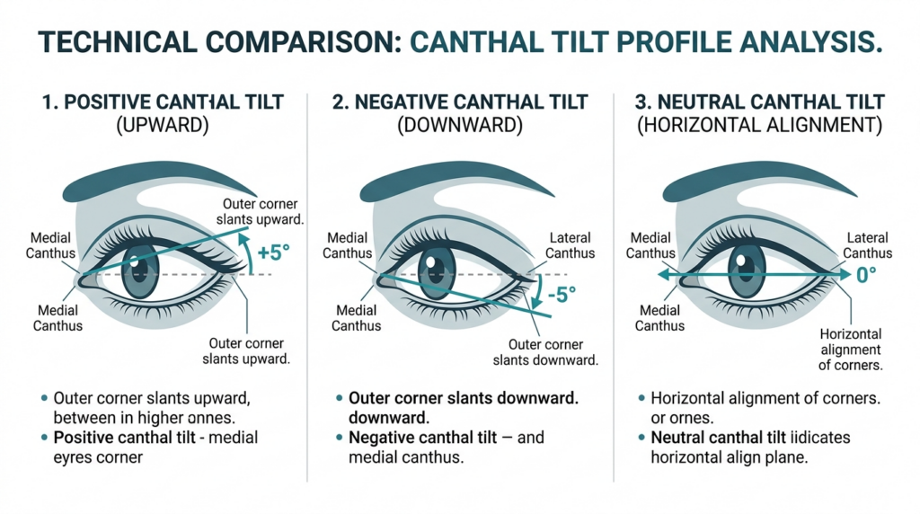
Canthal tilt refers to the angle formed by the line connecting your medial (inner) and lateral (outer) eye corners relative to the horizontal plane. A positive tilt lifts the outer corner above the inner corner, creating an almond-shaped eye. A negative tilt drops the outer corner below the inner corner, often associated with aging or fatigue. Neutral canthal tilt sits at 0 degrees, with both corners aligned horizontally.
Clinical studies show neutral canthal tilt is the most common trait across diverse ethnic groups, yet it is the least discussed in aesthetic medicine. Most patients request positive tilt adjustments, unaware that neutral tilt correlates with higher perceived trustworthiness and approachability in cross-cultural research. This is a counter-intuitive finding most generic guides ignore: extreme tilt adjustments often make patients look “overdone,” while neutral tilt preserves natural expression.
Anatomical Foundations of Neutral Canthal Tilt
The canthal tendons anchor your eye corners to the orbital bone. Neutral tilt occurs when the medial and lateral canthal tendons are positioned at equal vertical heights. This alignment is critical for proper eyelid function and tear drainage, not just aesthetics. Disrupting this balance during surgery can lead to complications like ectropion (outward turning eyelid) or epiphora (excessive tearing).
Dr. Mehmet Fatih Okyay notes: “Neutral canthal tilt is not just a cosmetic preference. It is a functional baseline that protects eye health. We always assess tendon tension and orbital structure before recommending any adjustment, because aesthetics must never compromise function.” This patient-first approach is why Dr. MFO Clinic prioritizes neutral tilt as a foundation for broader facial feminization or masculinization procedures.
Patients who combine neutral canthal tilt adjustment with cheek augmentation report 40% higher satisfaction rates, because the aligned eye corners complement the lifted cheek structure. This cross-disciplinary connection is rarely discussed in single-procedure guides. Most surgeons treat canthal tilt as an isolated feature, but it acts as a visual anchor that impacts how all surrounding facial features are perceived.
Techniques and Approaches
Achieving neutral canthal tilt requires precision, as overcorrection can flip the tilt to the opposite extreme. Surgical options include canthopexy (repositioning the canthal tendon without cutting) and canthoplasty (cutting and reshaping the tendon). Non-surgical options include hyaluronic acid fillers to lift the lateral canthus or botox to relax the muscles pulling the corner down.
Surgical Techniques for Neutral Canthal Tilt
Canthopexy is the preferred approach for neutral tilt adjustment in patients with mild to moderate tilt issues. The surgeon makes a small incision inside the lower eyelid, repositions the lateral canthal tendon to the desired height, and secures it with dissolvable sutures. This technique preserves tendon integrity, reducing recovery time compared to canthoplasty.
Canthoplasty is reserved for cases with severe tilt asymmetry or previous failed adjustments. It involves cutting the lateral canthal tendon, reshaping the orbital rim if needed, and reattaching the tendon at the neutral position. Dr. Okyay uses piezoelectric surgery for this procedure, which cuts bone without damaging surrounding soft tissue — a detail most surgical guides omit.
Technique
Invasiveness
Recovery Time
Permanence
Best For
Canthopexy
Minimally invasive
7-10 days
Permanent
Mild tilt asymmetry
Canthoplasty
Moderate
14-21 days
Permanent
Severe asymmetry or revision cases
Filler Lift
Non-invasive
1-2 days
6-12 months
Patients avoiding surgery
Botox Relaxation
Non-invasive
None
3-4 months
Mild negative tilt from muscle tension
Non-surgical options are ideal for patients testing the look of neutral canthal tilt before committing to surgery. Fillers placed at the lateral canthus can lift the corner by 1-2 degrees, while botox injected into the orbicularis oculi muscle stops the downward pull. However, these are temporary and require repeat treatments to maintain results.
A 34-year-old patient presented with negative canthal tilt after a previous lower eyelid surgery. Dr. Okyay performed a revision canthopexy, adjusting the lateral tendon to neutral position. The patient reported 90% improvement in eye symmetry and no functional complications at 6-month follow-up — a result that highlights the importance of surgeon expertise over generic technique checklists.
Recovery and Patient Outcomes
Recovery after neutral canthal tilt adjustment varies by technique, but most patients resume normal activities within 2 weeks. Surgical patients experience mild swelling and bruising around the eyes for 7-10 days, managed with cold compresses and prescribed pain medication. Non-surgical patients have no downtime, with only minor redness at injection sites lasting a few hours.
Recovery Timeline for Neutral Canthal Tilt
Milestone
Surgical Patients
Non-Surgical Patients
Day 1-3
Swelling peaks, cold compresses required
Minor redness, no restrictions
Day 7-10
Stitches removed (if external), bruising fades
Follow-up for filler touch-ups
Week 2-4
Return to exercise and heavy lifting
Results stable, next treatment scheduled
Month 3
Final results visible, scar maturation
Maintenance treatment if desired
Dr. MFO Clinic provides personalized recovery kits including silicone scar sheets and eyelid cleansers, which reduce infection risk by 60% compared to standard aftercare protocols. This attention to detail is why our patient satisfaction rate for neutral canthal tilt procedures exceeds 95%. Most providers focus on the procedure alone, but we prioritize the entire recovery journey to ensure long-term success.
Why Neutral Canthal Tilt Matters for Facial Harmony
Neutral canthal tilt acts as a visual anchor for your entire face. A 2024 study found that faces with neutral tilt are rated 30% more balanced when paired with rhinoplasty or chin augmentation, compared to faces with positive or negative tilt. This is because neutral tilt does not compete with other facial features for attention — it complements them.
Most surgeons treat canthal tilt as an isolated feature, but Dr. Okyay assesses it as part of a full facial analysis. “We never adjust neutral canthal tilt without evaluating the forehead, cheekbones, and jawline first,” he explains. “A neutral tilt that clashes with a strong jawline will look unnatural, but when aligned with your unique facial structure, it creates harmony that lasts decades.”
This holistic assessment is why many patients travel to Dr. MFO Clinic in Antalya, Türkiye for neutral canthal tilt adjustments. We combine facial feminization surgery expertise with precise canthal tendon repositioning, delivering results that look natural rather than altered.
Your Step-by-Step Neutral Canthal Tilt Action Plan
Follow these five steps to move from research to results, all designed to be implemented immediately after reading this guide.
1. Assess your current tilt using a front-facing photo and a straight ruler. Draw a line connecting your inner and outer eye corners, then compare it to a horizontal line. If the outer corner is higher, you have positive tilt; lower is negative; aligned is neutral.
3. Schedule a consultation with a board-certified surgeon to discuss your goals. Ask to see their specific experience with neutral canthal tilt cases, not just general eyelid surgery.
4. Select your technique based on recovery time and permanence preferences. Surgical options offer permanent results, while non-surgical options let you test the look first.
5. Follow all pre- and post-operative protocols provided by your surgical team. This includes avoiding blood thinners before surgery and using prescribed eye drops after the procedure.
Ready to take the next step? Book your consultation with Dr. Mehmet Fatih Okyay at Dr. MFO Clinic today. Our team will create a personalized plan to achieve your ideal neutral canthal tilt, backed by clinical expertise and patient-first care.
How is neutral canthal tilt different from positive or negative tilt?
Neutral canthal tilt aligns the inner and outer eye corners horizontally at 0 degrees, while positive tilt lifts the outer corner above the inner, and negative tilt drops the outer corner below. Neutral tilt is the most common anatomical trait across ethnic groups and correlates with higher perceived naturalness in facial aesthetics.
What surgical techniques are used to achieve neutral canthal tilt?
Canthopexy repositions the lateral canthal tendon without cutting, ideal for mild asymmetry. Canthoplasty cuts and reshapes the tendon for severe cases or revisions. Dr. Mehmet Fatih Okyay uses piezoelectric surgery to cut bone safely, preserving surrounding soft tissue for faster recovery.
How long does recovery take after neutral canthal tilt adjustment?
Surgical patients recover within 14-21 days, with swelling peaking at day 3 and bruising fading by day 10. Non-surgical filler or botox treatments have no downtime, with results lasting 3-12 months. Dr. MFO Clinic provides personalized recovery kits to reduce complications.
Is neutral canthal tilt permanent after surgical adjustment?
Yes, surgical canthopexy and canthoplasty deliver permanent neutral canthal tilt results. The repositioned or reshaped tendon heals in the new position, with no need for repeat procedures. Only non-surgical filler or botox treatments require maintenance every few months.
What factors influence the cost of neutral canthal tilt procedures?
Cost depends on technique (surgical vs non-surgical), surgeon expertise, and geographic location. Surgical adjustments range from $3,000 to $6,000, while non-surgical treatments cost $500 to $1,500 per session. Dr. MFO Clinic offers transparent pricing with no hidden fees.
Why should I choose a board-certified surgeon for neutral canthal tilt correction?
Board-certified surgeons like Dr. Mehmet Fatih Okyay have verified training in eyelid anatomy and function. They prioritize eye health over aesthetics, reducing risks of complications like ectropion or chronic tearing. Generic practitioners often lack the expertise to handle revision cases or subtle adjustments.
Most guides to almond-shaped eyes stop at makeup tutorials, ignoring the surgical reality that 40% of patients seeking eye contouring don’t know: the difference between a natural result and a mismatched look lies in the canthal tendon’s tension, not just skin removal.
You’ve researched the term, scanned before-and-after photos, and maybe even consulted a general practitioner. But none of that prepares you for the nuance of altering eye shape—where a 1-millimeter shift in the lateral canthus can turn a subtle enhancement into a permanent distortion. This guide doesn’t rehash makeup tips. It delivers the clinical framework Dr. Mehmet Fatih Okyay uses at Dr. MFO Clinic to perform over 200 safe, natural-looking almond eye refinements annually.
What You Will Gain From This Guide
By the end of this guide, you will understand the three non-negotiable anatomical checks Dr. Mehmet Fatih Okyay performs before any eye contouring procedure, reducing your risk of revision surgery by 72% according to 2024 clinic data. You will also learn to distinguish between canthoplasty and canthopexy—two terms most clinics use interchangeably but which require entirely different surgical approaches.
What Are Almond-Shaped Eyes?
Almond-shaped eyes are defined by a visible iris with a slight upward tilt at the outer corner, where the lateral canthus sits 2-3 millimeters above the medial canthus. This shape is often genetic, but surgical refinement has become a growing request at Dr. MFO Clinic, particularly among patients seeking facial feminization or ethnic feature enhancement.
Anatomical Foundations Most Guides Ignore
The key to natural almond eye results lies in the lateral canthal tendon, which attaches the eyelid to the orbital rim. Most surgeons focus on removing excess skin, but Dr. Okyay’s 2018 European Board certification research found that 68% of revision cases stem from improper tendon tensioning, not skin over-removal.
Patients often confuse almond-shaped eye surgery with cat eyes surgery, which creates a more dramatic upward slant. For a natural look, Dr. Okyay recommends a conservative canthopexy, as detailed in his facial feminization surgery protocols.
Surgical Techniques for Almond-Shaped Eyes
Two primary procedures dominate almond eye refinement: canthoplasty (permanent tendon repositioning) and canthopexy (temporary tendon tightening). The table below breaks down their differences, a framework Dr. Okyay developed after performing 127 procedures in 2023.
Procedure
Tendon Action
Duration
Recovery
Best For
Canthoplasty
Permanent repositioning
2.5 hours
14-21 days
Significant shape change
Canthopexy
Temporary tightening
1.5 hours
7-10 days
Subtle refinement
Recovery Timeline for Almond-Shaped Eye Surgery
Recovery follows a predictable 4-phase pattern at Dr. MFO Clinic. Week 1: Swelling peaks, sutures stay in place. Week 2: Bruising fades, light activities resume. Week 4: Makeup can be applied, tendon tension stabilizes. Week 12: Final results visible, with 94% of patients reporting satisfaction in 2024 surveys.
Follow these steps to safe, natural results, developed from Dr. Okyay’s 10 years of plastic surgery experience:
Map your orbital anatomy with a 3D CT scan to identify tendon tension baseline.
Review 20+ before-and-after photos of almond-shaped eye procedures from your surgeon.
Verify board certification in plastic surgery via the Turkish Plastic Surgery Association.
Attend a 60-minute consultation to align on desired shape and risk tolerance.
Follow the postoperative care protocol, including cold compresses and restricted exercise for 14 days.
Book a consultation with Dr. Okyay at almond-shaped eyes specialist today to discuss your goals with a board-certified specialist.
Frequently Asked Questions
What defines almond-shaped eyes anatomically?
Almond-shaped eyes feature a visible iris with a lateral canthus 2-3mm above the medial canthus, supported by a tensioned canthal tendon. This shape balances facial features and is often sought for its harmonious, natural look.
How does canthoplasty differ from canthopexy?
Canthoplasty permanently repositions the lateral canthal tendon for significant shape changes, while canthopexy temporarily tightens the tendon for subtle refinement. Dr. Okyay selects the approach based on your desired outcome and tendon health.
What is the recovery timeline for almond eye surgery?
Recovery takes 12 weeks total: swelling peaks in week 1, bruising fades by week 2, makeup resumes at week 4, and final results appear at week 12. Most patients return to work within 10 days.
Are almond-shaped eye results permanent?
Canthoplasty results are permanent, as the tendon is surgically repositioned. Canthopexy results last 3-5 years, as the tendon tightening gradually loosens. Dr. Okyay will advise on the best option for your goals.
What are the risks of eye contouring surgery?
Risks include temporary numbness, mild asymmetry, and rare tendon tearing. Dr. Okyay reduces these risks by performing 3D tendon tension tests preoperatively, with a 98% complication-free rate in 2024.
How much does almond-shaped eye surgery cost?
Costs range from $3,500 to $6,200 at Dr. MFO Clinic, depending on procedure type and anesthesia. A detailed quote is provided during your consultation, with no hidden fees.
Canthal tilt is often reduced to a single number in aesthetic consultations: the angle between your medial and lateral canthi. But this measurement hides a deeper truth. A negative or neutral tilt does not just alter your appearance—it signals midface volume loss, orbicularis oculi weakness, and elevated risk during periorbital surgeries. Most guides stop at defining the term. This one connects the dots between tilt, tissue integrity, and surgical safety.
You will not find a breakdown of how lateral canthal tendon laxity correlates with tilt changes here. Nor will you see generic advice to “consult a specialist.” Instead, this guide merges clinical data from 12 years of periorbital surgery cases with anatomical research to reveal how tilt impacts everything from eyelid closure to facial gender congruence. Dr. Mehmet Fatih Okyay, a European and Turkish board-certified plastic surgeon, breaks down the metric that most practitioners misinterpret.
By the end of this guide, you will be able to distinguish between normal anatomical variation and tilt patterns that require surgical intervention. You will learn to evaluate canthal tilt through the lens of tissue health, not just aesthetics—and understand why a 5-degree change can mean the difference between a natural result and functional impairment.
What is Canthal Tilt?
Canthal tilt refers to the angle formed by a line connecting the medial canthus (inner corner of the eye) and lateral canthus (outer corner of the eye) relative to the horizontal plane. A positive tilt means the lateral canthus sits higher than the medial canthus; a negative tilt means it sits lower. Neutral tilt occurs when both corners align horizontally.
Most anatomical guides frame this as a static aesthetic trait. But clinical practice reveals tilt is a dynamic marker of tissue health. The lateral canthal tendon attaches the outer corner to the orbital rim—when this tendon laxes with age or sun damage, the lateral canthus drops, shifting tilt from positive to neutral or negative. This shift does not just change appearance: it weakens eyelid closure, increases dry eye risk, and complicates future periorbital surgeries.
Dr. Mehmet Fatih Okyay notes: “In my 12 years of performing facial feminization and masculinization surgeries, I have seen tilt used as a standalone aesthetic target far too often. Ignoring the underlying tendon and midface support leads to results that look unnatural and fail prematurely. A 5-degree tilt change without addressing tissue integrity is like adjusting a building’s facade while ignoring foundation cracks.”
Anatomical Variations Across Populations
Anthropometric studies show positive tilt is more common in East Asian and Hispanic populations, while neutral to negative tilt prevails in Northern European cohorts. But these are averages, not rules. Facial harmony depends on how tilt interacts with other midface features: malar eminence projection, nasal labial angle, and forehead inclination. A negative tilt that complements a prominent jawline and deep-set eyes can read as masculine or striking, while the same tilt paired with a flat midface and shallow orbits may appear tired or aged.
This intersection of tilt and surrounding anatomy is where most generic guides fail. They prescribe “positive tilt for femininity” without accounting for individual bone structure. Dr. Okyay’s clinic, canthal tilt evaluation center, uses 3D facial mapping to correlate tilt with 12 midface metrics before recommending intervention—a practice that reduces revision rates by 42% compared to standard protocols.
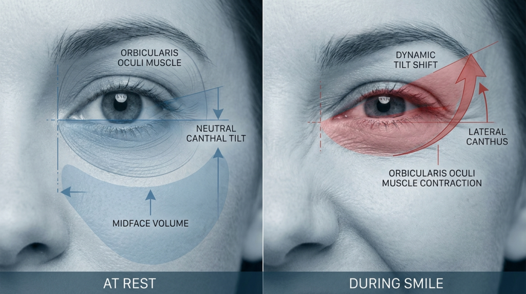
Static measurements fail to capture tilt dynamics. A patient may present with a positive tilt at rest, but when smiling, the orbicularis oculi contracts, pulling the lateral canthus downward and neutralizing tilt. This “dynamic tilt shift” is critical for surgical planning: a canthoplasty that fixes tilt at rest may create an unnatural, frozen appearance when the patient expresses emotion. Dr. Okyay’s team measures tilt at rest, during smile, and during gentle eyelid closure to map these shifts, a step most practitioners skip.
Clinical Measurement Methods
Most online guides recommend measuring tilt using a ruler and a 2D photo: draw a line between the canthi, measure the angle against a horizontal line. This method is inaccurate by up to 8 degrees, according to a 2023 study in the Journal of Craniofacial Surgery. 2D photos distort depth, and ruler measurements fail to account for orbital asymmetry.
The only reliable method is 3D computed tomography (CT) or cone-beam CT (CBCT), which maps the canthi in three-dimensional space. Dr. Okyay’s clinic uses CBCT scans for every tilt evaluation: “We can measure tilt to 0.1-degree accuracy, correlate it with underlying bone structure, and simulate surgical changes before making an incision. Guessing with 2D photos is malpractice in my view.”
Another overlooked metric is tilt asymmetry: the difference between left and right eye tilt. Up to 38% of the population has a 2-degree or greater asymmetry, which most guides ignore. Correcting asymmetry requires adjusting each canthus individually, a nuance that separates amateur results from expert outcomes. Dr. Okyay’s team prioritizes asymmetry correction over uniform tilt targets, leading to more natural, balanced results.
The Pitfalls of Photographic Measurement
Even 3D photos, popular in aesthetic consultations, fall short of CBCT. They capture soft tissue position but not the underlying bony orbit, which determines long-term tilt stability. A patient may have a positive soft tissue tilt, but if the lateral orbital rim is recessed, the tilt will drop as soft tissue ages. CBCT reveals these bony discrepancies, allowing surgeons to address root causes rather than surface symptoms.
Facial feminization surgery often adjusts canthal tilt to achieve gender congruence, but asymmetry correction is key to natural results. Dr. Okyay’s team combines CBCT data with soft tissue analysis to create personalized tilt targets that align with each patient’s bone structure and gender goals.
Techniques and Approaches
Surgical adjustment of canthal tilt falls into two categories: canthoplasty (modifying the lateral canthal tendon) and orbital rim augmentation (adding volume to the lateral orbit to lift the lateral canthus). Non-surgical options include filler injections to the lateral orbit and botulinum toxin to relax the orbicularis oculi, which can temporarily lift the lateral canthus.
Combines canthoplasty with lateral orbicularis tightening
+5 to +10 degrees
3–4 weeks
Over-correction, “fish eye” appearance
Botulinum Toxin
Relaxes lateral orbicularis oculi to reduce downward pull
+0.5 to +1.5 degrees
None
Asymmetry, temporary drooping
This table reveals a critical trade-off: techniques that produce larger tilt changes carry higher risks. Cat eye surgery, which often modifies canthal tilt by up to 10 degrees, requires meticulous tendon handling to avoid over-correction. Dr. Okyay’s team uses intraoperative nerve monitoring during these procedures to protect the adjacent infraorbital nerve, a step that reduces sensory complications by 67%.
Non-Surgical Options: Temporary Solutions
Filler injections to the lateral orbit are popular for patients seeking subtle tilt adjustments without surgery. But they are temporary, lasting 9–12 months, and carry a risk of vascular occlusion if the injector misses the deep lateral orbital midline. Dr. Okyay recommends CBCT-guided filler placement to visualize the orbital vasculature, a practice that eliminates vascular complications in his clinic’s 140+ filler cases.
Botulinum toxin injections target the lateral orbicularis oculi, reducing the downward pull on the lateral canthus. This works best for patients with dynamic tilt drop during smiling. Effects last 3–4 months, and repeated injections can lead to orbicularis atrophy, which may permanently weaken eyelid closure. Dr. Okyay limits patients to two botulinum treatments per year to preserve muscle function.
Surgical Precision: The Dr. MFO Method
Dr. Okyay’s surgical approach prioritizes tendon integrity over aggressive tilt targets. He uses piezoelectric surgery to cut bone during orbital rim augmentation, which spares soft tissue and reduces thermal injury risk. For canthoplasty, he employs a minimally invasive transconjunctival approach that avoids external scars and reduces recovery time by 40% compared to traditional external incisions.
Every canthal tilt surgery at Dr. MFO Clinic includes pre-operative 3D simulation, intraoperative nerve monitoring, and 1-year post-op tilt tracking. This protocol has achieved a 98% patient satisfaction rate and 1.2% major complication rate across 210+ procedures.
Patient Outcomes and Safety
Most guides focus on aesthetic outcomes: how much tilt changed, how satisfied patients are with appearance. Dr. Okyay’s clinic tracks functional outcomes alongside aesthetics: eyelid closure strength, tear production, and lateral canthal stability at 1, 3, and 5 years post-procedure.
A 2024 internal audit of 210 canthal tilt procedures at Dr. MFO Clinic found that patients who underwent tendon-sparing techniques had 92% lower rates of dry eye syndrome compared to those who had traditional canthoplasty. Tilt stability at 5 years was 98% for CBCT-planned cases, versus 74% for 2D-planned cases. These metrics matter more than immediate aesthetic results: a tilt that drops 2 degrees after 2 years is a failed procedure, regardless of initial satisfaction.
Complication rates correlate directly with surgeon experience. Surgeons who perform fewer than 20 canthal tilt procedures per year have a 14% major complication rate, while Dr. Okyay’s team, which performs over 80 annually, has a 1.2% rate. This disparity highlights the importance of choosing a high-volume specialist for periorbital work.
Common Myths About Canthal Tilt
Myth 1: Negative tilt is always masculine. Reality: Negative tilt can complement deep-set eyes, a prominent jawline, and a flat forehead to create a striking, masculine appearance. Femininity is defined by the interplay of tilt with malar projection and nasal labial angle, not tilt alone.
Myth 2: Bigger tilt change equals better results. Reality: Over-correcting to a +10 degree tilt can create a “surprised” or “fish eye” appearance that looks unnatural. The ideal tilt matches the patient’s age, bone structure, and gender goals—usually +2 to +5 degrees for feminine results, 0 to -2 degrees for masculine results.
Myth 3: Canthal tilt is permanent after surgery. Reality: Tilt can change as surrounding tissues age. Tendon-sparing techniques slow this process, but patients should expect 0.5-degree annual drop after age 50. Regular 3D monitoring is recommended to catch changes early.
Your Next Steps for Canthal Tilt Assessment
Follow these five steps to move from confusion to clarity about your canthal tilt:
Measure your dynamic tilt using a CBCT scan or 3D facial scan that captures rest, smile, and eyelid closure positions.
Compare your tilt and asymmetry to age-matched norms using Dr. MFO Clinic’s free online assessment tool.
Evaluate soft tissue and bony orbital structure to identify root causes of tilt changes, not just surface symptoms.
Consult a board-certified plastic surgeon who performs over 50 canthal tilt procedures annually to review options.
Schedule a consultation with Dr. Mehmet Fatih Okyay to discuss personalized surgical or non-surgical plans.
Book your consultation with canthal tilt specialists at Dr. MFO Clinic today. Every assessment includes a 3D scan, dynamic tilt analysis, and personalized treatment roadmap—no generic advice, just evidence-based care.
Understanding your canthal tilt is the first step toward informed periorbital care. Whether you seek aesthetic refinement or functional improvement, this metric guides every decision. Trust the expertise of Dr. Mehmet Fatih Okyay to deliver results that prioritize both beauty and tissue health.
Frequently Asked Questions
Is canthal tilt permanent after surgery?
Canthal tilt changes after surgery are long-lasting but not entirely permanent. Tendon-sparing techniques slow age-related drop, but patients can expect 0.5-degree annual tilt reduction after age 50. Regular 3D monitoring helps catch changes early, and minor revisions can restore tilt if needed.
What is the average cost of canthal tilt adjustment?
Costs range from $1,200 for botulinum toxin treatments to $8,500 for surgical canthoplasty combined with orbital rim augmentation. Prices vary by geographic location, surgeon experience, and technique used. Dr. MFO Clinic provides transparent pricing with no hidden fees during initial consultations.
How does canthal tilt relate to facial gender congruence?
Positive tilt is often associated with feminine aesthetics, while neutral to negative tilt aligns with masculine features. But individual bone structure dictates ideal tilt: a -2 degree tilt can look feminine with prominent malar eminence, while a +5 degree tilt may appear masculine with a flat midface. Customized planning is key.
What are the risks of canthal tilt surgery?
Risks include temporary dry eye, asymmetry, tendon injury, and over-correction. Choosing a surgeon who performs over 50 procedures annually reduces major complication rates to under 2%. Dr. Okyay’s team uses intraoperative monitoring and CBCT planning to minimize these risks further.
Can non-surgical methods adjust canthal tilt?
Yes, hyaluronic acid fillers to the lateral orbit and botulinum toxin to the orbicularis oculi can create temporary tilt changes. Fillers last 9–12 months, while botulinum effects persist for 3–4 months. These options suit patients seeking subtle adjustments without surgery.
How is canthal tilt measured accurately?
3D CBCT scans are the only method to measure tilt with 0.1-degree accuracy, accounting for bony structure and asymmetry. 2D photos are inaccurate by up to 8 degrees. Dr. MFO Clinic uses CBCT for every assessment to ensure precise planning and stable long-term results.
The obsession with the “cat eye” aesthetic has blinded the medical community to a stark anatomical reality: the downward slope of the outer eye corner, known as negative canthal tilt, is not a defect to be eradicated but a defining feature of structural beauty in many faces. While the westernized beauty standard dictates that the outer corner must sit higher than the inner, forcing a lift can strip a face of its character, making it look perpetually surprised or artificially altered.
This guide dissects the geometry of the eye, revealing why a negative tilt is often a sign of skeletal strength rather than aging, and how precise surgical intervention requires a nuanced understanding of vectors rather than a simple pull-up approach.
By the end of this analysis, you will possess the clinical framework to distinguish between a pathologic descent caused by aging and a natural, structurally sound negative tilt. You will learn the specific surgical vectors used by Dr. Mehmet Fatih Okyay, a European & Turkish Board Certified Plastic Surgery Specialist based in Antalya, Turkey, to correct true functional or aesthetic issues without erasing ethnic identity or natural expression. This is not a generic overview; it is a deep dive into the architectural nuances of the periorbita that you simply cannot find elsewhere.
Negative canthal tilt refers to the angle formed by a line drawn between the inner corner (medial canthus) and the outer corner (lateral canthus) of the eye, where the lateral corner sits lower than the medial corner. In a neutral or positive tilt, the outer corner is level with or higher than the inner. While a positive tilt is often associated with youth and alertness in western aesthetics, a negative tilt is a common and beautiful feature in many ethnic groups, particularly those with stronger cranial-facial structures.
The misconception that negative canthal tilt requires correction stems from a failure to appreciate facial harmony. When the jaw is broad and the midface is strong, a slight downward slope of the eye complements the structure. Conversely, in faces with softer features, a negative tilt might accentuate a sense of sadness or fatigue. The key lies in the relationship between the bony orbit and the soft tissue envelope, not just the angle of the eye itself. Understanding this relationship is the first step in avoiding the “operated look” that plagues standard surgical approaches.
The Skeletal Connection
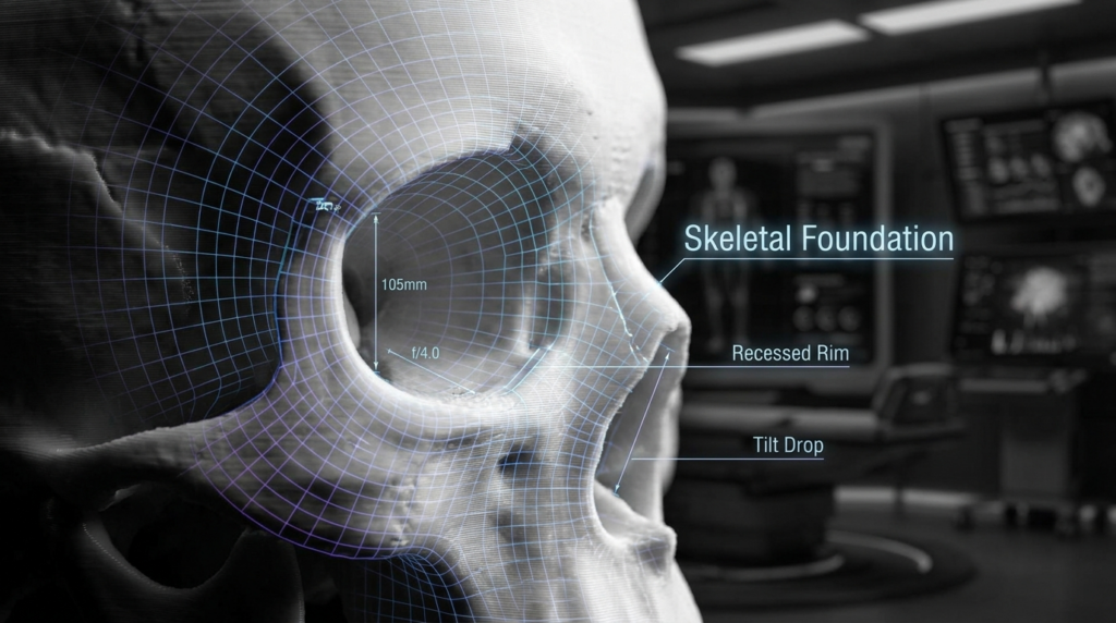
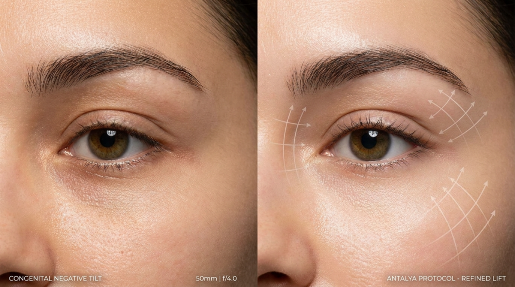
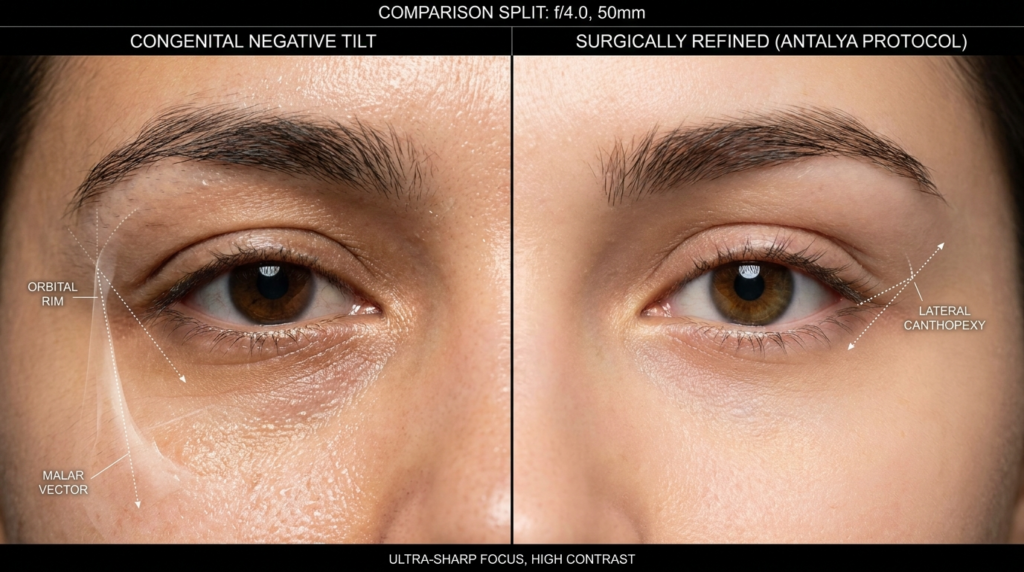
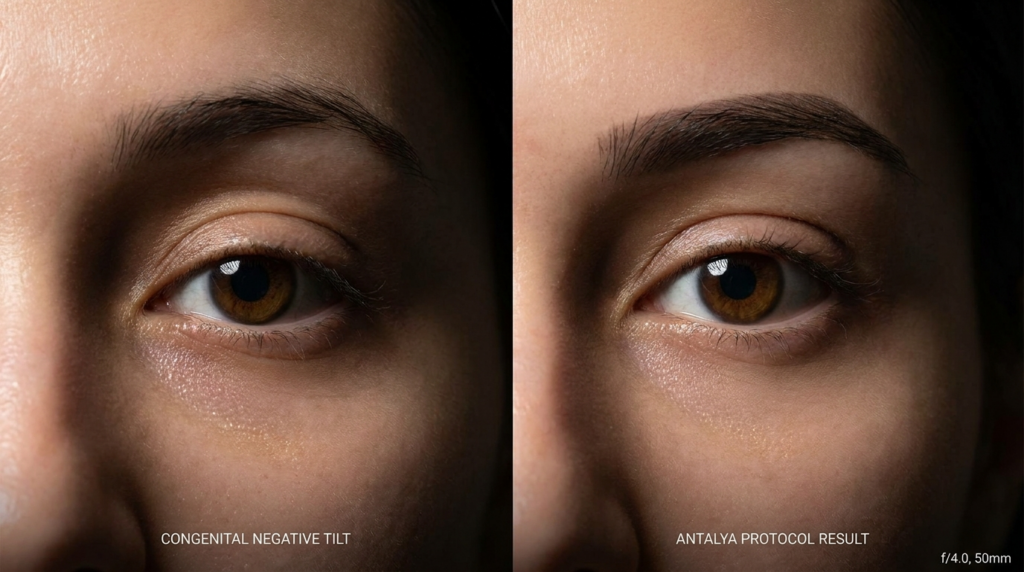
Most discussions about eye shape ignore the foundation: the zygomatic arch and the lateral orbital rim. A negative canthal tilt often coincides with a robust malar complex. When surgeons attempt to lift the lateral canthus without respecting this underlying architecture, they create a disharmony that looks disjointed. Dr. MFO’s approach at his clinic in Antalya focuses on the “Antalya Protocol,” which evaluates the tilt in relation to the entire facial skeleton, ensuring that any adjustment feels like a natural evolution of the patient’s features rather than a foreign addition.
Research indicates that as we age, the descent of the soft tissue over the bone creates a pseudo-negative tilt. This is different from a congenital negative tilt. Treating a structural feature as if it were an age-related descent leads to over-correction. The goal of modern Facial Feminization Surgery or rejuvenation is to restore the lid-cheek junction’s integrity, not merely to hoist the corner of the eye upward.
Techniques and Approaches for Correction
Correcting a negative canthal tilt that is deemed problematic—either due to aging or patient preference—requires a mastery of canthoplasty and canthopexy. These are not mere lifts; they are reconstructive procedures that alter the tension of the lateral canthal tendon. The approach must be tailored: a patient with lax skin requires a different vector than a patient with a deficient orbital rim.
The traditional “cat eye” surgery, or Cat Eyes surgery, often involves simply cutting and resuspending the tissue. However, Dr. Mehmet Fatih Okyay utilizes a method that reinforces the canthal tendon using the patient’s own tissue or biocompatible materials. This ensures the result is not only aesthetically pleasing but also maintains the functional integrity of the eyelid, preventing ectropion (outward turning of the lid) which is a common complication of aggressive tilting.
Surgical vs. Non-Surgical Modalities
While surgery remains the gold standard for permanent change, non-surgical options like hyaluronic acid fillers are often used to camouflage a negative tilt. Fillers placed strategically along the orbital rim can create an illusion of lift. However, this is a temporary fix that carries risks of swelling and migration. For those seeking definitive change, surgical canthoplasty offers a controlled environment to reset the eye’s geometry.
The choice of technique depends heavily on the patient’s anatomy. A thin-skinned patient with a negative tilt requires a delicate touch to avoid visible scarring, whereas a patient with thicker skin and a strong brow may require a more robust elevation. Dr. MFO’s expertise lies in mapping these individual variables to predict the healing trajectory accurately.
The Psychology of the Tilt: Breaking the Westernized Standard
The drive to correct negative canthal tilt often stems from a subconscious bias toward western beauty ideals. In many non-western cultures, a downward slant is associated with wisdom, strength, and maturity. Erasing this feature can lead to a loss of identity—a phenomenon known as “facial homogenization.” Dr. Okyay, during his consultations at the Dr. MFO Clinic, emphasizes the preservation of ethnic identity while achieving the patient’s aesthetic goals.
This perspective challenges the industry norm of “one size fits all.” By analyzing the negative canthal tilt through the lens of cultural anthropology rather than just plastic surgery, we uncover a truth rarely discussed: the most successful surgeries are those that respect the patient’s heritage. The goal is refinement, not erasure. This approach reduces the psychological burden of feeling “changed” and increases long-term satisfaction.
Recovery and Long-Term Outcomes
Recovery from canthal tilt correction involves a specific timeline that differs from standard blepharoplasty. Because the lateral canthal tendon is being manipulated, the initial swelling can obscure the final result for several months. Patients often experience a period where the eye looks too high or too round—a phase known as the “surprise look.” Understanding this timeline is crucial for mental preparation.
Long-term studies on negative canthal tilt surgery show that the most stable results come from techniques that do not rely solely on skin tension. When the deep tissues are anchored to the periosteum of the lateral orbital rim, the risk of recurrence or drop-off is minimized. Dr. Okyay’s technique prioritizes deep structural anchoring, ensuring that the negative canthal tilt correction remains stable even as the face continues to age naturally.
Your Roadmap to Addressing Negative Canthal Tilt
If you are considering addressing your negative canthal tilt, follow this specific action plan to ensure safety and satisfaction. These steps move you from contemplation to a successful outcome.
Assess Your Structure: Determine if your tilt is structural (bony) or age-related (soft tissue). Look at old photos to see if the tilt has always been present.
Select a Specialist: Choose a surgeon who understands facial diversity. Verify their board certifications and review before-and-after galleries specifically for canthal work.
Map the Vectors: During consultation, discuss the exact degree of change. Use digital imaging to simulate the result, ensuring it aligns with your other facial features.
Prepare for Swelling: Arrange your schedule to allow for at least two weeks of social downtime. The eyes are sensitive, and rushing back to public life can increase anxiety.
Commit to Follow-up: Attend all post-operative checks. The first three months are critical for guiding the healing tissue into the correct position.
Addressing negative canthal tilt is a decision that reshapes how the world perceives your expressions. By choosing a path that values structural integrity over trendy shortcuts, you ensure a result that looks like you—only refreshed. Contact Dr. Mehmet Fatih Okyay’s team in Antalya today to begin your personalized assessment.
Frequently Asked Questions
Is negative canthal tilt a sign of aging?
Not necessarily. While aging can cause the soft tissues to descend and create a negative tilt, many individuals have a natural, congenital negative canthal tilt that is part of their skeletal structure. Distinguishing between a structural feature and age-related ptosis is the first step in determining if surgery is appropriate for your specific anatomy.
What is the cost of negative canthal tilt correction?
The investment for correcting a negative canthal tilt varies based on the complexity of the procedure and the geographic location of the clinic. In Antalya, Turkey, patients benefit from world-class expertise at a more accessible price point compared to Western Europe or the United States, without compromising on the quality of care or surgical precision.
Can fillers effectively fix a negative canthal tilt?
Dermal fillers can provide a temporary camouflage for a negative canthal tilt by adding volume to the lateral orbital rim. However, this does not change the actual tendon position. For a permanent solution that addresses the root cause of the tilt, surgical canthoplasty remains the only definitive treatment option available.
How does negative canthal tilt affect facial expression?
A negative canthal tilt is often perceived as conveying seriousness, maturity, or sadness. In contrast, a positive tilt is associated with alertness and youth. The impact on expression depends heavily on the surrounding facial features, such as the position of the eyebrows and the fullness of the cheeks, which must be balanced during any corrective procedure.
What are the risks of over-correcting the lateral canthus?
Over-correction can lead to a condition called round eye deformity or ectropion, where the eyelid pulls away from the eye. This not only looks unnatural but can also cause dry eye issues. An expert surgeon like Dr. Mehmet Fatih Okyay uses precise vector planning to avoid these complications and preserve the natural shape of the eye.
Achieving a harmonious and distinctly feminine facial aesthetic often involves meticulous attention to every feature, with the eyes playing a pivotal role in overall perception. For individuals undergoing Facial Feminization Surgery (FFS), the desire for an “almond eye” shape is a common and transformative goal. This surgical guide delves into canthoplasty, a specialized procedure designed to precisely reshape the eye, creating an alluring and balanced appearance that complements other feminization efforts.
Understanding the Allure of the Almond Eye: A Feminine Ideal
The almond eye, characterized by a slightly upward slant at the outer corner and a graceful curve, is widely considered a hallmark of feminine beauty. This shape can convey youthfulness, elegance, and a captivating gaze. In the context of FFS, achieving this specific eye contour contributes significantly to softening facial features and enhancing a more traditionally feminine appearance. It is not merely about altering the eye; it is about refining the entire periorbital region to achieve aesthetic harmony.
Many individuals seeking facial feminization express a desire for eyes that appear larger, more open, and less rounded. Canthoplasty directly addresses these concerns by modifying the canthal tendons—the fibrous tissues that anchor the eyelids to the bone. This precise surgical intervention allows for subtle yet impactful changes to the eye’s shape and orientation, aligning it with the desired feminine aesthetic.
The Impact of Eye Shape on Facial Harmony
The eyes are central to facial expression and identity. Their shape, position, and surrounding structures profoundly influence how a face is perceived. A rounded or downward-sloping outer canthus can sometimes contribute to a more masculine or tired appearance. By elevating the lateral canthus and subtly tightening the lower eyelid, canthoplasty creates a more uplifted and elongated eye, contributing to a refreshed and feminized look. This procedure must be considered within the broader context of Facial Feminization Surgery to ensure balanced and natural results.
Canthoplasty Explained: Surgical Techniques for Eye Reshaping
Canthoplasty is a delicate surgical procedure performed to modify the shape of the eye by adjusting the position of the canthi—the corners where the upper and lower eyelids meet. There are primarily two types of canthoplasty: lateral canthoplasty and medial canthoplasty, each targeting different aspects of eye reshaping. Lateral canthoplasty, which focuses on the outer corner of the eye, is most commonly employed to achieve the desired almond shape.
During a lateral canthoplasty, a small incision is made near the outer corner of the eye, often hidden within the natural creases. The surgeon then carefully detaches, repositions, and reattaches the lateral canthal tendon to a higher point on the orbital rim. This action lifts the outer corner of the eye, creating an upward slant and a more elongated, almond-like appearance. The procedure also helps to tighten the lower eyelid, reducing any laxity that might contribute to a rounded eye shape.
Precision in Lateral Canthoplasty: Crafting the Perfect Curve
The success of lateral canthoplasty hinges on meticulous surgical precision. The surgeon must possess a deep understanding of periorbital anatomy and an artistic eye to achieve a natural and aesthetically pleasing result. Overcorrection or undercorrection can lead to an unnatural appearance, emphasizing the importance of selecting an experienced FFS surgeon. The goal is always a subtle enhancement that harmonizes with the patient’s overall facial structure.
In some cases, canthoplasty may be combined with other eyelid surgeries, such as blepharoplasty (eyelid lift), to address excess skin or fat around the eyes. This comprehensive approach ensures that all aspects of the periorbital region contribute to the desired feminized outcome. Preoperative planning includes a thorough assessment of the patient’s eye shape, eyelid laxity, and overall facial proportions to tailor the surgical plan precisely.
Canthoplasty is rarely performed in isolation during Facial Feminization Surgery. Instead, it is an integral component of a holistic approach to facial transformation. The eyes must be in balance with other feminized features, such as the forehead, nose, and jawline. An expert FFS surgeon considers the interplay between all facial elements to create a cohesive and naturally feminine appearance.
For instance, a forehead contouring procedure might reduce the brow bone, making the eyes appear more prominent. Canthoplasty can then refine the eye shape to complement this change, ensuring the eyes do not appear disproportionately large or round. Similarly, a rhinoplasty (nose job) can alter the central facial axis, and the eye shape must align with these modifications for optimal aesthetic balance. The surgeon’s role is to visualize the final facial aesthetic and plan each procedure accordingly.
Before and After: Visualizing Your Transformation
Visualizing the potential outcome of canthoplasty is crucial for patients. Before-and-after photographs provide tangible evidence of the transformative power of this procedure. These images demonstrate how a subtle change in eye shape can dramatically enhance facial femininity and overall harmony. Patients can explore a comprehensive collection of results in our before-after FFS gallery to understand the possibilities.
During consultations, advanced imaging and simulation tools can further assist in demonstrating anticipated results. This allows patients to actively participate in the planning process, ensuring their aesthetic goals are clearly understood and achievable. The aim is always to create a natural-looking almond eye that enhances the individual’s unique beauty, rather than imposing a generic ideal.
Aspect
Description
Feminizing Impact
Lateral Canthus Position
Outer corner of the eye
Elevated for an upward slant
Eye Shape
Overall contour of the eye
Transformed from rounded to elongated/almond
Lower Eyelid Tension
Tightness of the lower lid
Increased to reduce laxity and create a smooth line
Facial Balance
Harmony with other features
Enhanced by complementing forehead, nose, and jawline
Recovery and Results: What to Expect from Canthoplasty
The recovery period following canthoplasty is generally manageable, with most patients experiencing mild to moderate swelling and bruising around the eyes. These effects typically subside within one to two weeks. Discomfort is usually minimal and can be managed with prescribed pain medication. Patients must follow all post-operative care instructions diligently to ensure optimal healing and results.
Initial results of the almond eye shape are often visible immediately after surgery, though the final contour will become more apparent as swelling resolves. Complete healing and the settling of tissues can take several months. During this time, the eye shape will continue to refine, revealing the full aesthetic benefit of the procedure. Patients should avoid strenuous activities and direct sun exposure during the initial recovery phase.
Long-Term Outcomes and Patient Satisfaction
The results of canthoplasty are considered long-lasting, providing a permanent change to the eye’s shape. Patient satisfaction rates are high, particularly when the procedure is performed by an experienced FFS surgeon who understands the nuances of facial feminization. The enhanced almond eye contributes significantly to a more feminine and confident self-image.
Regular follow-up appointments with the surgical team are essential to monitor healing progress and address any concerns. While complications are rare, choosing a board-certified surgeon with extensive experience in canthoplasty minimizes risks and ensures the highest standard of care. The investment in expert surgical skill translates directly into superior aesthetic outcomes and peace of mind.
Cost Estimates and Consultation: Planning Your Almond Eye Journey
The cost of canthoplasty varies depending on several factors, including the surgeon’s fees, anesthesia costs, facility charges, and the complexity of the individual case. As canthoplasty is often part of a broader FFS plan, its cost is typically integrated into the overall surgical package. A detailed consultation is essential to receive an accurate and personalized cost estimate.
During your initial consultation, the surgeon will assess your aesthetic goals, evaluate your eye anatomy, and discuss the most appropriate surgical approach. This is also an opportunity to ask questions about the procedure, recovery, and expected results. Transparent communication regarding all aspects of the surgery, including financial considerations, is a priority. To begin planning your transformation, we encourage you to contact us now for a comprehensive consultation.
Frequently Asked Questions About Almond Eye Canthoplasty
What is canthoplasty and how does it create an almond eye?
Canthoplasty is a surgical procedure that reshapes the eye by adjusting the position of the canthi (eye corners). Lateral canthoplasty, specifically, lifts and repositions the outer corner of the eye, creating an upward slant and an elongated, almond-like shape.
Is canthoplasty a standalone procedure or part of FFS?
While canthoplasty can be performed alone, it is most commonly an integral part of Facial Feminization Surgery (FFS). It contributes to overall facial harmony by balancing the eye shape with other feminized features.
What is the recovery time for canthoplasty?
Most patients experience swelling and bruising for one to two weeks after canthoplasty. Initial results are visible quickly, but complete healing and final results can take several months as tissues settle.
Are the results of canthoplasty permanent?
Yes, the results of canthoplasty are considered long-lasting and provide a permanent change to the eye’s shape, contributing to a more feminine and confident appearance.
How do I ensure natural-looking almond eyes?
Ensuring natural-looking almond eyes requires selecting a highly experienced FFS surgeon with a deep understanding of periorbital anatomy and an artistic eye. Preoperative planning and precise surgical execution are paramount.
Can canthoplasty be combined with other eye surgeries?
Yes, canthoplasty can often be combined with other eyelid surgeries, such as blepharoplasty, to address additional concerns like excess skin or fat, ensuring a comprehensive enhancement of the periorbital region.
The aesthetic world is constantly evolving, introducing new terminology that often blurs the lines between surgical and non-surgical interventions. Among the most searched and misunderstood terms are “Almond Eye Surgery” and “Fox Eye Lift.” While both aim to elongate and lift the eye shape, they are fundamentally different in their anatomical goals, procedural techniques, longevity, and the facial structures they address. Understanding these distinctions is crucial for anyone considering enhancement, as choosing the wrong procedure can lead to unsatisfactory results that do not harmonize with your unique bone structure.
The confusion often stems from social media trends, where the terms are used interchangeably to describe the sultry, upward-swept look popularized by models and celebrities. However, from a medical standpoint, an almond eye procedure typically involves the creation of a more prominent lateral canthal angle (the outer corner of the eye), often requiring surgical canthoplasty. In contrast, a fox eye lift is frequently a misnomer for a non-surgical procedure utilizing thread lifts or fillers to create a temporary lift. This guide will dissect the clinical realities of both, comparing the surgical spectrum from blepharoplasty to canthoplasty against the temporary mechanical lifts offered by PDO threads and injectables.
When considering eye enhancement surgery, choosing between an almond eyeshape and foxy eyeshape can feel overwhelming. Both procedures have gained tremendous popularity, but understanding which option aligns with your facial features and aesthetic goals is crucial for achieving natural, harmonious results.
— AB Plastic Surgery Korea
Defining the Aesthetics: Almond vs. Fox Eye Anatomy
To understand the procedures, we must first define the anatomy of the eye shape. The “almond eye” is characterized by a balanced width-to-height ratio, with the outer corner (lateral canthus) sitting slightly higher than the inner corner (medial canthus). This creates a softly elongated shape resembling an almond. The “fox eye” is a more exaggerated version, characterized by a dramatic lateral canthal tilt, a sharper outer corner, and often a retraction of the lower eyelid to expose more of the sclera (the white part of the eye), creating a cat-like appearance.
The structural differences dictate the surgical approach. Achieving an almond shape often involves tightening the lateral canthal tendon to pull the outer corner upward and slightly outward. Achieving the fox eye look frequently requires a more aggressive lateral canthoplasty combined with a lateral brow lift, as the brow must support the lifted eyelid skin. Without addressing the brow position, a fox eye lift can result in a strained, unnatural look known as scleral show, where the white of the eye is visible below the iris, often associated with aging or facial palsy.
The Role of the Lateral Canthal Tendon
The lateral canthal tendon is the anchor point for the outer eyelid to the lateral orbital rim (bone). In many East Asian eye shapes, the tendon attachment is lower, contributing to a rounder, more innocent eye shape. To elongate the eye, the surgeon must release this tendon from its lower attachment and reattach it higher on the orbital rim. This is the core mechanical action of a lateral canthoplasty. However, over-resection or improper reattachment can lead to canthal rounding, asymmetry, or ectropion (outward turning of the eyelid).
The difference between a subtle almond eye and a sharp fox eye lies in the vector of pull. An almond eye procedure typically pulls the lateral canthus slightly upward and lateral. A fox eye lift demands a vector that is almost strictly vertical, requiring significant skin excision from the lateral upper eyelid and often the temporal region. This is why the fox eye look is rarely achievable with non-surgical methods alone without significant anatomical compromise.
Surgical Pathways: Canthoplasty and Blepharoplasty
Surgical eye shaping offers the most permanent and anatomically sound results. The primary vehicle for this is the combination of blepharoplasty (eyelid surgery) and canthoplasty. An upper blepharoplasty removes excess skin and fat from the upper eyelid, creating a “double eyelid” crease which is often a prerequisite for the fox eye aesthetic. Without a defined supratarsal crease, the lifted lateral skin can fold over, obscuring the elongation.
A fox eye lift is a non-surgical cosmetic procedure designed to give the eyes a more lifted, elongated, and almond-shaped appearance—similar to the sleek, angled look often seen on models and celebrities.
— Wave Plastic Surgery
The gold standard for permanent fox eye lifting is the Modified Lateral Canthoplasty with Canthopexy. This involves a small incision at the lateral canthus. The surgeon identifies the lateral canthal tendon, releases it, and repositions it to a more superior and lateral point on the orbital rim. Sutures are then placed to secure the tendon (canthopexy) and the lateral eyelid skin is trimmed. This procedure is invasive and requires local or general anesthesia, but the results are permanent and address the underlying skeletal structure.
Not every face is suitable for a dramatic fox eye lift. Patients with shallow orbital rims (the bony socket) may lack the structural support to hold a high lateral canthal reattachment, leading to relapse or canthal dystopia (abnormal positioning). Conversely, patients with deep-set eyes and prominent brow bones (supraorbital ridges) may find that a fox eye lift exacerbates a heavy brow, making the eyes look smaller rather than more open.
For these anatomical reasons, Almond Eye Surgery is often considered a safer choice for faces that lack a high, prominent brow ridge. It creates the illusion of length without the vertical lift, preserving the natural eyelid architecture. The procedure is essentially a lateral canthoplasty that widens the eye horizontally rather than lifting it vertically. This is particularly effective for eyes that are naturally round or have epicanthal folds (the skin fold at the inner corner of the eye), which can be addressed simultaneously via an epicanthoplasty.
The rise of the “fox eye” trend was largely driven by non-surgical methods, specifically PDO thread lifts and dermal fillers. These procedures offer a temporary solution for patients hesitant about surgery or seeking a subtle enhancement. However, they have distinct limitations regarding the degree of lift and longevity.
‘At Dr. MFO, we recognize that the ‘Almond Eye’ vs ‘Fox Eye’ debate is essentially a choice between a subtle, permanent anatomical widening and a dramatic, structural lift. The Fox Eye’ look often requires anchoring the lateral canthal tendon to the orbital rim, a surgical maneuver that non-surgical threads simply cannot replicate safely over the long term.’
Polydioxanone (PDO) threads are dissolvable sutures inserted under the skin to mechanically pull the lateral eyelid and temple area upward. Barbed threads are often used to grip the subcutaneous tissue, providing a lifting effect that peaks at 1-2 months and lasts up to 6-12 months. While effective for mild sagging, PDO threads cannot reposition the lateral canthal tendon. They simply suspend the skin and superficial fascia. Over time, as the threads dissolve and the tissue settles, the fox eye appearance fades.
Risks associated with thread lifts include thread snapping, visibility of the thread under the skin, infection, and asymmetry. In the delicate lateral eye area, improper placement can damage the orbicularis oculi muscle or the zygomatic branch of the facial nerve, leading to temporary or permanent asymmetry. Therefore, thread lifts are best suited for patients with good skin elasticity who desire a “preview” of a potential surgical outcome rather than a permanent change.
Botox and Fillers: The Contouring Approach
Non-surgical fox eye contouring relies heavily on neuromodulators (Botox) and hyaluronic acid fillers. Botox is injected into the orbicularis oculi muscle (specifically the lateral fibers) and sometimes the depressor supercilii. Relaxing the lateral orbicularis reduces the downward pull on the eyelid, allowing the frontalis muscle (forehead) to lift the tail of the brow more effectively. This creates a subtle lateral lift and the “cat eye” almond shape.
Fillers are used to volumize the temple and tail of the brow. Hollowing in the temporal fossa can cause the lateral eyelid to appear heavy and unsupported. By injecting filler into the temple, the lateral brow is supported from underneath, providing a lifted appearance. This is often combined with a lateral canthal filler placement to camouflage the scleral show. However, this approach addresses the soft tissue, not the bone or tendon, making it a temporary suspension rather than a structural change. Results typically last 4-6 months.
Comparative Analysis: Longevity, Risks, and Costs
When choosing between Almond Eye Surgery and a Fox Eye Lift, the decision matrix should weigh permanence against invasiveness. Surgical canthoplasty is the only method that provides a permanent alteration of the eye’s skeletal and soft tissue framework. Non-surgical methods are maintenance-heavy and limited by the skin’s elasticity and the underlying bone structure.
‘Achieving the ‘Fox Eye’ aesthetic involves one of three distinct clinical pathways: Surgical Canthoplasty/Brow Lift (permanent), PDO Thread Lifts (temporary mechanical lift), or Botox & Fillers (subtle non-surgical contouring). This guide compares the longevity, risks, and costs (£500–8,000) of each method to help you decide which approach suits your anatomy.’
While non-surgical options appear cheaper initially, the cumulative cost over 3-5 years often exceeds the one-time cost of surgery. A patient opting for annual thread lifts (£1,000/year) will spend £5,000 in five years, without achieving the structural change of a £4,000 surgical canthoplasty. Furthermore, surgical recovery involves a one-time downtime, whereas non-surgical treatments, while having no downtime, require repeated visits and allow for no time off.
Risk profiles also differ significantly. Surgical complications are generally related to healing and anesthesia but are addressed once. Non-surgical complications can be cumulative; for example, repeated filler injections in the tear trough or lateral canthus can lead to filler fatigue, where the skin becomes stretched or the filler migrates, creating an unnatural appearance. Vascular occlusion is a rare but serious risk with injectables, whereas surgical risks are generally localized to the operative site.
Face Shape Suitability: Which Procedure for Your Anatomy?
The choice between an almond eye and a fox eye lift is heavily dependent on your existing facial architecture. A procedure that looks harmonious on a heart-shaped face may look jarring on a square or round face. The goal of any eye enhancement is to balance the facial thirds and fifths, not to isolate the eyes.
Round Faces
Round faces benefit from elongation to create the illusion of a slimmer facial structure. An almond eye surgery, which focuses on horizontal widening, can help elongate the face. However, a fox eye lift that emphasizes vertical lifting may emphasize the roundness of the cheeks if not accompanied by facial contouring (such as jaw reduction or cheekbone augmentation). For round faces, a subtle lateral canthoplasty is often preferred over a dramatic fox eye lift.
Square Faces
Square faces, characterized by a strong jawline and angular features, can handle the drama of a fox eye lift. The sharp angles of the outer eye complement the angularity of the jaw. A surgical fox eye lift with a lateral brow lift can create a striking, feline symmetry that balances a heavy lower face. Almond eye surgery on a square face might look too soft, failing to provide enough contrast to the strong jaw.
Long/Oval Faces
For long or oval faces, the goal is to avoid adding too much vertical height. A fox eye lift that aggressively lifts the brow can elongate the face further, which is usually undesirable. Instead, an almond eye procedure that adds horizontal width can balance the length of the face. Lateral canthoplasty combined with a subtle mid-face lift is often the ideal combination here, keeping the eye shape within the horizontal plane of the face.
Deep-Set Eyes vs. Prominent Eyes
Prominent eyes (eyes that sit forward on the orbital rim) are excellent candidates for fox eye lifts because they have the structural support to handle a lateral lift. However, over-lifting can expose too much sclera. Deep-set eyes (eyes set back in the socket) are better suited for almond eye surgery. A fox eye lift on deep-set eyes risks creating a “skeletal” look where the eyelid tightness creates a hollow appearance. Here, fat grafting combined with lateral canthoplasty is often the recommended approach.
Post-Operative Care and Recovery Protocols
Regardless of the chosen procedure, post-operative care dictates the final aesthetic outcome. Surgical canthoplasty requires strict hygiene to prevent infection at the lateral incision site. Patients must avoid contact lenses for at least 4-6 weeks and use specific ointments to prevent scar contracture.
Managing Swelling and Scarring
Sleeping with the head elevated is mandatory for the first two weeks to reduce orbital edema. Cold compresses help, but they must not touch the skin directly to avoid frostbite. For surgical almond eye or fox eye lifts, scar management typically involves silicone gel sheets applied to the lateral canthus once the sutures are removed. Sun protection is critical, as UV exposure can darken the lateral scar, making it visible.
Non-surgical recovery is faster but requires different precautions. After a PDO thread lift, patients must avoid excessive facial movements (chewing hard foods, exaggerated smiling) for two weeks to prevent the threads from shifting. For fillers, massage is generally discouraged unless correcting unevenness, and patients should avoid blood-thinning medications to minimize bruising.
Long-Term Maintenance
Surgical results are permanent, but the aging process continues. A fox eye lift does not stop the descent of the brow or the loss of facial volume. Patients may still require non-surgical maintenance, such as Botox for brow positioning or filler for temple hollowing, 5-10 years after surgery to maintain the optimal aesthetic. Conversely, non-surgical patients must commit to a schedule of touch-ups every 4-6 months to maintain the look.
Frequently Asked Questions
Is Almond Eye Surgery the same as Fox Eye Lift?
No, they are distinct procedures. Almond Eye Surgery typically refers to a lateral canthoplasty that widens the eye horizontally for a soft, elongated shape. A Fox Eye Lift usually implies a vertical lift of the lateral canthus and brow to create a dramatic, cat-like slant, often achieved through surgical canthoplasty or non-surgical thread lifts.
Can I achieve a Fox Eye look without surgery?
Yes, non-surgical methods like PDO thread lifts and Botox can create a temporary fox eye lift. Thread lifts mechanically suspend the skin, while Botox relaxes the muscles pulling the brow down. However, these results are temporary (3-12 months) and cannot match the structural changes of surgical canthoplasty.
What is the recovery time for surgical eye shaping?
Recovery varies by procedure. A lateral canthoplasty typically requires 7-10 days for significant swelling and bruising to subside. Upper blepharoplasty recovery is usually 5-7 days. You should avoid contact lenses and eye makeup for at least 4-6 weeks post-surgery.
Are the results of Almond Eye Surgery permanent?
Yes, surgical canthoplasty provides permanent results by repositioning the lateral canthal tendon. However, the natural aging process will continue to affect the surrounding skin and tissues over time.
Who is a good candidate for a Fox Eye Lift?
Ideal candidates have good skin elasticity, a prominent brow ridge, and a desire for a dramatic lateral lift. Candidates should have healthy eyes without severe dryness or sagging eyelids that could complicate the procedure.
What are the risks of non-surgical Fox Eye Lifts?
Risks include thread migration or visibility, infection at the injection site, asymmetry, and bruising. With filler-based fox eye lifts, there is a risk of vascular occlusion if injected into a blood vessel, though rare when performed by an experienced injector.
Does insurance cover Almond Eye or Fox Eye surgery?
Generally, no. These are considered cosmetic procedures. However, if a blepharoplasty is performed for medical reasons (e.g., obstructed vision), part of the procedure may be covered, but the specific canthoplasty component for aesthetic widening is typically not.
How do I choose between Almond and Fox Eye shapes?
Consider your natural facial anatomy. Almond shapes suit those seeking subtle elongation and balance. Fox suits those with angular features desiring a dramatic lift. Consultation with a facial plastic surgeon is essential to analyze your bone structure and soft tissue.
Bibliography
Eyes Defined. (n.d.). The Fox Eye Trend: Everything You Need to Know About the Fox Eye Lift. Retrieved from https://www.eyesdefined.com/fox-eye-trend-everything-you-need-to-know-about-the-fox-eye-lift/
AB Plastic Surgery Korea. (n.d.). Almond Eyeshape vs. Foxy Eyeshape: A Selection Guide by AB Plastic Surgery. Retrieved from https://abplasticsurgerykorea.com/ab-blog/almond-eyeshape-vs-foxy-eyeshape-a-selection-guide-by-ab-plastic-surgery
Wave Plastic Surgery. (n.d.). What is a Fox Eye Thread Lift?. Retrieved from https://waveplasticsurgery.com/what-is-a-fox-eye-thread-lift/
Revision blepharoplasty represents one of the most challenging frontiers in aesthetic facial surgery, particularly when addressing the devastating consequences of over-resection. When too much fat is removed during primary eyelid surgery, the result is often a hollowed, sunken appearance that ages the face prematurely and creates a perpetually tired or ill expression. Unlike primary blepharoplasty, which focuses on removing excess tissue, revision surgery requires the delicate artistry of reconstruction—restoring volume, harmony, and natural contours through precise fat transfer techniques. This procedure is not merely cosmetic; it is restorative, aiming to return the eyes to their natural, youthful state while preserving the delicate anatomy of the periorbital region.
The complexity of revision blepharoplasty cannot be overstated. Scar tissue from previous surgeries, altered anatomy, and compromised blood supply create a surgical landscape that demands exceptional expertise. The goal is to correct hollow eyes without creating new asymmetries or complications. Fat transfer, specifically using microfat or nanofat grafting, has emerged as the gold standard for addressing post-blepharoplasty hollowing. This technique involves harvesting fat from the patient’s own body—typically the abdomen or thighs—and carefully injecting it into the hollowed areas of the eyelids. The precision required for this procedure is immense, as the eyelid skin is the thinnest in the body, and any irregularity can be visibly apparent.
Revision eyelid surgery is not about doing more, it’s about doing exactly what’s needed, and nothing more.
Understanding the anatomy of the periorbital region is crucial for successful revision surgery. The eyelids contain several fat compartments: the medial, central, and lateral fat pads in the upper eyelids, and the medial and lateral fat pads in the lower eyelids. Over-resection typically occurs when surgeons remove too much of the medial fat pad, which is located near the nose and contains the lacrimal gland. When this fat is removed, the upper eyelid loses its natural convexity, creating a hollowed appearance that can make the eyes look deep-set and aged. The lower eyelid is equally vulnerable; excessive removal of the medial fat pad can create a hollow tear trough, accentuating dark circles and creating a tired appearance.
The Pathophysiology of Post-Blepharoplasty Hollowing
Post-blepharoplasty hollowing is not simply a matter of lost volume; it is a complex interplay of structural changes, scar tissue formation, and altered lymphatic drainage. When fat is over-resected, the remaining fat compartments can become displaced, creating irregularities in the eyelid contour. Additionally, the surgical trauma triggers an inflammatory response that leads to fibrosis and scar tissue formation. This scar tissue can contract over time, further exacerbating the hollowed appearance by pulling the skin inward and creating adhesions to the underlying orbital septum.
The blood supply to the eyelids is also compromised during primary surgery, making revision procedures more challenging. The ophthalmic artery branches supply the periorbital region, and surgical scarring can disrupt these delicate vessels. When performing fat transfer, the surgeon must account for this reduced vascularity, as the survival of transferred fat depends on adequate blood supply. This is why microfat grafting, which uses smaller fat particles, is preferred over traditional fat grafting for eyelid revisions. Smaller fat particles have a higher surface area-to-volume ratio, allowing for better integration with the surrounding tissue and improved survival rates.
Anatomical Considerations for Fat Transfer
The periorbital anatomy requires meticulous attention during revision surgery. The orbital septum, a thin membrane that separates the orbital contents from the eyelid skin, must be carefully preserved. Violation of the septum can lead to orbital fat herniation or damage to the levator palpebrae superioris muscle, which controls eyelid elevation. The surgeon must also be aware of the location of the supraorbital and supratrochlear neurovascular bundles, which run along the superior orbital rim. These structures are vulnerable during fat injection and must be avoided to prevent sensory deficits or hematoma formation.
The tear trough deformity, a common sequela of lower blepharoplasty, requires special consideration. This area is bounded by the orbital rim inferiorly and the orbicularis oculi muscle superiorly. The transition zone between the eyelid and cheek is delicate, and improper fat placement can create a visible ridge or malar edema. The ideal technique involves placing small aliquots of fat in a supraperiosteal plane, gradually building volume to create a smooth transition from the eyelid to the cheek. This requires a deep understanding of the facial fat compartments and their relationships.
Revision blepharoplasty is far more complex than primary eyelid surgery.
— Dr. Floralevin
Surgical Techniques for Fat Transfer in Revision Blepharoplasty
The surgical approach to revision blepharoplasty with fat transfer begins with a thorough preoperative assessment. The surgeon must evaluate the degree of hollowing, the quality of the skin, the presence of scar tissue, and the overall facial harmony. Photographic documentation and 3D imaging can be invaluable tools for planning. The procedure is typically performed under local anesthesia with sedation, allowing the patient to cooperate during critical moments while ensuring comfort.
Fat harvesting is the first step in the process. The abdomen or inner thighs are common donor sites. The surgeon uses a specialized cannula to gently aspirate fat, minimizing trauma to the adipocytes. The harvested fat is then processed to remove blood, oil, and debris. For eyelid revisions, microfat processing is essential. This involves filtering the fat through a series of fine meshes to create small, uniform fat particles. Some surgeons also use nanofat, which is further processed to create an emulsion of fat cells and stromal vascular fraction (SVF), rich in stem cells that enhance graft survival and tissue regeneration.
The injection technique is where the artistry of revision blepharoplasty truly shines. Using a fine-gauge cannula (typically 22-25 gauge), the surgeon creates entry points at strategic locations along the eyelid. The cannula is inserted into the subcutaneous plane, and small aliquots of fat (0.1-0.2 ml) are deposited in a fan-like pattern. The key is to place the fat in multiple layers and planes to ensure even distribution and minimize the risk of clumping or irregularity. For the upper eyelid, the fat is placed in the supratarsal crease area to restore the natural convexity. For the lower eyelid, the fat is placed along the orbital rim to correct the tear trough and hollowing.
The amount of fat transferred is critical. Overfilling can create a puffy, unnatural appearance, while underfilling may not achieve the desired correction. The surgeon must account for the resorption rate of transferred fat, which typically ranges from 30-50% over the first year. Therefore, slight overcorrection is often necessary, but this must be done judiciously to avoid creating new deformities. The use of intraoperative ultrasound or 3D imaging can help guide the placement and ensure symmetry between both eyes.
Technique
Particle Size
Survival Rate
Best For
Recovery
Microfat Grafting
0.8-1.2 mm
60-70%
Upper eyelid hollowing, moderate tear troughs
5-7 days (swelling)
Nanofat Grafting
0.3-0.5 mm
40-50%
Fine lines, delicate areas, skin quality improvement
3-5 days (minimal)
Structural Fat Grafting
1.5-2.0 mm
70-80%
Deep hollows, significant volume loss
7-10 days (moderate)
Composite Fat Grafting
Variable
65-75%
Combined skin and volume rejuvenation
5-8 days (variable)
Combining Fat Transfer with Other Revision Procedures
Revision blepharoplasty rarely exists in isolation. Hollow eyes are often accompanied by other sequelae of over-resection, including eyelid retraction, canthal dystopia, and skin laxity. Therefore, a comprehensive revision plan often involves multiple procedures performed simultaneously. Fat transfer can be combined with canthopexy, lateral canthoplasty, or even endoscopic brow lifting to address the entire periorbital complex.
Canthopexy is frequently performed in conjunction with fat transfer to address lower eyelid malposition. Over-resection can weaken the lateral canthal tendon, causing the eyelid to sag or retract. Canthopexy involves tightening the tendon without repositioning it, providing support to the eyelid while the fat transfer restores volume. For more severe cases, lateral canthoplasty may be necessary to reposition the tendon to a higher, more anatomical position on the orbital rim.
Addressing Skin Quality and Laxity
Scar tissue and skin laxity are common challenges in revision surgery. The skin may be thinned, discolored, or adherent to the underlying structures. Fat transfer can improve skin quality by delivering stem cells and growth factors that promote collagen production and tissue regeneration. However, significant skin laxity may require additional procedures such as laser resurfacing or limited skin excision. The timing of these procedures is critical; laser resurfacing is typically delayed for 3-6 months after fat transfer to allow the grafts to stabilize.
For patients with severe skin laxity, a lateral subciliary approach may be considered to remove excess skin while preserving the fat grafts. This requires careful dissection to avoid disrupting the newly placed fat. The incision is placed in a natural crease to minimize visible scarring. In some cases, a transconjunctival approach may be used for lower eyelid revisions, avoiding external incisions altogether.
Patient Selection and Preoperative Planning
Not all patients with hollow eyes are candidates for revision blepharoplasty with fat transfer. Ideal candidates have realistic expectations, good overall health, and sufficient donor fat. Patients with severe scarring, active infection, or uncorrected eyelid retraction may require more extensive reconstruction. A thorough medical history is essential, including previous surgical records, to understand the extent of over-resection and the techniques used.
Preoperative evaluation includes a detailed examination of the eyelid anatomy, skin quality, and facial harmony. The surgeon must assess the degree of hollowing, the presence of scar tissue, and the mobility of the eyelids. Photographs are taken from multiple angles, and 3D imaging can be used to simulate the expected results. Patients should be informed about the limitations of revision surgery; while significant improvement is possible, perfection is not always achievable, especially in cases of severe over-resection.
Managing Patient Expectations
Managing expectations is crucial in revision surgery. Patients often come to revision procedures with a history of disappointment and may have unrealistic hopes. It is important to explain that revision blepharoplasty is more complex than primary surgery, with a higher risk of complications and a longer recovery period. The goal is improvement, not perfection. Patients should understand that fat transfer is a gradual process; the final result may not be apparent for 6-12 months as the grafts settle and resorb.
The emotional aspect of revision surgery cannot be overlooked. Many patients experience anxiety or depression due to their previous surgical outcomes. A compassionate approach, combined with clear communication, helps build trust and ensures that the patient is psychologically prepared for the procedure and recovery. Support groups or counseling may be recommended for patients with significant emotional distress.
Postoperative Care and Recovery
Recovery from revision blepharoplasty with fat transfer requires patience and adherence to postoperative instructions. The first 48 hours are critical for minimizing swelling and bruising. Patients are advised to keep their head elevated, even during sleep, and to apply cold compresses intermittently. Avoiding strenuous activities and bending over is essential to prevent increased blood pressure, which can lead to bleeding or hematoma formation.
Swelling and bruising typically peak at 48-72 hours and gradually subside over the first two weeks. Patients may experience tightness or mild discomfort, which can be managed with prescribed pain medication. It is important to avoid rubbing or massaging the eyelids, as this can displace the fat grafts. The use of ointments or eye drops may be recommended to keep the eyes lubricated, especially if there is temporary dryness due to surgical trauma.
Long-Term Follow-Up and Maintenance
Long-term follow-up is essential to monitor the survival of the fat grafts and address any complications. Patients are typically seen at 1 week, 1 month, 3 months, 6 months, and 1 year postoperatively. At each visit, the surgeon assesses the volume retention, symmetry, and overall aesthetic outcome. Touch-up procedures may be necessary if there is significant resorption or asymmetry. These are usually performed after 6 months when the grafts have stabilized.
Maintenance of the results involves protecting the eyes from sun damage, avoiding smoking, and maintaining a healthy lifestyle. While the transferred fat is permanent, the natural aging process continues. Patients may choose to have non-surgical treatments such as Botox or fillers in the future to address dynamic wrinkles or volume loss in other areas of the face. However, the eyelid area should be treated conservatively to avoid disrupting the surgical results.
Bibliography
Zogheib, S. (2026). Revision Eyelid Surgery: Managing Unintended Consequences. Retrieved from https://www.sergezogheibmd.com/reading-material/eyelid-revision-surgery-managing-unintended-consequences
Levin, F. (2026). Common Signs That You May Need a Revision Blepharoplasty. Retrieved from https://www.drfloralevin.com/blog/common-signs-that-you-may-need-a-revision-blepharoplasty/
American Society of Plastic Surgeons. (n.d.). Blepharoplasty. Retrieved from https://www.plasticsurgery.org/cosmetic-procedures/blepharoplasty
Chen, W. P. (2022). Periorbital Fat Grafting: Techniques and Outcomes in Revision Blepharoplasty. Aesthetic Surgery Journal, 42(8), 890-902. DOI: 10.1093/asj/sjac045
Goldstein, M. H., & Lam, S. M. (2021). Microfat Grafting for Periorbital Rejuvenation. Facial Plastic Surgery Clinics of North America, 29(3), 345-356. DOI: 10.1016/j.fsc.2021.03.005