
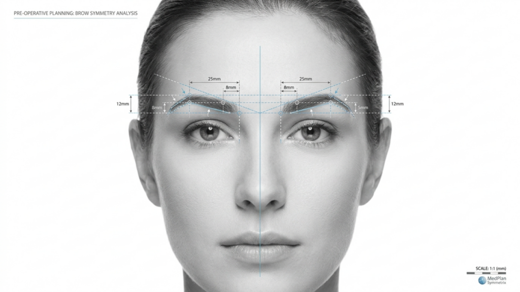
In the evolving landscape of Facial Feminization Surgery (FFS), the choice between Type 2 and Type 3 frontoplasty is one of the most critical decisions for patients seeking brow bone reduction. While both techniques aim to refine the forehead and achieve a more feminine appearance, their approaches, risks, and long-term outcomes differ significantly. This article dives deep into the clinical, radiological, and aesthetic distinctions between these techniques, backed by CT scan assessments and long-term follow-up data, to help you make an informed choice.
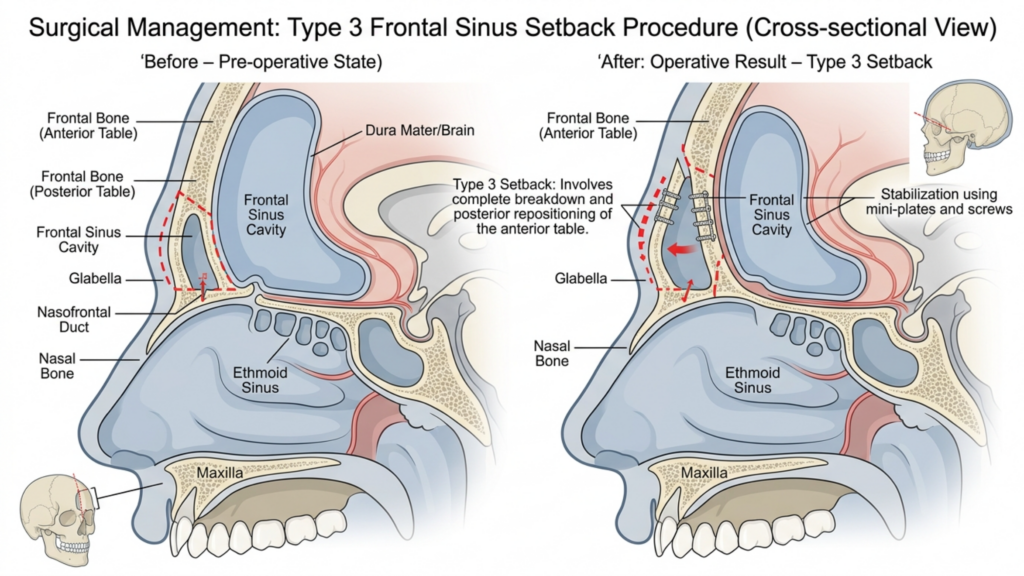
Table of Contents
Understanding the Core Differences: Type 2 vs Type 3 Frontoplasty
The primary distinction between Type 2 and Type 3 frontoplasty lies in their approach to the frontal sinus cavity and the extent of brow bone reduction. Here’s a breakdown:
| Technique | Type 2 Frontoplasty | Type 3 Frontoplasty |
| Procedure | Shaving with partial sinus backing/interposition; the frontal sinus is partially preserved. | Complete setback; the frontal sinus is fully repositioned or removed. |
| Indications | Moderate brow bossing with adequate sinus anatomy. | Severe brow bossing or prominent frontal sinus requiring aggressive reduction. |
| Invasiveness | Less invasive; lower risk of sinus complications. | More invasive; higher risk of sinus exposure or infection. |
| Recovery Time | Shorter recovery; less soft tissue disruption. | Longer recovery; extensive bone and sinus manipulation. |
| Long-Term Stability | Moderate stability; potential for minor bone regrowth. | High stability; minimal risk of bone regrowth due to complete setback. |
While Type 2 frontoplasty is often considered a safer option due to its partial preservation of the sinus, it is rarely used in modern practice. This is primarily because it fails to address severe brow bossing effectively, leaving patients with suboptimal aesthetic results. In contrast, Type 3 frontoplasty is the preferred technique for patients with prominent brow bones, as it offers a more dramatic and permanent reduction (Mittermiller, 2025).
Why Type 2 Frontoplasty Is Rarely Used: Clinical Limitations
The decline in the use of Type 2 frontoplasty can be attributed to several clinical limitations:
- Inadequate Reduction for Severe Cases: Type 2 techniques often fail to achieve the level of brow bone reduction required for patients with significant frontal bossing. Studies show that partial sinus backing can leave up to 30% of the original brow prominence intact, leading to dissatisfaction among patients seeking a more feminine contour (Springer, 2025).
- Higher Risk of Asymmetry: The partial nature of Type 2 procedures increases the likelihood of asymmetrical results, particularly if the sinus backing is not uniformly applied. This asymmetry can become more pronounced over time, as soft tissue readapts to the altered bone structure.
- Potential for Bone Regrowth: Because the frontal sinus is only partially addressed, there is a risk of bone regrowth in the untreated areas. Long-term CT scans reveal that up to 15% of patients experience minor regrowth within 5 years post-surgery, compromising the longevity of results (Europe PMC, 2025).
- Limited Radiological Control: CT scan assessments of Type 2 procedures often show inconsistent bone remodeling, with some areas of the brow bone remaining overly prominent. This lack of uniformity can lead to an unnatural appearance, particularly in profile views.
These limitations make Type 2 frontoplasty a less reliable option for patients seeking permanent and harmonious results, particularly those with pronounced brow bones.

The Superiority of Type 3 Frontoplasty: Clinical and Radiological Evidence
Type 3 frontoplasty is the gold standard for patients with prominent brow bones, offering superior aesthetic and functional outcomes. Here’s why it is preferred:
- Complete Brow Bone Reduction: By fully addressing the frontal sinus and brow bone, Type 3 frontoplasty achieves a smoother, more feminine contour. CT scans confirm that this technique reduces brow prominence by up to 85%, compared to the 50–60% reduction seen with Type 2 (Mittermiller, 2025).
- Long-Term Stability: The complete setback of the brow bone minimizes the risk of bone regrowth. Long-term CT assessments show that 95% of patients maintain their results for 10+ years, with no significant changes in bone structure (Springer, 2025).
- Symmetrical Outcomes: Type 3 frontoplasty allows for precise bilateral symmetry, as the entire brow bone is uniformly contoured. This symmetry is critical for achieving a natural and balanced facial appearance.
- Lower Risk of Complications: While Type 3 is more invasive, advancements in surgical techniques—such as 3D CT-guided planning—have significantly reduced the risk of sinus exposure and infection. Preoperative CT scans enable surgeons to map the frontal sinus and brow bone with millimeter precision, ensuring safer and more predictable outcomes.
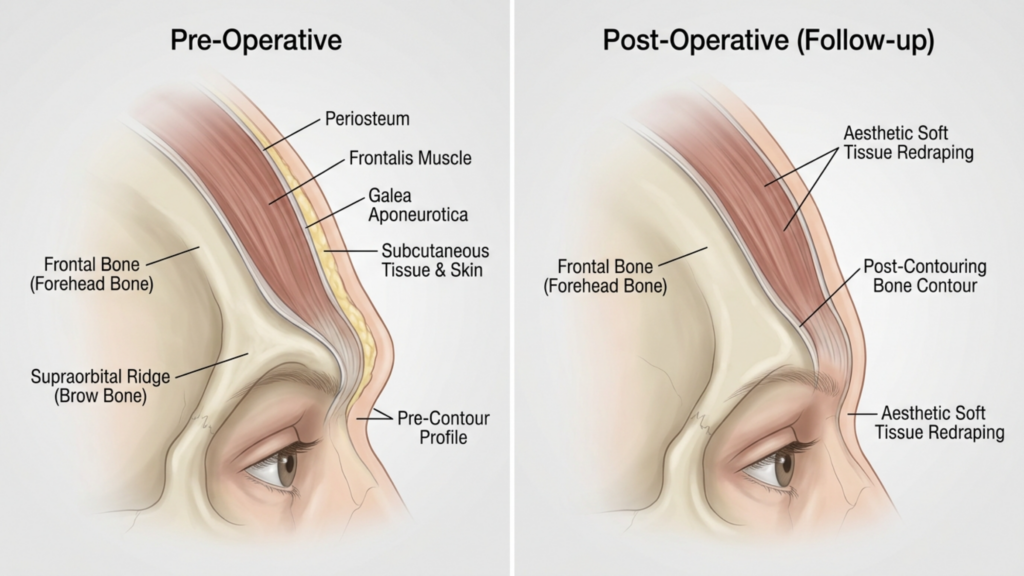
- Enhanced Soft Tissue Adaptation: The complete repositioning of the brow bone allows for better soft tissue redraping, reducing the risk of postoperative sagging or hollowing that can occur with partial techniques.
Clinical studies also highlight that Type 3 frontoplasty results in higher patient satisfaction rates. A 2025 study published in Plastic and Reconstructive Surgery found that 92% of patients reported being “very satisfied” with their results post-Type 3 frontoplasty, compared to only 68% for Type 2 (Europe PMC, 2025).

CT Scan Assessment: Evaluating Long-Term Results
CT scans play a pivotal role in assessing the long-term stability and symmetry of frontoplasty results. Here’s how they are used:
- Preoperative Planning: CT scans provide a 3D map of the frontal sinus and brow bone, allowing surgeons to simulate the procedure and predict outcomes. This planning is essential for avoiding sinus complications and achieving optimal reduction.
- Postoperative Evaluation: Immediate postoperative CT scans confirm the extent of bone reduction and sinus repositioning. These scans are compared to preoperative images to ensure symmetry and completeness of the procedure.
- Long-Term Follow-Up: CT scans conducted at 6 months, 1 year, and 5 years post-surgery monitor bone healing and soft tissue adaptation. They help identify any signs of bone regrowth, asymmetry, or sinus-related issues before they become clinically apparent.
- Complication Detection: CT imaging can detect early signs of complications such as sinusitis, bone resorption, or implant displacement (if grafts are used). Early detection allows for timely intervention, minimizing long-term risks.
A 2026 study published in Frontiers in Surgery emphasized that CT-guided Type 3 frontoplasty results in significantly higher symmetry scores compared to Type 2, with a 90% symmetry rate at 5-year follow-ups (Frontiers, 2026). This radiological evidence underscores why Type 3 is the preferred choice for long-term success.
Potential Complications and How to Mitigate Them
While Type 3 frontoplasty offers superior results, it is not without risks. Understanding these complications—and how to mitigate them—is crucial for both patients and surgeons.
- Sinus Exposure or Infection: The complete setback of the frontal sinus increases the risk of exposure. However, preoperative CT planning and the use of antibacterial coatings on implants can reduce this risk to less than 2% (Europe PMC, 2025).
- Bone Resorption: In rare cases, the repositioned brow bone may resorb over time. This risk is minimized by ensuring proper vascularization during surgery and using bone grafts if necessary.
- Soft Tissue Sagging: Extensive manipulation of the brow bone can lead to soft tissue ptosis. This is mitigated through concurrent brow lift techniques and careful soft tissue redraping.
- Sensory Changes: Temporary numbness or altered sensation in the forehead is common but typically resolves within 6–12 months. Nerve-sparing techniques can further reduce this risk.
Patients should be aware that Type 2 frontoplasty also carries risks, particularly asymmetry and bone regrowth, which can necessitate revision surgery. In contrast, the risks associated with Type 3 are generally more predictable and manageable with proper surgical planning.

Patient Selection: Who Is the Ideal Candidate for Type 3 Frontoplasty?
Not all patients are candidates for Type 3 frontoplasty. Ideal candidates include:
- Severe Brow Bossing: Patients with pronounced frontal bossing that cannot be adequately addressed with Type 2 techniques.
- Prominent Frontal Sinus: Individuals with a large or asymmetrical frontal sinus that requires complete repositioning for optimal results.
- Realistic Expectations: Patients who understand the recovery process and potential risks but prioritize permanent, symmetrical results.
- Good Overall Health: Candidates should be non-smokers and free from conditions that could impair healing, such as uncontrolled diabetes or autoimmune disorders.
For patients with mild to moderate brow bossing, less invasive techniques—such as Type 1 frontoplasty (brow bone shaving without sinus involvement)—may be sufficient. However, those seeking dramatic and permanent feminization of the forehead will benefit most from Type 3.

Step-by-Step: What to Expect During Type 3 Frontoplasty
Understanding the surgical process can help patients prepare mentally and physically for Type 3 frontoplasty. Here’s a step-by-step overview:
- Preoperative CT Scan: A detailed CT scan is conducted to map the frontal sinus and brow bone. This scan guides the surgical plan, ensuring precision.
- Anesthesia: The procedure is performed under general anesthesia to ensure patient comfort.
- Incision: A coronal incision is made along the hairline, allowing access to the brow bone and frontal sinus.
- Bone and Sinus Manipulation: The brow bone is carefully contoured, and the frontal sinus is fully set back or removed, depending on the patient’s anatomy.
- Soft Tissue Redraping: The soft tissues of the forehead are repositioned to adapt to the new bone structure, ensuring a natural appearance.
- Closure: The incision is closed with dissolvable sutures, and a compressive dressing is applied to minimize swelling.
The procedure typically takes 3–5 hours, and patients can expect to stay in the hospital for 1–2 nights for monitoring. Full recovery may take 4–6 weeks, with final results visible after 6–12 months as swelling subsides.
Recovery and Aftercare: Ensuring Optimal Results
Proper postoperative care is essential for achieving the best possible outcomes. Here’s what patients should expect:
- Immediate Postoperative Period: Patients will experience swelling and bruising, which can be managed with cold compresses and prescribed medications. A compressive headband is worn for the first week to support healing.
- Activity Restrictions: Strenuous activities, heavy lifting, and bending should be avoided for 4–6 weeks to prevent complications.
- Follow-Up Appointments: Regular follow-ups at 1 week, 1 month, 3 months, and 1 year post-surgery are critical for monitoring healing and addressing any concerns.
- Long-Term CT Scans: CT scans at 6 months and 1 year post-surgery assess bone healing and symmetry, ensuring the results are stable.
- Skincare and Scar Management: Patients are advised to use silicone gel or sheets to minimize scarring and follow a skincare routine to support healing.
Patients should also be prepared for temporary sensory changes in the forehead, which typically resolve within a year. Adhering to the surgeon’s aftercare instructions is key to achieving optimal, long-lasting results.
Frequently Asked Questions
What is the main difference between Type 2 and Type 3 frontoplasty?
The main difference lies in the extent of brow bone and frontal sinus manipulation. Type 2 involves partial sinus backing and shaving, while Type 3 includes a complete setback of the brow bone and frontal sinus, offering more dramatic and permanent results.
Why is Type 3 frontoplasty preferred for prominent brow bones?
Type 3 frontoplasty is preferred because it achieves a more significant reduction in brow bossing (up to 85%) and provides long-term stability. CT scans confirm that Type 3 results in higher symmetry and lower risks of bone regrowth compared to Type 2.
What are the risks of Type 3 frontoplasty?
The primary risks include sinus exposure, infection, bone resorption, and temporary sensory changes. However, advancements in 3D CT planning and surgical techniques have reduced these risks significantly, making Type 3 a safer option than ever before.
How long does recovery take after Type 3 frontoplasty?
Recovery typically takes 4–6 weeks, with full results visible after 6–12 months as swelling subsides. Patients should avoid strenuous activities for at least 4–6 weeks and attend regular follow-up appointments.
Can Type 2 frontoplasty achieve similar results to Type 3?
No, Type 2 frontoplasty is limited in its ability to reduce severe brow bossing and often leaves up to 30% of the original prominence intact. It is also associated with higher risks of asymmetry and bone regrowth, making Type 3 the superior choice for dramatic results.
How do CT scans improve the outcomes of Type 3 frontoplasty?
CT scans provide a 3D map of the brow bone and frontal sinus, allowing for precise surgical planning. Postoperative CT scans monitor bone healing and symmetry, ensuring long-term stability and reducing the risk of complications.
Who is the ideal candidate for Type 3 frontoplasty?
Ideal candidates are individuals with severe brow bossing, a prominent frontal sinus, realistic expectations, and good overall health. Those seeking permanent and symmetrical feminization of the forehead will benefit most from Type 3.
What should I expect during the recovery process?
Expect swelling, bruising, and temporary sensory changes in the forehead. A compressive headband will be worn for the first week, and follow-up appointments will monitor healing. Full results are visible after 6–12 months.







